a new platinum complex with tryptophan: synthesis, structural characterization, dft studies and...
TRANSCRIPT
Spectrochimica Acta Part A: Molecular and Biomolecular Spectroscopy 122 (2014) 209–215
Contents lists available at ScienceDirect
Spectrochimica Acta Part A: Molecular andBiomolecular Spectroscopy
journal homepage: www.elsevier .com/locate /saa
A new platinum complex with tryptophan: Synthesis, structuralcharacterization, DFT studies and biological assays in vitro over humantumorigenic cells
1386-1425/$ - see front matter � 2013 Elsevier B.V. All rights reserved.http://dx.doi.org/10.1016/j.saa.2013.11.044
Abbreviations: B3LYP, Becke 3-Lee Young Parr functional; DFT, density functional theory; DMEM, Dulbelcco’s modified eagle’s medium; DMSO, dimethylsulfoxQTOF-MS, electrospray ionization quadrupole time-of-flight mass spectrometry; FBS, fetal bovine serum; IR, infrared; LANL2DZ, Los Alamos National Laboratoversion on double zeta function; MTT, (3-(4,5-dimethylthiazol-2-yl)-2,5-diphenyl tetrazolium bromide; NMR, nuclear magnetic resonance; PCM, polarized continuumPES, potential energy surface; Pt-trp, Pt(II) complex with tryptophan; CP/MAS NMR, cross-polarization/magic angle spinning solid-state nuclear magnetic resonance;Time dependent density functional theory; TGA/DTA, Thermogravimetric and differential thermal analyses TGA/DTA; trp, L-Tryptophan; UV–Vis, Ultraviolespectroscopy.⇑ Corresponding author. Tel.: +55 19 35213130; fax: +55 19 35213023.
E-mail address: [email protected] (P.P. Corbi).
Marcos A. Carvalho a, Silvia M. Shishido b, Bárbara C. Souza a, Raphael E.F. de Paiva a, Alexandre F. Gomes c,Fábio C. Gozzo c, André L.B. Formiga d, Pedro P. Corbi a,⇑a Bioinorganic and Medicinal Chemistry Research Laboratory, Institute of Chemistry, University of Campinas – UNICAMP, P.O. Box 6154, 13083-970 Campinas, SP, Brazilb Laboratory of Bioassays and Signal Transduction, Department of Biochemistry, Institute of Biology, University of Campinas – UNICAMP, P.O. Box 6109, 13083-970 Campinas, SP,Brazilc Dalton Mass Spectrometry Laboratory, Organic Chemistry Department, Institute of Chemistry, University of Campinas – UNICAMP, P.O. Box 6154, 13083-970 Campinas, SP, Brazild Coordination Chemistry Laboratory, Institute of Chemistry, University of Campinas – UNICAMP, P.O. Box 6154, 13083-970 Campinas, SP, Brazil
h i g h l i g h t s
� A novel platinum(II) complex withtryptophan.� IR and solid-state NMR studies
permitted to confirm nitrogen andoxygen coordination.� The compound shows cytotoxic
activities over human tumorigeniccells.
g r a p h i c a l a b s t r a c t
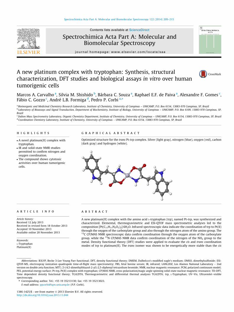
Optimized structure for the trans Pt-trp complex. Silver (light gray), nitrogen (blue), oxygen (red), carbon(dark gray) and hydrogen (white).
a r t i c l e i n f o
Article history:Received 12 July 2013Received in revised form 18 October 2013Accepted 10 November 2013Available online 20 November 2013
Keywords:L-TryptophanPlatinum(II)
a b s t r a c t
A new platinum(II) complex with the amino acid L-tryptophan (trp), named Pt-trp, was synthesized andcharacterized. Elemental, thermogravimetric and ESI-QTOF mass spectrometric analyses led to thecomposition [Pt(C11H11N2O2)2]�6H2O. Infrared spectroscopic data indicate the coordination of trp to Pt(II)through the oxygen of the carboxylate group and also through the nitrogen atom of the amino group. The13C CP/MAS NMR spectroscopic data confirm coordination through the oxygen atom of the carboxylategroup, while the 15N CP/MAS NMR data confirm coordination of the nitrogen of the NH2 group to themetal. Density functional theory (DFT) studies were applied to evaluate the cis and trans coordinationmodes of trp to platinum(II). The trans isomer was shown to be energetically more stable than the cis
ide; ESI-ry – 2nd
model;TD-DFT,t–visible

210 M.A. Carvalho et al. / Spectrochimica Acta Part A: Molecular and Biomolecular Spectroscopy 122 (2014) 209–215
ESI-QTOF-MSMolecular modelingAntitumoral assays
one. The Pt-trp complex was evaluated as a cytotoxic agent against SK-Mel 103 (human melanoma) andPanc-1 (human pancreatic carcinoma) cell lines. The complex was shown to be cytotoxic over the consid-ered cells.
� 2013 Elsevier B.V. All rights reserved.
Introduction
Metal ions present remarkable effects in many cellular pro-cesses including metabolism and cell division. These propertiesled the researchers to evaluate the effects of metal-based com-pounds for the treatment of several human diseases, includingthe treatment of cancer. Cisplatin, or cis-diamminedichoridoplati-num(II), is the most significant platinum-based anticancer drug.It is used for the treatment of cervix, head and neck, esophagealand small cell lung cancer, among others [1].
The notable antitumor activity of cisplatin led to the develop-ment of new drugs based on the cisplatin structure, aiming forthe reduction of the side effects of cisplatin, and also an improve-ment in the solubility and stability of the compounds in physiolog-ical conditions [2]. The ligand-exchange rate is one of the keyfactors regarding the activity of Pt(II) compounds against tumorcells. The ligand-exchange rate of Pt(II) compounds leads toa certain kinetic stability, with ligand-exchange reactions in orderof few minutes to days [3]. According to the literature, the mecha-nism of action of cisplatin is based on its interaction with DNAforming adducts. Cisplatin is activated intracellularly by hydrolysisof one of the two chloride ions, and covalently binds to DNA [4].
Despite the great dominance of nucleic acids, proteins are alsorelevant targets to platinum complexes with antitumor activity.Cisplatin, for instance, was investigated regarding its interactionswith serum albumin [5]. Platinum drugs were also investigatedregarding its interaction with hemoglobin [6]. In this context, thecharacterization of a platinum complex with tryptophan can beuseful for understanding the interaction of platinum with proteinsand even to evaluate its possible antitumor activity.
Nowadays, second and third generation platinum-based anti-cancer drugs such as carboplatin, oxaliplatin and nedaplatin havebeen applied as anticancer agents [7]. In vitro cytotoxic studies ofplatinum(II) iminophosphine complexes showed moderate activityand blocked the proliferation of esophageal cancer cells WHCO1and KYSE450, with the IC50 values of 5.5–9.5 lM, and2.2–7.6 lM, respectively [8].
In our research group, some platinum and palladium complexeswere synthesized and evaluated as anticancer and antibacterialdrugs. Castello et al. described a dimeric platinum(II) complex withmethionine sulfoxide, of composition [(C5H10NO3S)Pt(l-Cl)2Pt(C5-
H10NO3S)]�2.5H2O, which shows antibacterial activities againstPseudomonas aeruginosa bacterial cells [9]. Spera et al. publishedthe antibacterial studies of a palladium(II) complex with deoxyalliinthat shows in vitro antibacterial activities over pathogenic Gram-positive and Gram-negative bacterial strains [10]. This compoundwas also shown to possess antitumor activities over HeLa cancercells [11,12]. Also, Cavicchioli et al. described a platinum complexwith acesulfame of composition K2[PtCl2(ace)2], which was assayedin vitro against HeLa (human cervix cancer) cells, showing low activ-ity when compared to the vehicle-treated cells [13].
Amino acids are versatile ligands in coordination chemistry,forming chelates through the amino and carboxylate groups. Inparticular, tryptophan (trp), is an amino acid that possesses oneprimary amino group, one indolic group and one carboxylic group[14]. Several metal complexes with tryptophan have already beendescribed in the literature. A copper(II) complex with the aminoacid L-tryptophan, of composition [Cu(trp)2], was first prepared
by Wagner and Baran [15]. Kazachenko et al. [16] investigatedthe synthesis and antibacterial activities of silver complexes withthe amino acids histidine and tryptophan. Both compoundsshowed a good antibacterial activity against Gram-negative andGram-positive bacterial strains and low toxicity. More recentlyCarvalho et al. [17,18] carried out the synthesis of new palla-dium(II) and silver(I) complexes with tryptophan. The Pd(II) com-plex, of composition [Pd(C11H11N2O2)2]�2H2O shows coordinationof the ligand to Pd(II) though the nitrogen of the amino groupand the oxygen of carboxylate group, forming a square-planargeometry around the metal center. The complex was shown tobe active against Escherichia coli, P. aeruginosa and Staphylococcusaureus bacterial stains. The silver(I) complex with tryptophan, ofcomposition AgC11H11N2O2 shows coordination of the ligand toAg(I) ion through the nitrogen of the NH2 group and also by theoxygen of carboxylate group [18]. The compound was effectiveagainst S. aureus and Enterococcus faecalis (Gram-positive), andP. aeruginosa and E. coli (Gram-negative) bacterial strains. The com-plex was also cytotoxic against Panc-1 (human pancreatic carci-noma) and SK-Mel 103 (human melanoma) cells.
In this context, synthesis, spectroscopic characterization andnovel biological studies of a platinum(II) complex with tryptophan,with emphasis on its possible application as an antitumoral agent,are presented in this manuscript.
Experimental
Reagents and equipments
L-Tryptophan (98%) and potassium tetrachloroplatinate(II) 98%were purchased from Sigma–Aldrich Laboratories. Potassiumhydroxide (85%) was obtained from Fluka. Cisplatin, DMSO and(3-(4,5-dimethylthiazol-2-yl)-2,5-diphenyl tetrazolium bromide(MTT), were purchased from Sigma, and ethanol from Synth. Dul-belcco’s Modified Eagle’s Medium (DMEM), fetal bovine serumand antibiotics were purchased from Invitrogen (Gaithersburg,MD). Flasks and plates were purchased from TPP (Techno PlasticProducts – St. Louis, USA). Elemental analyses for carbon, hydrogenand nitrogen were performed using a CHNS/O Perkin Elmer 2400Analyzer. Infrared (IR) spectra from 4000 to 400 cm�1 of trp andthe Pt-trp complex were measured using an ABB Bomen MB SeriesModel B100 with resolution of 4 cm�1; samples were prepared asKBr pellets. The solid-state 13C{1H} nuclear magnetic resonancespectra were recorded on a Bruker 300 MHz, using the combina-tion of cross-polarization, proton decoupling and magic anglespinning (CP/MAS). The solid-state 15N{1H} nuclear magnetic reso-nance spectra were recorded on a Bruker 400 MHz, using thecombination of cross-polarization, proton decoupling and magicangle spinning (CP/MAS). Electrospray ionization quadrupoletime-of-flight mass spectrometry (ESI-QTOF-MS) measurementswere carried out in a Waters Synapt HDMS instrument (Manches-ter, UK). A sample of the Pt-trp complex was prepared in H2O/MeCN 50:50 with 0.1% (v/v) formic acid at a concentration of1 mg cm�3 and further diluted 100-fold in the same solventmixture. Resulting solutions were directly infused into the instru-ment’s ESI source at a flow rate of 15 lL min�1. Typical acquisitionconditions were capillary voltage 3 kV, sampling cone voltage 20 V,
M.A. Carvalho et al. / Spectrochimica Acta Part A: Molecular and Biomolecular Spectroscopy 122 (2014) 209–215 211
source temperature 100 �C, desolvation temperature 200 �C, conegas flow 30 L h�1, desolvation gas flow 900 L h�1, Trap and Transfercollision energies at 6 and 4 eV, respectively. ESI(+) mass spectra(fullscans) and fragment ion mass spectra for quadrupole-isolatedions were acquired in reflectron W-mode at a scan rate of 1 Hz. Forfragment ion spectrum experiment by collision-induced dissocia-tion (argon as collision gas), the desired ion was isolated in themass-resolving quadrupole, and the collision energy of the trap cellwas increased until sufficient fragmentation was observed. Prior toall analyses, the instrument was calibrated with phosphoric acidoligomers (H3PO4 0.05% v/v in H2O/MeCN 50:50) ranging fromm/z 99 to 1960.
Thermogravimetric and differential thermal analysis (TGA/DTA)were performed on a Simultaneous TGA/DTA SEIKO EXSTAR 6000Thermoanalyzer, using the following conditions: synthetic air,flow rate of 50 cm3 min�1 and heating rate of 10 �C min�1, from25 �C to 1000 �C. The residue of the thermal treatment was ana-lyzed on a Shimadzu XRD-6000 diffractometer (Cu Ka radiation,k = 1.54056 Å) with a graphite monochromator and at room tem-perature. The sample was scanned over the 2h range from 4� to 70�.
Synthesis of the complex
The platinum(II) complex with L-tryptophan was synthesizedby the reaction of 1.0 � 10�3 mol of a freshly prepared aqueouspotassium tryptophanate solution (10.0 cm3, pH = 10) with5.0 � 10�4 mol of an aqueous solution of K2PtCl4 (5.0 cm3). Thesynthesis of the complex was carried out with stirring at roomtemperature. After 20 h of constant stirring, the dark solid obtainedwas collected by filtration, washed with cold water and dried in adesiccator with P2O5. The yield was 67%. Anal. Calc. for [Pt(C11H11-
N2O2)2] 6H2O (%): C 37.2; H 4.83; N 7.89. Found (%): C 36.6; H 4.71;N 7.62. The complex is insoluble in water, chloroform, ethanol,methanol, acetone and hexane. Although the compound dissolvesin dimethylsulfoxide, 1H NMR studies suggests the decompositionof the complex in this solvent. The potassium tryptophanatesolution used in the synthesis of the platinum(II) complex was pre-pared in situ by the reaction of equivalent amounts of trp andpotassium hydroxide. No single crystals suitable for full X-ray crys-tallographic studies were obtained.
Molecular modeling
Geometric optimizations were carried out using GAMESS soft-ware [19] with a convergence criterion of 10�4 a.u. in a conjugategradient algorithm. The LANL2DZ [20] effective core potentialwas used for silver and the atomic 6-31G (d, p) basis set [21–24]for all other atoms. Density functional theory (DFT) calculationswere performed by using the B3LYP [25,26] gradient-correctedhybrid to solve the Kohn–Sham equations with a 10�5 a.u. conver-gence criterion for the density change. Two possible coordinationgeometries were evaluated, the cis and trans isomers. Also, thepolarized continuum model (PCM) [27] considering water as thecontinuum medium was used in order to take into accountthe zwitterionic form of tryptophan, since it is the main form ofthe molecule in the solid state. The effect of PCM was also evalu-ated considering the two coordination modes proposed. The finalgeometries were confirmed as minima of the potential energy sur-face (PES) with calculation of the Hessians showing no imaginaryfrequencies.
The harmonic vibrational frequencies and intensities were cal-culated at the same level of theory with the analytical evaluationof second derivatives of energy as a function of atomic coordinates,and the calculated intensities were used to generate the theoreticalspectra [28]. Frequencies were scaled by a factor of 0.9614, as rec-ommended by Scott and Radom [29]. Simulated vibrational spectra
were obtained from the sum of Lorentzian functions with 20 cm�1
half-bandwidths using the software Molden 4.7 [29].
Cell culture and cytotoxic assays
Balb/c 3T3 (mouse embryonic fibroblast) cells were purchasedfrom National Institute of Health-Baltimore, USA (NIH), whileSK-Mel 103 (human melanoma cells) was courtesy from Dr. SilvyaStuchi (University of São Paulo – USP, São Paulo, Brazil). The Panc-1(human pancreatic carcinoma) cells were purchased from Rio deJaneiro Cell Bank (Rio de Janeiro, Brazil). All cell lines were rou-tinely grown in Dulbelcco’s Modified Eagle’s Medium (DMEM) sup-plemented with 10% fetal bovine serum (FBS) and antibiotics(100 U mL�1 penicillin, 10 lg mL�1 streptomycin) in a humidifiedincubator with 5% carbon dioxide, at 37 �C. The values of concen-tration that lead to 50% of cell viability reduction (IC50) were usedas the parameter of cytotoxicity. Cisplatin was used as a positivecontrol.
Cells (1 � 105 cell mL�1) were plated in 96 well plates and 24 hlater the cells were exposed to a Pt-trp suspension containing 0.1to 2.0 mg mL�1of the complex. After 48 h the solutions were re-moved and 100 lL of MTT solution (0.5 mg mL�1 in FBS free cul-ture medium) was added to each well. After incubation for 2 h at37 �C, the MTT solution was removed and the formazan crystalswere solubilized in 100 lL of ethanol. The plate was shaken for5 min on a plate shaker and the absorbance was measured at570 nm in a microplate reader (Synergy HT, BioTek) [30].
Results and discussion
Thermal analysis
The thermogravimetric (TGA) data confirmed the compositionof the complex formulated as [Pt(C11H11N2O2)2]�6H2O. Water mol-ecules were lost in the range 25–250 �C. The oxidation of the ligandstarts at 270 �C. In the differential thermal analysis (DTA) we cansee one exothermic peak at 370 �C related to ligand oxidation.The residue of the thermal treatment at 1000 �C was identifiedby powder X-ray diffraction analysis as metallic Pt� [31]. Anal. Calc.for loss of (C11H11N2O2)2�6H2O: 72.5% Found: 68.5%. Anal. Calc. forresidue Pt�: 27.5%, Found: 31.5%. The TGA/DTA curves are shown inthe Supplementary Information #1.
13C and 15N NMR spectroscopic measurements
The structural formula of trp, with hydrogen and carbon atomnumbering was already published by Carvalho et al. [17]. For ana-lytical purposes, the structure is presented in Fig. 1 as earlyreported.
Since the complex is insoluble in all common solvents and inDMSO it shows decomposition with the appearance of broadenedsignals with poor resolution in the 1H NMR spectrum, the com-pound was studied by 13C and 15N NMR in the solid state.
The 13C and 15N chemical shifts for the complex were evaluatedin comparison to the data of free trp. The 13C and 15N chemicalshifts are shown in Table 1. The free ligand presents two reso-nances for COO�, Ca, and Cb, probably due to polymorphism [32].The carbon atom of the COO� group is the most shifted upon coor-dination. The carbon atom of the COO� is observed at 177.1 ppmfor trp, and it is shifted downfield to 185.5 ppm upon coordination.
The nitrogen of the primary amino group is shifted from43.4 ppm in the free ligand to �15.2 ppm in the Pt-trp complex,evidencing the coordination of the ligand to Pt(II) through the nitro-gen atom of NH2 group. Supplementary information #2 shows thesolid state 13C and 15N NMR spectra for trp and the Pt-trp complex.
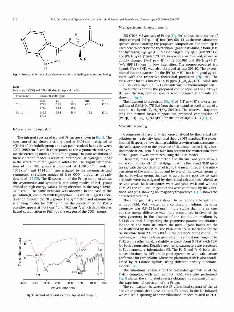
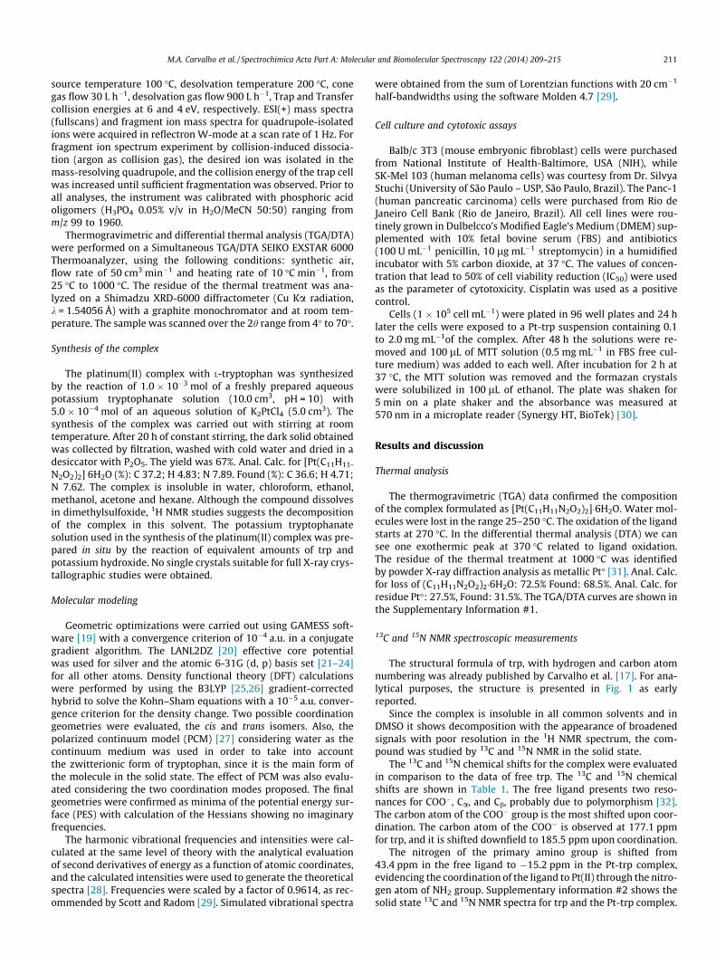
Fig. 1. Structural formula of trp showing carbon and hydrogen atoms numbering.
Table 1Solid-state 13C{1H} and 15N NMR data for trp and the Pt-trp.
Compounds Chemical shifts (ppm)
COO� Ca Cb NH2 NHTrp 177.1 56.2 29.5 43.3 126.3
175.7 53.5 28.8 – –
Pt-trp 185.5 56.3 28.4 �15.2 130.4176.1 – – – –
212 M.A. Carvalho et al. / Spectrochimica Acta Part A: Molecular and Biomolecular Spectroscopy 122 (2014) 209–215
Infrared spectroscopic data
The infrared spectra of trp and Pt-trp are shown in Fig. 2. Thespectrum of trp shows a strong band at 3403 cm�1 assigned tom(N–H) of the indolic group and two poor resolved bands between3090–2980 cm�1, which corresponds to the asymmetric and sym-metric stretching modes of the amino group. The poor resolution ofthese vibration modes is result of intermolecular hydrogen bondsin the structure of the ligand in solid state. The angular deforma-tion of the NH2 group is observed at 1590 cm�1. Bands at1668 cm�1 and 1414 cm�1 are assigned to the asymmetric andsymmetric stretching modes of free COO� group, as alreadydescribed [15,33]. The IR spectrum of the Pt-trp complex showsthe asymmetric and symmetric stretching modes of NH2 groupshifted to high energy values, being observed in the range 3290–3195 cm�1. The same behavior was observed in the case of thepalladium(II) complex with tryptophan [17] which suggests coor-dination through the NH2 group. The symmetric and asymmetricstretching modes for COO� cm�1 in the spectrum of the Pt-trpcomplex appear at 1448 cm�1 and 1625 cm�1, which also indicatesligand coordination to Pt(II) by the oxygen of the COO� group.
4000 3500 3000 2500 2000 1500 1000 500
C-HCOO -
NH2COO -
NH2
NH2
N-H
COO -
Tran
smitt
ance
(u.a
.)
Wavenumber cm-1
b
aN-H NH2 COO -
C-H
Fig. 2. Infrared vibrational spectra of trp (a) and Pt-trp (b).
Mass spectrometric measurements
ESI-QTOF-MS analysis of Pt-trp (Fig. 3A) shows the presence ofsingle charged [PtTrp2 + H]+ ions (m/z 601.13) as the most abundantspecies, demonstrating the proposed composition. The term trp isused here to describe the tryptophan ligand in its anionic form (lessone hydrogen, C11H11N2O�2 ). Single charged [Pt2Trp3]+ (m/z 997.17)and [Pt2Trp4 + H]+ (m/z 1203.27) ions were also observed, as well asdoubly charged [Pt2Trp3 + H]2+ (m/z 500.09) and [Pt3Trp4 + H]2+
(m/z 698.91) ions in low intensities. The monoprotonated trpligand, [Trp + 2H]+, was also observed at m/z 205.10. The experi-mental isotope pattern for the [PtTrp2 + H]+ ion is in good agree-ment with the respective theoretical prediction (Fig. 3B). Themass error for this ion was +4.15 ppm (C22H23N4O4Pt+, calcd. m/z602.1346, exp. m/z 602.1371), considering the monoisotopic ion.
To further confirm the proposed composition of the [PtTrp2 +H]+ ion, the fragment ion spectra were obtained. The results areshown in Fig. 4.
The fragment ion spectrum (Fig. 4) of [PtTrp2 + H]+ shows a neu-tral loss of C2H5NO2 (75 Da) from the trp ligand, as well as loss of aneutral trp ligand (C11H12N2O2, 204 Da). The observed fragmentions and neutral losses support the proposed composition of[PtTrp2 + H]+ (C22H23N4O4Pt+) for the ion of m/z 601.13 (Fig. 3).
Molecular modeling
Geometries of trp and Pt-trp were analyzed by theoretical cal-culations using density functional theory (DFT) studies. The exper-imental IR spectra show that trp exhibits a zwitterionic structure inthe solid state, due to the presence of the combination NHþ3 vibra-tion mode at 2070 cm�1. To take into account the zwitterionic formof the ligand, it was optimized using the PCM model.
Elemental, mass spectrometric and thermal analyses show amolar composition of 1:2 metal/ligand, while the IR and NMR spec-tra indicate the coordination of trp to the metal through the nitro-gen atom of the amino group and by one of the oxygen atoms ofthe carboxylate group. So, two structures are possible to existand both were investigated by theoretical calculations, labeled ascis and trans. Both structures were analyzed with and withoutPCM. All the equilibrium geometries were confirmed by the vibra-tional analysis, showing no imaginary frequencies. Fig. 5 shows theobtained structures.
The trans geometry was shown to be more stable with andwithout PCM. With water as a continuum medium, the transgeometry was 0.8452 kcal mol�1 more stable than the cis one,but the energy difference was more pronounced in favor of thetrans geometry in the absence of the continuum medium by8.5335 kcal mol�1. Regarding the geometric parameters obtainedfor the cis and trans structures, the metal–ligand bonds are themost affected by the PCM. The Pt–N distance is shortened for thecis structure from 2.10 to 2.08 Å in the presence of the continuummedium, while for the trans geometry it is almost unchanged. ThePt-O, on the other hand, is slightly relaxed (about 0.01 Å) with PCMfor both geometries. Detailed geometric parameters are presentedin Supplementary information #3. The Pt–N and Pt–O bond dis-tances obtained by DFT are in good agreement with calculationsperformed for carboplatin, where the platinum atom is also coordi-nated by N,O-donor ligands, using different density functionalmodels [34].
The vibrational analysis for the calculated geometries of thePt-trp complex, with and without PCM, was also performed.Fig. 6 shows the simulated spectra obtained in comparison withthe experimental spectrum of the Pt-trp.
The comparison between the IR vibrational spectra of the cisand trans geometries shows minor differences. In the far infrared,we can see a splitting of some vibrational modes related to Pt–O
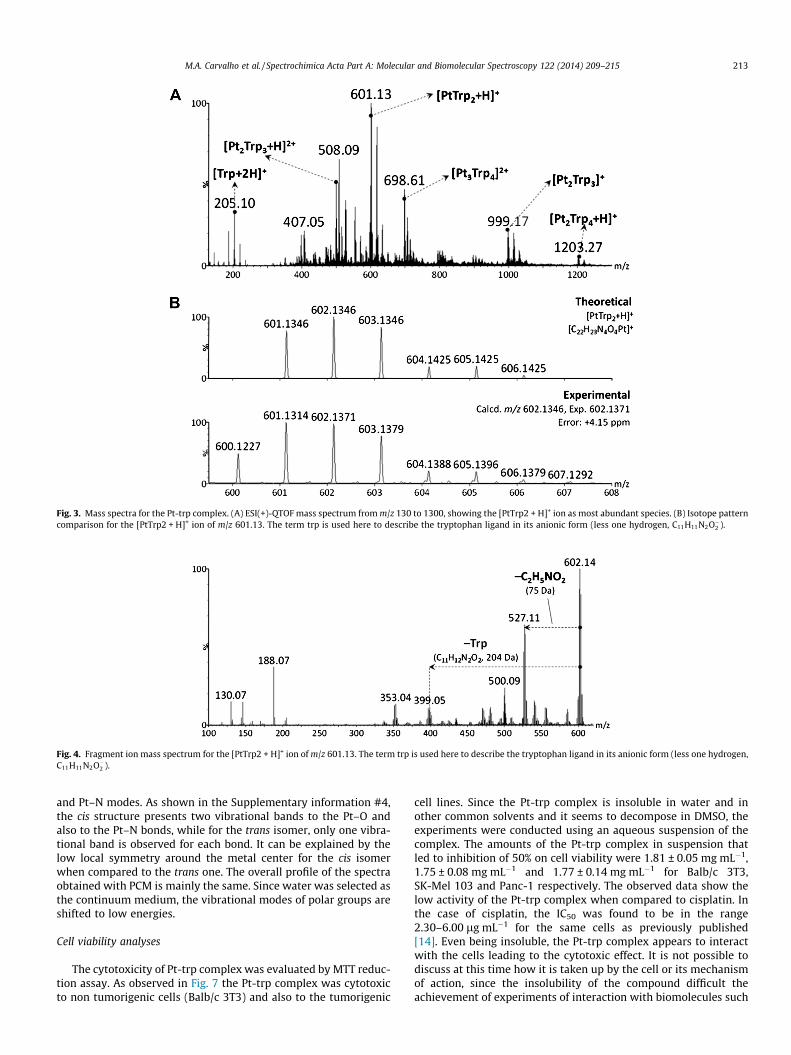
Fig. 3. Mass spectra for the Pt-trp complex. (A) ESI(+)-QTOF mass spectrum from m/z 130 to 1300, showing the [PtTrp2 + H]+ ion as most abundant species. (B) Isotope patterncomparison for the [PtTrp2 + H]+ ion of m/z 601.13. The term trp is used here to describe the tryptophan ligand in its anionic form (less one hydrogen, C11H11N2O�2 ).
Fig. 4. Fragment ion mass spectrum for the [PtTrp2 + H]+ ion of m/z 601.13. The term trp is used here to describe the tryptophan ligand in its anionic form (less one hydrogen,C11H11N2O�2 ).
M.A. Carvalho et al. / Spectrochimica Acta Part A: Molecular and Biomolecular Spectroscopy 122 (2014) 209–215 213
and Pt–N modes. As shown in the Supplementary information #4,the cis structure presents two vibrational bands to the Pt–O andalso to the Pt–N bonds, while for the trans isomer, only one vibra-tional band is observed for each bond. It can be explained by thelow local symmetry around the metal center for the cis isomerwhen compared to the trans one. The overall profile of the spectraobtained with PCM is mainly the same. Since water was selected asthe continuum medium, the vibrational modes of polar groups areshifted to low energies.
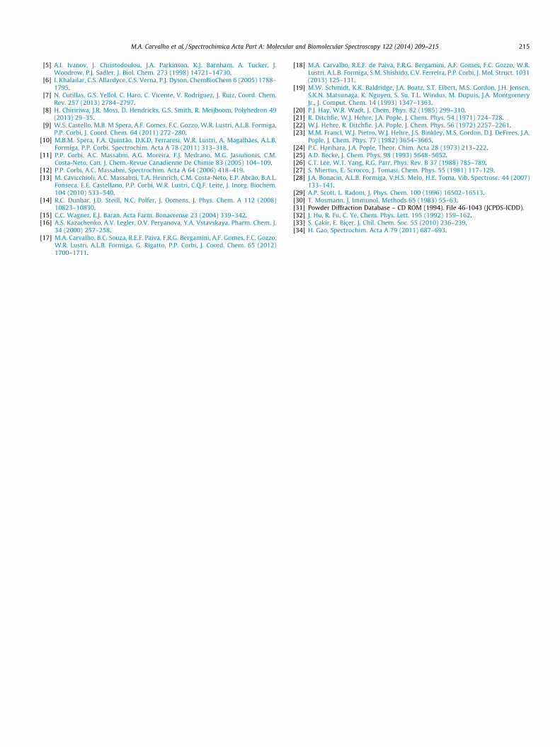
Cell viability analyses
The cytotoxicity of Pt-trp complex was evaluated by MTT reduc-tion assay. As observed in Fig. 7 the Pt-trp complex was cytotoxicto non tumorigenic cells (Balb/c 3T3) and also to the tumorigenic
cell lines. Since the Pt-trp complex is insoluble in water and inother common solvents and it seems to decompose in DMSO, theexperiments were conducted using an aqueous suspension of thecomplex. The amounts of the Pt-trp complex in suspension thatled to inhibition of 50% on cell viability were 1.81 ± 0.05 mg mL�1,1.75 ± 0.08 mg mL�1 and 1.77 ± 0.14 mg mL�1 for Balb/c 3T3,SK-Mel 103 and Panc-1 respectively. The observed data show thelow activity of the Pt-trp complex when compared to cisplatin. Inthe case of cisplatin, the IC50 was found to be in the range2.30–6.00 lg mL�1 for the same cells as previously published[14]. Even being insoluble, the Pt-trp complex appears to interactwith the cells leading to the cytotoxic effect. It is not possible todiscuss at this time how it is taken up by the cell or its mechanismof action, since the insolubility of the compound difficult theachievement of experiments of interaction with biomolecules such
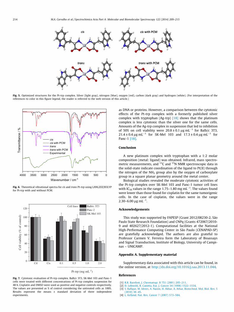
Fig. 5. Optimized structures for the Pt-trp complex. Silver (light gray), nitrogen (blue), oxygen (red), carbon (dark gray) and hydrogen (white). (For interpretation of thereferences to color in this figure legend, the reader is referred to the web version of this article.)
4000 3500 3000 2500 2000 1500 1000 500 0
Tran
smitt
ance
/ %
Wavenumber / cm-1
ciscis with PCMtranstrans with PCM
Experimental
Fig. 6. Theoretical vibrational spectra for cis and trans Pt-trp using LANL2DZ/B3LYPfor Pt-trp with and without PCM.
Ctl Cis 0.1 0.5 1.0 2.00
20
40
60
80
100
120
Cel
l via
bilit
y (%
of
cont
rol)
Pt-trp (mg mL-1)
Cell lines Balb/c 3T3 Panc-2 SK-Mel 103
Fig. 7. Cytotoxic evaluation of Pt-trp complex. Balb/c 3T3, SK-Mel 103 and Panc-1cells were treated with different concentrations of Pt-trp complex suspension for48 h. Cisplatin and DMSO were used as positive and negative controls respectively.The values are presented as % of control considering the untreated cells as 100%.Results represent the means ± standard deviation of three independentexperiments.
214 M.A. Carvalho et al. / Spectrochimica Acta Part A: Molecular and Biomolecular Spectroscopy 122 (2014) 209–215
as DNA or proteins. However, a comparison between the cytotoxiceffects of the Pt-trp complex with a formerly published silvercomplex with tryptophan (Ag-trp) [18] shows that the platinumcomplex is less cytotoxic than the silver one for the same cells.Amounts of the Ag-trp complex in suspension that led to inhibitionof 50% on cell viability were 20.8 ± 0.1 lg mL�1 for Balb/c 3T3,21.4 ± 0.4 lg mL�1 for SK-Mel 103 and 17.3 ± 0.4 lg mL�1 forPanc-1 [18].
Conclusion
A new platinum complex with tryptophan with a 1:2 molarcomposition (metal: ligand) was obtained. Infrared, mass spectro-metric measurements, and 13C and 15N NMR spectroscopic data inthe solid-state indicate coordination of the ligand to Pt(II) throughthe nitrogen of the NH2 group also by the oxygen of carboxylategroup in a square planar geometry around the metal center.
Biological studies revealed the moderate cytotoxic activities ofthe Pt-trp complex over SK-Mel 103 and Panc-1 tumor cell lineswith IC50 values in the range 1.75–1.80 mg mL�1. The values foundwere lower than those found for cisplatin for the same tumorigeniccells. In the case of cisplatin, the values were in the range2.30–6.00 lg mL�1.
Acknowledgements
This study was supported by FAPESP (Grant 2012/08230-2, SãoPaulo State Research Foundation) and CNPq (Grants 472067/2010-9 and 402627/2012-1). Computational facilities at the NationalHigh-Performance Computing Center in São Paulo (CENAPAD-SP)are gratefully acknowledged. The authors are also grateful toProfessor Carmen V. Ferreira form the Laboratory of Bioassaysand Signal Transduction, Institute of Biology, University of Campi-nas – UNICAMP.
Appendix A. Supplementary material
Supplementary data associated with this article can be found, inthe online version, at http://dx.doi.org/10.1016/j.saa.2013.11.044.
References
[1] R.R. Barefoot, J. Chromatogr. B 751 (2001) 205–211.[2] D. Lebwohl, R. Canetta, Eur. J. Cancer 34 (1998) 1522–1534.[3] S. Rafique, M. Idrees, A. Nasim, H. Akbar, A. Athar, Biotechnol. Mol. Biol. Rev. 5
(2010) 38–45.[4] L. Kelland, Nat. Rev. Cancer 7 (2007) 573–584.
M.A. Carvalho et al. / Spectrochimica Acta Part A: Molecular and Biomolecular Spectroscopy 122 (2014) 209–215 215
[5] A.I. Ivanov, J. Christodoulou, J.A. Parkinson, K.J. Barnham, A. Tucker, J.Woodrow, P.J. Sadler, J. Biol. Chem. 273 (1998) 14721–14730.
[6] I. Khalailar, C.S. Allardyce, C.S. Verna, P.J. Dyson, ChemBioChem 6 (2005) 1788–1795.
[7] N. Cutillas, G.S. Yellol, C. Haro, C. Vicente, V. Rodriguez, J. Ruiz, Coord. Chem.Rev. 257 (2013) 2784–2797.
[8] H. Chiririwa, J.R. Moss, D. Hendricks, G.S. Smith, R. Meijboom, Polyhedron 49(2013) 29–35.
[9] W.S. Castello, M.B. M Spera, A.F. Gomes, F.C. Gozzo, W.R. Lustri, A.L.B. Formiga,P.P. Corbi, J. Coord. Chem. 64 (2011) 272–280.
[10] M.B.M. Spera, F.A. Quintão, D.K.D. Ferraresi, W.R. Lustri, A. Magalhães, A.L.B.Formiga, P.P. Corbi, Spectrochim. Acta A 78 (2011) 313–318.
[11] P.P. Corbi, A.C. Massabni, A.G. Moreira, F.J. Medrano, M.G. Jasiulionis, C.M.Costa-Neto, Can. J. Chem.-Revue Canadienne De Chimie 83 (2005) 104–109.
[12] P.P. Corbi, A.C. Massabni, Spectrochim. Acta A 64 (2006) 418–419.[13] M. Cavicchioli, A.C. Massabni, T.A. Heinrich, C.M. Costa-Neto, E.P. Abrão, B.A.L.
Fonseca, E.E. Castellano, P.P. Corbi, W.R. Lustri, C.Q.F. Leite, J. Inorg. Biochem.104 (2010) 533–540.
[14] R.C. Dunbar, J.D. Steill, N.C. Polfer, J. Oomens, J. Phys. Chem. A 112 (2008)10823–10830.
[15] C.C. Wagner, E.J. Baran, Acta Farm. Bonaerense 23 (2004) 339–342.[16] A.S. Kazachenko, A.V. Legler, O.V. Peryanova, Y.A. Vstavskaya, Pharm. Chem. J.
34 (2000) 257–258.[17] M.A. Carvalho, B.C. Souza, R.E.F. Paiva, F.R.G. Bergamini, A.F. Gomes, F.C. Gozzo,
W.R. Lustri, A.L.B. Formiga, G. Rigatto, P.P. Corbi, J. Coord. Chem. 65 (2012)1700–1711.
[18] M.A. Carvalho, R.E.F. de Paiva, F.R.G. Bergamini, A.F. Gomes, F.C. Gozzo, W.R.Lustri, A.L.B. Formiga, S.M. Shishido, C.V. Ferreira, P.P. Corbi, J. Mol. Struct. 1031(2013) 125–131.
[19] M.W. Schmidt, K.K. Baldridge, J.A. Boatz, S.T. Elbert, M.S. Gordon, J.H. Jensen,S.K.N. Matsunaga, K. Nguyen, S. Su, T.L. Windus, M. Dupuis, J.A. MontgomeryJr., J. Comput. Chem. 14 (1993) 1347–1363.
[20] P.J. Hay, W.R. Wadt, J. Chem. Phys. 82 (1985) 299–310.[21] R. Ditchfie, W.J. Hehre, J.A. Pople, J. Chem. Phys. 54 (1971) 724–728.[22] W.J. Hehre, R. Ditchfie, J.A. Pople, J. Chem. Phys. 56 (1972) 2257–2261.[23] M.M. Francl, W.J. Pietro, W.J. Hehre, J.S. Binkley, M.S. Gordon, D.J. DeFrees, J.A.
Pople, J. Chem. Phys. 77 (1982) 3654–3665.[24] P.C. Harihara, J.A. Pople, Theor. Chim. Acta 28 (1973) 213–222.[25] A.D. Becke, J. Chem. Phys. 98 (1993) 5648–5652.[26] C.T. Lee, W.T. Yang, R.G. Parr, Phys. Rev. B 37 (1988) 785–789.[27] S. Miertus, E. Scrocco, J. Tomasi, Chem. Phys. 55 (1981) 117–129.[28] J.A. Bonacin, A.L.B. Formiga, V.H.S. Melo, H.E. Toma, Vib. Spectrosc. 44 (2007)
133–141.[29] A.P. Scott, L. Radom, J. Phys. Chem. 100 (1996) 16502–16513.[30] T. Mosmann, J. Immunol. Methods 65 (1983) 55–63.[31] Powder Diffraction Database – CD ROM (1994). File 46-1043 (JCPDS-ICDD).[32] J. Hu, R. Fu, C. Ye, Chem. Phys. Lett. 195 (1992) 159–162.[33] S. Çakir, E. Biçer, J. Chil. Chem. Soc. 55 (2010) 236–239.[34] H. Gao, Spectrochim. Acta A 79 (2011) 687–693.