a non-canonical peptide synthetase adenylates 3-methyl-2-oxovaleric ... · a non-canonical peptide...
TRANSCRIPT
2766
A non-canonical peptide synthetase adenylates3-methyl-2-oxovaleric acid for auriculamide biosynthesisDaniel Braga1,2, Dirk Hoffmeister1 and Markus Nett*3
Letter Open Access
Address:1Friedrich-Schiller-Universität Jena, Department PharmaceuticalMicrobiology at the Hans-Knöll-Institute, Winzerlaer Strasse 2, 07745Jena, Germany, 2Friedrich-Schiller-Universität Jena, Junior ResearchGroup Synthetic Microbiology at the Hans-Knöll-Institute,Adolf-Reichwein-Strasse 23, 07745 Jena, Germany and 3Departmentof Biochemical and Chemical Engineering, Technical Biology,Technical University Dortmund, Emil-Figge-Strasse 66, 44227Dortmund, Germany
Email:Markus Nett* - [email protected]
* Corresponding author
Keywords:adenylation; auriculamide; biosynthesis; Herpetosiphon; nonribosomalpeptide synthetase
Beilstein J. Org. Chem. 2016, 12, 2766–2770.doi:10.3762/bjoc.12.274
Received: 02 September 2016Accepted: 07 December 2016Published: 16 December 2016
This article is part of the Thematic Series "Chemical biology".
Guest Editor: H. B. Bode
© 2016 Braga et al.; licensee Beilstein-Institut.License and terms: see end of document.
AbstractAuriculamide is the first natural product known from the predatory bacterium Herpetosiphon aurantiacus. It is composed of three
unusual building blocks, including the non-proteinogenic amino acid 3-chloro-L-tyrosine, the α-hydroxy acid L-isoleucic acid, and
a methylmalonyl-CoA-derived ethane unit. A candidate genetic locus for auriculamide biosynthesis was identified and encodes four
enzymes. Among them, the non-canonical 199 kDa four-domain nonribosomal peptide synthetase, AulA, is extraordinary in that it
features two consecutive adenylation domains. Here, we describe the functional characterization of the recombinantly produced
AulA. The observed activation of 3-methyl-2-oxovaleric acid by the enzyme supports the hypothesis that it participates in the bio-
synthesis of auriculamide. An artificially truncated version of AulA that lacks the first adenylation domain activated this substrate
like the full-length enzyme which shows that the first adenylation domain is dispensable. Additionally, we provide evidence that the
enzyme tolerates structural variation of the substrate. α-Carbon substituents significantly affected the substrate turnover. While all
tested aliphatic α-keto acids were accepted by the enzyme and minor differences in chain size and branches did not interfere with
the enzymatic activity, molecules with methylene α-carbons led to low turnover. Such enzymatic plasticity is an important attribute
to help in the perpetual search for novel molecules and to access a greater structural diversity by mutasynthesis.
2766
FindingsHerpetosiphon aurantiacus is a filamentous, Gram-negative
bacterium with a facultative saprophytic predatory behaviour
[1,2]. For a more profound insight into the predation strategies
among bacteria, along with the underlying chemistry, the com-
plete genome of H. aurantiacus 114-95T (ATCC 23779, DSM
785) was sequenced and analysed [3]. Present as one circular

Beilstein J. Org. Chem. 2016, 12, 2766–2770.
2767
Figure 1: Herpetosiphon natural products auriculamide (1) and siphonazole (2).
Figure 2: Organisation of the aul biosynthetic gene cluster. Circles illustrate the domain architecture of the NRPSs and the PKS present therein.Domains are abbreviated as A, adenylation; ACP, acyl carrier protein; AT, acyl transferase; C, condensation; KR, ketoreductase; KS, ketosynthase;PCP, peptidyl carrier protein; TE, thioesterase. The gene aulD encodes a type II thioesterase.
chromosome and two circular plasmids, the 6.8 Mb genome of
H. aurantiacus encodes as many as 14 biosynthesis gene clus-
ters corresponding to 6.6% (0.45 Mb) of the genome. This
capacity highlights this microorganism as a promising source of
natural products. Genes for nonribosomal peptide synthetases
(NRPSs) were found to be preponderant, either solely or organ-
ised in combination with polyketide synthase (PKS) genes,
representing four and five clusters, respectively. Two PKS and
three putative bacteriocin gene clusters complete the total set
involved in the biosynthesis of natural products. Contrasting the
high number of biosyntheses deduced from genomic data,
knowledge on the actual natural products is limited. Recently,
the dipeptide auriculamide (1, Figure 1), and the diterpene
O-methylkolavelool were observed in cultures of H. auranti-
acus 114-95T, providing initial evidence for the assumed sec-
ondary metabolome of this species [4-6]. Within the entire
genus, 1 is only the second PKS/NRPS-derived molecule to be
described, following the report on siphonazole (2, Figure 1) [7].
Retrobiosynthetic analysis allowed the identification of a
14,130 bp-gene cluster, now referred to as aul-cluster
(Figure 2), which putatively encodes two NRPSs (AulA and
AulB) and one PKS (AulC) possessing domains that collec-
tively allow and plausibly explains the assembly of 1. A gene
for a type-II thioesterase is also found at the 3’ portion of the
aul cluster that may help unload misacylated carrier protein
domains [8,9].
Contrasting the standard layout of NRPSs, the amino acid se-
quence of one of the deduced NRPSs, termed AulA (1818 aa,
199 kDa), reveals the peculiar chimeric A1-A2-KR-PCP archi-
tecture (Figure 2) [10]. Of particular interest, the occurrence of
two sequential adenylation (A) domains is a very rare feature
and only preceded by PyrG from Streptomyces pyridomyceticus
[11].
Since the lack of a genetic system for H. aurantiacus makes the
use of reverse genetics prohibitive, we sought to provide
biochemical evidence for the participation of this unusual NRPS
in the biosynthesis of 1. AulA is suggested to incorporate
L-isoleucic acid (= 2-hydroxy-3-methylvaleric acid). The
domain architecture indicates the substrate undergoes no other
chemical modification besides a reductive step after being teth-
ered to the PCP domain by the PKS-type ketoreductase domain
(KR), as reported for other natural products, such as pyrido-
mycin [11], cereulide, valinomycin [12], and bacillaene [13].
Hence, the molecule to be recognized and activated by AulA
would be 3-methyl-2-oxovaleric acid (3).
Seminal work with gramicidin synthetase from Bacillus brevis
led to the identification of ten positions within an A domain
(PheA), collectively referred to as nonribosomal code [14], that
control substrate selectivity. Further research started to estab-
lish a relationship between this code and structural require-
Beilstein J. Org. Chem. 2016, 12, 2766–2770.
2768
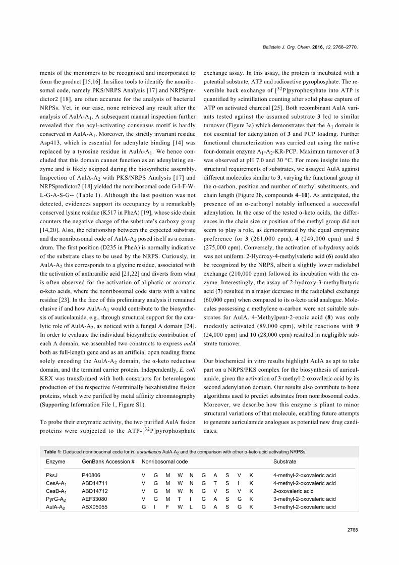
Table 1: Deduced nonribosomal code for H. aurantiacus AulA-A2 and the comparison with other α-keto acid activating NRPSs.
Enzyme GenBank Accession # Nonribosomal code Substrate
PksJ P40806 V G M W N G A S V K 4-methyl-2-oxovaleric acidCesA-A1 ABD14711 V G M W N G T S I K 4-methyl-2-oxovaleric acidCesB-A1 ABD14712 V G M W N G V S V K 2-oxovaleric acidPyrG-A2 AEF33080 V G M T I G A S G K 3-methyl-2-oxovaleric acidAulA-A2 ABX05055 G I F W L G A S G K 3-methyl-2-oxovaleric acid
ments of the monomers to be recognised and incorporated to
form the product [15,16]. In silico tools to identify the nonribo-
somal code, namely PKS/NRPS Analysis [17] and NRPSpre-
dictor2 [18], are often accurate for the analysis of bacterial
NRPSs. Yet, in our case, none retrieved any result after the
analysis of AulA-A1. A subsequent manual inspection further
revealed that the acyl-activating consensus motif is hardly
conserved in AulA-A1. Moreover, the strictly invariant residue
Asp413, which is essential for adenylate binding [14] was
replaced by a tyrosine residue in AulA-A1. We hence con-
cluded that this domain cannot function as an adenylating en-
zyme and is likely skipped during the biosynthetic assembly.
Inspection of AulA-A2 with PKS/NRPS Analysis [17] and
NRPSpredictor2 [18] yielded the nonribosomal code G-I-F-W-
L-G-A-S-G-- (Table 1). Although the last position was not
detected, evidences support its occupancy by a remarkably
conserved lysine residue (K517 in PheA) [19], whose side chain
counters the negative charge of the substrate’s carboxy group
[14,20]. Also, the relationship between the expected substrate
and the nonribosomal code of AulA-A2 posed itself as a conun-
drum. The first position (D235 in PheA) is normally indicative
of the substrate class to be used by the NRPS. Curiously, in
AulA-A2 this corresponds to a glycine residue, associated with
the activation of anthranilic acid [21,22] and diverts from what
is often observed for the activation of aliphatic or aromatic
α-keto acids, where the nonribosomal code starts with a valine
residue [23]. In the face of this preliminary analysis it remained
elusive if and how AulA-A1 would contribute to the biosynthe-
sis of auriculamide, e.g., through structural support for the cata-
lytic role of AulA-A2, as noticed with a fungal A domain [24].
In order to evaluate the individual biosynthetic contribution of
each A domain, we assembled two constructs to express aulA
both as full-length gene and as an artificial open reading frame
solely encoding the AulA-A2 domain, the α-keto reductase
domain, and the terminal carrier protein. Independently, E. coli
KRX was transformed with both constructs for heterologous
production of the respective N-terminally hexahistidine fusion
proteins, which were purified by metal affinity chromatography
(Supporting Information File 1, Figure S1).
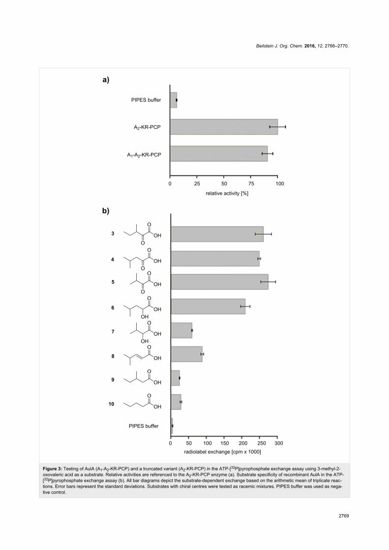
To probe their enzymatic activity, the two purified AulA fusion
proteins were subjected to the ATP-[32P]pyrophosphate
exchange assay. In this assay, the protein is incubated with a
potential substrate, ATP and radioactive pyrophosphate. The re-
versible back exchange of [32P]pyrophosphate into ATP is
quantified by scintillation counting after solid phase capture of
ATP on activated charcoal [25]. Both recombinant AulA vari-
ants tested against the assumed substrate 3 led to similar
turnover (Figure 3a) which demonstrates that the A1 domain is
not essential for adenylation of 3 and PCP loading. Further
functional characterization was carried out using the native
four-domain enzyme A1-A2-KR-PCP. Maximum turnover of 3
was observed at pH 7.0 and 30 °C. For more insight into the
structural requirements of substrates, we assayed AulA against
different molecules similar to 3, varying the functional group at
the α-carbon, position and number of methyl substituents, and
chain length (Figure 3b, compounds 4–10). As anticipated, the
presence of an α-carbonyl notably influenced a successful
adenylation. In the case of the tested α-keto acids, the differ-
ences in the chain size or position of the methyl group did not
seem to play a role, as demonstrated by the equal enzymatic
preference for 3 (261,000 cpm), 4 (249,000 cpm) and 5
(275,000 cpm). Conversely, the activation of α-hydroxy acids
was not uniform. 2-Hydroxy-4-methylvaleric acid (6) could also
be recognized by the NRPS, albeit a slightly lower radiolabel
exchange (210,000 cpm) followed its incubation with the en-
zyme. Interestingly, the assay of 2-hydroxy-3-methylbutyric
acid (7) resulted in a major decrease in the radiolabel exchange
(60,000 cpm) when compared to its α-keto acid analogue. Mole-
cules possessing a methylene α-carbon were not suitable sub-
strates for AulA. 4-Methylpent-2-enoic acid (8) was only
modestly activated (89,000 cpm), while reactions with 9
(24,000 cpm) and 10 (28,000 cpm) resulted in negligible sub-
strate turnover.
Our biochemical in vitro results highlight AulA as apt to take
part on a NRPS/PKS complex for the biosynthesis of auricul-
amide, given the activation of 3-methyl-2-oxovaleric acid by its
second adenylation domain. Our results also contribute to hone
algorithms used to predict substrates from nonribosomal codes.
Moreover, we describe how this enzyme is pliant to minor
structural variations of that molecule, enabling future attempts
to generate auriculamide analogues as potential new drug candi-
dates.
Beilstein J. Org. Chem. 2016, 12, 2766–2770.
2769
Figure 3: Testing of AulA (A1-A2-KR-PCP) and a truncated variant (A2-KR-PCP) in the ATP-[32P]pyrophosphate exchange assay using 3-methyl-2-oxovaleric acid as a substrate. Relative activities are referenced to the A2-KR-PCP enzyme (a). Substrate specificity of recombinant AulA in the ATP-[32P]pyrophosphate exchange assay (b). All bar diagrams depict the substrate-dependent exchange based on the arithmetic mean of triplicate reac-tions. Error bars represent the standard deviations. Substrates with chiral centres were tested as racemic mixtures. PIPES buffer was used as nega-tive control.
Beilstein J. Org. Chem. 2016, 12, 2766–2770.
2770
Supporting InformationSupporting Information File 1Complete experimental details.
[http://www.beilstein-journals.org/bjoc/content/
supplementary/1860-5397-12-274-S1.pdf]
AcknowledgementsD.B. gratefully acknowledges a doctoral fellowship from the
International Leibniz Research School (ILRS Mibintact). We
thank Wiebke Hanke and Dr. Hirokazu Kage for a preliminary
analysis of AulA.
References1. Holt, J.; Lewin, R. J. Bacteriol. 1968, 95, 2407–2408.2. Jurkevitch, E. Microbe 2007, 2, 67–73. doi:10.1128/microbe.2.67.13. Kiss, H.; Nett, M.; Domin, N.; Martin, K.; Maresca, J. A.; Copeland, A.;
Lapidus, A.; Lucas, S.; Berry, K. W.; Glavina Del Rio, T.; Dalin, E.;Tice, H.; Pitluck, S.; Richardson, P.; Bruce, D.; Goodwin, L.; Han, C.;Detter, J. C.; Schmutz, J.; Brettin, T.; Land, M.; Hauser, L.;Kyrpides, N. C.; Ivanova, N.; Göker, M.; Woyke, T.; Klenk, H. P.;Bryant, D. A. Stand. Genomic Sci. 2011, 5, 356–370.doi:10.4056/sigs.2194987
4. Schieferdecker, S.; Domin, N.; Hoffmeier, C.; Bryant, D. A.; Roth, M.;Nett, M. Eur. J. Org. Chem. 2015, 3057–3062.doi:10.1002/ejoc.201500181
5. Nakano, C.; Oshima, M.; Kurashima, N.; Hoshino, T. ChemBioChem2015, 16, 772–781. doi:10.1002/cbic.201402652
6. Korp, J.; Vela Gurovic, M. S.; Nett, M. Beilstein J. Org. Chem. 2016,12, 594–607. doi:10.3762/bjoc.12.58
7. Nett, M.; Erol, Ö.; Kehraus, S.; Köck, M.; Krick, A.; Eguereva, E.;Neu, E.; König, G. M. Angew. Chem., Int. Ed. 2006, 45, 3863–3867.doi:10.1002/anie.200504525
8. Schwarzer, D.; Mootz, H. D.; Linne, U.; Marahiel, M. A.Proc. Natl. Acad. Sci. U. S. A. 2002, 99, 14083–14088.doi:10.1073/pnas.212382199
9. Pfeifer, B.; Hu, Z.; Licari, P.; Khosla, C. Appl. Environ. Microbiol. 2002,68, 3287–3292. doi:10.1128/AEM.68.7.3287-3292.2002
10. Marchler-Bauer, A.; Derbyshire, M. K.; Gonzales, N. R.; Lu, S.;Chitsaz, F.; Geer, L. Y.; Geer, R. C.; He, J.; Gwadz, M.; Hurwitz, D. I.;Lanczycki, C. J.; Lu, F.; Marchler, G. H.; Song, J. S.; Thanki, N.;Wang, Z.; Yamashita, R. A.; Zhang, D.; Zheng, C.; Bryant, S. H.Nucleic Acids Res. 2014, 43 (Suppl. D1), D222–D226.doi:10.1093/nar/gku1221
11. Huang, T.; Li, L.; Brock, N. L.; Deng, Z.; Lin, S. ChemBioChem 2016,17, 1421–1425. doi:10.1002/cbic.201600156
12. Magarvey, N. A.; Ehling-Schulz, M.; Walsh, C. T. J. Am. Chem. Soc.2006, 128, 10698–10699. doi:10.1021/ja0640187
13. Calderone, C. T.; Bumpus, S. B.; Kelleher, N. L.; Walsh, C. T.;Magarvey, N. A. Proc. Natl. Acad. Sci. U. S. A. 2008, 105,12809–12814. doi:10.1073/pnas.0806305105
14. Conti, E.; Stachelhaus, T.; Marahiel, M. A.; Brick, P. EMBO J. 1997, 16,4174–4183. doi:10.1093/emboj/16.14.4174
15. Stachelhaus, T.; Mootz, H. D.; Marahiel, M. A. Cell Chem. Biol. 1999,6, 493–505. doi:10.1016/S1074-5521(99)80082-9
16. Challis, G. L.; Ravel, J.; Townsend, C. A. Cell Chem. Biol. 2000, 7,211–224. doi:10.1016/S1074-5521(00)00091-0
17. Bachmann, B. O.; Ravel, J. Methods Enzymol. 2009, 458, 181–217.doi:10.1016/S0076-6879(09)04808-3
18. Röttig, M.; Medema, M. H.; Blin, K.; Weber, T.; Rausch, C.;Kohlbacher, O. Nucleic Acids Res. 2011, 39 (Suppl. 2), W362–W367.doi:10.1093/nar/gkr323
19. Kalb, D.; Lackner, G.; Hoffmeister, D. Fungal Biol. Rev. 2013, 27,43–50. doi:10.1016/j.fbr.2013.05.002
20. Schwarzer, D.; Finking, R.; Marahiel, M. A. Nat. Prod. Rep. 2003, 20,275–287. doi:10.1039/b111145k
21. Ames, B. D.; Walsh, C. T. Biochemistry 2010, 49, 3351–3365.doi:10.1021/bi100198y
22. Gao, X.; Chooi, Y.-H.; Ames, B. D.; Wang, P.; Walsh, C. T.; Tang, Y.J. Am. Chem. Soc. 2011, 133, 2729–2741. doi:10.1021/ja1101085
23. Wackler, B.; Lackner, G.; Chooi, Y. H.; Hoffmeister, D. ChemBioChem2012, 13, 1798–1804. doi:10.1002/cbic.201200187
24. Kalb, D.; Lackner, G.; Rappe, M.; Hoffmeister, D. ChemBioChem 2015,16, 1426–1430. doi:10.1002/cbic.201500190
25. Linne, U.; Marahiel, M. A. Methods Enzymol. 2004, 388, 293–315.doi:10.1016/S0076-6879(04)88024-8
License and TermsThis is an Open Access article under the terms of the
Creative Commons Attribution License
(http://creativecommons.org/licenses/by/4.0), which
permits unrestricted use, distribution, and reproduction in
any medium, provided the original work is properly cited.
The license is subject to the Beilstein Journal of Organic
Chemistry terms and conditions:
(http://www.beilstein-journals.org/bjoc)
The definitive version of this article is the electronic one
which can be found at:
doi:10.3762/bjoc.12.274