a novel dna-binding protein, phar, plays a central role in...
TRANSCRIPT
A Novel DNA-Binding Protein, PhaR, Plays a Central Role in theRegulation of Polyhydroxyalkanoate Accumulation and GranuleFormation in the Haloarchaeon Haloferax mediterranei
Shuangfeng Cai,a,b Lei Cai,a,c Dahe Zhao,a,b Guiming Liu,a,b Jing Han,a Jian Zhou,a Hua Xianga,b
State Key Laboratory of Microbial Resources, Institute of Microbiology, Chinese Academy of Sciences, Beijing, People’s Republic of Chinaa; University of Chinese Academyof Sciences, Beijing, People’s Republic of Chinab; School of Food Science and Biotechnology, Zhejiang Gongshang University, Hangzhou, People’s Republic of Chinac
Polyhydroxyalkanoates (PHAs) are synthesized and assembled as PHA granules that undergo well-regulated formation in manymicroorganisms. However, this regulation remains unclear in haloarchaea. In this study, we identified a PHA granule-associatedregulator (PhaR) that negatively regulates the expression of both its own gene and the granule structural gene phaP in the sameoperon (phaRP) in Haloferax mediterranei. Chromatin immunoprecipitation-quantitative PCR (ChIP-qPCR) assays demon-strated a significant interaction between PhaR and the phaRP promoter in vivo. Scanning mutagenesis of the phaRP promoterrevealed a specific cis-element as the possible binding position of the PhaR. The haloarchaeal homologs of the PhaR contain anovel conserved domain that belongs to a swapped-hairpin barrel fold family found in AbrB-like proteins. Amino acid substitu-tion indicated that this AbrB-like domain is critical for the repression activity of PhaR. In addition, the phaRP promoter had aweaker activity in the PHA-negative strains, implying a function of the PHA granules in titration of the PhaR. Moreover, the H.mediterranei strain lacking phaR was deficient in PHA accumulation and produced granules with irregular shapes. Interestingly,the PhaR itself can promote PHA synthesis and granule formation in a PhaP-independent manner. Collectively, our results dem-onstrated that the haloarchaeal PhaR is a novel bifunctional protein that plays the central role in the regulation of PHA accumu-lation and granule formation in H. mediterranei.
Polyhydroxyalkanoates (PHAs) are biodegradable polyesterssynthesized by most genera of bacteria (1, 2) and some archaea
(3–5). PHAs are accumulated as storage compounds of energy andcarbon under imbalanced growth conditions (i.e., when nutrientssuch as nitrogen, phosphorus, or oxygen are limited but the car-bon sources are in excess) (6).
PHAs are often deposited in the cytoplasm as water-insolubleinclusions that are called PHA granules (6). Native PHA granulesare found to be composed of 97.5% PHA, 2% proteins, and likelysome amount of lipids (7). At least four types of proteins werefound to be the PHA granule-associated proteins (PGAPs) in bac-teria: PHA synthases, PHA depolymerases, regulators, and struc-tural proteins (phasins [PhaPs]) (8, 9). In recent years, increasingnew roles have been found for the PGAPs. Besides the classicalphasin role of preventing PHA granules from coalescing, two dis-tinct phasin-like proteins, PhaM and PhaF, have also been char-acterized as being crucial for granule distribution during cell divi-sion (10, 11).
The PGAPs play important roles in PHA synthesis, PHA utili-zation, and granule formation and distribution (8, 9, 12), amongwhich the regulatory proteins are responsible for ensuring theproper formation of PHA granules by influencing the expressionof both phasins and themselves (13–17). A classic regulationmodel was presented in a poly(3-hydroxybutyrate) (PHB [a typeof PHA])-accumulating bacterium, Ralstonia eutropha H16 (9).Briefly, the cytoplasmic regulator PhaR could bind to the pro-moter of phaP as well as the promoter of its own gene to represstheir transcription. When cells start accumulating PHA, PhaR at-taches to the PHA granules, which results in a lower cytoplasmicPhaR level. The block of the expression of phaP and phaR is re-leased, and the cells start synthesizing more PhaP and PhaR to coatthe growing PHA granules. PhaP is usually more abundant than
PhaR and possesses a higher hydrophobic affinity to PHA gran-ules. When the PHA granules reach a proper size, there is no moreroom on PHA granules for the excess PhaR to attach. The cyto-plasmic PhaR concentration returns to a higher level to resumethe repression of the transcription of both phaP and phaR. Thistight regulation by PhaR ensures a well-organized granule forma-tion process, in which sufficient PhaP proteins are produced tocoat the newly synthesized PHAs, with few free PhaP present in thecytoplasm (9).
Unlike bacterial PGAPs, there has been little study of the archaealPGAPs until recently. In our previous studies, five PGAPs were iden-tified in a haloarchaeon, Haloferax mediterranei, which accumu-lates poly(3-hydroxybutyrate-co-3-hydroxyvalerate) (PHBV [atype of PHA]) and shows potential for industrial applications (18–20). Besides the PHA synthase subunits (PhaC and PhaE) and aputative enoyl coenzyme A (enoyl-CoA) hydratase (MaoC), twoconserved hypothetical proteins (encoded by HFX_5218 and
Received 3 September 2014 Accepted 21 October 2014
Accepted manuscript posted online 24 October 2014
Citation Cai S, Cai L, Zhao D, Liu G, Han J, Zhou J, Xiang H. 2015. A novel DNA-binding protein, PhaR, plays a central role in the regulation ofpolyhydroxyalkanoate accumulation and granule formation in the haloarchaeonHaloferax mediterranei. Appl Environ Microbiol 81:373–385.doi:10.1128/AEM.02878-14.
Editor: M. Kivisaar
Address correspondence to Hua Xiang, [email protected].
Supplemental material for this article may be found at http://dx.doi.org/10.1128/AEM.02878-14.
Copyright © 2015, American Society for Microbiology. All Rights Reserved.
doi:10.1128/AEM.02878-14
January 2015 Volume 81 Number 1 aem.asm.org 373Applied and Environmental Microbiology
on June 25, 2018 by guesthttp://aem
.asm.org/
Dow
nloaded from
HFX_5219) were also separated from the PHA granules of H.mediterranei. The protein encoded by HFX_5219 was identified tobe the major phasin (PhaP) that could prevent the aggregation ofPHA granules (20). HFX_5218 encodes a small protein that wastemporarily named GAP12 (12.0 kDa). The gap12 gene was re-vealed to be cotranscribed with phaP, but its function is still un-known (20). Characterization of the GAP12 separated from PHAgranules might provide important hints for the exploration of theregulation of PHA biosynthesis and granule formation in halo-archaea.
In this study, using a combined approach of gene expression,gene knockout, promoter activity analysis, and a chromatin im-munoprecipitation-quantitative PCR (ChIP-qPCR) assay, theGAP12 protein was identified as a regulator and renamed PhaR,which directly binds to the promoter of phaRP and negativelyregulates this operon. In addition, the cis-elements of the phaRPpromoter were identified by site-directed mutagenesis, and theeffects of PhaR on the PHA accumulation and granule formationwere further demonstrated by gas chromatography and electronmicroscopy analyses. Therefore, the identification and character-ization of the haloarchaeal type of phasin regulator PhaR, which isphylogenetically distinct from the bacterial counterpart, have pro-vided new insights into the regulation of PHA synthesis in halo-archaea.
MATERIALS AND METHODSStrains and culture conditions. The strains used in this study are listed inTable 1. Escherichia coli JM109 was used for cloning procedures and wasgrown in lysogeny broth (LB) medium at 37°C (21). H. mediterraneiDF50, a uracil-auxotrophic (�pyrF) strain of H. mediterranei ATCC33500 (22), and its derivative mutants were cultivated at 37°C in nutrient-rich AS-168L medium (20). H. mediterranei strains carrying expressionplasmids were cultivated in AS-168SYL medium (with yeast extract omit-ted from AS-168L) (20). For PHA accumulation analysis, the culture pro-cedures were similar to those described previously (20). Briefly, H. medi-terranei was first grown in AS-168L for 2 days and then was inoculatedinto a modified PHA production medium, named MGF medium, con-taining (per liter) 110 g NaCl, 9.6 g MgCl2, 14.4 g MgSO4, 5 g KCl, 1 gCaCl2, 3 g yeast extract, 2 g NH4Cl, 0.0375 g KH2PO4, 10 g glucose, 15 gPIPES [piperazine-N,N=-bis(2-ethanesulfonic acid)], 0.008 g NH4
�-Fe(III) citrate, and 1 ml trace element solution SL-6 (pH 7.2) (18). ForAS-168SYL seed cultures, yeast extract was also omitted from the MGFmedium. When needed, ampicillin, uracil, and 5-fluoroorotic acid (5-FOA) were added to the medium at final concentrations of 100, 50, and250 mg/liter, respectively.
Construction of mutants. The plasmids and primers used in thisstudy are listed in Tables 1 and 2, respectively. The in-frame gene deletionand gene expression in H. mediterranei were carried out as previouslydescribed (20, 22, 23).
For construction of the green fluorescent protein (GFP) reporter plas-mid pRF, a 168-bp DNA fragment upstream of the phaR open readingframe (ORF) (the promoter of the phaRP operon, named PphaRP) waslinked with the coding region of a soluble modified red-shifted greenfluorescent protein (smRSGFP [simply named “GFP” here]) (24), andwas cloned into the expression plasmid pWL502 (20). For another re-porter plasmid, pEF, the PphaRP fragment in pRF was replaced by a 189-bpDNA fragment upstream of the phaE ORF. To introduce mutations intoDNA fragments, the site-directed mutagenesis was performed using aDpnI-mediated method as described previously (25). Briefly, the 168-bpPphaRP fragment was cloned into the pGEM-T Easy vector (Promega), andthe resultant plasmid, pT-Rpro, was used as the PCR template for thesite-directed mutagenesis of PphaRP. The PphaRP fragments with desired
mutations together with the gfp fragment were subcloned into pWL502 togenerate the plasmids pD41, pD86, and pM1 to pM16, respectively.
For the amino acid residue substitutions in PhaR, a knock-in plasmid,pR-IN, which possesses a 1.2-kb DNA fragment containing the phaR ORFand its upstream and downstream regions, was used as the template. Thesubstitutions were introduced independently into each plasmid by DpnI-mediated site-directed mutagenesis. The resultant plasmids (e.g., pR-IN-E24A) were, respectively, transferred into the H. mediterranei strainwith phaR deleted and integrated into the chromosome to generate thecorresponding strains, which express the desired PhaR mutants (e.g.,PhaRE24A).
For the overexpression of phaR or phaP, each ORF region was insertedinto the pSCM307 plasmid (26). Then, the corresponding fragments con-taining both the hsp5 promoter region and the ORF region of phaP (orphaR) were subcloned into the plasmid pWL502 (or pRF), resulting inplasmid pHP (or pHRRF). For the complementary expression of phaR orphaRP, a fragment containing the region of the native promoter and theORF of phaR (or the ORFs of phaRP) was cloned into pWL502 to generateplasmid pWLR (or pWLRP).
All PCR-amplified sequences were verified by DNA sequencing. Theplasmid transformation of H. mediterranei was performed by a polyeth-ylene glycol-mediated transformation method (27).
Promoter activity assays. H. mediterranei strains harboring the GFPreporter plasmids were cultured at 37°C in AS-168SYL medium. Cells(100 �l/well) from certain growth phases were transferred into 96-wellplate to measure the turbidity (optical density at 600 nm [OD600]) and thefluorescence intensity (excitation, 488 nm; emission, 509 nm) using aSynergy H4 hybrid microplate reader (BioTek Instruments, Inc., Win-ooski, VT) (24, 28), with AS-168SYL medium serving as the blank control.The fluorescence intensity was normalized against the cell density (perOD600 of 0.1) and expressed as relative fluorescence units (RFU). At leastthree independent biological replicates were performed.
RNA extraction, CR-RT-PCR, and Northern blot analysis. The totalRNA of H. mediterranei cells in late-exponential phase was extracted withTRIzol reagent (Invitrogen) (29).
For the identification of the transcription start site (TSS), the circular-ized RNA reverse transcription-PCR (CR-RT-PCR) method was carriedout as previously described (30). After the reverse transcription of self-ligated RNA with random hexamer primers, the cDNA was used as thePCR template. The PCR products amplified with the primer pair phaRP-CRRT-F/phaRP-CRRT-R were cloned into the pGEM-T Easy vector todetermine the TSS by DNA sequencing.
For Northern blot analysis, 4 �g of each RNA sample was separated ona 4% denaturing polyacrylamide gel (7 M urea, 0.5� Tris-borate-EDTA[TBE] buffer) and transferred onto the nylon membranes using a semidrytransfer cell (Bio-Rad). The probes used to detect the expression of phaPand the internal control 7S RNA were amplified by the primer pairs phaP-NB-F/phaP-NB-R and 7S-NB-F/7S-NB-R, respectively. The probes werelabeled with biotin-11-dUTP (R0081; Thermo Scientific) by PCR. Afterthe cross-linking of RNA onto the membranes by UV, the membraneswere hybridized with the labeled probes. The prehybridization, hybridiza-tion, and washing procedures were performed as previously described(29). The biotin was detected using the Pierce chemiluminescent nucleicacid detection module (Thermo Scientific, Rockford, IL).
Protein expression and purification, antiserum preparation, andWestern blot analysis. The coding regions of phaR and phaP amplified bythe primer pairs PhaR-28a-F/PhaR-28a-R and PhaP-28a-F/PhaP-28a-R,respectively, were inserted into the vector pET-28a (Novagen). The resul-tant expression plasmids, pET-28aR and pET-28aP, were transferred intoE. coli BL21(DE3), respectively. Expression and purification of the PhaR-His6 and PhaP-His6 proteins, as well as the preparation of the correspond-ing antisera were performed as previously described (31).
For Western blot analysis, the cells cultivated in MGF medium werecollected in the stationary phase (after cultivation for approximately 3days). The cell pellets were dissolved in 8 M urea buffer and homogenized
Cai et al.
374 aem.asm.org January 2015 Volume 81 Number 1Applied and Environmental Microbiology
on June 25, 2018 by guesthttp://aem
.asm.org/
Dow
nloaded from
TABLE 1 Strains and plasmids used in this study
Strain or plasmid Relevant characteristicsSource orreference
StrainsE. coli
JM109 recA1 supE44 endA1 hsdR7 gyrA96 relA1 thi 21BL21(DE3) F� ompT hsdSB(rB
� mB�) dcm gal (DE3) Novagen
H. mediterraneiDF50 pyrF deletion mutant of H. mediterranei ATCC 33500 22�phaP mutant phaP deletion mutant of H. mediterranei DF50 20�phaR mutant phaR deletion mutant of H. mediterranei DF50 This study�phaRP mutant phaRP deletion mutant of H. mediterranei DF50 This study�phaEC mutant phaEC deletion mutant of H. mediterranei DF50 This study�phaPEC mutant phaPEC deletion mutant of H. mediterranei DF50 This study�phaRPEC mutant phaRPEC deletion mutant of H. mediterranei DF50 This studyE24A DF50 strain with PhaR carrying E24A mutation This studyQ28A DF50 strain with PhaR carrying Q28A mutation This studyQ30A DF50 strain with PhaR carrying Q30A mutation This studyR32A DF50 strain with PhaR carrying R32A mutation This studyK67A DF50 strain with PhaR carrying K67A mutation This studyR75A DF50 strain with PhaR carrying R75A mutation This studyE82A DF50 strain with PhaR carrying E82A mutation This studyR83A DF50 strain with PhaR carrying R83A mutation This study
PlasmidspHFX 4.0-kb integration vector containing pyrF and its native promoter, Ampr 22pWL502 7.8-kb expression vector containing pyrF and its native promoter, Ampr 20pSCM307 8.2-kb shuttle vector containing promoter of hsp5 of Halobacterium sp. strain NRC-1, Ampr 26pJAM1020 10.7-kb expression plasmid containing smRSGFP gene, Ampr 24pM1915 8.8-kb expression vector pWL502 containing smRSGFP gene and mutated promoter of PTS 28pGEM-T Easy 3.0-kb cloning vector, Ampr PromegapET-28a 5.4-kb IPTG-inducible expression vector with His6 tag NovagenpDR 5.6-kb integration vector of pHFX for knockout of phaR This studypDRP 5.6-kb integration vector of pHFX for knockout of phaRP This studypDEC 5.3-kb integration vector of pHFX for knockout of phaEC This studypDPEC 5.4-kb integration vector of pHFX for knockout of phaPEC This studypDRPEC 5.4-kb integration vector of pHFX for knockout of phaRPEC This studypR-IN 5.2-kb integration vector of pHFX for knock-in of phaR This studypRF 8.7-kb expression vector pWL502 containing smRSGFP gene and the phaRP promoter (�151 to �17) This studypEF 8.7-kb expression vector pWL502 containing smRSGFP gene and phaEC promoter This studypWLR 8.4-kb expression vector pWL502 containing phaR and phaRP promoter This studypWLP 8.5-kb expression vector pWL502 containing phaP and phaRP promoter 20pWLRP 8.8-kb expression vector pWL502 containing phaRP and phaRP promoter This studypHP 8.4-kb expression vector pWL502 containing phaP and hsp5 promoter This studypHRRF 9.2-kb pRF-derived vector for additional expression of phaR under hsp5 promoter This studypRmyc 8.3-kb pWL502 derived vector, expressing phaR-myc under mutated PTS promoter from pM1915 This studypT-Rpro 2.9-kb pGEM-T Easy-derived cloning vector of phaRP promoter This studypM1 to pM16 8.7-kb pRF-derived vectors with mutations introduced into phaRP promoter This studypD41 8.6-kb pRF-derived vector with phaRP promoter truncated (�41 to �17) This studypD86 8.6-kb pRF-derived vector with phaRP promoter truncated (�86 to �17) This studypET-28aR 5.7-kb pET-28a-derived vector, expressing phaR-His6 This studypET-28aP 5.8-kb pET-28a derived vector, expressing phaP-His6 This studypR-IN-E24A 5.2-kb integration vector of pHFX for knock-in of phaR with E24A mutation This studypR-IN-Q28A 5.2-kb integration vector of pHFX for knock-in of phaR with Q28A mutation This studypR-IN-Q30A 5.2-kb integration vector of pHFX for knock-in of phaR with Q30A mutation This studypR-IN-R32A 5.2-kb integration vector of pHFX for knock-in of phaR with R32A mutation This studypR-IN-K67A 5.2-kb integration vector of pHFX for knock-in of phaR with K67A mutation This studypR-IN-R75A 5.2-kb integration vector of pHFX for knock-in of phaR with R75A mutation This studypR-IN-E82A 5.2-kb integration vector of pHFX for knock-in of phaR with E82A mutation This studypR-IN-R83A 5.2-kb integration vector of pHFX for knock-in of phaR with R83A mutation This study
Haloarchaeal PHA Granule-Associated Regulator PhaR
January 2015 Volume 81 Number 1 aem.asm.org 375Applied and Environmental Microbiology
on June 25, 2018 by guesthttp://aem
.asm.org/
Dow
nloaded from
TABLE 2 Oligonucleotides used in this study
Primer 5=¡3= sequencea
Gene knockout and knock-inphaR-DF1 ATAGGTACCCGGTGTCACCTGGATTphaR-DR1 ATAGGATCCGTCGTTCGTCATCTCCTphaR-DF2 TATGGATCCAGTGAACAAGCCAACCCphaR-DR2 ATACTGCAGGGTCTCCTCTATCTCCTGTphaRP-DF1 GATGGTACCACCATCGGCGTTCGTAAphaRP-DR1 GCAGGATCCCTCCTAACTCGGTGTTGTphaRP-DF2 TCTGGATCCCTACAGGAGATAGAGGAGphaRP-DR2 CGACAAGCTTCTTCGTTTGGGGTTTTGCphaEC-DF1 GATGGTACCCGATGGGTGACTTCCphaEC-DR1 TCTGGATCCCCGACAGACTACTCCGphaEC-DF2 TTAGGATCCCGTGGGTTGAACAGGphaEC-DR2 GCCGAAGCTTGATAGCACAGCGAAAphaPEC-DF1 ATAGGTACCCCTCGTCTCCGTCCAGTCphaPEC-DR1 GAGGGATCCTCACTCATTTGAATCACCR-IN-F ATAGGTACCCGAGTCGTCGTAGGCAR-IN-R ATAGGATCCACTACTCCGGCGTGTC
Gene complementary expressionphaR-ex-F GCAGGTACCCTTATGTACTTCGGTATGTGphaR-ORF-R GTCGGATCCTCACTCATTTGAATCACCACphaP-ORF-R TATGGATCCTCTCGGGCGGGCTAAA
Gene overexpressionphaR-ORF-F GTACTCGCATATGACGAACGACTCAAACGATGCphaR-ORF-R GTCGGATCCTCACTCATTTGAATCACCACphaP-ORF-F GTACTCGCATATGAGTGAACAAGCCAACCCphaP-ORF-R TATGGATCCTCTCGGGCGGGCTAAA
Promoter GFP fusion reporterphaR-Pro-F CAGGGTACCCCCAACTTATGTACTTCGphaR-Pro-R CGCAAGCTTGTCGTTCGTCATCTCCTgfp-ORF-F CCCAAGCTTAGTAAAGGAGAAGAACTTTTCACgfp-ORF-R CGGGATCCTTATTTGTATAGTTCATCCATGCphaE-Pro-F CTAGGTACCGAGGAGAACGCAGACGphaE-Pro-R CGCAAGCTTTTGTGACATGGGCATA
PhaR-His6 and PhaP-His6 expressionphaR-28a-F GTACTCGCATATGACGAACGACTCAAACGphaR-28a-R GGACTCGAGTCACTCATTTGAATCACCAphaP-28a-F GTACTCGCATATGAGTGAACAAGCCAACCphaP-28a-R AGACTCGAGCTACTCCGGCGTGTCTGGT
PhaR-Myc fusion expressionM1915-Pro-F CGGGGTACCCGAGGTAACCACTGTACGM1915-Pro-R CATGCCATGGCATAGTGTTGCCAACCCTCTGCphaR-ORF-F2 CATGCCATGGACGAACGACTCAAACGATphaR-ORF-R2 CGCAAGCTTCTCATTTGAATCACCACGmyc1-ct-F AGCTTGAGCAGAAGCTCATCAGCGAGGAGGATCTGTGAGmyc1-ct-R GATCCTCACAGATCCTCCTCGCTGATGAGCTTCTGCTCA
Northern blot probephaP-NB-F ACAAGCCAACCCATTCAphaP-NB-R CCAGGTCTGTTCGGTCAT7S-NB-F TAGGTCGGGCAGTTA7S-NB-R GCGACGCACGTCCGATGGT
CR-RT-PCR and qRT-PCRphaRP-CRRT-F CCTGGGATGTCATGGAAGphaRP-CRRT-R GCTGTCTGAAACACCCGTAC16S-qF CGTCCGCAAGGATGAAA16S-qR CAGCGTCGTGGTAAGGT
(Continued on following page)
Cai et al.
376 aem.asm.org January 2015 Volume 81 Number 1Applied and Environmental Microbiology
on June 25, 2018 by guesthttp://aem
.asm.org/
Dow
nloaded from
by ultrasonication. Cell debris was removed by centrifugation. The pro-tein concentrations in the extracts were measured with a bicinchoninicacid protein assay kit (Applygen Technologies, Inc., Beijing, China). Foreach set of experiments, the same amount of total proteins (30 �g fordetection of PhaP and 100 �g for detection of PhaR and Myc-tag) wasresolved by 14% SDS-PAGE and analyzed by Western blotting with anti-bodies against PhaR, PhaP, or Myc tag (M20002; Abmart), performed aspreviously described (31).
ChIP and qPCR assays. For chromatin immunoprecipitation (ChIP)analysis, a C-terminal tagged PhaR-Myc protein was expressed in a phaRknockout mutant. The cells from the late-exponential-phase culture of H.mediterranei were used for ChIP analysis according to Wilbanks et al. (32).Briefly, cells were fixed in 1% (vol/vol) formaldehyde for 10 min and thentreated with 125 mM glycine for 5 min and washed twice with TBSL buffer(20). Approximately 1010 cells were resuspended in 700 �l cold lysis buffer(32) containing 1� protease inhibitor cocktail (PIC [Sigma]). The lysatewas then sonicated (4 s on/5 s off, 4-min cycles, 20% power setting) usinga JY92-IIDN ultrasonic homogenizer (Ningbo Scientz Biotechnology Co.,Ltd., Ningbo, China) to shear the DNAs to lengths between 200 and 1,000bp. After centrifugation (at 16,000 � g at 4°C) for 3 min, 50 �l of thesupernatant was saved as the input sample. The rest of the supernatant wasmixed with 2 �l of anti-Myc antibody and 30 to �40 �l of protein A
Sepharose CL-4B beads (GE Healthcare), which were preblocked with 5mg/ml bovine serum albumin (BSA) in phosphate-buffered saline. Themixture was incubated overnight at 4°C. The following bead-washingsteps and the elution step (with 50 �l of elution buffer) were performedexactly like the protocol reported by Wilbanks et al. (32). Tris-EDTA(TE)-SDS (32) (180 �l) was added to the eluates (20 �l) and the inputsample (20 �l), respectively, to reverse the cross-linking by incubationovernight at 65°C. The DNA was purified with an E.Z.N.A. Cycle-Pure kit(Omega Bio-Tek).
The ChIP DNA fractions were analyzed by using quantitative real-time PCR (qPCR) to measure the enrichment of genomic DNA regions ofinterest with the primers shown in Table 2. The qPCR was performed onRotor-Gene Q real-time cycler (Qiagen, Valencia, CA) with Kapa SYBRFast qPCR master mix (2�) (KM4101; Kapa Biosystems) using a three-step PCR procedure (including initial denaturation at 95°C for 5 minfollowed by 40 cycles of denaturation at 95°C for 15 s, annealing at 54°Cfor 20 s, and synthesis at 72°C for 30 s). Product specificity was confirmedby melting curve analysis. A 16S rRNA gene region (positions�836 to�980 relative to the TSS) was used to normalize the qPCR results of eachChIP sample. The enrichment of DNA fragments was analyzed with theinput DNA samples serving as controls. Samples were analyzed in tripli-
TABLE 2 (Continued)
Primer 5=¡3= sequencea
phaR-Pro-qF CCCAACTTATGTACTTCGGphaR-Pro-qR GTCGTTCGTCATCTCCTAglpR-Pro-qF CCGTTTCTCGTTCAGTTTCglpR-Pro-qR CCTCGTTAGGTGGATGGTA
Truncation of phaRP promoterphaR-Pro-41F CAGGGTACCCGAAGGGAACATATATGphaR-Pro-86F CAGGGTACCCGGCTTCTACACCATACphaR-Pro-R CGCAAGCTTGTCGTTCGTCATCTCCT
Site-directed mutation of phaRP promoterb
M1-F CGAAGGGAACATATATGTTACTGACCGTACAACACCGAGTTAGGAGM2-F CGAAGGGAACATATATTGGACTGCAGGTACAACACM3-F TGTCGAAGGGAACATAGCGTGGACTGCAGGTACAACACM4-F CCACTAAATGGTGTCGCATGTCACATATATGTTACTGCAGM5-F CCATCTGATACCACTAACGCTGTGCGAAGGGAACATATAM6-F GATACCATCTGATACCAAGCCATGGTGTCGAAGGGAM7-F CCATACGATACCATCTGCTCAACCTAAATGGTGTCGAAGM8-F ACACCATACGATACCAGAGTATACCACTAAATGGTGTM9-F GCTTCTACACCATACGCGCAACTCTGATACCACTAAATGM10-F CGGCTTCTACACCATCATATACCATCTGATACCACM11-F GATTTTTGCCGGCTTCTCACAACGACGATACCATCTGATACM13-F GGGGATTTTTGCCGGCGGAGACACCATACGATACCM14-F CAGGCAGGGGATTTTTGACTGATTCTACACCATACGATACCM15-F TGCCAGGCAGGGGCTGTGTGCCGGCTTCTACACM16-F ACTGAGTGCCAGGCCGTGTATTTTTGCCGGCTTCT
PhaR point mutationb
E24A-F GATGCAGAAAGCCAGCGCTGAGTTCACCCAACAACQ28A-F CAGCGAAGAGTTCACCGCTCAACAACTCCGTCTGTQ30A-F AGAGTTCACCCAACAAGCACTCCGTCTGTTCGAR32A-F CACCCAACAACAACTCGCTCTGTTCGAACAACTGK67A-F CAAACCGCGGTCTTCGCAACGCGCGTGCAGAR75A-F TGCAGAGTGGGGGGGCTATCAGCATCCCCGACE82A-F CATCCCCGACGCTGCGCGCGATGCCCTCR83A-F ATCCCCGACGCTGAGGCTGATGCCCTCGACATC
a Sequences representing restriction sites are underlined.b The substituted nucleotides in the sense primers (M1-F to M16-F and E24A-F to R83A-F) are indicated by boldface letters. The corresponding antisense primers are not listedbecause they are exactly the reverse complements of the sense primers.
Haloarchaeal PHA Granule-Associated Regulator PhaR
January 2015 Volume 81 Number 1 aem.asm.org 377Applied and Environmental Microbiology
on June 25, 2018 by guesthttp://aem
.asm.org/
Dow
nloaded from
cate from three independent ChIP assays, and representative results fromone biological replicate are presented.
PHA accumulation assay and TEM studies. After a 3-day’s cultiva-tion in MGF medium, the cells in the stationary phase were harvested forthe PHA accumulation assay or for transmission electron microscopy(TEM) analysis. The PHA content and PHA composition analyses usinggas chromatography were performed as previously described (31). ThePHA granule morphology was observed through TEM analysis with aworkflow in accordance with the procedure described previously (20, 33).Photomicrographs were taken with a Philips JEOL-1400 electron micro-scope.
Bioinformatics analysis. The codon adaptation index (CAI) was an-alyzed on the CAIcal server (http://genomes.urv.es/CAIcal/).
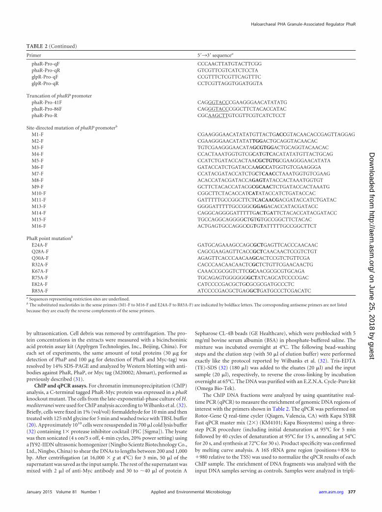
RESULTSIdentification of the AbrB-like protein PhaR and the promoterof the phaRP operon in H. mediterranei. We have previouslyreported a small protein GAP12 (110 amino acids [aa]), which waspresent in abundance on the PHA granule of H. mediterranei, withits coding gene cotranscribed with the phasin gene phaP (20). Thegap12-phaP operon is located upstream of the phaEC operon thatencodes the two subunits of PHA synthase (Fig. 1A). Conserveddomain search analysis at NCBI revealed that the C-terminal por-tion of GAP12 has a putative DNA-binding domain consisting ofswapped-hairpin barrel fold, which shows low homology (with anE value of 1.82e�05) to the corresponding domain of the AbrB(antibiotic resistance protein B) superfamily of regulators (34),implying a regulatory-related function of GAP12. Thus, theGAP12 was renamed PhaR, and the gap12-phaP operon was re-named the phaRP operon (Fig. 1A).
To explore the function of PhaR and its expression profiles, aGFP-based reporter system was constructed to conveniently mon-itor the activity of the phaRP promoter (PphaRP). First, the TSS ofthe phaRP cotranscript was analyzed using a CR-RT-PCR ap-proach. An adenine residue at 5 bp upstream of the initiator ATGcodon was determined as the TSS of phaRP, which revealed anextended 5=-untranslated region (5=-UTR) with the sequenceAGGAG (Fig. 1B). A 168-bp region (positions �151 to �17 rela-tive to the TSS of phaRP) joined with the gfp gene was used toconstruct the PphaRP-gfp-fused reporter plasmid pRF (Fig. 1B andFig. 2A). After plasmid pRF had been transferred into H. mediter-ranei DF50 (22), GFP could be visualized by fluorescence micros-copy (see Fig. S1 in the supplemental material). The activity of thePphaRP promoter was evaluated by quantifying the fluorescencesignal with a fluorescence microplate reader.
The promoter activities of the PphaRP were monitored duringcell growth. As PHA is actively accumulated in the cells in thestationary phase and the promoter activity of PphaRP is more stableat this phase, we primarily show or discuss the data obtained in thestationary phase. Remarkable fluorescence intensity (reachingvalues of over 10,000 RFU) was detected in the DF50/pRF strain,while the promoter of the phaEC operon only showed a weakersignal (up to approximately 500 RFU in the DF50/pEF strain) (Fig.1C). These results reveal that PphaRP exhibits a strong activity thatis consistent with the high abundance of the PhaP protein.
Modulation of phaRP expression and its promoter activityby PhaR and PHA accumulation. Since PhaR is associated withthe PHA granules, first the effect of PHA production on the phaRPexpression level was analyzed. The PHA-accumulating haloar-chaea possess a conserved pha gene cluster (phaR-phaP-phaE-phaC), including two operons, phaRP and phaEC, in H. mediter-
ranei (Fig. 1A) (20). The deletion of the PHA synthase operon(phaEC) makes cells incapable of synthesizing PHA (35). The pRFplasmid was transferred to three PHA-negative mutants, includ-ing the �phaEC, �phaPEC, and �phaRPEC strains, and the ex-pression levels of GFP were evaluated. The �phaEC and �phaPECmutants both showed an approximately 2-fold decrease in theactivity of the PphaRP promoter compared with that of the samepromoter in DF50 (Fig. 1D), implying that the expression ofphaRP could be activated by the presence of PHA. In contrast, thefurther deletion of the phaR gene in �phaPEC, which resulted inthe third PHA-negative mutant �phaRPEC strain, caused an in-crease of more than 4-fold in the activity of PphaRP (Fig. 1D). Theseresults suggest that when cells do not synthesize PHA, the expres-sion of phaRP is suppressed, whereas the knockout of the phaRgene could relieve this suppression effect. Thus, the PhaR could bea negative regulator of the phaRP operon.
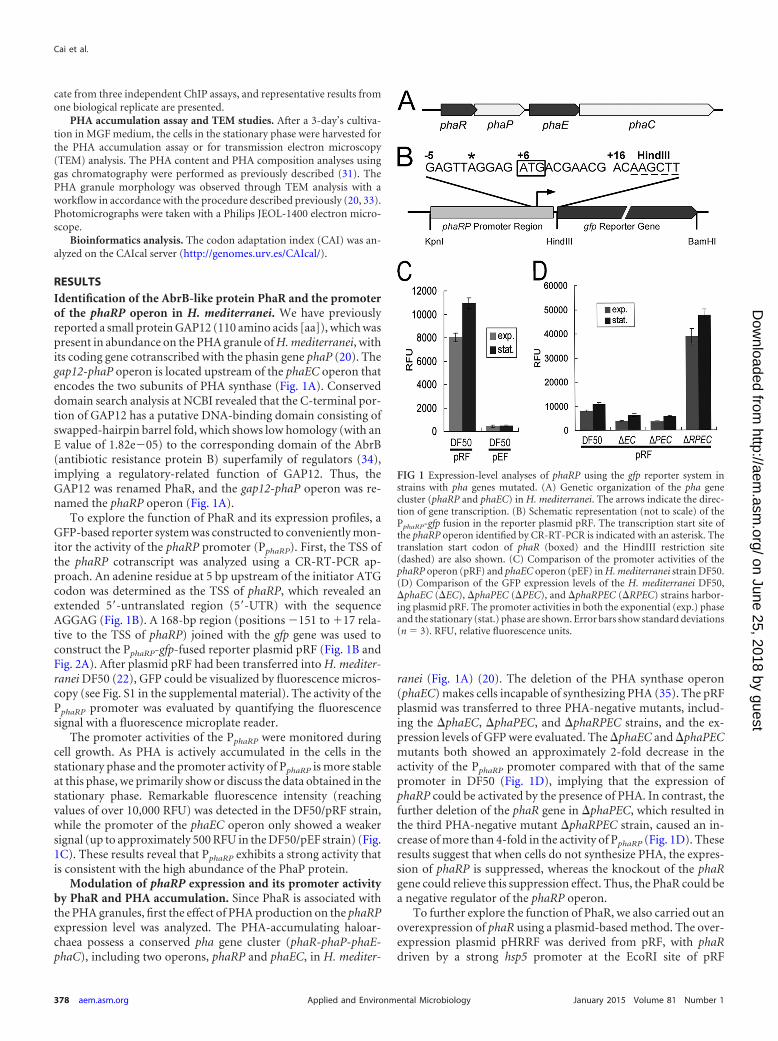
To further explore the function of PhaR, we also carried out anoverexpression of phaR using a plasmid-based method. The over-expression plasmid pHRRF was derived from pRF, with phaRdriven by a strong hsp5 promoter at the EcoRI site of pRF
FIG 1 Expression-level analyses of phaRP using the gfp reporter system instrains with pha genes mutated. (A) Genetic organization of the pha genecluster (phaRP and phaEC) in H. mediterranei. The arrows indicate the direc-tion of gene transcription. (B) Schematic representation (not to scale) of thePphaRP-gfp fusion in the reporter plasmid pRF. The transcription start site ofthe phaRP operon identified by CR-RT-PCR is indicated with an asterisk. Thetranslation start codon of phaR (boxed) and the HindIII restriction site(dashed) are also shown. (C) Comparison of the promoter activities of thephaRP operon (pRF) and phaEC operon (pEF) in H. mediterranei strain DF50.(D) Comparison of the GFP expression levels of the H. mediterranei DF50,�phaEC (�EC), �phaPEC (�PEC), and �phaRPEC (�RPEC) strains harbor-ing plasmid pRF. The promoter activities in both the exponential (exp.) phaseand the stationary (stat.) phase are shown. Error bars show standard deviations(n � 3). RFU, relative fluorescence units.
Cai et al.
378 aem.asm.org January 2015 Volume 81 Number 1Applied and Environmental Microbiology
on June 25, 2018 by guesthttp://aem
.asm.org/
Dow
nloaded from
(Fig. 2A). The plasmid pHRRF was transferred into the DF50strain to generate the DF50/pHRRF strain, and the influence ofPhaR on the expression of phaP was investigated. The enhancedexpression of phaR in DF50/pHRRF strain was revealed by West-ern blotting using anti-PhaR antibody, whereas the expressionlevel of the major phasin PhaP was analyzed with anti-PhaP anti-body (Fig. 2B). As shown in the Western blot results, overpro-duced PhaR in the cells strongly reduced the amount of PhaP (Fig.2B). Further Northern blot analysis with a phaP-specific probeshowed that in contrast to the high abundance of phaRP transcriptin the DF50/pRF strain, only a negligible amount of phaRP mRNAwas detected in the phaR overexpression strain DF50/pHRRF (Fig.2C). These data indicate that PhaR controls the amount of thePhaP protein by inhibiting the expression of phaRP at the tran-scriptional level. Concomitant with these observations, in theDF50/pHRRF strain, the fluorescence signal driven by the PphaRP
promoter was strongly decreased to be a faint signal (200 to 300
RFU) by the larger amount of the PhaR protein (Fig. 2D), con-firming the repression role of PhaR.
In conclusion, these results reveal that absence of PhaR couldenhance the expression of phaRP, while excess of PhaR could re-duce the expression, demonstrating that PhaR is a transcriptionalrepressor of both itself and phaP. In addition, the presence of PHAalso could active the expression of phaRP, indicating that a PhaRtitration effect of PHA granules plays an important role in the finemodulation of phaRP expression.
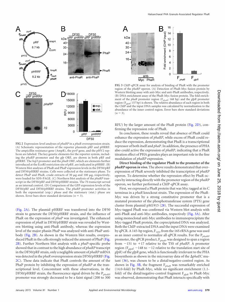
Direct binding of the regulator PhaR to the promoter of thephaRP operon in vivo. The above results demonstrated that over-expression of PhaR severely inhibited the transcription of phaRPoperon. To determine whether the repression effect by PhaR oc-curs by interacting directly with the promoter region of the phaRPoperon, we further performed a ChIP-qPCR assay.
First, we expressed a PhaR protein that was Myc tagged at its Cterminus in a phaR knockout strain. The expression of the PhaR-Myc was driven by a strong constitutive promoter that is amutated promoter of the phosphotransferase system (PTS) genecluster from plasmid pM1915 (28). The successful expression ofMyc-tagged PhaR was confirmed via Western blot analysis withanti-PhaR and anti-Myc antibodies, respectively (Fig. 3A). Afterusing monoclonal anti-Myc antibodies to immunoprecipitate theMyc-tagged PhaR protein, the coprecipitated DNA was analyzed.Both the ChIP-extracted DNA and the input DNA were examinedby qPCR. A 145-bp region, F16S, from the 16S rRNA gene was usedas an inner control to normalize all qPCR results. For the PphaRP
promoter, the qPCR product, FphaR, was designed to span a regionfrom �151 to �17 relative to the TSS of phaRP. A promoterregion (FglpR; �148 to �12 relative to the translation start site ofglpR) of the glpR gene, which is functionally irrelevant to the PHAbiosynthesis as shown in the microarray data of the �phaEC mu-tant (36), was chosen to be a distal/negative-control region. Asshown in Fig. 3B, the fragment FphaR was remarkably enriched(14.0-fold) by PhaR-Myc, while no significant enrichment (1.1-fold) of the distal/negative-control fragment FglpR to PhaR-Mycwas observed, demonstrating that PhaR interacts specifically with
FIG 2 Expression-level analyses of phaRP in a phaR overexpression strain.(A) Schematic representation of the reporter plasmids pRF and pHRRF.The ampicillin resistance gene (AmpR), the pyrF gene, and the pHV2 rep-licon are labeled. The key genetic elements for the reporter system, includ-ing the phaRP promoter and the gfp ORF, are shown in both pRF andpHRRF. The hsp5 promoter and the phaR ORF, which are elements furtherintroduced at the EcoRI restriction site of pRF, are indicated in pHRRF. (B)Western blot analyses of PhaR and PhaP expression levels in the DF50/pRFand DF50/pHRRF strains. Cells were collected at the stationary phase. Todetect PhaP and PhaR, crude extracts of 30 �g and 100 �g, respectively,were loaded for SDS-PAGE. (C) Northern blot analysis of the phaRP tran-script in the DF50/pRF and DF50/pHRRF strains. The 7S transcript servedas an internal control. (D) Comparison of the GFP expression levels of theDF50/pRF and DF50/pHRRF strains. The phaRP promoter activities inboth the exponential (exp.) phase and the stationary (stat.) phase areshown. Error bars show standard deviations (n � 3).
FIG 3 ChIP-qPCR assay for analysis of binding of PhaR with the promoterregion of the phaRP operon. (A) Detection of PhaR-Myc fusion protein byWestern blotting assay with anti-Myc and anti-PhaR antibodies, respectively.(B) DNA enrichment assay of the PhaR-Myc fusion protein. The fold enrich-ment of the phaR promoter region (FphaR; 168 bp) and the glpR promoterregion (FglpR; 137 bp) is shown. The relative abundance of each region in boththe ChIP and the input DNA samples was calculated by normalization to theabundance of the inner control region. Error bars show standard deviations(n � 3).
Haloarchaeal PHA Granule-Associated Regulator PhaR
January 2015 Volume 81 Number 1 aem.asm.org 379Applied and Environmental Microbiology
on June 25, 2018 by guesthttp://aem
.asm.org/
Dow
nloaded from
the phaRP promoter in vivo. These data indicated that PhaR reg-ulates phaRP expression directly through its interaction with thepromoter of the phaRP operon.
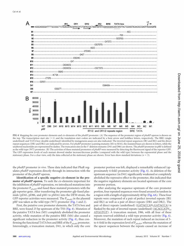
Identification of a specific negative cis-element in the pro-moter of phaRP operon. To seek the cis-elements important forthe inhibition of phaRP expression, we introduced mutations intothe promoter PphaRP and fused these mutated promoters with thegfp reporter gene. After transferring the promoter-gfp-fused plas-mids (pD41, pD86, and pM1 to pM16) into the DF50 strain, theGFP reporter activities were measured. The PphaRP in the plasmidpRF was taken as the wild-type (WT) promoter (Fig. 1 and 2).
First, the putative core promoter elements, the TATA box andBRE, were found 25 bp upstream of the TSS (Fig. 4). Mutation ofthe putative TATA box (M3) completely abolished the promoteractivity, while mutation of the putative BRE (M4) also caused asignificant reduction in the promoter activity (Fig. 4), thus con-firming the functional TATA box and BRE of the PphaRP promoter.Interestingly, a truncation mutant, D41, in which only the core
promoter portion was left, displayed a remarkably enhanced (ap-proximately 4-fold) promoter activity (Fig. 4). As deletion of theupstream sequence (in D41) significantly weakened or completelyabolished the repressive effect to the promoter, this indicated thatthe negative regulatory elements are located upstream of the corepromoter portion.
After analyzing the sequence upstream of the core promoterportion, four repeated sequences were found arrayed in tandem ina region with a length of approximately 40 bp (Fig. 4A). These fourrepeats were composed of a pair of perfect inverted repeats (IR1and IR2) as well as a pair of direct repeats (DR1 and DR2). Thepair of direct repeats (underlined) (GATACCAN3GATACCA) isflanked by the pair of inverted repeats (underlined) (ACACCATN23
ATGGTGT). A truncation mutant, D86, with all four of theserepeats reserved exhibited a wild-type promoter activity (Fig. 4).Moreover, the mutation of each repeat induced an increase of 3-to 4-fold in the expression level of PphaRP, whereas the mutation ofthe spacer sequences between the repeats caused an increase of
FIG 4 Mapping the core promoter elements and cis-elements of the phaRP promoter. (A) The sequence of the promoter region of phaRP operon is shown onthe top. The transcription start site (�1) and the translation start codon are indicated by a bent arrow and boldface letters, respectively. The BRE (singleunderlined) and TATA box (double underlined) identified by mutagenesis assays are also indicated. The inverted repeat sequences (IR1 and IR2) and the directrepeat sequences (DR1 and DR2) are indicated by arrows. For phaRP promoter scanning mutants (M1 to M16), the mutated bases are shown in letters, while theunaltered nucleotides are represented by dashes. The truncation sites in the 5= deletion mutants (D41 and D86) are shown. The phaRP promoter in pRF is definedas the wild-type (WT) promoter. (B) The activities of these mutated promoters of phaRP were measured by detecting the fluorescent signal of the reporter GFP.The GFP expression levels of each mutant showed similar increase/decrease profiles (compared with the wild type) between the exponential phase and thestationary phase. For a clear view, only the data collected at the stationary phase are shown. Error bars show standard deviations (n � 3).
Cai et al.
380 aem.asm.org January 2015 Volume 81 Number 1Applied and Environmental Microbiology
on June 25, 2018 by guesthttp://aem
.asm.org/
Dow
nloaded from
approximately 2-fold (Fig. 4). Other mutations introduced intothe sequence upstream of this region had almost no effect on thepromoter activity (Fig. 4). These results show that each of thesefour repeated sequences is essential for the repression regulationof phaRP, suggesting that the region containing these repeats(IR1/IR2 and DR1/DR2) is the negative cis-element and would bethe binding position of the transcriptional repressor.
Since PhaR was identified to be a transcriptional repressor ofthe phaRP operon and to interact directly with its promoter invivo, it is most likely that these four repeated sequences of thenegative cis-element serve as the binding sites of PhaR. To furthersupport this hypothesis, the reporter plasmids with mutations onthese four repeats, including pM5, pM7, pM9, pM11, and pD41,were transferred into the �phaRPEC strains, in which the PhaRwas absent, and the promoter activities were measured. As shownin Fig. 5, mutations of those repeats all resulted in high levels ofpromoter activity similar to that of the wild-type (WT) promoterin pRF. Small increases in the promoter activity were observed inthe �phaRPEC strains containing the plasmid pM5 or pD41,which might be caused by the alteration of the sequence of IR2 thatis adjacent to the BRE region. This might promote the recruitmentof TFB to BRE and thereby slightly enhanced the transcriptionalactivity. The mutations of the IR1/IR2 and DR1/DR2 sequencesdid not cause a significant further enhancement of the expressionof phaRP in the absence of PhaR, indicating that the four repeatedunits of the cis-element might be the binding sites of PhaR.
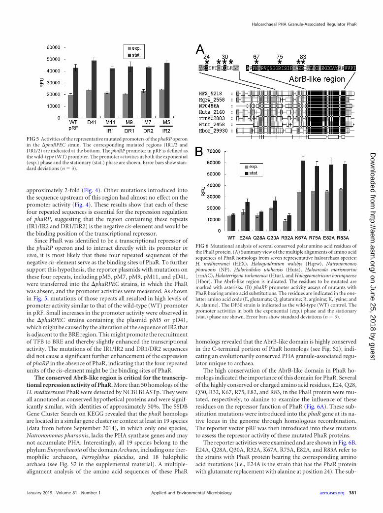
The conserved AbrB-like region is critical for the transcrip-tional repression activity of PhaR. More than 50 homologs of theH. mediterranei PhaR were detected by NCBI BLASTp. They wereall annotated as conserved hypothetical proteins and were signif-icantly similar, with identities of approximately 50%. The SSDBGene Cluster Search on KEGG revealed that the phaR homologsare located in a similar gene cluster or context at least in 19 species(data from before September 2014), in which only one species,Natronomonas pharaonis, lacks the PHA synthase genes and maynot accumulate PHA. Interestingly, all 19 species belong to thephylum Euryarchaeota of the domain Archaea, including one ther-mophilic archaeon, Ferroglobus placidus, and 18 halophilicarchaea (see Fig. S2 in the supplemental material). A multiple-alignment analysis of the amino acid sequences of these PhaR
homologs revealed that the AbrB-like domain is highly conservedin the C-terminal portion of PhaR homologs (see Fig. S2), indi-cating an evolutionarily conserved PHA granule-associated regu-lator unique to archaea.
The high conservation of the AbrB-like domain in PhaR ho-mologs indicated the importance of this domain for PhaR. Severalof the highly conserved or charged amino acid residues, E24, Q28,Q30, R32, K67, R75, E82, and R83, in the PhaR protein were mu-tated, respectively, to alanine to examine the influence of theseresidues on the repressor function of PhaR (Fig. 6A). These sub-stitution mutations were introduced into the phaR gene at its na-tive locus in the genome through homologous recombination.The reporter vector pRF was then introduced into these mutantsto assess the repressor activity of these mutated PhaR proteins.
The reporter activities were examined and are shown in Fig. 6B.E24A, Q28A, Q30A, R32A, K67A, R75A, E82A, and R83A refer tothe strains with PhaR protein bearing the corresponding aminoacid mutations (i.e., E24A is the strain that has the PhaR proteinwith glutamate replacement with alanine at position 24). The sub-
FIG 5 Activities of the representative mutated promoters of the phaRP operonin the �phaRPEC strain. The corresponding mutated regions (IR1/2 andDR1/2) are indicated at the bottom. The phaRP promoter in pRF is defined asthe wild-type (WT) promoter. The promoter activities in both the exponential(exp.) phase and the stationary (stat.) phase are shown. Error bars show stan-dard deviations (n � 3).
FIG 6 Mutational analysis of several conserved polar amino acid residues ofthe PhaR protein. (A) Summary view of the multiple alignments of amino acidsequences of PhaR homologs from seven representative haloarchaea species:H. mediterranei (HFX), Haloquadratum walsbyi (Hqrw), Natronomonaspharaonis (NP), Halorhabdus utahensis (Huta), Haloarcula marismortui(rrnAC), Haloterrigena turkmenica (Htur), and Halogeometricum borinquense(Hbor). The AbrB-like region is indicated. The residues to be mutated aremarked with asterisks. (B) phaRP promoter activity assays of mutants withPhaR bearing amino acid substitutions. The residues are indicated in the one-letter amino acid code (E, glutamate; Q, glutamine; R, arginine; K, lysine; andA, alanine). The DF50 strain is indicated as the wild-type (WT) control. Thepromoter activities in both the exponential (exp.) phase and the stationary(stat.) phase are shown. Error bars show standard deviations (n � 3).
Haloarchaeal PHA Granule-Associated Regulator PhaR
January 2015 Volume 81 Number 1 aem.asm.org 381Applied and Environmental Microbiology
on June 25, 2018 by guesthttp://aem
.asm.org/
Dow
nloaded from
stitutions of alanine for the glutamate, glutamine, and arginineresidues in the N-terminal portion of PhaR in the E24A, Q28A,Q30A, and R32A strains only slightly affected the PphaRP activity.In contrast, the K67A, R75A, E82A, and R83A mutants, in whichmutations occurred in the C-terminal conserved domain of PhaR,all showed a remarkable increase (2- to 4-fold) in PphaRP activity.These results revealed that PhaR mutants with amino acid substi-tution in the N-terminal portion still exhibited the capability tosuppress the corresponding promoter, whereas, the mutation ofthe AbrB-like domain of PhaR could significantly weaken or abol-ish the repressor function of PhaR. These results show that theAbrB-like domain is crucial for the repressor role of PhaR.
It is noteworthy that the expression of the mutated PhaR wasdriven by the native promoter; thus, the mutant strains mightpossess different quantities of the mutated PhaR proteins. How-ever, this would not affect our above conclusions on the crucialamino acids of the PhaR. For example, the K67A mutant, whichhad a high promoter activity of phaRP operon, would also pro-duce more PhaRK67A. Nevertheless the overproduced PhaRK67A
did not result in an enhanced repressive effect on the activity of thePphaRP in the reporter system, further indicating the weakenedrepressor effect of PhaRK67A.
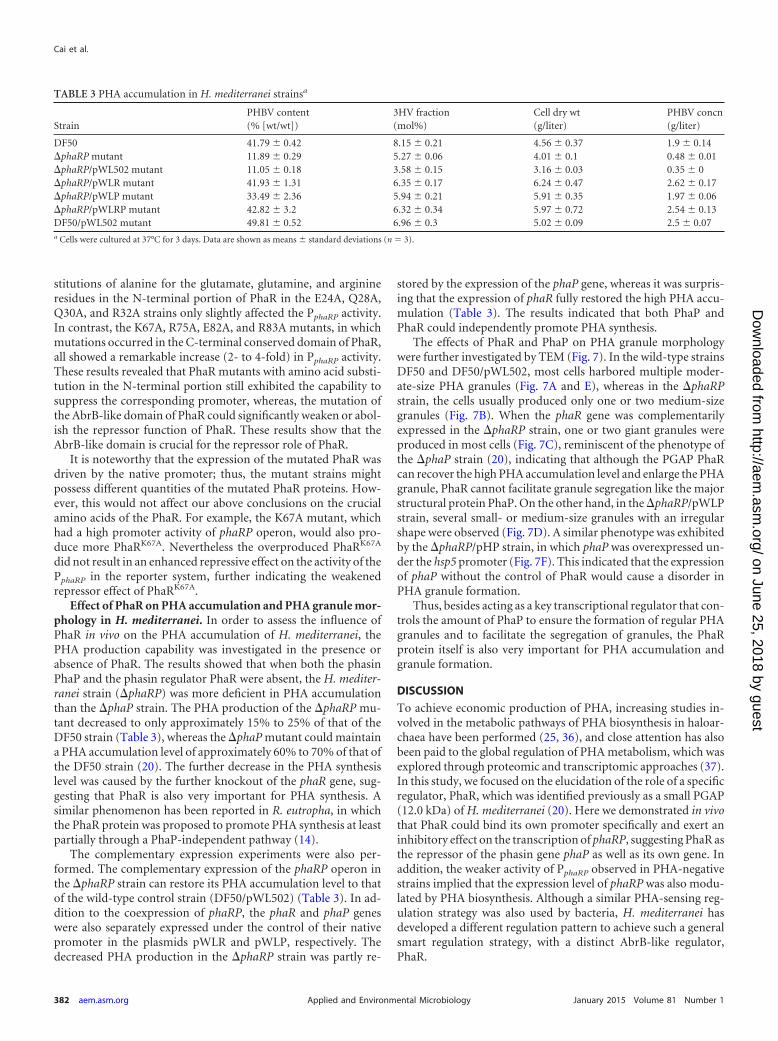
Effect of PhaR on PHA accumulation and PHA granule mor-phology in H. mediterranei. In order to assess the influence ofPhaR in vivo on the PHA accumulation of H. mediterranei, thePHA production capability was investigated in the presence orabsence of PhaR. The results showed that when both the phasinPhaP and the phasin regulator PhaR were absent, the H. mediter-ranei strain (�phaRP) was more deficient in PHA accumulationthan the �phaP strain. The PHA production of the �phaRP mu-tant decreased to only approximately 15% to 25% of that of theDF50 strain (Table 3), whereas the �phaP mutant could maintaina PHA accumulation level of approximately 60% to 70% of that ofthe DF50 strain (20). The further decrease in the PHA synthesislevel was caused by the further knockout of the phaR gene, sug-gesting that PhaR is also very important for PHA synthesis. Asimilar phenomenon has been reported in R. eutropha, in whichthe PhaR protein was proposed to promote PHA synthesis at leastpartially through a PhaP-independent pathway (14).
The complementary expression experiments were also per-formed. The complementary expression of the phaRP operon inthe �phaRP strain can restore its PHA accumulation level to thatof the wild-type control strain (DF50/pWL502) (Table 3). In ad-dition to the coexpression of phaRP, the phaR and phaP geneswere also separately expressed under the control of their nativepromoter in the plasmids pWLR and pWLP, respectively. Thedecreased PHA production in the �phaRP strain was partly re-
stored by the expression of the phaP gene, whereas it was surpris-ing that the expression of phaR fully restored the high PHA accu-mulation (Table 3). The results indicated that both PhaP andPhaR could independently promote PHA synthesis.
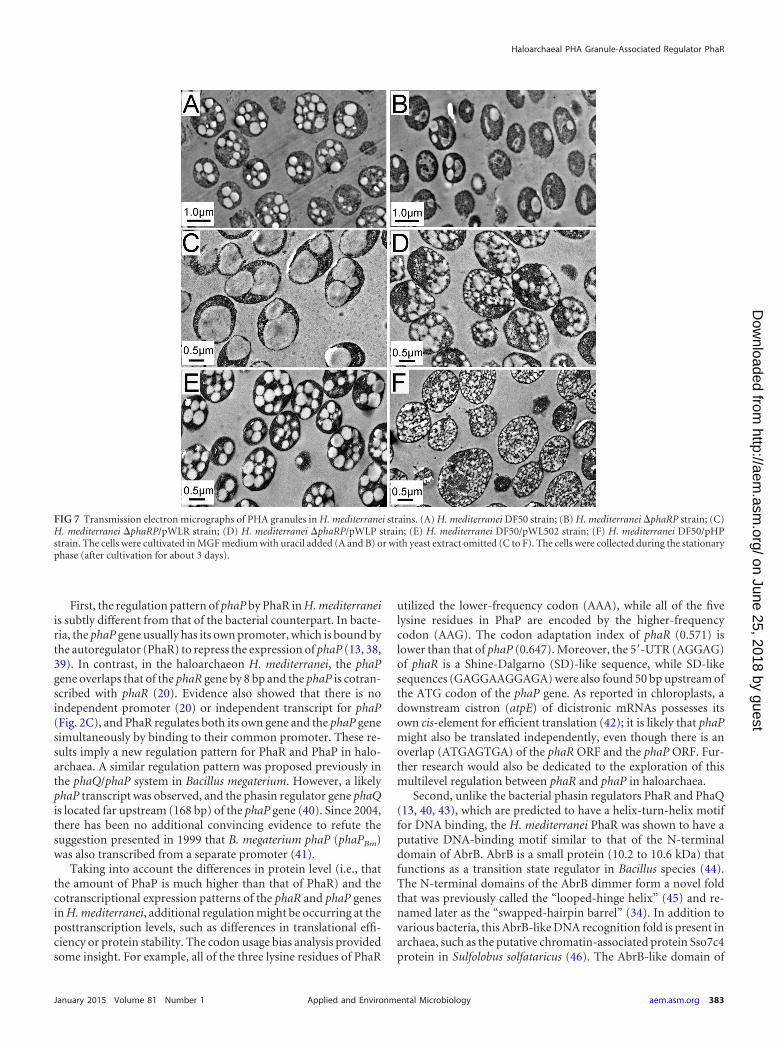
The effects of PhaR and PhaP on PHA granule morphologywere further investigated by TEM (Fig. 7). In the wild-type strainsDF50 and DF50/pWL502, most cells harbored multiple moder-ate-size PHA granules (Fig. 7A and E), whereas in the �phaRPstrain, the cells usually produced only one or two medium-sizegranules (Fig. 7B). When the phaR gene was complementarilyexpressed in the �phaRP strain, one or two giant granules wereproduced in most cells (Fig. 7C), reminiscent of the phenotype ofthe �phaP strain (20), indicating that although the PGAP PhaRcan recover the high PHA accumulation level and enlarge the PHAgranule, PhaR cannot facilitate granule segregation like the majorstructural protein PhaP. On the other hand, in the �phaRP/pWLPstrain, several small- or medium-size granules with an irregularshape were observed (Fig. 7D). A similar phenotype was exhibitedby the �phaRP/pHP strain, in which phaP was overexpressed un-der the hsp5 promoter (Fig. 7F). This indicated that the expressionof phaP without the control of PhaR would cause a disorder inPHA granule formation.
Thus, besides acting as a key transcriptional regulator that con-trols the amount of PhaP to ensure the formation of regular PHAgranules and to facilitate the segregation of granules, the PhaRprotein itself is also very important for PHA accumulation andgranule formation.
DISCUSSION
To achieve economic production of PHA, increasing studies in-volved in the metabolic pathways of PHA biosynthesis in haloar-chaea have been performed (25, 36), and close attention has alsobeen paid to the global regulation of PHA metabolism, which wasexplored through proteomic and transcriptomic approaches (37).In this study, we focused on the elucidation of the role of a specificregulator, PhaR, which was identified previously as a small PGAP(12.0 kDa) of H. mediterranei (20). Here we demonstrated in vivothat PhaR could bind its own promoter specifically and exert aninhibitory effect on the transcription of phaRP, suggesting PhaR asthe repressor of the phasin gene phaP as well as its own gene. Inaddition, the weaker activity of PphaRP observed in PHA-negativestrains implied that the expression level of phaRP was also modu-lated by PHA biosynthesis. Although a similar PHA-sensing reg-ulation strategy was also used by bacteria, H. mediterranei hasdeveloped a different regulation pattern to achieve such a generalsmart regulation strategy, with a distinct AbrB-like regulator,PhaR.
TABLE 3 PHA accumulation in H. mediterranei strainsa
StrainPHBV content(% [wt/wt])
3HV fraction(mol%)
Cell dry wt(g/liter)
PHBV concn(g/liter)
DF50 41.79 0.42 8.15 0.21 4.56 0.37 1.9 0.14�phaRP mutant 11.89 0.29 5.27 0.06 4.01 0.1 0.48 0.01�phaRP/pWL502 mutant 11.05 0.18 3.58 0.15 3.16 0.03 0.35 0�phaRP/pWLR mutant 41.93 1.31 6.35 0.17 6.24 0.47 2.62 0.17�phaRP/pWLP mutant 33.49 2.36 5.94 0.21 5.91 0.35 1.97 0.06�phaRP/pWLRP mutant 42.82 3.2 6.32 0.34 5.97 0.72 2.54 0.13DF50/pWL502 mutant 49.81 0.52 6.96 0.3 5.02 0.09 2.5 0.07a Cells were cultured at 37°C for 3 days. Data are shown as means standard deviations (n � 3).
Cai et al.
382 aem.asm.org January 2015 Volume 81 Number 1Applied and Environmental Microbiology
on June 25, 2018 by guesthttp://aem
.asm.org/
Dow
nloaded from
First, the regulation pattern of phaP by PhaR in H. mediterraneiis subtly different from that of the bacterial counterpart. In bacte-ria, the phaP gene usually has its own promoter, which is bound bythe autoregulator (PhaR) to repress the expression of phaP (13, 38,39). In contrast, in the haloarchaeon H. mediterranei, the phaPgene overlaps that of the phaR gene by 8 bp and the phaP is cotran-scribed with phaR (20). Evidence also showed that there is noindependent promoter (20) or independent transcript for phaP(Fig. 2C), and PhaR regulates both its own gene and the phaP genesimultaneously by binding to their common promoter. These re-sults imply a new regulation pattern for PhaR and PhaP in halo-archaea. A similar regulation pattern was proposed previously inthe phaQ/phaP system in Bacillus megaterium. However, a likelyphaP transcript was observed, and the phasin regulator gene phaQis located far upstream (168 bp) of the phaP gene (40). Since 2004,there has been no additional convincing evidence to refute thesuggestion presented in 1999 that B. megaterium phaP (phaPBm)was also transcribed from a separate promoter (41).
Taking into account the differences in protein level (i.e., thatthe amount of PhaP is much higher than that of PhaR) and thecotranscriptional expression patterns of the phaR and phaP genesin H. mediterranei, additional regulation might be occurring at theposttranscription levels, such as differences in translational effi-ciency or protein stability. The codon usage bias analysis providedsome insight. For example, all of the three lysine residues of PhaR
utilized the lower-frequency codon (AAA), while all of the fivelysine residues in PhaP are encoded by the higher-frequencycodon (AAG). The codon adaptation index of phaR (0.571) islower than that of phaP (0.647). Moreover, the 5=-UTR (AGGAG)of phaR is a Shine-Dalgarno (SD)-like sequence, while SD-likesequences (GAGGAAGGAGA) were also found 50 bp upstream ofthe ATG codon of the phaP gene. As reported in chloroplasts, adownstream cistron (atpE) of dicistronic mRNAs possesses itsown cis-element for efficient translation (42); it is likely that phaPmight also be translated independently, even though there is anoverlap (ATGAGTGA) of the phaR ORF and the phaP ORF. Fur-ther research would also be dedicated to the exploration of thismultilevel regulation between phaR and phaP in haloarchaea.
Second, unlike the bacterial phasin regulators PhaR and PhaQ(13, 40, 43), which are predicted to have a helix-turn-helix motiffor DNA binding, the H. mediterranei PhaR was shown to have aputative DNA-binding motif similar to that of the N-terminaldomain of AbrB. AbrB is a small protein (10.2 to 10.6 kDa) thatfunctions as a transition state regulator in Bacillus species (44).The N-terminal domains of the AbrB dimmer form a novel foldthat was previously called the “looped-hinge helix” (45) and re-named later as the “swapped-hairpin barrel” (34). In addition tovarious bacteria, this AbrB-like DNA recognition fold is present inarchaea, such as the putative chromatin-associated protein Sso7c4protein in Sulfolobus solfataricus (46). The AbrB-like domain of
FIG 7 Transmission electron micrographs of PHA granules in H. mediterranei strains. (A) H. mediterranei DF50 strain; (B) H. mediterranei �phaRP strain; (C)H. mediterranei �phaRP/pWLR strain; (D) H. mediterranei �phaRP/pWLP strain; (E) H. mediterranei DF50/pWL502 strain; (F) H. mediterranei DF50/pHPstrain. The cells were cultivated in MGF medium with uracil added (A and B) or with yeast extract omitted (C to F). The cells were collected during the stationaryphase (after cultivation for about 3 days).
Haloarchaeal PHA Granule-Associated Regulator PhaR
January 2015 Volume 81 Number 1 aem.asm.org 383Applied and Environmental Microbiology
on June 25, 2018 by guesthttp://aem
.asm.org/
Dow
nloaded from
H. mediterranei PhaR is highly conserved in its archaeal homologs.Several charged residues in this domain, including the positivelycharged Lys67, Arg75, and Arg83 residues, were identified to becrucial, which indicated that these positively charged residuesmight contribute to DNA binding by interacting with the nega-tively charged DNA and demonstrated a critical involvement ofthe AbrB-like domain in the negative regulatory role of the PhaRin H. mediterranei. AbrB and AbrB-like proteins usually functionas dimers or tetramers (44, 47). In bacteria, PhaR was also shownto form a tetramer in vitro (43). In the promoter region of thephaRP operon, we identified a specific negative cis-element com-posed of four repeat sequences in tandem (Fig. 4), which are verylikely the binding sites of H. mediterranei PhaR. In combinationwith the oligomer character of bacterial PhaR and AbrB, it is im-plied that the AbrB-like protein PhaR might also function as anoligomer, such as a dimer or a tetramer, to bind the four repeatDNA sequences. Therefore, although they have different DNA-binding motifs, both the haloarchaeal and bacterial PhaRs mayregulate the phasin genes by a similar dose-dependent mecha-nism.
Notably, in addition to the regulation mechanism, the centralrole of PhaR in PHA accumulation and granule formation in H.mediterranei was further addressed in this study. When both of thetwo major PGAPs PhaR and PhaP were absent, there would be adeficiency in the protection layer between the PHA granule andthe cytoplasm, and therefore the cells of the �phaRP strain couldonly accumulate a small amount of PHA (Table 3 and Fig. 7B).When only phaR was expressed in the �phaRP strain, PHA accu-mulation returned to a wild-type level, and the cells synthesizedgranules larger than those of the �phaRP strain (Table 3 and Fig.7C). It is speculated that PhaR might facilitate PHA synthesis in aPhaP-independent mechanism, such as by promoting PHA syn-thase activity or by acting as the major protein to form a boundaryto protect both the PHA and the cytoplasmic protein from unspe-cific binding. The reason why the �phaRP/pWLR strain had ahigher PHA accumulation level than the �phaP strain might bethe larger amount of PhaR proteins produced by the increasedphaR gene copy number, as the complementary expression wascarried out through a plasmid-based method that increased thegene copy number. When the repressor PhaR was absent, the tran-scriptional repression effect was released. The sole expression ofphaP under its native promoter would produce excess PhaP pro-tein. The disordered PHA granule morphology displayed in the�phaRP/pWLP strain and the DF50/pHP strain (Fig. 7D and F)indicates that the proper amount of PhaP is critical to the forma-tion of regular PHA granules and that it is important to keep theexpression of phaP under the control of PhaR. Therefore, H. medi-terranei PhaR, which is essential for the control of the expressionof phaP, plays a very important role in maintaining proper granuleformation.
In summary, our results reveal a novel phasin regulator, PhaR,with a novel regulation pattern in haloarchaea. We demonstratedthat in addition to acting as a phasin regulator to control PHAgranule morphology, H. mediterranei PhaR can also facilitate PHAbiosynthesis in a PhaP-independent manner. It is noteworthy thatmutation of the promoter of the phaRP operon has also generatedseveral very strong promoters that would have potential applica-tion in genetic engineering in haloarchaea. Therefore, this studyhas provided not only new insights into the regulation of PHAsynthesis and granule formation in H. mediterranei but also the
tools and targets for the further exploration and engineering ofPHA metabolism in haloarchaea.
ACKNOWLEDGMENTS
We thank Jingnan Liang (Institute of Microbiology, Chinese Academy ofSciences) for technical assistance in the transmission electron microscopyexperiments.
This work was supported by grants 31330001 and 31370096 from theNational Natural Science Foundation of China.
REFERENCES1. Lee SY. 1996. Bacterial polyhydroxyalkanoates. Biotechnol Bioeng 49:1–
14. http://dx.doi.org/10.1002/(SICI)1097-0290(19960105)49:11::AID-BIT1�3.3.CO;2-1.
2. Steinbüchel A, Füchtenbusch B. 1998. Bacterial and other biologicalsystems for polyester production. Trends Biotechnol 16:419 – 427. http://dx.doi.org/10.1016/S0167-7799(98)01194-9.
3. Fernandez-Castillo R, Rodriguez-Valera F, Gonzalez-Ramos J, Ruiz-Berraquero F. 1986. Accumulation of poly(beta-hydroxybutyrate) by ha-lobacteria. Appl Environ Microbiol 51:214 –216.
4. Han J, Hou J, Liu H, Cai S, Feng B, Zhou J, Xiang H. 2010. Widedistribution among halophilic archaea of a novel polyhydroxyalkano-ate synthase subtype with homology to bacterial type III synthases.Appl Environ Microbiol 76:7811–7819. http://dx.doi.org/10.1128/AEM.01117-10.
5. Legat A, Gruber C, Zangger K, Wanner G, Stan-Lotter H. 2010. Iden-tification of polyhydroxyalkanoates in Halococcus and other haloarchaealspecies. Appl Microbiol Biotechnol 87:1119 –1127. http://dx.doi.org/10.1007/s00253-010-2611-6.
6. Anderson AJ, Dawes EA. 1990. Occurrence, metabolism, metabolic role,and industrial uses of bacterial polyhydroxyalkanoates. Microbiol Rev 54:450 – 472.
7. Griebel R, Smith Z, Merrick JM. 1968. Metabolism of poly-beta-hydroxybutyrate. I. Purification, composition, and properties of nativepoly-beta-hydroxybutyrate granules from Bacillus megaterium. Biochem-istry 7:3676 –3681.
8. Jendrossek D, Pfeiffer D. 2014. New insights in formation of polyhy-droxyalkanoate (PHA) granules (carbonosomes) and novel functions ofpoly(3-hydroxybutyrate) (PHB). Environ Microbiol 16:2357–2373. http://dx.doi.org/10.1111/1462-2920.12356.
9. Pötter M, Steinbüchel A. 2005. Poly(3-hydroxybutyrate) granule-associated proteins: impacts on poly(3-hydroxybutyrate) synthesis anddegradation. Biomacromolecules 6:552–560. http://dx.doi.org/10.1021/bm049401n.
10. Pfeiffer D, Wahl A, Jendrossek D. 2011. Identification of a multifunc-tional protein, PhaM, that determines number, surface to volume ratio,subcellular localization and distribution to daughter cells of poly(3-hydroxybutyrate), PHB, granules in Ralstonia eutropha H16. Mol Micro-biol 82:936 –951. http://dx.doi.org/10.1111/j.1365-2958.2011.07869.x.
11. Galán B, Dinjaski N, Maestro B, de Eugenio LI, Escapa IF, Sanz JM,García JL, Prieto MA. 2011. Nucleoid-associated PhaF phasin drivesintracellular location and segregation of polyhydroxyalkanoate granulesin Pseudomonas putida KT2442. Mol Microbiol 79:402– 418. http://dx.doi.org/10.1111/j.1365-2958.2010.07450.x.
12. Rehm BH. 2006. Genetics and biochemistry of polyhydroxyalkanoategranule self-assembly: the key role of polyester synthases. Biotechnol Lett28:207–213. http://dx.doi.org/10.1007/s10529-005-5521-4.
13. Pötter M, Madkour MH, Mayer F, Steinbüchel A. 2002. Regulation ofphasin expression and polyhydroxyalkanoate (PHA) granule formation inRalstonia eutropha H16. Microbiology 148:2413–2426.
14. York GM, Stubbe J, Sinskey AJ. 2002. The Ralstonia eutropha PhaRprotein couples synthesis of the PhaP phasin to the presence of polyhy-droxybutyrate in cells and promotes polyhydroxybutyrate production. JBacteriol 184:59 – 66. http://dx.doi.org/10.1128/JB.184.1.59-66.2002.
15. Tian JM, He AM, Lawrence AG, Liu PH, Watson N, Sinskey AJ, StubbeJ. 2005. Analysis of transient polyhydroxybutyrate production in Wauter-sia eutropha H16 by quantitative Western analysis and transmission elec-tron microscopy. J Bacteriol 187:3825–3832. http://dx.doi.org/10.1128/JB.187.11.3825-3832.2005.
16. Yamada M, Yamashita K, Wakuda A, Ichimura K, Maehara A, MaedaM, Taguchi S. 2007. Autoregulator protein PhaR for biosynthesis of poly-
Cai et al.
384 aem.asm.org January 2015 Volume 81 Number 1Applied and Environmental Microbiology
on June 25, 2018 by guesthttp://aem
.asm.org/
Dow
nloaded from
hydroxybutyrate [P(3HB)] possibly has two separate domains that bind tothe target DNA and P(3HB): functional mapping of amino acid residuesresponsible for DNA binding. J Bacteriol 189:1118 –1127. http://dx.doi.org/10.1128/JB.01550-06.
17. Pötter M, Müller H, Steinbüchel A. 2005. Influence of homologousphasins (PhaP) on PHA accumulation and regulation of their expressionby the transcriptional repressor PhaR in Ralstonia eutropha H16. Micro-biology 151:825– 833. http://dx.doi.org/10.1099/mic.0.27613-0.
18. Zhao D, Cai L, Wu J, Li M, Liu H, Han J, Zhou J, Xiang H. 2013.Improving polyhydroxyalkanoate production by knocking out the genesinvolved in exopolysaccharide biosynthesis in Haloferax mediterranei.Appl Microbiol Biotechnol 97:3027–3036. http://dx.doi.org/10.1007/s00253-012-4415-3.
19. Koller M, Hesse P, Bona R, Kutschera C, Atlic A, Braunegg G. 2007.Potential of various archae- and eubacterial strains as industrial polyhy-droxyalkanoate producers from whey. Macromol Biosci 7:218 –226. http://dx.doi.org/10.1002/mabi.200600211.
20. Cai S, Cai L, Liu H, Liu X, Han J, Zhou J, Xiang H. 2012. Identifi-cation of the haloarchaeal phasin (PhaP) that functions in polyhy-droxyalkanoate accumulation and granule formation in Haloferaxmediterranei. Appl Environ Microbiol 78:1946 –1952. http://dx.doi.org/10.1128/AEM.07114-11.
21. Sambrook J, Russell DW. 2001. Molecular cloning: a laboratory manual,3rd ed. Cold Spring Harbor Laboratory Press, Cold Spring Harbor, NY.
22. Liu H, Han J, Liu X, Zhou J, Xiang H. 2011. Development of pyrF-basedgene knockout systems for genome-wide manipulation of the archaea Ha-loferax mediterranei and Haloarcula hispanica. J Genet Genomics 38:261–269. http://dx.doi.org/10.1016/j.jgg.2011.05.003.
23. Krebs MP, Mollaaghababa R, Khorana HG. 1993. Gene replacement inHalobacterium halobium and expression of bacteriorhodopsin mutants.Proc Natl Acad Sci U S A 90:1987–1991. http://dx.doi.org/10.1073/pnas.90.5.1987.
24. Reuter CJ, Maupin-Furlow JA. 2004. Analysis of proteasome-dependentproteolysis in Haloferax volcanii cells, using short-lived green fluorescentproteins. Appl Environ Microbiol 70:7530 –7538. http://dx.doi.org/10.1128/AEM.70.12.7530-7538.2004.
25. Hou J, Feng B, Han J, Liu HL, Zhao DH, Zhou J, Xiang H. 2013.Haloarchaea l- type beta-ketothio lases involved in poly(3-hydroxybutyrate-co-3-hydroxyvalerate) synthesis in Haloferax mediterra-nei. Appl Environ Microbiol 79:5104 –5111. http://dx.doi.org/10.1128/AEM.01370-13.
26. Miao D, Sun C, Xiang H. 2009. Construction and application of a novelshuttle expression vector based on haloarchaeal plasmid pSCM201. WeiSheng Wu Xue Bao 49:1040 –1047. (In Chinese.)
27. Cline SW, Lam WL, Charlebois RL, Schalkwyk LC, Doolittle WF. 1989.Transformation methods for halophilic archaebacteria. Can J Microbiol35:148 –152. http://dx.doi.org/10.1139/m89-022.
28. Cai L, Cai SF, Zhao DH, Wu JH, Wang L, Liu XQ, Li M, Hou J, ZhouJ, Liu JF, Han J, Xiang H. 2014. Analysis of the transcriptional regulatorGlpR, promoter elements, and posttranscriptional processing involved infructose-induced activation of the phosphoenolpyruvate-dependentsugar phosphotransferase system in Haloferax mediterranei. Appl EnvironMicrobiol 80:1430 –1440. http://dx.doi.org/10.1128/AEM.03372-13.
29. Lu Q, Han J, Zhou L, Coker JA, DasSarma P, DasSarma S, Xiang H.2008. Dissection of the regulatory mechanism of a heat-shock responsivepromoter in haloarchaea: a new paradigm for general transcription factordirected archaeal gene regulation. Nucleic Acids Res 36:3031–3042. http://dx.doi.org/10.1093/nar/gkn152.
30. Kuhn J, Binder S. 2002. RT-PCR analysis of 5= to 3=-end-ligated mRNAsidentifies the extremities of cox2 transcripts in pea mitochondria. NucleicAcids Res 30:439 – 446. http://dx.doi.org/10.1093/nar/30.2.439.
31. Han J, Lu Q, Zhou L, Zhou J, Xiang H. 2007. Molecular characterizationof the phaECHm genes, required for biosynthesis of poly(3-hydroxybutyrate) in the extremely halophilic archaeon Haloarcula maris-mortui. Appl Environ Microbiol 73:6058 – 6065. http://dx.doi.org/10.1128/AEM.00953-07.
32. Wilbanks EG, Larsen DJ, Neches RY, Yao AI, Wu CY, Kjolby RAS,
Facciotti MT. 2012. A workflow for genome-wide mapping of archaealtranscription factors with ChIP-seq. Nucleic Acids Res 40:e74. http://dx.doi.org/10.1093/nar/gks063.
33. Tian J, Sinskey AJ, Stubbe J. 2005. Kinetic studies of polyhydroxybu-tyrate granule formation in Wautersia eutropha H16 by transmission elec-tron microscopy. J Bacteriol 187:3814 –3824. http://dx.doi.org/10.1128/JB.187.11.3814-3824.2005.
34. Coles M, Djuranovic S, Söding J, Frickey T, Koretke K, Truffault V,Martin J, Lupas AN. 2005. AbrB-like transcription factors assume aswapped hairpin fold that is evolutionarily related to double-psi beta bar-rels. Structure 13:919 –928. http://dx.doi.org/10.1016/j.str.2005.03.017.
35. Lu QH, Han J, Zhou LG, Zhou J, Xiang H. 2008. Genetic and biochem-ical character izat ion of the poly(3-hydroxybutyrate-co-3-hydroxyvalerate) synthase in Haloferax mediterranei. J Bacteriol 190:4173– 4180. http://dx.doi.org/10.1128/JB.00134-08.
36. Han J, Hou J, Zhang F, Ai GM, Li M, Cai SF, Liu HL, Wang L, WangZJ, Zhang SL, Cai L, Zhao DH, Zhou J, Xiang H. 2013. Multiplepropionyl coenzyme A-supplying pathways for production of the bioplas-tic poly(3-hydroxybutyrate-co-3-hydroxyvalerate) in Haloferax mediter-ranei. Appl Environ Microbiol 79:2922–2931. http://dx.doi.org/10.1128/AEM.03915-12.
37. Liu H, Luo Y, Han J, Wu J, Wu Z, Feng D, Cai S, Li M, Liu J, Zhou J,Xiang H. 2013. Proteome reference map of Haloarcula hispanica andcomparative proteomic and transcriptomic analysis of polyhydroxyal-kanoate biosynthesis under genetic and environmental perturbations. JProteome Res 12:1300 –1315. http://dx.doi.org/10.1021/pr300969m.
38. Maehara A, Taguchi S, Nishiyama T, Yamane T, Doi Y. 2002. Arepressor protein, PhaR, regulates polyhydroxyalkanoate (PHA) synthesisvia its direct interaction with PHA. J Bacteriol 184:3992– 4002. http://dx.doi.org/10.1128/JB.184.14.3992-4002.2002.
39. Chou ME, Yang MK. 2010. Analyses of binding sequences of the PhaRprotein of Rhodobacter sphaeroides FJ1. FEMS Microbiol Lett 302:138 –143. http://dx.doi.org/10.1111/j.1574-6968.2009.01836.x.
40. Lee TR, Lin JS, Wang SS, Shaw GC. 2004. PhaQ, a new class of poly-beta-hydroxybutyrate (PHB)-responsive repressor, regulates phaQ andphaP (phasin) expression in Bacillus megaterium through interaction withPHB. J Bacteriol 186:3015–3021. http://dx.doi.org/10.1128/JB.186.10.3015-3021.2004.
41. McCool GJ, Cannon MC. 1999. Polyhydroxyalkanoate inclusion body-associated proteins and coding region in Bacillus megaterium. J Bacteriol181:585–592.
42. Suzuki H, Kuroda H, Yukawa Y, Sugiura M. 2011. The downstream atpEcistron is efficiently translated via its own cis-element in partially overlap-ping atpB-atpE dicistronic mRNAs in chloroplasts. Nucleic Acids Res 39:9405–9412. http://dx.doi.org/10.1093/nar/gkr644.
43. Maehara A, Doi Y, Nishiyama T, Takagi Y, Ueda S, Nakano H, YamaneT. 2001. PhaR, a protein of unknown function conserved among short-chain-length polyhydroxyalkanoic acids producing bacteria, is a DNA-binding protein and represses Paracoccus denitrificans phaP expression invitro. FEMS Microbiol Lett 200:9-15. http://dx.doi.org/10.1111/j.1574-6968.2001.tb10685.x.
44. Chumsakul O, Takahashi H, Oshima T, Hishimoto T, Kanaya S,Ogasawara N, Ishikawa S. 2011. Genome-wide binding profiles of theBacillus subtilis transition state regulator AbrB and its homolog Abh re-veals their interactive role in transcriptional regulation. Nucleic Acids Res39:414 – 428. http://dx.doi.org/10.1093/nar/gkq780.
45. Vaughn JL, Feher V, Naylor S, Strauch MA, Cavanagh J. 2000. NovelDNA binding domain and genetic regulation model of Bacillus subtilistransition state regulator abrB. Nat Struct Biol 7:1139 –1146. http://dx.doi.org/10.1038/81999.
46. Hsu CH, Wang AH. 2011. The DNA-recognition fold of Sso7c4 suggestsa new member of SpoVT-AbrB superfamily from archaea. Nucleic AcidsRes 39:6764 – 6774. http://dx.doi.org/10.1093/nar/gkr283.
47. Yao F, Strauch MA. 2005. Independent and interchangeable multim-erization domains of the AbrB, Abh, and SpoVT global regulatory pro-teins. J Bacteriol 187:6354 – 6362. http://dx.doi.org/10.1128/JB.187.18.6354-6362.2005.
Haloarchaeal PHA Granule-Associated Regulator PhaR
January 2015 Volume 81 Number 1 aem.asm.org 385Applied and Environmental Microbiology
on June 25, 2018 by guesthttp://aem
.asm.org/
Dow
nloaded from