a novel dna repair disorder with thrombocytopenia...
TRANSCRIPT

A Novel DNA Repair Disorder With Thrombocytopenia,
Nephrosis, and Features Overlapping Cockayne Syndrome
Article (Published Version)
http://sro.sussex.ac.uk
Forsythe, Elizabeth, Wild, Ruth, Sellick, Gabrielle, Houlston, Richard S., Lehmann, Alan R and Wakeling, Emma (2009) A Novel DNA Repair Disorder With Thrombocytopenia, Nephrosis, and Features Overlapping Cockayne Syndrome. American Journal of Medical Genetics Part A, 149A (10). pp. 2075-2079. ISSN 1552-4825
This version is available from Sussex Research Online: http://sro.sussex.ac.uk/23101/
This document is made available in accordance with publisher policies and may differ from the published version or from the version of record. If you wish to cite this item you are advised to consult the publisher’s version. Please see the URL above for details on accessing the published version.
Copyright and reuse: Sussex Research Online is a digital repository of the research output of the University.
Copyright and all moral rights to the version of the paper presented here belong to the individual author(s) and/or other copyright owners. To the extent reasonable and practicable, the material made available in SRO has been checked for eligibility before being made available.
Copies of full text items generally can be reproduced, displayed or performed and given to third parties in any format or medium for personal research or study, educational, or not-for-profit purposes without prior permission or charge, provided that the authors, title and full bibliographic details are credited, a hyperlink and/or URL is given for the original metadata page and the content is not changed in any way.

NEW SYNDROME
A Novel DNA Repair Disorder With Thrombocytopenia,Nephrosis, and Features OverlappingCockayne SyndromeElizabeth Forsythe,1 Ruth Wild,2 Gabrielle Sellick,2 Richard S. Houlston,2 Alan R. Lehmann,3
and Emma Wakeling1*1Kennedy Galton Centre, North West Thames Regional Genetics Service, The North West London Hospitals NHS Trust, Middlesex, United Kingdom
2Section of Cancer Genetics, Institute of Cancer Research, Sutton, Surrey, United Kingdom
3Genome Damage and Stability Centre, University of Sussex, Falmer, Brighton, United Kingdom
Received 22 September 2008; Accepted 20 May 2009
We report on four siblings with Cockayne-like syndrome with
thrombocytopenia and nephrotic syndrome. The parents were
healthy and consanguineous, consistent with an autosomal
recessive mode of disease inheritance. UV irradiation of fibro-
blasts revealed an intermediate sensitivity between normal and
standard Cockayne syndrome (CS) control cells. A genome-wide
linkage scan conducted using Affymetrix 10K arrays provided
exclusion of the known CS genes in the family, and evidence that
the disease genemaps to 1p33-p31.1. Thrombocytopenia has not
previously been linked with CS, but two patients with CS in
association with nephrotic syndrome have previously been
documented and the phenotypes are compared with the patients
described here. We suggest that this Cockayne-like phenotype
with thrombocytopenia and nephrotic syndromemay be a novel
DNA repair disorder, and propose that further investigation of
other affected families may help identify the causative genetic
defect. � 2009 Wiley-Liss, Inc.
Key words: autosomal recessive inheritance; Cockayne syn-
drome; DNA repair disorders; linkage analysis; nephrotic syn-
drome; thrombocytopenia; UV irradiation
INTRODUCTION
Cockayne syndrome (CS) is a rare autosomal recessive disorder
with variable expression. The phenotype classically becomes
evident in early childhood, and patients present with hyperpig-mentation, sunken eyes secondary to subcutaneous lipoatrophy,
failure to thrive, short stature and microcephaly. Neurological
sequelae include progressive ataxia, global developmental delay,
sensorineural deafness and tremors [Cockayne, 1936; Nance and
Berry, 1992; Lehmann et al., 1993; Tan et al., 2005].While approxi-
mately 10% of patients with CS develop renal pathology, this rarely
leads to serious complications [Hirooka et al., 1988; Sato et al., 1988;
Reiss et al., 1996]. Renal changes may vary significantly betweenaffected individuals but typically include tubulointerstitial inflam-
mation, interstitial fibrosis and tubular atrophy [Hirooka et al.,
1988; Funaki et al., 1996Q1]—all consistentwithnonspecific end-stage
renal disease. A small simplified glomerular structure with wrin-
kling of the basementmembrane appears to be specific toCockayne
Syndrome [Funaki et al., 1996]. Thrombocytopenia has not, to our
knowledge, been reported previously in association with CS.
A diagnosis of CS is made on clinical features and can be
confirmed by UV irradiation of fibroblasts from the patient. CScells fail to restore normal levels of RNA synthesis following UV
irradiation when compared to control fibroblasts [Mayne and
Lehmann, 1982]. Mutations in genes CSA and CSB, coding for
proteins involved in transcription-coupled repair, are thought to be
responsible for the Cockayne phenotype in the majority of cases
[Lehmann, 2003]. There is poor genotype/ phenotype correlation
[Mallery et al., 1998], and identical mutations in CSB may be
associatedwith either CS or xerodermapigmentosum (XP) - anotherDNA repair disorder [Lehmann, 2003]. Mutations in genes XPB,
XPD, and XPG can result in a mixed clinical phenotype with
neurological features of CS and skin abnormalities associated with
XP [Lehmann, 2003].
*Correspondence to:
Emma Wakeling, Kennedy Galton Centre, North West Thames Regional
Genetics Service, The North West London Hospitals NHS Trust,
Middlesex, United Kingdom. E-mail: [email protected]
Published online 00 Month 2009 in Wiley InterScience
(www.interscience.wiley.com)
DOI 10.1002/ajmg.a.32995
How to Cite this Article:Forsythe E, Wild R, Sellick G, Houlston RS,LehmannAR,WakelingE. 2009.AnovelDNA
repair disorder with thrombocytopenia,
nephrosis and features overlapping Cockayne
syndrome.
Am J Med Genet Part A 9999:1–5.
� 2009 Wiley-Liss, Inc. 1AJMA080679:R3ð32995Þ

Here we report on four siblings affected by a Cockayne-likedisorder associated with nephrotic syndrome and thrombocytope-
nia, and an intermediate fibroblast response to UV irradiation.
CLINICAL REPORT
The parents of the four affected children are healthy, Pakistani first
cousins (Fig. 1). The two older siblings (III.1 and III.2) were first
referred for genetic evaluation aged 6 and 5, respectively. They
were noted to have a distinctive phenotype with severe global
developmental delay, short stature and similar dysmorphic fea-
tures: progeroid facies with microcephaly, frontal bossing, sunken
eyes, highnasal bridge and large, prominent, low-set ears. Therewasno history of photosensitivity, poor vision or deafness.
Patient 1The eldest sibling (III.1, Fig. 1) weighed 2.8 kg (9th centile) at birth.
At 6 years, OFC was 47.5 cm, height 93 cm, and weight 13.2 kg; allwell below the 0.4th centile. In addition to the dysmorphic features
he shared with his sister, he also had keratinized, purple, nodular
lesions on his forehead, the exact nature of which could not be
identified. Development was slowly progressive with no loss of
skills. At presentation, aged 6, he was able to walk up stairs with two
feet per step, draw a circle, had goodpincer grip andhad a few single
recognizable words. Previous diagnostic investigations had been
inconclusive. He was noted to have a normal karyotype. Comput-erized tomography (CT) of his brain at the age of 2 revealed
‘‘moderately prominent ventricles and sulci’’ but was otherwise
normal with no evidence of basal ganglia calcification. Urine amino
acids were normal and congenital infection screenwas negative. He
had further investigations at the age of 7, during admission to
hospital for steroid-unresponsive nephrotic syndrome. A renal
biopsy identified glomerular sclerosis, tubular atrophy, interstitial
fibrosis, and hyaline thickening of the arteriolar wall, consistentwith the non-specific end-stage renal pathology typically observed
in CS. The patient was thrombocytopenic on admission (plateletcount: 50� 109/L) and had a history of easy bruising. A bone
marrow biopsy showed hypocellular particulate bonemarrow with
representation of all three hematopoietic precursor cell lines in-
cluding erythrocytes, leucocytes, and thrombocytes. A blood smear
was normal except for a few spherocytes, consistent with mild
hemolysis. The hematological findings were reviewed and felt to be
consistent with a hereditary thrombocytopenia. Normal sensitivity
to diepoxybutane excluded Fanconi anemia. Generalized osteopo-rosis and delayed bone age was apparent on skeletal survey.
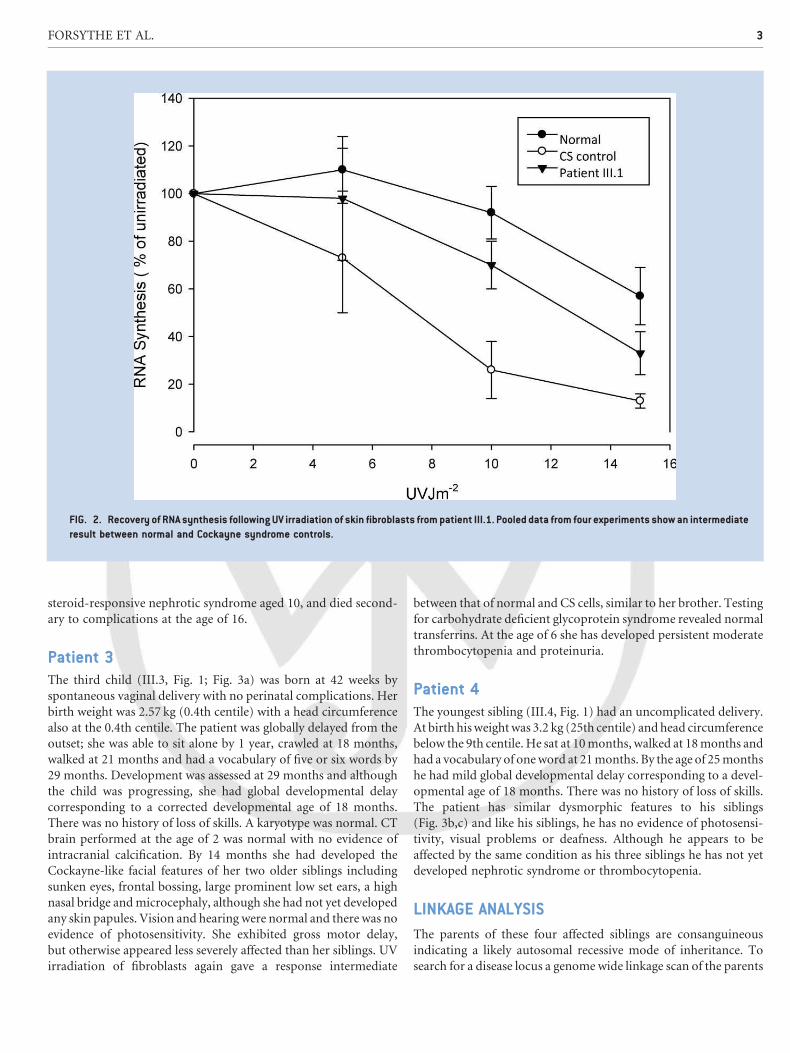
Recovery of RNA synthesis following UV irradiation of skin
fibroblasts gave an intermediate result between normal and known
CS controls (Fig. 2). At the age of 8 he died secondary to compli-
cations of nephrotic syndrome.
Patient 2The second child (III.2, Fig. 1) was born at 41 weeks by normal
vaginal delivery,weighing 3.1 kg (50th centile). Theneonatal period
was complicated by hypotonia, and she required 2 days admission
to the Special Care Baby Unit. At presentation aged 5, OFC was
44.5 cm and length 87 cm. At age 7 all growth parameters werebelow the 0.4th centile. Like her brother, this patient had slowly
progressive global developmental delay with no loss of skills. She
walked at 21 months, and by age 7 she had severe developmental
delay particularly affecting speech with only a few single words. A
skeletal survey revealeddelayedbone agewithnon-specific changes.
Borderline growth hormone response was noted, and the patient
was started on replacement therapy at the age of 9. She subsequently
developed nodular lesions on her skin similar to those of her elderbrother andpersistent thrombocytopenia.Normal sister chromatid
exchanges for Bloom syndrome, diepoxybutane sensitivity and
chromosomal ionizing radiation excluded Bloom syndrome,
ataxia telangiectasia and Fanconi anemia, respectively. Attempts
to establish a fibroblasts culture for UV sensitivity testing were
unsuccessful, andparents declined a second attempt. She developed
FIG. 1. Pedigree showing the four affected siblings (III.1, III.2, III.3, III.4) as filled black symbols. No other familymembers are known to be affected and
are represented asunfilled symbols. Circles denote females and squares denotemales. Individuals of unknowngender are represented bya rhombus.
2 AMERICAN JOURNAL OF MEDICAL GENETICS PART A
steroid-responsive nephrotic syndrome aged 10, and died second-
ary to complications at the age of 16.
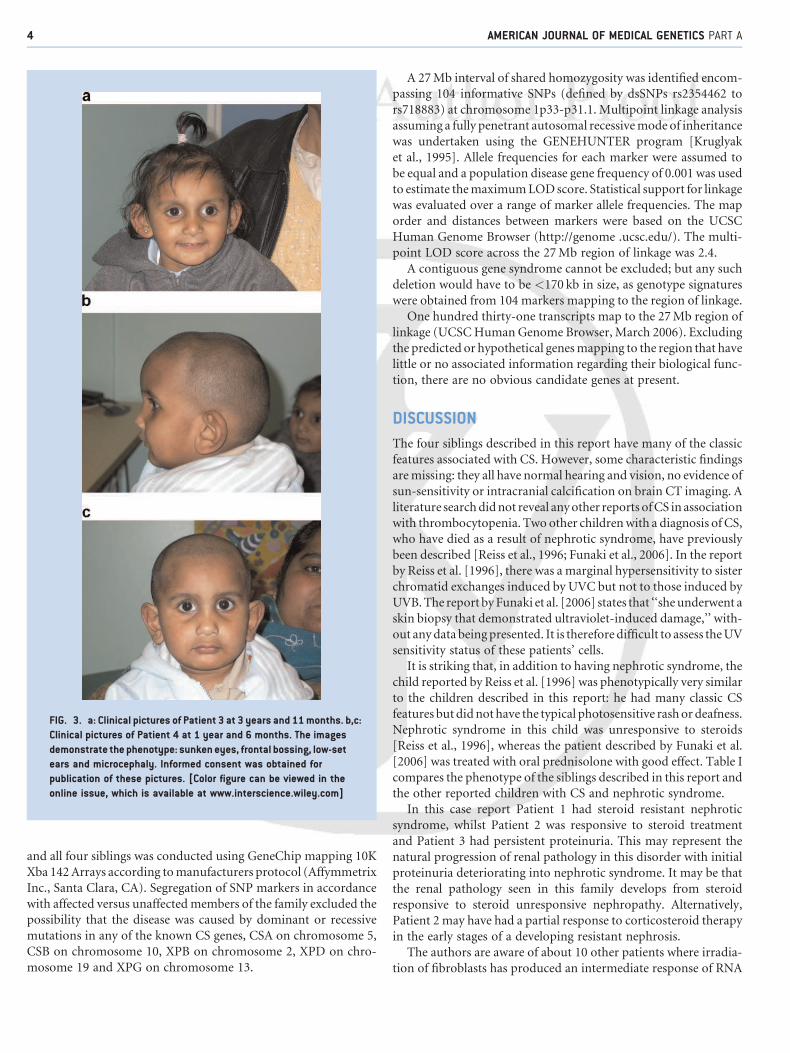
Patient 3The third child (III.3, Fig. 1; Fig. 3a) was born at 42 weeks by
spontaneous vaginal delivery with no perinatal complications. Her
birth weight was 2.57 kg (0.4th centile) with a head circumference
also at the 0.4th centile. The patient was globally delayed from theoutset; she was able to sit alone by 1 year, crawled at 18 months,
walked at 21 months and had a vocabulary of five or six words by
29 months. Development was assessed at 29 months and although
the child was progressing, she had global developmental delay
corresponding to a corrected developmental age of 18 months.
There was no history of loss of skills. A karyotype was normal. CT
brain performed at the age of 2 was normal with no evidence of
intracranial calcification. By 14 months she had developed theCockayne-like facial features of her two older siblings including
sunken eyes, frontal bossing, large prominent low set ears, a high
nasal bridge andmicrocephaly, although she had not yet developed
any skin papules. Vision and hearing were normal and there was no
evidence of photosensitivity. She exhibited gross motor delay,
but otherwise appeared less severely affected than her siblings. UV
irradiation of fibroblasts again gave a response intermediate
between that of normal and CS cells, similar to her brother. Testing
for carbohydrate deficient glycoprotein syndrome revealed normal
transferrins. At the age of 6 she has developed persistent moderate
thrombocytopenia and proteinuria.
Patient 4The youngest sibling (III.4, Fig. 1) had an uncomplicated delivery.
At birth hisweightwas 3.2 kg (25th centile) andhead circumferencebelow the 9th centile. He sat at 10months, walked at 18months and
had a vocabulary of oneword at 21months. By the age of 25months
he had mild global developmental delay corresponding to a devel-
opmental age of 18 months. There was no history of loss of skills.
The patient has similar dysmorphic features to his siblings
(Fig. 3b,c) and like his siblings, he has no evidence of photosensi-
tivity, visual problems or deafness. Although he appears to be
affected by the same condition as his three siblings he has not yetdeveloped nephrotic syndrome or thrombocytopenia.
LINKAGE ANALYSIS
The parents of these four affected siblings are consanguineous
indicating a likely autosomal recessive mode of inheritance. To
search for a disease locus a genome wide linkage scan of the parents
FIG. 2. Recovery of RNA synthesis following UV irradiation of skin fibroblasts frompatient III.1. Pooled data from four experiments showan intermediate
result between normal and Cockayne syndrome controls.
FORSYTHE ET AL. 3
and all four siblings was conducted using GeneChip mapping 10K
Xba 142Arrays according tomanufacturers protocol (Affymmetrix
Inc., Santa Clara, CA). Segregation of SNP markers in accordance
with affected versus unaffectedmembers of the family excluded thepossibility that the disease was caused by dominant or recessive
mutations in any of the known CS genes, CSA on chromosome 5,
CSB on chromosome 10, XPB on chromosome 2, XPD on chro-
mosome 19 and XPG on chromosome 13.
A 27Mb interval of shared homozygosity was identified encom-passing 104 informative SNPs (defined by dsSNPs rs2354462 to
rs718883) at chromosome 1p33-p31.1. Multipoint linkage analysis
assuming a fully penetrant autosomal recessivemode of inheritance
was undertaken using the GENEHUNTER program [Kruglyak
et al., 1995]. Allele frequencies for each marker were assumed to
be equal and a population disease gene frequency of 0.001 was used
to estimate themaximumLODscore. Statistical support for linkage
was evaluated over a range of marker allele frequencies. The maporder and distances between markers were based on the UCSC
Human Genome Browser (http://genome .ucsc.edu/). The multi-
point LOD score across the 27Mb region of linkage was 2.4.
A contiguous gene syndrome cannot be excluded; but any such
deletion would have to be <170 kb in size, as genotype signatures
were obtained from 104 markers mapping to the region of linkage.
One hundred thirty-one transcripts map to the 27Mb region of
linkage (UCSCHuman Genome Browser, March 2006). Excludingthe predicted or hypothetical genesmapping to the region that have
little or no associated information regarding their biological func-
tion, there are no obvious candidate genes at present.
DISCUSSION
The four siblings described in this report have many of the classic
features associated with CS. However, some characteristic findings
aremissing: they all have normal hearing and vision, no evidence of
sun-sensitivity or intracranial calcification on brain CT imaging. A
literature searchdidnot reveal anyother reports ofCS in association
with thrombocytopenia. Two other childrenwith a diagnosis of CS,who have died as a result of nephrotic syndrome, have previously
been described [Reiss et al., 1996; Funaki et al., 2006]. In the report
by Reiss et al. [1996], there was a marginal hypersensitivity to sister
chromatid exchanges induced by UVC but not to those induced by
UVB.The report byFunaki et al. [2006] states that ‘‘sheunderwent a
skin biopsy that demonstrated ultraviolet-induced damage,’’ with-
out anydata beingpresented. It is therefore difficult to assess theUV
sensitivity status of these patients’ cells.It is striking that, in addition to having nephrotic syndrome, the
child reported by Reiss et al. [1996] was phenotypically very similar
to the children described in this report: he had many classic CS
features butdidnothave the typical photosensitive rashor deafness.
Nephrotic syndrome in this child was unresponsive to steroids
[Reiss et al., 1996], whereas the patient described by Funaki et al.
[2006] was treated with oral prednisolone with good effect. Table I
compares the phenotype of the siblings described in this report andthe other reported children with CS and nephrotic syndrome.
In this case report Patient 1 had steroid resistant nephrotic
syndrome, whilst Patient 2 was responsive to steroid treatment
and Patient 3 had persistent proteinuria. This may represent the
natural progression of renal pathology in this disorder with initial
proteinuria deteriorating into nephrotic syndrome. It may be that
the renal pathology seen in this family develops from steroid
responsive to steroid unresponsive nephropathy. Alternatively,Patient 2 may have had a partial response to corticosteroid therapy
in the early stages of a developing resistant nephrosis.
The authors are aware of about 10 other patients where irradia-
tion of fibroblasts has produced an intermediate response of RNA
FIG. 3. a: Clinical pictures of Patient 3 at 3 years and 11months. b,c:
Clinical pictures of Patient 4 at 1 year and 6 months. The images
demonstrate the phenotype: sunkeneyes, frontal bossing, low-set
ears and microcephaly. Informed consent was obtained for
publication of these pictures. [Color figure can be viewed in the
online issue, which is available at www.interscience.wiley.com]
4 AMERICAN JOURNAL OF MEDICAL GENETICS PART A

synthesis recovery following UV irradiation (A.R. Lehmann, un-
published work). Limited information exists regarding phenotype
and results ofmolecular diagnostic testing in these patients. At least
three of the patients appear to have a clear Cockayne-like pheno-type. However, none are known to have developed nephrotic
syndrome.
Collectively these data suggest that the CS-like phenotype we
have identified in four affected individuals from a single family
represents adistinctive syndrome, possibly a consequenceof anovel
DNA repair disorder. As the parents of the patients we report were
consanguineous, autosomal recessive inheritance is most likely,
though other modes of transmission, including mitochondrialinheritance, remain a possibility. On the basis of linkage data it
appears that the disease genemaps to 1p. Investigation of additional
affected families should, as well as further elucidate this emerging
phenotype, facilitate identification of the causal gene.
ACKNOWLEDGMENTS
We are grateful to the family for their participation and support of
this article.
REFERENCES
Cockayne EA. 1936. Dwarfismwith retinal atrophy and deafness. Arch DisChild 11:1–8.
Funaki S, Takahashi S, Murakami H, Harada K, Tikamura H. 2006.Cockayne syndrome with recurrent acute tubulointerstitial nephritis.Pathol Int 56:678–682.
Hirooka M, Hirota M, Kamada M. 1988. Renal lesions in Cockaynesyndrome. Pediatr Nephrol 2:239–243.
Kruglyak L, DalyMJ, Lander ES. 1995. Rapidmultipoint linkage analysis ofrecessive traits innuclear families, including homozygositymapping.AmJ Hum Genet 56:519–527.
Lehmann AR. 2003. DNA repair-deficient diseases, xeroderma pigmento-sum, Cockayne syndrome and trichothiodystrophy. Biochimie85:1101–1111.
Lehmann AR, Thompson AF, Harcourt SA, Stefanini M, Norris PG. 1993.Cockayne’s syndrome: Correlation of clinical features with cellularsensitivity of RNA synthesis to UV irradiation. J Med Genet 30:679–682.
Mallery DL, Tanganelli B, Colella S, Steingrimsdottir H, van Gool AJ,Troelstra C, Stefanini M, Lehmann AR. 1998. Molecular analysis ofmutations in the CSB (ERCC6) gene in patients with Cockayne syn-drome. Am J Hum Genet 62:77–85.
Mayne LV, Lehmann AR. 1982. Failure of RNA synthesis to recoverafter UV irradiation: An early defect in cells from individuals withCockayne’s syndrome and xeroderma pigmentosum. Cancer Res42:1473–1478.
NanceMA, Berry SA. 1992. Cockayne syndrome: Review of 140 cases. Am JMed Genet 42:68–84.
Reiss U, Hofweber K, Herterich R,Waldherr R, Bohnert E, Jung E, ScharerK. 1996. Nephrotic syndrome, hypertension, and adrenal failure inatypical Cockayne Syndrome. Pediatr Nephrol 10:602–605.
Sato H, Saito T, Kurosawa K, Ootaka T, Furuyama T, YoshinagaK. 1988. Renal lesions in Cockayne’s syndrome. Clin Nephrol 29:206–220.
Tan WH, Baris H, Robson CD, Kimonis VE. 2005. Cockayne syndrome:The developing phenotype. Am J Med Genet Part A 135A:214–216.
Q1: Please add in the reference list.
TABLE I. Phenotype Comparison Between Classic CS, Children Described in the Literature With CS and Nephrotic Syndrome, and the Patients
Described in This Report
Cockaynesyndrome
Patientreportedby Reiss
et al. [1996]
Patientreportedby Funaki
et al. [2006] III.1 III.2 III.3 III.4Developmental delay þ þ þ þ þ þ þHyperpigmentation þ þ þ þ þ � �Loss of subcutaneoustissue around the eye
þ þ þ þ þ þ þ
Microcephaly þ þ þ þ þ þ þSmall stature þ þ þ þ þ þ þPeripheral neuropathy þ � þ � � � �Optic abnormalities þ � þ � � � �Deafness þ � � � � � �Nephrotic syndrome � þ þ þ þ þ �Sun-sensitive rash þ � þ Late onset Late onset � �Response to UV irradiation Confirms Possible increase
in sister chromaticexchange
Ambiguous IntermediateRNA repair
Not done IntermediateRNA repair
Notdone
Age at death Mean 12.3 (5) 6 12 7 16 Alive Alive
FORSYTHE ET AL. 5

111 R IV ER STREET, H O BO KEN , N J 07030
***IMMEDIATE RESPONSE REQUIRED***
Your article will be published online via Wiley's EarlyView® service (www.interscience.wiley.com) shortly after receipt of
corrections. EarlyView® is Wiley's online publication of individual articles in full text HTML and/or pdf format before release of
the compiled print issue of the journal. Articles posted online in EarlyView® are peer-reviewed, copyedited, author corrected,
and fully citable. EarlyView® means you benefit from the best of two worlds--fast online availability as well as traditional, issue-
based archiving.
READ PROOFS CAREFULLY
• This will be your only chance to review these proofs.
• Please note that the volume and page numbers shown on the proofs are for position only.
ANSWER ALL QUERIES ON PROOFS (Queries for you to answer are attached as the last page of your proof.)
• Mark all corrections directly on the proofs. Note that excessive author alterations may ultimately result in delay of
publication and extra costs may be charged to you.
CHECK FIGURES AND TABLES CAREFULLY (Color figures will be sent under separate cover.)
• Check size, numbering, and orientation of figures.
• All images in the PDF are downsampled (reduced to lower resolution and file size) to facilitate Internet delivery.
These images will appear at higher resolution and sharpness in the printed article.
• Review figure legends to ensure that they are complete.
• Check all tables. Review layout, title, and footnotes.
COMPLETE REPRINT ORDER FORM
• Fill out the attached reprint order form. It is important to return the form even if you are not ordering reprints. You
may, if you wish, pay for the reprints with a credit card. Reprints will be mailed only after your article appears in
print. This is the most opportune time to order reprints. If you wait until after your article comes off press, the
reprints will be considerably more expensive.
RETURN PROOFS
REPRINT ORDER FORM
CTA (If you have not already signed one)
RETURN IMMEDIATELY AS YOUR ARTICLE WILL BE POSTED IN ORDER OF RECEIPT.
QUESTIONS? Christopher Sannella, Production Editor
Phone: 201-748-5949E-mail: [email protected] to journal acronym and article production number
(i.e., AJMA 00-0001 for American Journal of Medical Genetics ms 00-0001).
AJMA, e-proof:
C1/Wiley-Liss
A. COPYRIGHT
1. The Contributor assigns to Wiley-Blackwell, during the full term of copy-
right and any extensions or renewals, all copyright in and to the Contribution,
and all rights therein, including but not limited to the right to publish, repub-
lish, transmit, sell, distribute and otherwise use the Contribution in whole or in
part in electronic and print editions of the Journal and in derivative works
throughout the world, in all languages and in all media of expression now
known or later developed, and to license or permit others to do so.
2. Reproduction, posting, transmission or other distribution or use of the final
Contribution in whole or in part in any medium by the Contributor as permit-
ted by this Agreement requires a citation to the Journal and an appropriate
credit to Wiley-Blackwell as Publisher, and/or the Society if applicable, suitable
in form and content as follows: (Title of Article, Author, Journal Title and
Volume/Issue, Copyright © [year], copyright owner as specified in the Journal).
Links to the final article on Wiley-Blackwell’s website are encouraged where
appropriate.
B. RETAINED RIGHTS
Notwithstanding the above, the Contributor or, if applicable, the Contributor’s
Employer, retains all proprietary rights other than copyright, such as patent
rights, in any process, procedure or article of manufacture described in the
Contribution.
C. PERMITTED USES BY CONTRIBUTOR
1. Submitted Version. Wiley-Blackwell licenses back the following rights to
the Contributor in the version of the Contribution as originally submitted for
publication:
a. After publication of the final article, the right to self-archive on the Con-
tributor’s personal website or in the Contributor’s institution’s/employer’s
institutional repository or archive. This right extends to both intranets and
the Internet. The Contributor may not update the submission version or
replace it with the published Contribution. The version posted must contain
a legend as follows: This is the pre-peer reviewed version of the following
article: FULL CITE, which has been published in final form at [Link to final
article].
b. The right to transmit, print and share copies with colleagues.
2. Accepted Version. Re-use of the accepted and peer-reviewed (but not
final) version of the Contribution shall be by separate agreement with Wiley-
Blackwell. Wiley-Blackwell has agreements with certain funding agencies
governing reuse of this version. The details of those relationships, and other
offerings allowing open web use, are set forth at the following website:
http://www.wiley.com/go/funderstatement. NIH grantees should check the
box at the bottom of this document.
3. Final Published Version. Wiley-Blackwell hereby licenses back to the
Contributor the following rights with respect to the final published version of
the Contribution:
a. Copies for colleagues. The personal right of the Contributor only to send
or transmit individual copies of the final published version in any format to
colleagues upon their specific request provided no fee is charged, and
further-provided that there is no systematic distribution of the Contribu-
tion, e.g. posting on a listserve, website or automated delivery.
b. Re-use in other publications. The right to re-use the final Contribution or
parts thereof for any publication authored or edited by the Contributor
(excluding journal articles) where such re-used material constitutes less
than half of the total material in such publication. In such case, any modifi-
cations should be accurately noted.
c. Teaching duties. The right to include the Contribution in teaching or
training duties at the Contributor’s institution/place of employment includ-
ing in course packs, e-reserves, presentation at professional conferences,
in-house training, or distance learning. The Contribution may not be used
in seminars outside of normal teaching obligations (e.g. commercial semi-
nars). Electronic posting of the final published version in connection with
teaching/training at the Contributor’s institution/place of employment is
permitted subject to the implementation of reasonable access control
mechanisms, such as user name and password. Posting the final published
version on the open Internet is not permitted.
d. Oral presentations. The right to make oral presentations based on the
Contribution.
4. Article Abstracts, Figures, Tables, Data Sets, Artwork and Selected
Text (up to 250 words).
a. Contributors may re-use unmodified abstracts for any non-commercial
purpose. For on-line uses of the abstracts, Wiley-Blackwell encourages but
does not require linking back to the final published versions.
b. Contributors may re-use figures, tables, data sets, artwork, and selected
text up to 250 words from their Contributions, provided the following
conditions are met:
(i) Full and accurate credit must be given to the Contribution.
(ii) Modifications to the figures, tables and data must be noted.
Otherwise, no changes may be made.
(iii) The reuse may not be made for direct commercial purposes, or for
financial consideration to the Contributor.
(iv) Nothing herein shall permit dual publication in violation of journal
ethical practices.
COPYRIGHT TRANSFER AGREEMENT
Date: Contributor name:
Contributor address:
Manuscript number (Editorial office only):
Re: Manuscript entitled
(the “Contribution”)
for publication in (the “Journal”)
published by (“Wiley-Blackwell”).
Dear Contributor(s):
Thank you for submitting your Contribution for publication. In order to expedite the editing and publishing process and enable Wiley-Blackwell to
disseminate your Contribution to the fullest extent, we need to have this Copyright Transfer Agreement signed and returned as directed in the Journal’s
instructions for authors as soon as possible. If the Contribution is not accepted for publication, or if the Contribution is subsequently rejected, this
Agreement shall be null and void. Publication cannot proceed without a signed copy of this Agreement.
CTA-A
D. CONTRIBUTIONS OWNED BY EMPLOYER
1. If the Contribution was written by the Contributor in the course of the
Contributor’s employment (as a “work-made-for-hire” in the course of
employment), the Contribution is owned by the company/employer which
must sign this Agreement (in addition to the Contributor’s signature) in the
space provided below. In such case, the company/employer hereby assigns to
Wiley-Blackwell, during the full term of copyright, all copyright in and to the
Contribution for the full term of copyright throughout the world as specified in
paragraph A above.
2. In addition to the rights specified as retained in paragraph B above and the
rights granted back to the Contributor pursuant to paragraph C above, Wiley-
Blackwell hereby grants back, without charge, to such company/employer, its
subsidiaries and divisions, the right to make copies of and distribute the final
published Contribution internally in print format or electronically on the Com-
pany’s internal network. Copies so used may not be resold or distributed externally.
However the company/employer may include information and text from the
Contribution as part of an information package included with software or
other products offered for sale or license or included in patent applications.
Posting of the final published Contribution by the institution on a public access
website may only be done with Wiley-Blackwell’s written permission, and payment
of any applicable fee(s). Also, upon payment of Wiley-Blackwell’s reprint fee,
the institution may distribute print copies of the published Contribution externally.
E. GOVERNMENT CONTRACTS
In the case of a Contribution prepared under U.S. Government contract or
grant, the U.S. Government may reproduce, without charge, all or portions of
the Contribution and may authorize others to do so, for official U.S. Govern-
ment purposes only, if the U.S. Government contract or grant so requires. (U.S.
Government, U.K. Government, and other government employees: see notes
at end)
F. COPYRIGHT NOTICE
The Contributor and the company/employer agree that any and all copies of
the final published version of the Contribution or any part thereof distributed
or posted by them in print or electronic format as permitted herein will include
the notice of copyright as stipulated in the Journal and a full citation to the
Journal as published by Wiley-Blackwell.
G. CONTRIBUTOR’S REPRESENTATIONS
The Contributor represents that the Contribution is the Contributor’s original
work, all individuals identified as Contributors actually contributed to the Con-
tribution, and all individuals who contributed are included. If the Contribution
was prepared jointly, the Contributor agrees to inform the co-Contributors of
the terms of this Agreement and to obtain their signature to this Agreement or
their written permission to sign on their behalf. The Contribution is submitted
only to this Journal and has not been published before. (If excerpts from copy-
righted works owned by third parties are included, the Contributor will obtain
written permission from the copyright owners for all uses as set forth in Wiley-
Blackwell’s permissions form or in the Journal’s Instructions for Contributors,
and show credit to the sources in the Contribution.) The Contributor also
warrants that the Contribution contains no libelous or unlawful statements,
does not infringe upon the rights (including without limitation the copyright,
patent or trademark rights) or the privacy of others, or contain material or
instructions that might cause harm or injury.
CHECK ONE BOX:
Contributor-owned work
Contributor’s signature Date
Type or print name and title
Co-contributor’s signature Date
Type or print name and title
Company/Institution-owned work
Company or Institution (Employer-for-Hire) Date
Authorized signature of Employer Date
U.S. Government work Note to U.S. Government Employees
A contribution prepared by a U.S. federal government employee as part of the employee’s official duties, or
which is an official U.S. Government publication, is called a “U.S. Government work,” and is in the public
domain in the United States. In such case, the employee may cross out Paragraph A.1 but must sign (in the
Contributor’s signature line) and return this Agreement. If the Contribution was not prepared as part of the
employee’s duties or is not an official U.S. Government publication, it is not a U.S. Government work.
U.K. Government work Note to U.K. Government Employees
(Crown Copyright) The rights in a Contribution prepared by an employee of a U.K. government department, agency or other
Crown body as part of his/her official duties, or which is an official government publication, belong to the
Crown. U.K. government authors should submit a signed declaration form together with this Agreement.
The form can be obtained via http://www.opsi.gov.uk/advice/crown-copyright/copyright-guidance/
publication-of-articles-written-by-ministers-and-civil-servants.htm
Other Government work Note to Non-U.S., Non-U.K. Government Employees
If your status as a government employee legally prevents you from signing this Agreement, please contact
the editorial office.
NIH Grantees Note to NIH Grantees
Pursuant to NIH mandate, Wiley-Blackwell will post the accepted version of Contributions authored by NIH
grant-holders to PubMed Central upon acceptance. This accepted version will be made publicly available
12 months after publication. For further information, see www.wiley.com/go/nihmandate.
ATTACH ADDITIONAL SIGNATURE
PAGES AS NECESSARY
(made-for-hire in thecourse of employment)
CTA-A

These proofs have been typeset using figure files transmitted to production when this article was accepted for publication.Please review all figures and note your approval with your submitted proof corrections. You may contact the journalproduction editor via e-mail at [email protected] if you wish to discuss specific concerns.
Because of the high cost of color printing, we can only print figures in color if authors cover the expense. If you havesubmitted color figures, please indicate your consent to cover the cost on the table listed below by marking the box corresponding to the approved cost on the table.
Please note, all color images will be reproduced online in Wiley InterScience at no charge, whether or not you opt for colorprinting.
You will be invoiced for color charges once the article has been published in print.
Failure to return this form with your article proofs may delay the publication of your article.
JOURNAL MS. NO. NO. COLOR PAGES
MANUSCRIPT TITLE
AUTHOR(S)
No. Color Pages Color Charge No. Color Pages Color Charge No. Color Pages Color Charge
1 $950 5 $3400 9 $58502 $1450 6 $3900 10 $63503 $1950 7 $4400 11 $68504 $2450 8 $4900 12 $7350
***Contact [email protected] for a quote if you have more than 12 pages of color***
Please print my figures color Please print my figures in black and white
Please print the following figures in color
and convert these figures to black and white
Approved by
Billing Address E-mail
Telephone
Fax
AMERICAN JOURNAL OF MEDICAL GENETICS
PART A

111 RIVER STREET, H O BO KEN , N J 07030
To: Mr. Christopher Sannella
Phone: 201-748-5949
Fax: 201-748-6281
From:
Date:
Pages including
this cover page:
Comments:

C1
REPRINT BILLING DEPARTMENT •• 111 RIVER STREET, HOBOKEN, NJ 07030
PHONE: (201) 748-8789; FAX: (201) 748-6281
E-MAIL: [email protected]
PREPUBLICATION REPRINT ORDER FORM
Please complete this form even if you are not ordering reprints. This form MUST be returned with your corrected proofsand original manuscript. Your reprints will be shipped approximately 4 weeks after publication. Reprints ordered after printingwill be substantially more expensive.
JOURNAL VOLUME ISSUE
TITLE OF MANUSCRIPT
MS. NO. NO. OF PAGES AUTHOR(S)
No. of Pages 100 Reprints 200 Reprints 300 Reprints 400 Reprints 500 Reprints
$ $ $ $ $1-4 336 501 694 890 1052
5-8 469 703 987 1251 1477
9-12 594 923 1234 1565 1850
13-16 714 1156 1527 1901 2273
17-20 794 1340 1775 2212 264821-24 911 1529 2031 2536 3037
25-28 1004 1707 2267 2828 3388
29-32 1108 1894 2515 3135 3755
33-36 1219 2092 2773 3456 4143
37-40 1329 2290 3033 3776 4528
**REPRINTS ARE ONLY AVAILABLE IN LOTS OF 100. IF YOU WISH TO ORDER MORE THAN 500 REPRINTS, PLEASE CONTACT OUR REPRINTSDEPARTMENT AT (201) 748-8891 FOR A PRICE QUOTE.
Please send me _____________________ reprints of the above article at $
Please add appropriate State and Local Tax (Tax Exempt No.____________________) $
for United States orders only.Please add 5% Postage and Handling $
TOTAL AMOUNT OF ORDER** $**International orders must be paid in currency and drawn on a U.S. bank
Please check one: Check enclosed Bill me Credit CardIf credit card order, charge to: American Express Visa MasterCard
Credit Card No Signature Exp. Date
BILL TO: SHIP TO: (Please, no P.O. Box numbers)Name Name
Institution Institution
Address Address
Purchase Order No. Phone Fax
Softproofing for advanced Adobe Acrobat Users - NOTES toolNOTE: ACROBAT READER FROM THE INTERNET DOES NOT CONTAIN THE NOTES TOOL USED IN THIS PROCEDURE.
Acrobat annotation tools can be very useful for indicating changes to the PDF proof of your article.By using Acrobat annotation tools, a full digital pathway can be maintained for your page proofs.
The NOTES annotation tool can be used with either Adobe Acrobat 4.0, 5.0 or 6.0. Other annotation tools are also available in Acrobat 4.0, but this instruction sheet will concentrateon how to use the NOTES tool. Acrobat Reader, the free Internet download software from Adobe,DOES NOT contain the NOTES tool. In order to softproof using the NOTES tool you must havethe full software suite Adobe Acrobat 4.0, 5.0 or 6.0 installed on your computer.
Steps for Softproofing using Adobe Acrobat NOTES tool:
1. Open the PDF page proof of your article using either Adobe Acrobat 4.0, 5.0 or 6.0. Proofyour article on-screen or print a copy for markup of changes.
2. Go to File/Preferences/Annotations (in Acrobat 4.0) or Document/Add a Comment (in Acrobat6.0 and enter your name into the “default user” or “author” field. Also, set the font size at 9 or 10point.
3. When you have decided on the corrections to your article, select the NOTES tool from theAcrobat toolbox and click in the margin next to the text to be changed.
4. Enter your corrections into the NOTES text box window. Be sure to clearly indicate where thecorrection is to be placed and what text it will effect. If necessary to avoid confusion, you canuse your TEXT SELECTION tool to copy the text to be corrected and paste it into the NOTEStext box window. At this point, you can type the corrections directly into the NOTES text
box window. DO NOT correct the text by typing directly on the PDF page.
5. Go through your entire article using the NOTES tool as described in Step 4.
6. When you have completed the corrections to your article, go to File/Export/Annotations (inAcrobat 4.0) or Document/Add a Comment (in Acrobat 6.0).
7. When closing your article PDF be sure NOT to save changes to original file.
8. To make changes to a NOTES file you have exported, simply re-open the original PDFproof file, go to File/Import/Notes and import the NOTES file you saved. Make changes and re-export NOTES file keeping the same file name.
9. When complete, attach your NOTES file to a reply e-mail message. Be sure to include yourname, the date, and the title of the journal your article will be printed in.