a novel histidine kinase inhibitor regulating development
TRANSCRIPT

A novel histidine kinase inhibitorregulating development inBacillus subtilisLing Wang, Roberto Grau, Marta Perego, and James A. Hoch1
Division of Cellular Biology, Department of Molecular and Experimental Medicine, The Scripps Research Institute,La Jolla, California 92037 USA
Kinase A is the sensor histidine kinase responsible for processing postexponential phase information andproviding phosphate input to the phosphorelay that activates developmental transcription via phosphorylatedSpo0A. A protein inhibitor, KipI, of kinase A was discovered encoded in an operon of genes of unknownfunction but regulated by the availability of fixed nitrogen. KipI is a potent inhibitor of theautophosphorylation reaction of kinase A but does not inhibit phosphate transfer to the Spo0F responseregulator once kinase A is phosphorylated. KipI is an inhibitor of the catalytic domain of kinase A affectingthe ATP/ADP reactions and not the phosphotransferase functions of this domain. The inhibitory activity ofKipI is counteracted by the product of another gene in the operon, KipA. This protein may bind to KipI,preventing its function as an inhibitor of kinase A. KipI may be the first representative of a new class of signaltransduction inhibitors that function by direct interaction with the catalytic domain of histidine kinases tocounteract signals influencing the ‘‘sensor’’ domain of such kinases. This inhibitor represents yet another wayby which the phosphorelay signal transduction system is affected by negative regulators under the control ofmetabolic, environmental, or cell cycle influences antithetical to the initiation of developmental transcription.
[Key Words: Phosphorelay; sporulation; histidine kinase inhibitor; signal transduction; kinase A; Bacillussubtilis]
Received June 30, 1997; revised version accepted August 5, 1997.
The initiation of developmental transcription in sporu-lation of Bacillus subtilis represents a cellular commit-ment to a process that requires the coordination of amyriad of cellular events to assure that they occur in thecorrect order and at the correct time. Commitment toinitiate this complex process and abandon vegetativegrowth and division is not made lightly and involvesanalysis of many signals that communicate the status ofmetabolism, the environment, and the cell cycle (Hoch1993). How a cell interprets this information and how itis used to decide between vegetative growth and sporu-lation is only now being revealed. Many of the signals,both positive and negative, that affect this decision areinterpreted through the phosphorelay signal transduc-tion system (Burbulys et al. 1991). The phosphorelay isan extended version of the familiar two-component sig-nal transduction systems used extensively in bacteria toperceive and transduce a variety of signals (Parkinsonand Kofoid 1992). Perception is the province of a histi-dine kinase that acts as a signal receptor and promotesthe transduction of information to chemical energy byits regulation of the autophosphorylation activity of the
kinase (Ninfa and Magasanik 1986). The kinase-boundphosphate is transferred to a response regulator proteinmated specifically and usually exclusively to the kinase.Phosphorylation of the response regulator activates itsfunctions—normally transcription regulation. The phos-phorelay differs from this paradigm in that the responseregulator Spo0F receives phosphate from two differentkinases, KinA and KinB, and Spo0F is not a transcriptionfactor but only an intermediate in the ultimate activa-tion of a transcription factor (Burbulys et al. 1991; Trachand Hoch 1993). This factor, Spo0A, is the recipient ofthe phosphate from Spo0F by means of a response regu-lator phosphotransferase, Spo0B, unique to the phos-phorelay. Since originally discovered in the sporulationsystem of B. subtilis (Burbulys et al. 1991), phosphore-lays have been described in other bacteria, yeast, andfungi (Posas et al. 1996; Uhl and Miller 1996).
Why use a multicomponent phosphorelay in place of atwo-component system when the end product, an acti-vated transcription factor, is the same in both? The ra-tionale originally proposed was that a multicomponentsystem provided more targets for regulation of the finalphosphorylation level of the transcription factor (Burbu-lys et al. 1991). Subsequent events have shown that thisis likely to be true. Regulation of the phosphorelay isnow known to occur not only at the level of phosphate
1Corresponding author.E-MAIL [email protected]; FAX (619) 784-7966.
GENES & DEVELOPMENT 11:2569–2579 © 1997 by Cold Spring Harbor Laboratory Press ISSN 0890-9369/97 $5.00 2569
Cold Spring Harbor Laboratory Press on January 3, 2019 - Published by genesdev.cshlp.orgDownloaded from

input by control of the kinases but also at the level of theresponse regulators Spo0F and Spo0A by regulated de-phosphorylation (Perego and Hoch 1996b; Perego et al.1996). Spo0A∼P is subject to dephosphorylation by theSpo0E phosphatase (Ohlsen et al. 1994) and Spo0F∼P isthe substrate for two of the Rap family of phosphatasesRapA and RapB (Perego et al. 1994). Because Spo0F∼P andSpo0A∼P are connected by the Spo0B phosphotransfer-ase, which is freely reversible, dephosphorylation of onecomponent rapidly results in lowered phosphate levelsin the other.
The transcription of the genes for these phosphatasesis tightly regulated by physiological processes inimicalto sporulation (Perego and Hoch 1996a). RapB is inducedby glucose in exponential growth, and RapA is regulatedby the ComA transcription factor that induces compe-tence, a physiological state in which sporulation is notdesirable. Therefore, signals from cellular processes thatare not compatible with sporulation prevent its occur-rence by dephosphorylating the phosphorelay. The phos-phorelay is best thought of as a signal integration circuit,in which positive signals regulating kinases and negativesignals regulating phosphatases compete to influence theoutput of the system, the phosphorylation level of theSpo0A transcription factor (Ohlsen et al. 1994). Thephosphorelay is not unlike a mitogen-activated protein(MAP) kinase cascade, where kinases and phosphatasescompete in a like manner to regulate the output from thesystem.
Are all of the signals that influence the sporulationphosphorelay acting via known channels of phosphateingress or egress? Have we exhausted the means bywhich two-component systems and phosphorelays maybe regulated? The experiments described in this commu-nication show that a new type of inhibitor of such sys-tems can be found that acts directly on the catalyticdomain of the kinase rather than on its signal perceptiondomain. Such an inhibitor may be a paradigm for newmechanisms of signal transduction control.
Results
Cloning a kinA Inhibitory DNA fragment
Negative regulators of the phosphorelay (e.g., phospha-
tases) that act by dephosphorylating the signal transduc-tion components are sporulation inhibitors if overex-pressed by means of placing the gene coding for them ona multicopy plasmid. This observation suggested to usthat expression of a gene encoding an inhibitor of phos-phate input, that is, a kinase inhibitor, should show thesame phenotype when expressed from a multicopy vec-tor. To search for such inhibitors, a library of B. subtilischromosomal DNA in the shuttle vector pHT315 wasproduced and yielded a series of sporulation-defectivemutants when transformed into the sporulating B. sub-tilis strain JH642 (Table 1). One of these mutants carrieda plasmid, pRM90, that gave sporulation-defective trans-formants when purified and retransformed into thesporulation-proficient strain JH642, and the transfor-mants were identical in phenotype to those of a kinA-defective mutant. Comparison of the sporulation fre-quency of strains bearing pRM90 to the same strainscarrying the parental plasmid pHT315 showed thatpRM90 reduced sporulation of the wild-type strain to thelevel of a kinA mutant (Table 2). PRM90 did not furtherreduce the sporulation in a kinA strain, whereas whenpRM90 was transformed into a kinB mutant strain(JH19980), the transformants showed a stage 0 sporula-tion defect typical of a double mutant kinA, kinB strain(Table 2). These two kinases account for virtually all ofthe sporulation under these conditions. These resultssuggest that KinA was the target of pRM90 inhibition.
When the nucleotide sequence of the chromosomalDNA fragment cloned in plasmid pRM90 was deter-mined, the results revealed a portion of the B. subtilisgenome previously identified by the B. subtilis genomesequencing project (Fig. 1). This region is organized in anoperon (see below) originally defined by six genes (seeMaterials and Methods) designated ycsF, ycsG, ycsI,ycsJ, ycsO, and ycsK (Akagawa et al. 1995).
The first gene, ycsF, encodes a protein of 257 aminoacids, similar (34% identity, 57% similarity) to theLamB protein (262 amino acids) of Aspergillus nidulans(Richardson et al. 1992). LamB in fungi seems to be re-quired for the utilization of lactam rings as a nitrogensource. The second gene, ycsG, codes for a highly hydro-phobic protein of 404 amino acids with 11 potentialtransmembrane domains. This protein shows similarity
Table 1. B. subtilis strains used in this study
Strain Genotypea
JH642 wild typeJH19980 kinB::tetJH19087 kinA::spcJH19108 kinB::tet, kipA::catb
JH19112 kinB::tet, kipI::catb
JH19115 kinB::tet, amyE::pRM111spcJH19117 kinB::tet, kipR::catb, amyE::pRM111spcJH19168 kinB::tet, DkipIJH19190 kinB::tet, tnrA, amyE::pRM111spcJH19192 kinB::tet, tnrA, kipR::cat, amyE::pRM111spc
aAll strains also carry the trpC2, phe-1 markers.bPresumed polar insertions on downstream genes in the operon.
Table 2. Effect of multicopy plasmid pRM90 on sporulation
Straina
(relevantgenotype) Plasmid
Viablecells/ml Spores/ml
Percentspores
JH642 (wild type) pHT315 2.0 × 10⁸ 3.0 × 10⁷ 15.0pRM90 3.6 × 10⁸ 1.4 × 10⁶ 0.4
JH19087 (kinA) pHT315 3.9 × 10⁸ 2.3 × 10⁶ 0.6pRM90 3.6 × 10⁸ 1.8 × 10⁶ 0.5
JH19980 (kinB) pHT315 3.6 × 10⁸ 5.0 × 10⁷ 14.0pRM90 4.8 × 10⁸ 4.0 × 10³ 0.0008
aStrains carrying the multicopy plasmids were grown in 3 ml ofSSM containing erythromycin at 25 µg/ml for 36 hr before plat-ing.
Wang et al.
2570 GENES & DEVELOPMENT
Cold Spring Harbor Laboratory Press on January 3, 2019 - Published by genesdev.cshlp.orgDownloaded from

(21% identity, 49% similarity) to bacterial permeasessuch as BraB (branched chain amino acid transport sys-tem II) of Pseudomonas aeruginosa (437 amino acids)(Hoshino et al. 1990). Gene three, ycsI, encodes a productof 263 amino acids with similarity (45% identity, 62%similarity) to a transmembrane protein of 301 amino ac-ids from Schizosaccharomyces pombe whose function isunknown (accession no. Q09674).
Genes four and five, kipI and kipA, were designated asycsJ in the original sequencing study (Akagawa et al.1995). Our sequencing and gene expression results indi-cate that ycsJ is actually two genes that produce twoproteins. Sequencing of this region from two wild-typeBacillus strains (see Materials and Methods) also gavetwo genes that expressed two proteins. The products ofthe kipI (240 amino acids) and kipA (337 amino acids)genes are both distantly related to the ureamidolyase en-zyme of Saccharomyces cereviseae (Genbauffe and Coo-per 1991), which is a single protein in yeast and is muchbigger (1835 amino acids) than the sum of kipI and kipA(577 amino acids). Two genes (HI1731 and HI1730)whose products are highly similar to kipI and kipA havebeen identified in Haemophilus influenzae, and they areclosely linked to LamB and BraB homologs (Fleischmannet al. 1995).
The sixth gene, ycsO, now renamed kipR, codes for aprotein of 247 amino acids whose sequence contains ahelix–turn–helix motif typical of DNA-binding proteins.Results reported below show KipR to be a regulator ofthe kip gene-containing operon.
The seventh gene, ycsK, codes for a protein of 213amino acids with no significant homology to any otherprotein in the databank. This gene is followed by a pu-tative transcription terminator.
Gene amplification and inactivation analyses
Plasmid pRM90 contains the carboxyl end of the ycsIgene, the kipI gene, and a truncated kipA gene (Fig. 1). To
determine the gene responsible for the sporulation phe-notype, deletions were made in the plasmid. A deletionof kipI in plasmid pRM92 (Fig. 1) was 1200-fold less ef-fective at preventing sporulation than pRM90 (Table 3).Deletion of a large portion of kipA in pRM91 was with-out effect on sporulation inhibition. However, if a com-plete copy of the kipA gene was added to pRM90, theplasmid pRM94 was much less effective in preventingsporulation. Deletion of kipI from such a plasmid,pRM93, resulted in enhanced sporulation relative tostrains bearing pHT315. This enhancement is likely at-tributable to KipA neutralization of the chromosomallyencoded KipI. Thus, the sporulation inhibitor is encodedin kipI, and its effects are neutralized in strains bearingkipA in addition to kipI on multicopy plasmids.
Deletion of the chromosomal copy of the kipI genewith or without deletion of the kipA gene enhancessporulation (strain JH19168; Table 4). However, deletionof kipA alone decreased sporulation 4- to 5-fold and 300-fold if glucose was added to the culture medium (strainJH19108; Table 4). Data presented below will show thatglucose is an inducer of the kip gene-containing operon.Under glucose conditions, the kipA insertional inactiva-tion strain JH19108 mimics a strain with a multicopykipI. Thus, the kipI gene product is likely to be the in-
Figure 1. Restriction map of the chromosomalregion carrying the operon containing the kipgenes. Arrows indicate the lengths of the variousgenes. The position of putative promoters are in-dicated by small arrows. Putative transcriptionterminators are also shown. The fragments usedin plasmid constructions are indicated by thelines. (D) Internal deletions. Abbreviations for re-striction enzymes are (A) ApaI; (B) BbsI (notunique); (C) ClaI; (E) EcoRI; (Eg) EagI; (H) HindIII;(N) NotI; (P) PflMI; (S) SstI; and (Su) Sau3A (notunique).
Table 3. Effect of deletions in pRM90 on sporulation of akinB strain
PlasmidaSporulation
phenotype (plates)Relative sporulation
frequency (liquid)
pHT315 Spo+ 1pRM90 Spo0 0.0008pRM91 Spo0 0.0008pRM92 Spo+ 1pRM93 Spo++ 2.27pRM94 Spo± 0.56
aParental strain for all plasmids was JH19980 kinB::tet.
Novel inhibitor of kinase A
GENES & DEVELOPMENT 2571
Cold Spring Harbor Laboratory Press on January 3, 2019 - Published by genesdev.cshlp.orgDownloaded from

hibitor of sporulation, and the genetics are consistentwith the product of the kipA gene product being an in-hibitor of KipI or at least preventing or reversing thesporulation inhibition by KipI. Note that the insertionalinactivation strains JH19108 and JH19112 may also pre-vent expression of the kipR regulator gene (described be-low), but this regulator has little effect in the nitrogen-rich medium used in the experiments described in Table4. Compare strains JH19112 and JH19168 where the lat-ter strain has a nonpolar deletion of kipI and the resultsare the same. To test whether kipI was a direct inhibitorof KinA, the KipI and KipA proteins were purified andtested for their effects on the phosphorelay reactions.
Effect of KipI and KipA on KinA autophosphorylation
The kipI gene and a complete copy of the kipA gene wereamplified by PCR from chromosomal DNA and sub-cloned independently into the pET vector expressionsystem with His-tagged extensions. After the sequenceof the genes was verified, each protein was expressed inEscherichia coli and purified by Ni–NTA affinity chro-matography. KipI was obtained as a soluble protein inthis system, whereas KipA was only partially soluble andmostly present in inclusion bodies. The purified proteinsgave the expected molecular masses when analyzed bySDS-PAGE: KipI was 26,720 daltons, and KipA was36,931 daltons.
The effect of the proteins on KinA activity was ana-lyzed in the standard phosphorylation reaction with[g-32P]ATP (Fig. 2A). KipI at 4 µM concentration stronglyinhibited the autophosphorylation of 0.5 µM KinA. Theaddition of 6 µM KipA to this reaction partially overcamethe inhibition by KipI. When 2 µM Spo0F was added tothe reaction along with KinA, both Spo0F and KinA werelabeled. However, when added, KipI strongly inhibitedthe accumulation of Spo0F∼P and no labeled KinA wasobserved. The addition of KipA alone to the autophos-phorylation of KinA or to the phosphorylation of Spo0Fhad no effect. However, KipA addition partially reversedthe inhibition by KipI of Spo0F∼P accumulation. Theseresults suggested that KipI inhibits the autophosphory-lation of KinA and this inhibition may be partially pre-vented by KipA. These data are entirely consistent withthe in vivo genetic data.
To gain some insight into the site of action of KipI, akinase domain fragment of KinA consisting of the last
217 carboxy-terminal residues (L. Wang and J.A. Hoch, inprep.), was tested for inhibition by KipI. The kinase Acatalytic domain is deficient in autophosphorylation,but the residual activity in the truncated protein is in-hibited by KipI (Fig. 2B). Therefore, the site of KinA in-hibition by KipI is the catalytic domain. The inhibitionof Spo0F∼P accumulation by KipI may be only related tothe effect of KipI on the autophosphorylation of KinA,but the present data do not exclude an effect on the phos-photransferase reaction. Titration of KipI against 0.5 µM
KinA showed half-maximal inhibition at 1–2 µM KipI(Fig. 3). The inhibition is noncompetitive with respect toATP (data not shown).
KipI does not inhibit NRII kinase of E. coli
To determine whether KipI was specific for KinA orwhether it was a general kinase inhibitor, the effect ofKipI was tested on purified NRII kinase of E. coli. KipI ata concentration sufficient to inhibit the autophosphory-lation of KinA was without effect on the autophosphory-lation of NRII. Titration of KipI against NRII at a higherconcentration had no effect on the NRII reaction (Fig. 4).
Figure 2. Effect of KipI and KipA on KinA and KinA-C auto-phosphorylation. Reactions were performed as described in Ma-terials and Methods. KinA (A) and KinA-C (B) were used at 0.5µM while Spo0F, KipI, and KipA were added at final concentra-tions of 2, 4, and 6 µM, respectively. (+) The proteins present ineach reaction.
Table 4. Effect of kipI and kipA chromosomal genotype and glucose on sporulation
Strain
Relevant genotype − GlucosePercentspores
+ GlucosePercentsporeskipI kipA viable cells/ml spores/ml viable cells/ml spores/ml
JH19980 + + 4.9 × 10⁸ 1.2 × 10⁸ 24.5 1.6 × 10⁹ 9.8 × 10⁵ 0.06JH19108 + − 4.6 × 10⁸ 2.4 × 10⁷ 5.2 2.2 × 10⁹ 5.0 × 10³ 0.0002JH19112 − − 4.5 × 10⁸ 1.6 × 10⁸ 35.5 1.4 × 10⁹ 1.2 × 10⁶ 0.08JH19168 − + 4.9 × 10⁸ 1.9 × 10⁸ 38.8 1.6 × 10⁹ 2.8 × 10⁶ 0.17
Cultures were grown in SSM at 37°C with or without glucose at 2% (wt/vol) final concentration. Samples were tested for sporeformation after ∼16 hr of growth. Results are the average of two independent experiments.
Wang et al.
2572 GENES & DEVELOPMENT
Cold Spring Harbor Laboratory Press on January 3, 2019 - Published by genesdev.cshlp.orgDownloaded from
Effect of KipI on reverse reactions of KinA
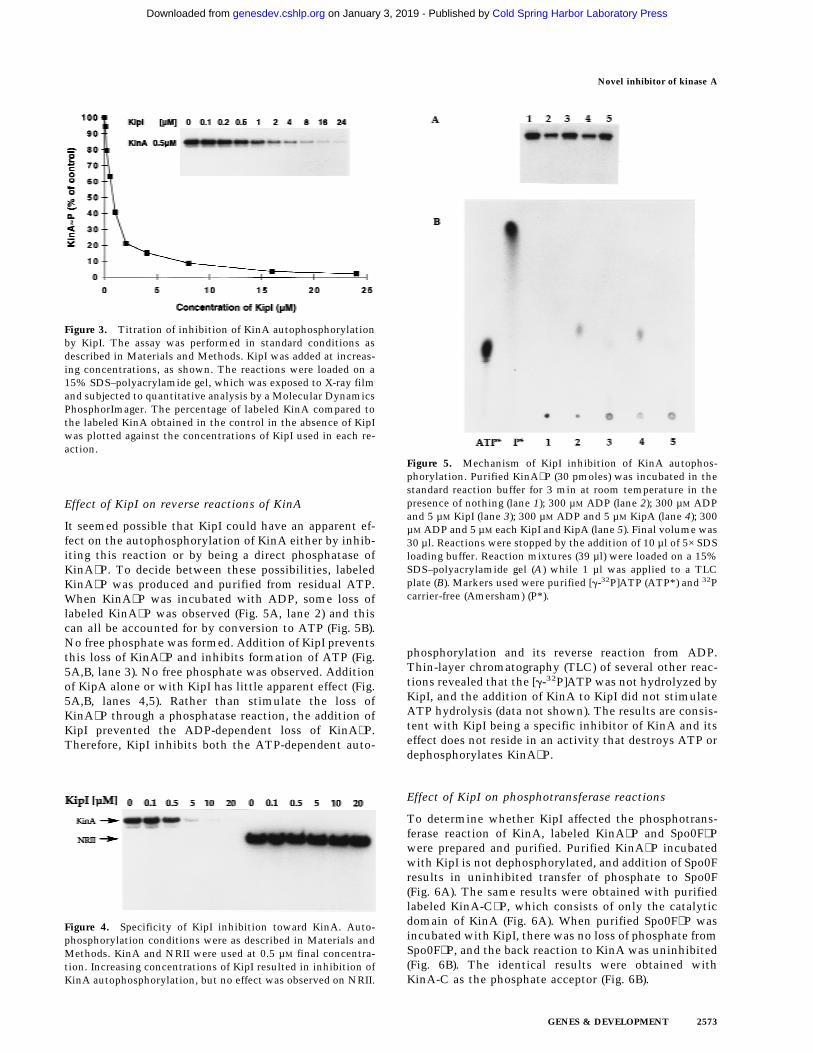
It seemed possible that KipI could have an apparent ef-fect on the autophosphorylation of KinA either by inhib-iting this reaction or by being a direct phosphatase ofKinA∼P. To decide between these possibilities, labeledKinA∼P was produced and purified from residual ATP.When KinA∼P was incubated with ADP, some loss oflabeled KinA∼P was observed (Fig. 5A, lane 2) and thiscan all be accounted for by conversion to ATP (Fig. 5B).No free phosphate was formed. Addition of KipI preventsthis loss of KinA∼P and inhibits formation of ATP (Fig.5A,B, lane 3). No free phosphate was observed. Additionof KipA alone or with KipI has little apparent effect (Fig.5A,B, lanes 4,5). Rather than stimulate the loss ofKinA∼P through a phosphatase reaction, the addition ofKipI prevented the ADP-dependent loss of KinA∼P.Therefore, KipI inhibits both the ATP-dependent auto-
phosphorylation and its reverse reaction from ADP.Thin-layer chromatography (TLC) of several other reac-tions revealed that the [g-32P]ATP was not hydrolyzed byKipI, and the addition of KinA to KipI did not stimulateATP hydrolysis (data not shown). The results are consis-tent with KipI being a specific inhibitor of KinA and itseffect does not reside in an activity that destroys ATP ordephosphorylates KinA∼P.
Effect of KipI on phosphotransferase reactions
To determine whether KipI affected the phosphotrans-ferase reaction of KinA, labeled KinA∼P and Spo0F∼Pwere prepared and purified. Purified KinA∼P incubatedwith KipI is not dephosphorylated, and addition of Spo0Fresults in uninhibited transfer of phosphate to Spo0F(Fig. 6A). The same results were obtained with purifiedlabeled KinA-C∼P, which consists of only the catalyticdomain of KinA (Fig. 6A). When purified Spo0F∼P wasincubated with KipI, there was no loss of phosphate fromSpo0F∼P, and the back reaction to KinA was uninhibited(Fig. 6B). The identical results were obtained withKinA-C as the phosphate acceptor (Fig. 6B).
Figure 4. Specificity of KipI inhibition toward KinA. Auto-phosphorylation conditions were as described in Materials andMethods. KinA and NRII were used at 0.5 µM final concentra-tion. Increasing concentrations of KipI resulted in inhibition ofKinA autophosphorylation, but no effect was observed on NRII.
Figure 5. Mechanism of KipI inhibition of KinA autophos-phorylation. Purified KinA∼P (30 pmoles) was incubated in thestandard reaction buffer for 3 min at room temperature in thepresence of nothing (lane 1); 300 µM ADP (lane 2); 300 µM ADPand 5 µM KipI (lane 3); 300 µM ADP and 5 µM KipA (lane 4); 300µM ADP and 5 µM each KipI and KipA (lane 5). Final volume was30 µl. Reactions were stopped by the addition of 10 µl of 5× SDSloading buffer. Reaction mixtures (39 µl) were loaded on a 15%SDS–polyacrylamide gel (A) while 1 µl was applied to a TLCplate (B). Markers used were purified [g-32P]ATP (ATP*) and 32Pcarrier-free (Amersham) (P*).
Figure 3. Titration of inhibition of KinA autophosphorylationby KipI. The assay was performed in standard conditions asdescribed in Materials and Methods. KipI was added at increas-ing concentrations, as shown. The reactions were loaded on a15% SDS–polyacrylamide gel, which was exposed to X-ray filmand subjected to quantitative analysis by a Molecular DynamicsPhosphorImager. The percentage of labeled KinA compared tothe labeled KinA obtained in the control in the absence of KipIwas plotted against the concentrations of KipI used in each re-action.
Novel inhibitor of kinase A
GENES & DEVELOPMENT 2573
Cold Spring Harbor Laboratory Press on January 3, 2019 - Published by genesdev.cshlp.orgDownloaded from

Therefore, KipI affects the KinA autophosphorylationreaction and its reverse reaction. It is not an inhibitor ofthe reverse phosphotransferase reaction and likely doesnot inhibit the forward reaction. It is not a phosphataseof KinA∼P or of Spo0F∼P and does not stimulate a latentautophosphatase activity of KinA.
Catabolite induction of kip genes
Transcriptional analysis of the kip gene-containing op-eron was carried out by assaying b-galactosidase activityof a promoter–lacZ fusion construction (pRM111; Fig. 1)cloned in the transcriptional fusion vector pJM116 andintegrated ectopically in the amyE locus (see Materialsand Methods). In the nutrient broth-based Schaeffer’ssporulation medium (SSM), kip transcription increasedduring the logarithmic phase, reaching a maximum levelat T0 (15 Miller units), thereafter decreasing to ∼7 Millerunits between T3 and T4 (Fig. 7). Because the sporulationdefect of a kipA deletion strain was enhanced by theaddition of glucose, transcription of the operon was ana-lyzed in the presence of sugars. The addition of glucoseat 1% final concentration to SSM strongly induced tran-scription of the operon in early exponential phase, but asharp decrease reproducibly occurred at T−1 (Fig. 7). Simi-lar induction was also observed when fructose at 1% orglycerol at 2% was added to SSM. The same constructionwas also placed isotopically on the chromosome by
means of a pJM783 integrative plasmid, and the samepatterns of transcription were observed.
The induction of the operon by glucose provided ameans to determine the boundaries of the operon. Tran-scriptional lacZ fusions to kipI, kipR, and ycsK wereconstructed in pJM783 and isotopically integrated in theB. subtilis chromosome as described in Materials andMethods. b-Galactosidase assays were carried out inSSM in the presence of 1% glucose. These lacZ fusionsshowed the same pattern of induction observed withplasmid pRM111, although the maximum level of tran-scription gradually decreased the farther the distancefrom the promoter (data not shown). No transcriptionalactivity was obtained from plasmid pRM116 carrying alacZ fusion to the 38 end of a putative Rho-independentterminator of transcription, located downstream of ycsK(Fig. 1). Furthermore, transcription of ycsL, locateddownstream of the terminator, showed a totally differentregulatory pattern (data not shown). This analysis sug-gests that the operon containing the kip genes comprisesseven cistrons in a single transcriptional unit induced byseveral carbon sources.
Nitrogen control of kip gene expression
When the B. subtilis strain JH19115 carrying the pro-moter–lacZ fusion construct pRM111 was grown in SSM
Figure 7. Expression of the operon containing the kip genes insporulation medium. The wild-type strain JH19115 harboringthe lacZ fusion construct plasmid pRM111 was grown in SSM(s), in SSM supplemented with 1% glucose (m), and in SSMsupplemented with 1% glucose and 0.3% glutamine (.). Time0 represents the transition from vegetative to sporulation phase.b-Galactosidase was expressed as Miller units (Miller 1972).
Figure 6. KipI does not prevent the phosphotransferase reac-tion from KinA/KinA-C∼P to Spo0F and from Spo0F∼P to KinA/KinA-C. (A) Purified KinA∼P or KinA-C∼P (30 pmoles) was in-cubated for 3 min at room temperature in standard reactionbuffer. The presence of Spo0F (5 µM) and/or KipI (5 µM) in thereaction is indicated (+). (B) Spo0F∼P (50 pmoles) was incubatedin standard reaction buffer with KinA (5 µM) or KinA-C (5 µM) inthe presence of KipI (5 µM) as indicated (+).
Wang et al.
2574 GENES & DEVELOPMENT
Cold Spring Harbor Laboratory Press on January 3, 2019 - Published by genesdev.cshlp.orgDownloaded from

in the presence of 0.3% glutamine, induction by glucosewas prevented (Fig. 7). Strain JH19115 was grown in Spiz-izen salts minimal medium in the presence of differentnitrogen sources to examine the basis for this nitrogeneffect. In a good nitrogen source, such as NH4
+, very littletranscription was observed. However, poor nitrogensources such as allantoin, allowed higher levels of tran-scription from the promoter–lacZ construct pRM111.Expression in different nitrogen sources increased as fol-lows: NH4
+ < arginine < glutamine < urea < proline = gamino butyric acid = asparagine < allantoin < glutamate(data not shown). When a good and a poor nitrogensource were both present in the medium, the good nitro-gen source effect was dominant.
Many nitrogen-regulated genes in B. subtilis are tran-scriptionally activated by the TnrA regulator, which re-sponds to poor nitrogen conditions (Wray et al. 1996). Wetested whether the enhanced transcription of the kipgene-containing operon under these conditions was a re-sult of TnrA action by studying transcription in a tnrAdeletion strain. Cells were grown in minimal glucosemedia with glutamate as a nitrogen source, and b-galac-tosidase activity driven by a single-copy chromosomalpromoter–lacZ construct (pRM111) was assayed. Dele-tion of kipR increased the final level of transcription>10-fold (Fig. 8). Deleting tnrA in this kipR mutant pre-
vented most of this increase, indicating that TnrA is anactivator of kip operon transcription and the loss of re-pression regulation by KipR allows TnrA to activatetranscription of the operon. In the absence of TnrA, KipRrepression of kip transcription is essentially complete.
Discussion
The regulation of the activity of KinA is of paramountimportance for the initiation of developmental transcrip-tion at the onset of sporulation. KinA is functional at theend of exponential growth and must respond to thosecellular signals that would promote sporulation at theexpense of further growth. Essentially all of the phos-phate that is sequestered by the phosphorelay compo-nents at this time arises from the action of KinA. Manyof the mechanisms by which KinA is regulated still re-main obscure, although the proteins described here mustplay a role in controlling KinA activity under somephysiological conditions. The operon in which KipI andKipA reside is induced by glucose when readily availablesources of nitrogen, such as glutamine or ammonia, arescarce. This operon is regulated by TnrA activationand by KipR repression. TnrA positively regulates anumber of genes and operons coding for proteins thatdegrade nitrogen-containing compounds (Ferson et al.1996; Wray et al. 1996). TnrA-dependent promotersare characterized by a common upstream sequence(TGTNAN7TNACA), two of which are present in thepromoter for the kip gene-containing operon, from theputative −60 to −30 region. Nitrogen-limited TnrA acti-vation is clearly antagonized by KipR repression, whichresponds to some other function of the operon. In addi-tion, there is some induction by catabolites that has anunknown basis. It seems likely that the genes of thisoperon must code for a system to take up and degradesome nitrogenous compound, perhaps with a lactamring, but its identity is obscure. Regardless, some physi-ological situations responsible for induction must becontrary to sporulation, as the product of induction,KipI, is a potent inhibitor of KinA activity.
KipI is an inhibitor of the autophosphorylation ofKinA by ATP. It is also an inhibitor of the reverse reac-tion from KinA∼P to KinA and ATP. KipI does not in-hibit by dephosphorylating the substrate or the product.Inhibition is not attributable to stimulation of a phos-phatase activity of the kinase similar to that of the ac-tion of PII on NRII of nitrogen regulation (Ninfa et al.1995). It also does not stimulate the reverse reaction,giving an apparent inhibition. KipI targets the carboxy-terminal catalytic domain where it interferes with theATP-dependent reactions of the kinase. However, theinhibition is noncompetitive with respect to ATP. Be-cause KipI is an inhibitor of the autophosphorylation ofthe isolated catalytic domain of KinA, its activity doesnot depend on the amino-terminal ‘‘sensor’’ domain ofthis kinase. Whether the amino-terminal domain of thiskinase plays any role in the binding of KipI cannot bedetermined. A detailed kinetic comparison of the effectsof KipI on intact and amino-terminal truncated KinA
Figure 8. Regulation of expression by KipR and TnrA. b-Ga-lactosidase activity of the lacZ fusion construct carried by plas-mid pRM111 was assayed in the wild-type strain JH19115 (s),the kipR mutant JH19117 (h), in the tnrA mutant JH19190 (d),and in the kipR, tnrA double mutant JH19192 (j). Cells weregrown in Spizizen salts minimal medium supplemented withglutamate, as described in Materials and Methods. Time andunits as in Fig. 7.
Novel inhibitor of kinase A
GENES & DEVELOPMENT 2575
Cold Spring Harbor Laboratory Press on January 3, 2019 - Published by genesdev.cshlp.orgDownloaded from

would be fruitless, as all amino-terminal truncated ver-sions of KinA are defective in the autophosphorylationreaction (L. Wang and J.A. Hoch, unpubl.). It seems mostlikely that the sensor domain recognizes somethingother than KipI and acts to regulate the kinase in re-sponse to other signals.
In contrast to its effects on the autophosphorylationreaction, KipI appears to have little effect on the phos-photransferase reaction of KinA. Purified KinA∼P trans-fers its phosphate to Spo0F unimpeded by KipI. Simi-larly, purified Spo0F∼P transfers phosphate back to KinAor to an amino-terminal truncated KinA regardless of thepresence of KipI. Thus, KipI does not interfere with thecapacity of the kinase to recognize its cognate responseregulator or to transfer phosphate on and off the reactivehistidine of KinA. This suggests that the structure of thecatalytic domain is such that the autophosphorylationand response regulator recognition portions of the mol-ecule are independent and are not integrated or overlap-ping.
Genetic studies indicated that KipA modulated the in-hibitory activity of KipI on sporulation. Overexpressionof KipI inhibited sporulation, whereas overexpression ofKipA enhanced sporulation and a strain producing bothproteins from a plasmid was only slightly sporulationdefective. KipA produced from the expression vector inE. coli was only partially effective in reversing the KipIeffects in vitro. This discrepancy between in vivo and invitro results is likely to be a result of expressing KipA inE. coli, where it is mainly present in inclusion bodies.Although KipA was purified from the soluble fraction, itseems likely that most of this protein is not in a nativeconformation and does not have native activities. BothKipI and KipA have been produced in the same cell froman expression vector (L. Wang, R. Grau, M. Perego, andJ.A. Hoch, unpubl.). In this case, a His-tagged KipI andKipA copurified on a Ni–NTA column, indicating thatKipI and KipA may form a complex. Regardless ofwhether these data explain the activity of KipA on KipIinhibition in vitro, the genetic evidence that KipA coun-teracts KipI inhibition is compelling.
What the interaction of KipI with both KinA and KipAhas to do with the actual role of KipI in vivo is still amystery. KipI and KipA were identified in the B. subtilissequencing project as homologous to ureamidolyase of S.cereviseae (Akagawa et al. 1995). Homologs of KipI andKipA were also found in the H. influenzae genome andidentified with the yeast enzyme (Fleischmann et al.1995). Ureamidolyase catalyzes the ATP-dependent deg-radation of urea to NH3 and CO2 via allophanic acid(Whitney and Cooper 1973). The available evidence isnot in agreement with KipI and KipA being a bacterialform of ureamidolyase. Neither KipI nor KipA is biotin-ylated whether assayed in B. subtilis or when expressedin E. coli. No ureamidolyase activity can be detected inpurified KipI and KipA. A urease-defective mutant of B.subtilis cannot be made to use urea as a nitrogen sourceeven if the operon bearing KipI and KipA is induced.These unpublished experiments are not consistent witha urea-degrading role for KipI and KipA. The function of
the operon encoding KipI and KipA is obscure, but theoperon is regulated by available nitrogen in a mannervery similar to that of ureamidolyase in S. cereviseae(Magasanik 1992; Cunnigham et al. 1994).
KipI acts as an anti-kinase and KipA could be thoughtof as an anti-anti-kinase. Because kipI and kipA are tran-scriptionally coregulated, it seems unlikely that expres-sion controls determine the activity of the anti-kinase. Itis reasonable to assume that KipI–KipA interaction regu-lates anti-kinase activity, and this interaction may besubject to effector molecules and/or covalent modifica-tion.
It is generally believed that the catalytic domain ofhistidine kinases is controlled by the sensor domain,which acts as the information recognition and process-ing site. To our knowledge, KipI is the first inhibitor oftwo-component histidine kinases directed toward thecatalytic domain. The effects of KipI on KinA suggestthat the catalytic domain may not be entirely passive insignal transduction. Inhibitors of the catalytic domainmay act to block or counteract signals that influence thesensor domain. Perhaps two-component systems de-signed to recognize the cellular level of a single metabo-lite such as available nitrogen may only need a sensordomain to interact with a single effector. The phosphore-lay is a signal integration circuit that regulates a cellulardevelopmental event, and many signals can and do in-fluence the decision to initiate development. The cellhas evolved several mechanisms to cope with these sig-nals other than regulation of the sensor domain. Somesignals manifest in the activation of phosphatases thatdephosphorylate the response regulator components ofthe phosphorelay (Perego et al. 1994). The signals trans-duced by phosphatases are believed to be negative influ-ences on the developmental process that function bycounteracting phosphate input into the phosphorelay byKinA (Ohlsen et al. 1994). Because phosphatases playthis role, it should not be surprising that the cell woulduse inhibitors such as KipI to counteract phosphate in-put by KinA.
The rationale for a cell using the phosphorelay to con-trol development has been postulated to stem from thepotential of this type of signal transduction system forregulation by a variety of signals promoting eithergrowth or sporulation (Burbulys et al. 1991). Becausesporulation initiation is seemingly only dependant onraising the level of phosphorylation of the Spo0A tran-scription factor, inhibition by phosphatases or enzymeinhibitors should prevent the process. It should not beunexpected that other components of the phosphorelayare regulated by dephosphorylation or inhibition of func-tion.
Materials and methods
Bacterial strains and growth conditions
The B. subtilis strains used in this study are described in TableI [the tnrA deletion strain SF706T was obtained from SusanFisher (Ferson et al. 1996)]. For sporulation efficiency, B. subtilisstrains were grown in Schaeffer’s sporulation medium (Schaef-
Wang et al.
2576 GENES & DEVELOPMENT
Cold Spring Harbor Laboratory Press on January 3, 2019 - Published by genesdev.cshlp.orgDownloaded from

fer et al. 1965) and then treated with CHCl3 or heated at 80°Cfor 10 min before plating. Transformation of B. subtilis wascarried out as described (Anagnostopoulos and Spizizen 1961).
b-Galactosidase in B. subtilis strains harboring lacZ fusionswere assayed as described previously, and the specific activitywas expressed in Miller units (Miller 1972). Cells were grown inSchaeffer’s sporulation medium or Spizizen’s minimal medium(Spizizen 1958) without (NH4)2SO4 and supplemented with 20mM MgSO4, 1% glucose, 0.005% each tryptophan and phenyl-alanine, and 20 mM nitrogen source.
Plasmids and strains constructions
Plasmid pRM90 was obtained from a B. subtilis JH642 chromo-somal library constructed by ligating partial Sau3A restrictionfragments in the multiple cloning site of the pHT315 shuttlevector (Arantes and Lereclus 1991; V. Dartois, T. Djava-khishvili, and J.A. Hoch, unpubl.). Plasmid pRM91 was derivedfrom pRM90 by deletion of the fragment comprised of the ApaIsite in kipA and the EcoRI site in the multiple cloning site.Plasmid pRM92 was obtained by religating plasmid pRM90 di-gested previously with EagI to create a 500-bp internal deletionin the KipI coding gene. The multicopy vector pRM94 carryingboth kipI and kipA coding sequences was obtained by replacingthe ApaI–EcoRI fragment of pRM90 with a ApaI–EcoRI frag-ment containing the entire kipA gene obtained by PCR ampli-fication of chromosomal DNA. The kipA multicopy plasmidpRM93 was obtained by digestion of pRM94 with EagI and re-ligation to delete the EagI fragment internal to kipI.
The entire operon containing the kip genes and its boundarieswere amplified by PCR using the Expand High Fidelity PCRsystem (Boehringer Mannheim) from chromosomal DNA ofstrain JH642 with the oligonucleotides 58-AGACATGGTACC-AGGAAACGGCTGATTATATCACG-38 KpnI and 58-AAAT-ATCTGCAGCTTGTGGAGTGGGAAACACTTG-38 PstI. Thefragment obtained (6908 bp) was digested with KpnI, EcoRI, andPstI to generate four fragments of 2, 1.45, 1.6, and 1.8 kbp,respectively. Each fragment was cloned in the shuttle vectorpHT315 (Arantes and Lereclus 1991) to generate plasmids thatwere subjected to partial nucleotide sequence.
Plasmids pRM107, pRM108, and pRM109 were constructedin the integrative vector pJM103 (Perego 1993). Their lineariza-tion and transformation in B. subtilis-competent cells resultedin integration by a double crossover event that yielded strainsJH19112, JH19108, and JH19117, respectively. In these strains,all of the genes in the operon located downstream of the site ofintegration may also be inactivated because of a polar effect.
For the construction of strain JH19168 (kipI−kipA+), plasmidpRM106, constructed in the integrative vector pJM103, was in-tegrated in the chromosome of JH19980 via single crossing-overby transformation and selection for Cm resistance (CmR) (5 µg/ml). Two Spo+ transformants were grown for several days inLuria Bertani (LB) medium without antibiotic to allow sponta-neous excision of the plasmid from the chromosome. The cul-tures were grown to stationary phase and then diluted 1:100 infresh medium every 12 hr for 5 days before plating serial dilu-tions on LB agar plates. The colonies obtained were screened forCm sensitivity (CmS). Three colonies out of 200 checked wereCmS. Their chromosomal DNA was used in PCR reactionsusing oligonucleotides 58-TGAGAATCATGACTGTACGATA-TCAAATCGAAC-38 and 58-TCCCGTCTCGAGTGTCAGCC-ATTCCATCTTCATTCTCGTTTTCAGTTC-38 to amplify thekipI region and verify the presence of the internal deletion of∼0.6 kb. The three CmS colonies analyzed yielded a PCR prod-uct 0.6 kb smaller than the control chromosomal DNA from
JH19980, (kipI+, kipA+). One of these CmS colonies was desig-nated JH19168 and used in further studies.
Transcriptional lacZ fusion plasmids were constructed invectors pJM783 for isotopic integrations and pJM116 for ectopicintegrations in the amyE locus (Perego 1993). Plasmid pRM111was constructed by cloning a PCR-generated KpnI–HindIII frag-ment into pJM116. Plasmid pRM112 carries the same fragmentbut in the lacZ fusion vector pJM783.
Nucleotide sequence
Extensive nucleotide sequence of the operon containing the kipgenes was carried out to define the precise end points of eachopen reading frame. The regions comprising nucleotides 7300–9060 and 10350–13000 in the original sequence (GenBank ac-cession no. d38161) were resequenced from fragments obtainedby PCR amplification of the chromosomal DNA of strainJH642. Corrections have been submitted to the databank as anupdate to the original sequence. To ensure that kipI and kipAwere two separate genes, the chromosomal region containingboth genes was amplified by PCR reaction from chromosomalDNA extracted from two natural Bacillus isolates (Bacillusnatto from a commercial Natto and B. subtilis polish, a strain inour collection). The fragments were cloned in the pET16b ex-pression vector, sequenced, and then transferred to the E. coliBL21 (DE3) strain for protein expression and purification. Theresults (not shown) confirmed that in these Bacillus naturalisolates, KipI and KipA are two distinct proteins coded by twoseparate genes.
Expression and purification of KipI and KipA
The coding sequences for kipI and kipA were amplified by PCRreaction from chromosomal DNA of the wild-type strain JH642.kipI was cloned as a NcoI–XhoI fragment in the NcoI–XhoI sitesof the pET28a expression vector (Novagen), which promotes thefusion of the carboxy-terminal end of the protein with six his-tidine residues. The kipA gene was cloned in the NdeI–BamHIsites of the pET16b vector (Novagen), thereby adding 10 histi-dine codons to the 58 end of the gene. Plasmids were constructedin E. coli DH5a and, after sequence verification, transferred inthe E. coli expression host BL21(DE3) plysS. Cells carrying thepET28a–KipI or the pET16b–KipA expression vectors weregrown in LB medium supplemented with kanamycin (30 µg/ml)or ampicillin (100 µg/ml), respectively. At an OD600 of 0.6, cellswere induced by the addition of 1 mM IPTG and allowed to growfor an additional 3 hr at 37°C before harvesting. The His-taggedproteins were purified by affinity chromatography on Ni–NTAagarose columns (Qiagen), in 20 mM Tris (pH 8.0), 10 mM KCl,and 1 mM PMSF using a 0–150 mM gradient of imidazole.
Phosphorylation assay conditions
Phosphorylation assays were performed in 30 µl volume in thestandard reaction buffer (50 mM EPPS at pH 8.0, 0.1 mM EDTA,20 mM MgCl2, 5% glycerol). KinA was added to a final concen-tration of 0.5 µM. The reactions were initiated by the addition of5 µl of a mixture of [g-32P]ATP and unlabeled ATP to reach afinal concentration of 300 µM. Reactions were carried out for 3min at room temperature and stopped by the addition of 12 µl of5× SDS loading buffer (25 mM Tris-HCl at pH 6.8, 1.5% SDS, 5%b-mercaptoethanol, 10% glycerol, 0.02% Bromophenol blue).Reactions were placed in dry ice before loading onto 15% SDS–polyacrylamide gels. Electrophoresis was carried out at constantvoltage (200 V) for ∼2.5 hr. Where necessary, the lower portionof the gel was removed to reduce the background, which is
Novel inhibitor of kinase A
GENES & DEVELOPMENT 2577
Cold Spring Harbor Laboratory Press on January 3, 2019 - Published by genesdev.cshlp.orgDownloaded from

attributable to unincorporated [g-32P]ATP. Gels were dried andexposed for 1 hr to Kodak AR films.
Purification of KinA∼P and Spo0F∼P
Phosphorylated KinA and Spo0F were purified as follows: 5 µM
KinA or KinA-C and 25 µM Spo0F were labeled by the standardphosphorylation assay in 1 ml volume. After 30 min incubationat room temperature, the mixtures were applied to an FPLCS-100 Sephacryl column (1.6 × 64 cm). Gel filtration-purifiedKinA∼P, KinA-C∼P, and Spo0F∼P were checked on a 15% SDS–polyacrylamide gel that had been exposed to autoradiography.KinA-C, spanning from residue 389 to 606, was expressed in thepET16b expression vector (L. Wang, in prep.). KinA and Spo0Fwere purified as described previously (Perego et al. 1989; Zapf etal. 1996).
Analytical TLC
TLC was performed according to Randerath and Randerath(1967), with some modifications. KinA∼P (30 pmoles) was incu-bated with 5 µM KipI and/or KipA in the standard assay bufferfor 5 min at room temperature. ADP at 300 µM final concentra-tion was added to initiate the reactions. Two microliters of stopbuffer (5× loading buffer) was added after 3 min incubation, and1 µl from each reaction was applied to PEI cellulose F layerplates (Alltech). Plates were developed in 1.6 M LiCl for ∼45 minand then dried and exposed overnight to X-ray films. Reactionswere also loaded on 15% SDS–polyacrylamide gels and exposedto X-ray films as well.
Acknowledgments
This research was supported in part by grant GM19416 from theNational Institute of General Medical Sciences (National Insti-tutes of Health, U.S. Public Health Service). L.W. and R.G. con-tributed equally to this work.
The publication costs of this article were defrayed in part bypayment of page charges. This article must therefore be herebymarked ‘‘advertisement’’ in accordance with 18 USC section1734 solely to indicate this fact.
References
Akagawa, E., K. Kurita, T. Sugawara, K. Nakamura, Y. Kasahara,N. Ogasawara, and K. Yamane. 1995. Determination of a17484 bp nucleotide sequence around the 39° region of theBacillus subtilis chromosome and similarity analysis of theproducts of putative ORFs. Microbiology 141: 3241–3245.
Anagnostopoulos, C. and J. Spizizen. 1961. Requirements fortransformation in Bacillus subtilis. J. Bacteriol. 81: 741–746.
Arantes, O. and D. Lereclus. 1991. Construction of cloning vec-tors for Bacillus thuringiensis. Gene 108: 115–119.
Burbulys, D., K.A. Trach, and J.A. Hoch. 1991. The initiation ofsporulation in Bacillus subtilis is controlled by a multicom-ponent phosphorelay. Cell 64: 545–552.
Cunnigham, T.S., R.A. Dorrington, and T.G. Cooper. 1994. TheUGA4 UASNTR site required for GLN3-dependent transcrip-tional activation also mediates DAL80-responsive regula-tion and DAL80 protein binding in Saccharomyces cerevi-siae. J. Bacteriol. 176: 4718–4725.
Ferson, A.E., L.V. Wray, and S.H. Fisher. 1996. Expression of theBacillus subtilis gabP gene is regulated independently inrepsonse to nitrogen and amino acid availability. Mol. Mi-crobiol. 22: 693–701.
Fleischmann, R.D., M.D. Adams, O. White, R.A. Clayton, E.F.Kirkness, A.R. Kerlavage, C.J. Bult, J. Tomb, B.A. Dougherty,J.M. Merrick et al. 1995. Whole-genome random sequencingand assembly of Haemophilus influenzae Rd. Science269: 496–512.
Genbauffe, F.S. and T.G. Cooper. 1991. The urea amidolyase(DUR1,2) gene of Saccharomyces cerevisiae. J. DNA Seq.Map. 2: 19–32.
Hoch, J.A. 1993. Regulation of the phosphorelay and the initia-tion of sporulation in Bacillus subtilis. Annu. Rev. Micro-biol. 47: 441–465.
Hoshino, T., K. Kose, and Y. Uratani. 1990. Cloning and nucleo-tide sequence of the gene braB coding for the sodium-coupledbranched-chain amino acid carrier in Pseudomonas aerugi-nosa PAO. Mol. & Gen. Genet. 220: 461–467.
Magasanik, B. 1992. Regulation of nitrogen utilization. In Themolecular and cellular biology of the yeast Saccharomycescerevisiae: Metabolism and gene expression (ed. Strathermet al.), pp. 283–317. Cold Spring Harbor Laboratory Press,Cold Spring Harbor, NY.
Miller, J.H. 1972. Experiments in molecular genetics, pp. 352–355. Cold Spring Harbor Laboratory, Cold Spring Harbor,NY.
Ninfa, A.J. and B. Magasanik. 1986. Covalent modification ofthe glnG product, NRI, by the glnL product, NRII, regulatesthe transcription of the glnALG operon in Escherichia coli.Proc. Natl. Acad. Sci. 83: 5909–5913.
Ninfa, A.J., M.R. Atkinson, E.S. Kamberov, J. Feng, and E.G.Ninfa. 1995. Two-component signal transduction. In Con-trol of nitrogen assimilation by the NRI-NRII two-compo-nent system of enteric bacteria (ed. J.A. Hoch and T.J. Sil-havy), pp. 67–88. ASM Press, Washington, D.C.
Ohlsen, K.L., J.K. Grimsley, and J.A. Hoch. 1994. Deactivationof the sporulation transcription factor Spo0A by the Spo0Eprotein phosphatase. Proc. Natl. Acad. Sci. 91: 1756–1760.
Parkinson, J.S. and E.C. Kofoid. 1992. Communication modulesin bacterial signaling proteins. Annu. Rev. Genet. 26: 71–112.
Perego, M. 1993. Bacillus subtilis and other Gram-positive bac-teria: Biochemistry, physiology, and molecular genetics. InIntegrational vectors for genetic manipulation in Bacillussubtilis (ed. A.L. Sonenshein, J.A. Hoch, and R. Losick), pp.615–624. American Society for Microbiology, Washington,D.C.
Perego, M. and J.A. Hoch. 1996a. Cell-cell communication regu-lates the effects of protein aspartate phosphatases on thephosphorelay controlling development in Bacillus subtilis.Proc. Natl. Acad. Sci. 93: 1549–1553.
———. 1996b. Protein aspartate phosphatases control the out-put of two-component signal transduction systems. TrendsGenet. 12: 97–101.
Perego, M., S.P. Cole, D. Burbulys, K.A. Trach, and J.A. Hoch.1989. Characterization of the gene for a protein kinase whichphosphorylates the sporulation-regulatory proteins Spo0Aand Spo0F of Bacillus subtilis. J. Bacteriol. 171: 6187–6196.
Perego, M., C.G. Hanstein, K.M. Welsh, T. Djavakhishvili, P.Glaser, and J.A. Hoch. 1994. Multiple protein aspartate phos-phatases provide a mechanism for the integration of diversesignals in the control of development in Bacillus subtilis.Cell 79: 1047–1055.
Perego, M., P. Glaser, and J.A. Hoch. 1996. Aspartyl-phosphatephosphatases deactivate the response regulator componentsof the sporulation signal transduction system in Bacillussubtilis. Mol. Microbiol. 19: 1151–1157.
Posas, F., S.M. Wurgler-Murphy, T. Maeda, E.A. Witten, T.C.Thai, and J. Saito. 1996. Yeast HOG1 MAP kinase cascade is
Wang et al.
2578 GENES & DEVELOPMENT
Cold Spring Harbor Laboratory Press on January 3, 2019 - Published by genesdev.cshlp.orgDownloaded from

regulated by a multistep phosphorelay mechanism in theSLN1-YPD1-SSK1 ‘‘two-component’’ osmosensor. Cell 86:865–875.
Randerath, K. and E. Randerath. 1967. Thin-layer separationmethods for nucleic acid derivatives. Methods Enzymol.12: 323–347.
Richardson, I.B., M.E. Katz, and M.J. Hynes. 1992. Molecularcharacterization of the lam locus and sequences involved inregulation by the AmdR protein of Aspergillus nidulans.Mol. Cell. Biol. 12: 337–346.
Schaeffer, P., J. Millet, and J. Aubert. 1965. Catabolic repressionof bacterial sporulation. Proc. Natl. Acad. Sci. 54: 701–711.
Spizizen, J. 1958. Transformation of biochemically deficientstrains of Bacillus subtilis by deoxyribonucleate. Proc. Natl.Acad. Sci. 44: 1072–1075.
Trach, K.A. and J.A. Hoch. 1993. Multisensory activation of thephosphorelay initiating sporulation in Bacillus subtilis:Identification and sequence of the protein kinase of the al-ternate pathway. Mol. Microbiol. 8: 69–79.
Uhl, M.A. and J.F. Miller. 1996. Integration of multiple domainsin a two-component sensor protein: The Bordetella pertussisBvgAS phosphorelay. EMBO J. 15: 1028–1036.
Whitney, P.A. and T.G. Cooper. 1973. Urea Carboxylase fromSaccharomyces cerevisiae. J. Biol. Chem. 248: 325–330.
Wray, L.V., A.E. Ferson, K. Rohrer, and S.H. Fisher. 1996. TnrA,a transcription factor required for global nitrogen regulationin Bacillus subtilis. Proc. Natl. Acad. Sci. 93: 8841–8845.
Zapf, J.W., J.A. Hoch, and J.M. Whiteley. 1996. A novel phos-phorylation activity of the Bacillus subtilis sporulation pro-tein Spo0F that employs phosphoramidate substrates. J. Biol.Chem. 35: 2926–2933.
Novel inhibitor of kinase A
GENES & DEVELOPMENT 2579
Cold Spring Harbor Laboratory Press on January 3, 2019 - Published by genesdev.cshlp.orgDownloaded from

10.1101/gad.11.19.2569Access the most recent version at doi: 11:1997, Genes Dev.
Ling Wang, Roberto Grau, Marta Perego, et al.
subtilis BacillusA novel histidine kinase inhibitor regulating development in
References
http://genesdev.cshlp.org/content/11/19/2569.full.html#ref-list-1
This article cites 27 articles, 11 of which can be accessed free at:
License
ServiceEmail Alerting
click here.right corner of the article or
Receive free email alerts when new articles cite this article - sign up in the box at the top
Cold Spring Harbor Laboratory Press
Cold Spring Harbor Laboratory Press on January 3, 2019 - Published by genesdev.cshlp.orgDownloaded from