a pet activation study of dynamic mechanical allodynia in
TRANSCRIPT

A PET activation study of dynamic mechanical allodynia in patientswith mononeuropathy
P. Petrovica, M. Ingvara,*, S. Stone-Elandera, b, K.M. Peterssona, P. Hanssonc
aClinical Neurophysiology, Department of Clinical Neuroscience, Karolinska Institute, Karolinska Hospital, Stockholm, SwedenbKarolinska Pharmacy, Karolinska Institute, Karolinska Hospital, Stockholm, Sweden
cNeurogenic Pain Unit, Multidisciplinary Pain Center and Department of Rehabilitation Medicine, Karolinska Institute, Karolinska Hospital,
Stockholm, Sweden
Received 28 September 1998; received in revised form 27 May 1999; accepted 8 June 1999
Abstract
The objective of this study was to investigate the central processing of dynamic mechanical allodynia in patients with mononeuropathy.
Regional cerebral blood ¯ow, as an indicator of neuronal activity, was measured with positron emission tomography. Paired comparisons
were made between three different states; rest, allodynia during brushing the painful skin area, and brushing of the homologous contralateral
area. Bilateral activations were observed in the primary somatosensory cortex (S1) and the secondary somatosensory cortex (S2) during
allodynia compared to rest. The S1 activation contralateral to the site of the stimulus was more expressed during allodynia than during
innocuous touch. Signi®cant activations of the contralateral posterior parietal cortex, the periaqueductal gray (PAG), the thalamus bilaterally
and motor areas were also observed in the allodynic state compared to both non-allodynic states. In the anterior cingulate cortex (ACC) there
was only a suggested activation when the allodynic state was compared with the non-allodynic states. In order to account for the individual
variability in the intensity of allodynia and ongoing spontaneous pain, rCBF was regressed on the individually reported pain intensity, and
signi®cant covariations were observed in the ACC and the right anterior insula. Signi®cantly decreased regional blood ¯ow was observed
bilaterally in the medial and lateral temporal lobe as well as in the occipital and posterior cingulate cortices when the allodynic state was
compared to the non-painful conditions. This ®nding is consistent with previous studies suggesting attentional modulation and a central
coping strategy for known and expected painful stimuli. Involvement of the medial pain system has previously been reported in patients with
mononeuropathy during ongoing spontaneous pain. This study reveals a bilateral activation of the lateral pain system as well as involvement
of the medial pain system during dynamic mechanical allodynia in patients with mononeuropathy. q 1999 International Association for the
Study of Pain. Published by Elsevier Science B.V.
Keywords: Positron emission tomography; Regional cerebral blood ¯ow; Brain activation; Pain; Painful mononeuropathy; Dynamic mechanical allodynia
1. Introduction
Pain is a complex phenomenon involving sensory-discri-
minative, cognitive-evaluative and affective-motivational
dimensions, which are processed in parallel (Melzack and
Casey, 1968). It is currently believed that the lateral pain
system is more involved in sensory-discriminative aspects
of pain processing whereas the medial system is more
involved in processing the affective-motivational compo-
nent (Albe-Fessard et al., 1985; Vogt et al., 1993; Willis,
1995). Functional neuroimaging studies have identi®ed
several networks, which are involved in pain processing in
man. Acute phasic noxious stimulation has been used in
most of these studies. Apart from some inconsistencies,
there is a general consensus that such stimuli activate the
anterior cingulate cortex (ACC) and the mid-/anterior insula
of the medial pain system as well as the primary- and
secondary somatosensory cortices (S1 and S2) of the lateral
pain system (Jones et al., 1991; Talbot et al., 1991; Casey et
al., 1994, 1996; Coghill et al., 1994; Apkarian, 1995; Davis
et al., 1995, 1997; Hsieh, 1995; Vogt et al., 1996). These
®ndings support the hypothesis that the experience of pain is
processed in a parallel interactive and distributed fashion.
The pathophysiology of neuropathic pain due to periph-
eral nerve injury is not completely understood (Bennett,
1994). The peripheral nerve lesion may result in different
clinical manifestations such as spontaneous ongoing pain
Pain 83 (1999) 459±470
0304-3959/99/$20.00 q 1999 International Association for the Study of Pain. Published by Elsevier Science B.V.
PII: S0304-3959(99)00150-5
www.elsevier.nl/locate/pain
* Corresponding author. Section of Clinical Neurophysiology, Depart-
ment of Clinical Neuroscience, Karolinska Hospital, 171 76 Stockholm,
Sweden. Tel.: 146-8-5177-5134; fax: 146-8-344-146.
E-mail address: [email protected] (M. Ingvar)

and, in a minority of cases, allodynia (Hansson and Kinn-
man, 1996). The most common type of allodynia is pain due
to a light dynamic mechanical stimulus (Hansson and Kinn-
man, 1996). We have previously reported changes in regio-
nal cerebral blood ¯ow (rCBF) following the alleviation of
pain in patients suffering from ongoing spontaneous pain
due to chronic painful mononeuropathy (Hsieh et al.,
1995a). Increased activity of the ACC and the anterior
insula of the medial pain system were observed during spon-
taneous ongoing pain, which suggests an increased tone in
the affective-motivational dimension of the pain experience.
In accordance with other positron emission tomography
(PET) studies of ongoing neuropathic pain, we also
observed a decreased activity in the thalamus contralateral
to the painful neuropathy (Di Piero et al., 1991; Iadarola et
al., 1995). However, altered activity of the primary or
secondary somatosensory cortex has not been reported in
studies of ongoing neuropathic pain.
The objective of the present study was to examine the
central processing of allodynia evoked by a dynamic
mechanical stimulus. Dynamic mechanical allodynia is
usually said to have an explosive, non-physiological char-
acter, which is often accompanied by aftersensations (Hans-
son, 1994). Affective and sometimes vegetative responses
are reported during allodynia. Clinical and experimental
data suggest that dynamic mechanical allodynia involves
activation of low threshold A-beta mechanoreceptive affer-
ents (Ab -®bres) (Gracely et al., 1992; Bennett, 1994).
Several possible pathophysiological mechanisms have
been disclosed; e.g. peripheral crosstalk between Ab -®bres
and nociceptive ®bres, opening of previously silent
synapses in the spinal cord bridging the mechanoreceptive
and the nociceptive system, sprouting of mechanoreceptive
®bres in the dorsal horn to establish new synaptic connec-
tions between the large ®bre system and nociceptive
neurons, and sensitization of spinal dorsal horn neurons
(Hansson and Kinnman, 1996). Thus, dynamic mechanical
allodynia is likely to be composed by signals reaching the
brain from both the nociceptive- and the mechanoreceptive
systems. Therefore, we hypothesized that the activation of
the primary somatosensory cortex would be more expressed
during the allodynic experience than during a non-painful
tactile sensation elicited by the same stimulus outside the
allodynic area. We also hypothesized that tactile allodynia
would activate the medial pain system re¯ecting the affec-
tive component of the painful experience.
2. Methods
2.1. Patients
Five patients with mononeuropathy and dynamic
mechanical allodynia in the lower extremity participated
in the study (Table 1). The patients were included if they
had had at least a 6-month period of a lower extremity
mononeuropathy and dynamic mechanical allodynia
following a peripheral nerve lesion, with or without ongoing
spontaneous pain. The allodynic experience had to be repro-
ducible in clinical testing regarding intensity without any
signi®cant habituation during brushing for 60 s.
As described in Table 1, three of the patients had right-
sided mononeuropathy and two had left-sided mononeuro-
pathy. Two of the patients also reported spontaneous
ongoing pain (one with a right-sided nerve-lesion and one
with a left-sided nerve-lesion). One patient was on medica-
tion (see Table 1) during the study. They were all right-
handed (Edinburgh handiness inventory) and reported no
history of major psychiatric disorder or head trauma.
None ful®lled the criteria for depression although one was
in the border zone according to MADRS depression inven-
tory (Montgomery and AÊ sberg, 1979). The local Ethics and
Radiation safety committees at the Karolinska Hospital
approved all procedures. Informed consent was given by
all the subjects.
2.2. PET scanning
Repeated measurements of rCBF (12 scans/subject, 4
scans/state) were made using an Ecat Exact HR PET scan-
ner in 3D-sampling mode and 500 MBq bolus injections of
[15O]butanol producing 60 s tracer uptake images (Berridge
et al., 1990; Ingvar et al., 1994; Wienhard et al., 1994).
Scatter correction was performed and a 2D-transmission
scan was used for attenuation correction. To ensure that
the radioactivity levels in the subjects had returned to back-
ground before starting a new scan, at least 10 min elapsed
between successive scans. Individual plaster head support
was made for each patient to minimize head movements
during the PET imaging (BergstroÈm et al., 1981).
2.3. Experimental design
The patients were scanned in three different conditions
(eyes closed):
P. Petrovic et al. / Pain 83 (1999) 459±470460
Table 1
Clinical characteristics of the patients with mononeuropathy and dynamic mechanical allodynia included in this study
Patient Age Sex Injured nerve Etiology Duration (years) Spontaneous ongoing pain Treatment
MCL 28 M Left sup. Peroneal Traumatic/ Surgical 2 No None
LW 42 F Left Sural Entrapment/Surgical 7 Yes Ketobemidon
DS 27 M Right Femoral/Saphenus Traumatic 6 No None
YZ 47 F Right Saphenus Surgical 4 No None
MC 58 F Right Cut. Fem. Lat. Surgical 6 Yes None
1. Reference condition in which the patients were lying still
and were instructed to relax and not to think or do
anything in particular but relax (Rest).
2. Brush stimuli were induced by lightly stroking of the
allodynic skin with a soft camel hair brush with a
diameter of 0.5 cm (Allodynia). The stimulation was
performed with a rate of approximately 1 stroke/s. It
began immediately following the injection of the ¯ow
tracer and stopped 45 s. later.
3. Brush stimuli as above on the contralateral homologous
area to the allodynic region (Contralateral touch).
These states were scanned in the order: A-B-A-C-B-C-C-
B-C-A-B-A. Prior to each scan the patients were informed
which condition to expect. The subjects were instructed to
use a numerical rating scale in which 100 equals the highest
imaginable total pain intensity and 0 no pain at all. After
each allodynic period the patients were asked to verbally
rate the maximum total pain intensity during provocation. If
present, they also rated the pain intensity of the spontaneous
ongoing pain immediately before each scan. Following the
®nal scan the subjects were interviewed in detail about the
sessions and their pain ratings were reviewed and
con®rmed.
2.4. Data analysis
In order to analyze all subjects as a group, the PET
images of the two patients with left-sided mononeuropathy
were mirrored across the midline. In addition, the data were
analyzed separately for the three patients with right-sided
nerve lesions and for the two patients with left-sided nerve
lesions to compare the results with the group analysis. The
PET images were realigned, spatially normalized and trans-
formed into an approximate Talairach±Tournoux stereotac-
tic space (Talairach and Tournoux, 1988), 3D Gaussian
®ltered (FWHM � 16 mm) and proportionally scaled to
account for global confounders using the SPM95 (Friston
et al., 1995). The coordinates of local maxima refer to the
approximate Talairach±Tournoux space. The anatomical
designations used below refer to the Karolinska Computer-
ized Brain Atlas (Greitz et al., 1991).
The data analysis was performed in three steps. First, the
contrasts Allodynia - Rest, Allodynia - Contralateral touch,
Rest - Allodynia, Contralateral touch - Allodynia and
Contralateral touch - Rest were analyzed, using a multi-
subject with replications design (3 conditions, 5 blocks
(subjects)). The rCBF increases were investigated in a
pre-de®ned pain network based on previous functional
imaging studies of pain (Di Piero et al., 1991, 1994; Jones
et al., 1991; Talbot et al., 1991; Apkarian et al., 1992, 1995;
Casey et al., 1994, 1996; Coghill et al., 1994; Davis et al.,
1995; Drevets et al., 1995; Hsieh, 1995; Hsieh et al.,
1995a,b; Iadarola et al., 1995; Craig et al., 1996; Vogt et
al., 1996). The pain network included the contralateral S1
and, bilaterally, the thalamus, S2, insula, ACC and the peri-
aqueductal gray (PAG). Similarly, the contralateral S1 and
S2 were chosen for the innocuous somatosensory control
condition based on previous functional imaging studies of
non-painful vibratory sensibility (Fox et al., 1987; Burton et
al., 1993; Coghill et al., 1994). The location of the second-
ary somatosensory cortex (S2) has been de®ned anatomi-
cally in the dorsal bank of the lateral sulcus in the parietal
operculum of the monkey (Roberts and Akert, 1963). This
corresponds to the part of BA43/40 which is situated in the
human operculum, and functional activations in these
regions during pain studies have been regarded as S2 acti-
vations (Talbot et al., 1991). Activations in the prede®ned
regions were considered signi®cant if Z $ 3:09 (or
P # 0:001, uncorrected). In addition, a global search was
performed and activations containing signi®cant local
maxima (P # 0:05, corrected for multiple non-independent
comparisons) that were not part of the pre-de®ned pain
matrix are given in Tables 4 and 5.
Finally, in order to investigate whether allodynia
provokes a more expressed activation in S1 compared to
an identical tactile stimulation of the skin contralateral to
the allodynic area, we placed a spherical ROI with a
diameter of 5 mm in the postcentral gyrus contralateral to
the respective site of stimulation. The exact positioning of
the ROIs was guided by the regional analysis of the activa-
tion in the right postcentral gyrus evoked by the non-painful
stimulation condition (see Fig. 3). At this site, the ROI
representing innocuous touch (S1right) was chosen and the
corresponding contralateral region (S1left), representing the
allodynia, was chosen by mirroring the ROI across the
midline. A two factor ANOVA (with subject and state as
independent variables) was then performed between the
increases in rCBF due to allodynia in the left S1 ROI
(S1leftallodynia 2 S1leftrest) and the increases in rCBF due to
P. Petrovic et al. / Pain 83 (1999) 459±470 461
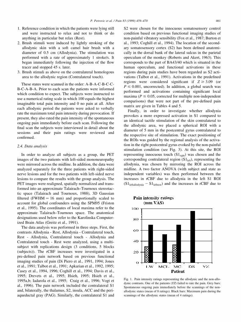
Fig. 1. Pain intensity ratings representing the allodynic and the non-allo-
dynic contrasts. One of the patients (YZ) failed to rate the pain. Grey bars:
Spontaneous ongoing pain immediately before the scannings of the non-
allodynic states (mean of 8 ratings). Black bars: Maximum pain during the
scannings of the allodynic states (mean of 4 ratings).
non-painful touch in the right S1 ROI
(S1righttouch 2 S1rightrest). Adjusted rCBF data for the ROIs
were used for this analysis.
3. Results
3.1. Behavioral results
In spite of instructions to try to avoid movements all but
one patient were unable to control minor muscular activity
in the extremities and face when the allodynic area was
brushed. This movement was observed as occasional muscle
contractions of the extremities and in the face during allo-
dynia. All patients rated the maximum total pain intensity as
being greater than 50/100 mm during the painful brushing
except for the patient who verbally failed to report the pain
intensity (Fig. 1). They also reported brush-evoked pain
only from the site of stimulation. Only two of the patients
reported spontaneous ongoing pain. Due to technical
problems, the heart rate of two patients could not be
continuously monitored. For the remaining three patients
the heart-rate during the scans increased signi®cantly in
the allodynic state compared to the other two states (mean
P. Petrovic et al. / Pain 83 (1999) 459±470462
Table 2
Increases in rCBF during allodynia for the group of patients with mononeuropathy (the data were mirrored for the patients with left-sided pain). The search
volume was restricted to the prede®ned pain matrixa
XYZ-coordinates Z-score rCBF-increase (%)
Thalamus
Thalamus sin
Allodynia vs. Rest: 2 10, 2 22, 4 3.83 S 3.4
Allodynia vs. Contralateral Touch: 2 12, 2 20, 4 4.53 S 4.1
Thalamus dx
Allodynia vs. Rest: 8, 2 22, 4 3.39 S 3.2
Allodynia vs. Contralateral Touch: 8, 2 20, 4 3.70 S 3.5
Lateral pain system
S1 sin
Allodynia vs. Rest: 2 16, 2 44, 56 6.89 S 9.0
2 22, 2 50, 52 6.83 S 7.3
Allodynia vs. Contralateral Touch: 2 16, 2 44, 56 7.03 S 9.3
2 20, 2 50, 52 6.23 S 6.7
S2 sin
Allodynia vs. Rest: 2 54, 2 38, 20 5.14 S 5.4
Allodynia vs. Contralateral Touch: 2 54, 2 36, 20 3.47 S 3.3
S2 dx
Allodynia vs. Rest: 42, 2 36, 16 4.01 S 2.2
54, 2 42, 20 3.82 S 3.7
Allodynia vs. Contralateral Touch: ± ± ± ±
Medial pain system
Anterior insula sin
Allodynia vs. Rest: ± ± ± ±
Allodynia vs. Contralateral Touch: ± ± ± ±
Anterior insula dx
Allodynia vs. Rest: 30, 16, 4 2.96 2.2
Allodynia vs. Contralateral Touch: 36, 12, 4 1.73 1.2
ACC (BA32/24)
Allodynia vs. Rest: 2 6, 14, 32 2.58 2.0
Allodynia vs. Contralateral Touch: 2 10, 12, 28 3.22 S 2.9
2 2, 16, 32 3.18 S 2.1
PAG/brainstem
Allodynia vs. Rest: Signi®cant activation
without any peak
activated voxel
Allodynia vs. Contralateral Touch: 4, 2 26, 2 4 3.75 S 3.2
a The locations of the maximally activated voxels are given in the coordinates of the Talairach±Tournoux atlas (Talairach and Tournoux, 1988). The search
for the exact location of the maximal activation was performed in the CBA atlas (Greitz et al., 1991). S � Significant activation. ACC � Anterior cingulate
cortex. PAG � Periaqueductal gray. S1 � Primary somatosensory cortex. S2 � Secondary somatosensory cortex.

heart-rate during allodynia � 74:12; mean heart-rate during
conterlateral touch � 63:26; mean heart-rate during
rest � 61:14; assessed by two factor ANOVA with state
and subject as independent variables; P-value , 0:0001;
F1;27 � 133:4).
3.2. Results of the search in the prede®ned matrix
Activations of the lateral pain system (with 9% rCBF
increase in the contralateral S1) and the thalamus bilaterally
were observed during allodynia (Table 2; Fig. 2A). No
signi®cant activation was found in the ACC or the anterior
insula of the medial pain system when the allodynic state
was compared to the non-allodynic states except for an ACC
activation just above the signi®cance level in the contrast
Allodynia - Contralateral touch. Non-painful touch acti-
vated a mirror site to one of the allodynia-induced maxima
in the S1 and also activated the contralateral S2 (Table 3).
Allodynia provocation and contralateral touch evoked
activations in mirror sites in the S1, as anatomically de®ned
P. Petrovic et al. / Pain 83 (1999) 459±470 463
Table 3
Increases in rCBF during non-painful brushing for the group of patients with mononeuropathy (the data were mirrored for the patients with left-sided pain). The
search volume was restricted to S1 and S2 contralateral to the stimulated lega
XYZ-coordinates Z-score rCBF increase (%)
S1 dx
Contralateral Touch vs. Rest: 14, 2 52, 48 3.20 S 2.4
S2 dx
Contralateral Touch vs. Rest: 42, 2 36, 12 4.15 S 2.2
a The locations of the maximally activated voxels are given in the coordinates of the Talairach±Tournoux space (Talairach and Tournoux, 1988). The search
for the exact location of the maximal activation was performed in the CBA atlas (Greitz et al., 1991). S � Significantactivation. S1 � Primary somatosensory
cortex. S2 � Secondary somatosensory cortex.
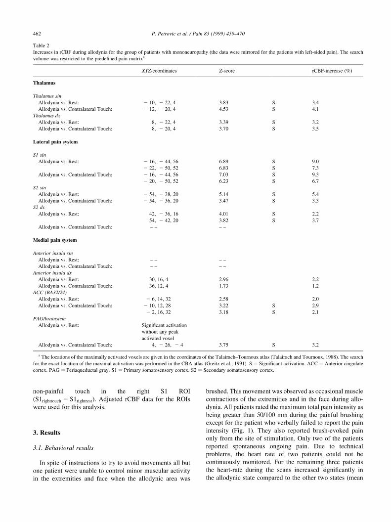
Fig. 2. (A) SPM (Statistical parametric mapping) results of increased rCBF in the S1, the S2, the brainstem and the cerebellum during Allodynia vs. Rest.
Although there was a contralateral dominance, the activation was bilateral. (B) SPM results of decreased activity bilaterally in the medial temporal lobe and
lateral temporal regions during Allodynia vs. Contralateral touch. All the data were thresholded at uncorrected P-value � 0:01. The images are shown in
neurological convention; left is left and right is right in the coronal section and the horizontal section is shown from above.
by the CBA (Fig. 3). The allodynic activation was situated
deep in the medial-dorsal parts of the left postcentral gyrus
(S1), most extensively within the borders of this gyrus but
also extending into the left pre-central gyrus (M1).
3.3. Additional rCBF-changes in the global search
All additional changes in rCBF during allodynia revealed
by the SPM analysis are presented in Tables 4 and 5, and
Fig. 2. The ipsilateral S1 was also activated during the allo-
dynic state vs. rest, although the most intensely elicited sites
were contralateral to the stimulation. Activations were also
observed in various motor regions, although no local maxi-
mum was observed in the primary motor cortex. Bilaterally
deactivated areas included the medial temporal lobe, the
lateral temporal lobe and the posterior occipital lobe.
3.4. Regional S1 ANOVA
The rCBF increase was more expressed during allodynia
than during touch in the corresponding contralateral soma-
totopic S1 ROI (7.1% vs. 2.5%). This difference was signif-
icant for the ®ve subjects when tested by two factor
ANOVA between the increases due to non-painful touch
in the right S1 ROI and the increases due to allodynia in
the left S1 ROI (P-value , 0:0003; F1:30 � 17:2).
3.5. Separate analyses of the left- and right
mononeuropathy-sided patients
During allodynia, increased activity was found bilaterally
in S1 and S2 of the lateral pain system and also in the
contralateral thalamus for the left- and right- sided mono-
neuropathy patients. Decreased activity was observed bilat-
erally in the medial temporal lobe, the occipital cortex and
the lateral temporal lobe for both groups. These results for
the two subgroups are in agreement with those from the
group analysis of all ®ve patients (data available upon
request).
4. Discussion
This paper deals with dynamic mechanical allodynia, i.e.
pain due to normally non-painful touch, in patients with
mononeuropathy. The patients included in this study were
carefully matched, which is of importance when averaging
the results across subjects. The group results obtained from
mirroring the data of the two patients with left-sided lesions
must, however, be interpreted with some caution, i.e. only
general conclusions can be made about the activations
outside the prede®ned pain matrix. However, the same
pattern of activations and deactivations were also revealed
by the separate analyses of the patients with left- and right-
sided mononeuropathy. The rCBF changes revealed by the
group and subgroup analyses were predominantly bilateral.
There is, of course, a possibility that the mirroring process
may mask unilateral rCBF changes.
It is our experience that for most patients with clinically
signi®cant tactile allodynia it is impossible to suppress all
movements during provocation. To reduce the number of
P. Petrovic et al. / Pain 83 (1999) 459±470464
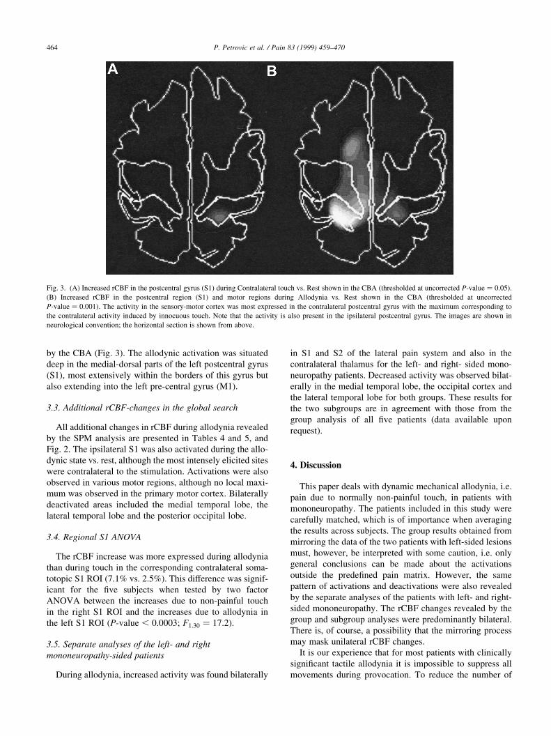
Fig. 3. (A) Increased rCBF in the postcentral gyrus (S1) during Contralateral touch vs. Rest shown in the CBA (thresholded at uncorrected P-value � 0:05).
(B) Increased rCBF in the postcentral region (S1) and motor regions during Allodynia vs. Rest shown in the CBA (thresholded at uncorrected
P-value � 0:001). The activity in the sensory-motor cortex was most expressed in the contralateral postcentral gyrus with the maximum corresponding to
the contralateral activity induced by innocuous touch. Note that the activity is also present in the ipsilateral postcentral gyrus. The images are shown in
neurological convention; the horizontal section is shown from above.
motor activations to a minimum the subjects were told to try
to avoid movements during the stimulation. However, occa-
sional muscular activity was observed in all but one of the
patients during the scanning period. Separating movement
related activations from pain related activations is a poten-
tial problem in all functional imaging studies of pain since
any observed movement or unobserved muscle tension may
activate the postcentral gyrus in concert with the motor
cortex (Colebatch et al., 1991; Hsieh et al., 1995b). In addi-
tion, movement intention/preparation may increase the
activity in the motor cortex (Hsieh et al., 1994; Deiber et
al., 1996). The same potential problem applies to S2 since
movement may activate this region (Weiller et al., 1996).
Movement related activity in the somatosensory regions is
not static or simply additive. Passive movements activate S2
signi®cantly more than active movements (Weiller et al.,
1996). The activity in these regions is also dependent on
which sensory channel is attended (Ghatan et al., 1995;
Shulman et al., 1997; Blakemore et al., 1998; Petrovic et
al., 1998). Thus, it is not known how, or if, movement
interacts with intense pain in S1 or S2 when attention is
directed to the painful stimulus. However, there are several
factors suggesting that the activation in the postcentral gyrus
was mainly related to a sensory response to allodynia.
First, the most expressed activation of the sensory-motor
cortex was located in the postcentral gyrus on the border to
the posterior parietal cortex, extending into the motor cortex
(Fig. 3). There was no local maximum in the pre-central
region. In comparison, in recent studies of motor activity
there are local maxima in the pre-central region or in the
central sulcus (Dettmers et al., 1995; Stephan et al., 1995;
Fink et al., 1997).
Secondly, the extensive S1-activation during allodynia
had one maximum at an almost identical but contralateral
coordinate as compared to the non-painful stimulation ( 222, 2 50, 52 during Allodynia vs. Rest and 14, 2 52, 48
during Touch vs. Rest; Fig. 3). This suggests that the activa-
tions stem from the same peripheral site.
Thus, we conclude that the increased S1 activity during
allodynia pertains primarily to the allodynic response but
observable or unobservable motor activity can not be
completely disregarded as a factor in the activity increase.
This problem is shared with most studies of pain where
subjects are awake and conscious (Coghill et al., 1994;
Casey et al., 1996; Rainville et al., 1997; Peyron et al.,
1998). To resolve this uncertainty, future studies need to
investigate this possible confound with, e.g. electromyogra-
phy (EMG).
Soft brushing on the non-allodynic side provoked a non-
painful, low-intensity tactile sensation. In the regional
analysis of increases during innocuous touch, activations
of the contralateral S1 and S2 were observed, which is in
agreement with previous PET studies of somatotactile
sensation (Fox et al., 1987; Burton et al., 1993; Coghill et
al., 1994).
Brushing the allodynic region provoked an acute painful
sensation in the leg with a maximum total pain intensity
which the patients rated between 50±100/100 mm. Signi®-
cant bilateral activations of S1 and S2 were observed during
the allodynic state compared to the habitual state. The ROI
analysis of the S1 con®rmed that the activation during the
allodynic sensation was signi®cantly more expressed than
the activations during non-painful touch sensation provoked
by the same stimulus but in the opposite leg (7.1% vs. 2.5%
rCBF increase).
Neurons responding to noxious stimuli have been found
in the primary and secondary somatosensory cortex of
primates (Robinson and Burton, 1980a; Kenshalo and Isen-
see, 1983; Dong et al., 1989). A study of primates with a
damaged primary somatosensory cortex and a case report of
a patient with a tumor affecting the secondary somatosen-
sory cortex have revealed clear de®cits in pain discrimina-
tion (Kenshalo et al., 1991; Greenspan and Win®eld, 1992)
which supports the hypothesis that the lateral pain system
plays an important role in the sensory/discriminative aspects
of pain processing (Kenshalo et al., 1980; Kenshalo and
Isensee, 1983; Albe-Fessard et al., 1985; Chung et al.,
1986; Friedman and Murray, 1986; Vogt et al., 1993;
Apkarian and Shi, 1994; Willis, 1995). Several functional
imaging studies of phasic heat pain, tonic pain and electri-
cally induced pain support the involvement of S1, S2 and
the thalamus in pain processing (Talbot et al., 1991; Casey
et al., 1994; Coghill et al., 1994; Hsieh, 1995; Casey et al.,
P. Petrovic et al. / Pain 83 (1999) 459±470 465
Table 4
Additional increases in rCBF outside the prede®ned pain network during
allodynia for the group of patients with mononeuropathy (the data were
mirrored for the patients with left-sided pain)a
XYZ-coordinates Z-scores Corrected
P-value
Allodynia vs. Rest
SMC/PMC/SMA/PPC
BA7 sin 2 26, 2 52, 48 5.73 0.000
BA6 sin 2 14, 2 8, 52 5.22 0.000
S1 dx 14, 2 48, 52 4.57 0.009
Cerebellum/Thalamus
Cerebellum sin 2 30, 2 50, 2 28 5.73 0.000
Vermis 0, 2 60, 2 12 5.29 0.000
Cerebellum dx 30, 2 50, 2 28 4.92 0.002
Allodynia vs.
Contralateral Touch
PMC/SMA
BA 6 sin 2 12, 2 6, 52 5.45 0.000
Cerebellum/Thalamus
Vermis 2 2, 2 60, 2 12 6.62 0.000
Cerebellum sin 2 28, 2 54, 2 28 5.79 0.000
Cerebellum dx 32, 2 46, 2 28 5.24 0.000
Cerebellum dx 34, 2 66, 2 28 4.38 0.018
a The locations of the maximally activated voxels are given in the coor-
dinates of the Talairach±Tournoux space (Talairach and Tournoux, 1988).
The search for the exact location of the maximal activation was performed
in the CBA atlas (Greitz et al. 1991). PMC � Premotor cortex. PPC �Posterior parietal cortex. SMA � Supplementary motor areas. S1 �Primary somatosensory cortex.

1996; Craig et al., 1996; Rainville et al., 1997). The studies
above indicate that the increased activity in the lateral pain
system during allodynia may contribute to the painful
experience. It is also likely that extensive activation of S1
and S2 represents a co-activation of the mechanoreceptive
and the nociceptive systems, consistent with previous
suggestions from the experimental literature (Hansson and
Kinnman, 1996). The results of the present study are in line
with studies of experimental allodynia and allodynia follow-
ing Wallenberg infarct where increased activity in S1 and
S2 were observed (Iadarola et al., 1998; Peyron et al., 1998).
The extensive activation of the lateral pain system in
dynamic mechanical allodynia contrasts with the lack of
activation in these regions during spontaneous ongoing
pain in patients with mononeuropathy (Hsieh et al.,
1995a) as well as in other ongoing neuropathic pain states
(Di Piero et al., 1991). The discrepancy may re¯ect that
provoked allodynia represents an acute exacerbation of the
pain that enhances the spatial localizing component. The
rate of change in pain intensity as a factor determining the
P. Petrovic et al. / Pain 83 (1999) 459±470466
Table 5
Decreases in rCBF during allodynia for the group of patients with mononeuropathy (the data were mirrored for the patients with left-sided pain)a
XYZ-coordinates Z-scores Corrected P-value
Rest vs. Allodynia
Medial temporal lobe dx/Lateral temporal lobe dx
Hippocampus dx 26, 2 22, 2 12 5.81 0.000
Amygdala/Hippocampus dx 22, 2 14, 2 12 5.74 0.000
Superior TG (BA38) dx 44, 2 8, 2 12 4.96 0.002
Middle TG (BA21) dx 56, 2 46, 2 12 4.90 0.002
Heschl's G/Planum temp (BA41/42) dx 54, 2 16, 4 4.54 0.010
Medial temporal lobe sin
Hippocampal gyrus/Uncus sin 2 36, 2 20, 2 20 4.35 0.021
Hippocampal gyrus/Uncus sin 2 34, 2 22, 2 16 4.33 0.02
Lateral temporal lobe sin
Superior TG/Heschl's G (BA22/41) sin 2 54, 2 14, 2 4 4.62 0.007
Occipital lobe sin
BA17/18 sin 2 18, 2 84, 2 8 5.00 0.001
BA18 sin 2 26, 2 92, 4 4.31 0.024
Occipital lobe/PCC
BA18 dx 24, 2 58, 4 5.00 0.001
PCC (BA29/30) sin 2 14, 2 48, 8 4.89 0.002
BA 19 dx 20, 2 80, 24 4.83 0.003
Precuneus (BA31) dx 18, 2 66, 12 4.42 0.016
BA 19 dx 34, 2 78, 24 4.25 0.030
BA 18/19 dx 36, 2 84, 2 4 4.22 0.034
Contralateral Touch vs. Allodynia
Medial temporal lobe dx/Lateral temporal lobe dx/ACC sin
Hippocampus/Amygdala dx 26, 2 22, 2 12 6.07 0.000
Middle TG (BA21/37) dx 54, 2 44, 2 12 5.37 0.000
Middle/Superior TG (BA21/22) dx 54, 2 36, 0 5.31 0.000
Middle/Superior TG (BA21/38) dx 44, 2 14, 2 16 5.21 0.000
Heschl's G (BA41) dx 54, 2 14, 0 4.78 0.003
Inferior ACC (BA32) sin 2 4, 34, 2 4 4.16 0.042
Medial temporal lobe sin
Hippocampal gyrus/Uncus sin 2 32, 2 18, 2 20 4.84 0.003
Hippocampus/Hippocampal gyrus sin 2 30, 2 20, 2 16 4.78 0.003
Amygdala/Uncus sin 2 22, 2 2, 2 16 4.52 0.010
Lateral temporal lobe sin
Middle TG (BA21) sin 2 58, 2 40, 2 12 5.47 0.000
Superior TG (BA22) sin 2 58, 2 20, 2 4 4.24 0.031
Superior TG/Heschl's G (BA22/41) sin 2 56, 2 16, 2 4 4.18 0.04
Occipital lobe/PCC/Lateral temporal lobe dx
PCC/Precuneus (BA23/31) 6, 2 56, 32 5.22 0.000
Middle TG (BA19) dx 48, 2 66, 20 5.08 0.001
BA 19 dx 34, 2 78, 24 4.62 0.007
BA 19 dx 18, 2 80, 28 4.21 0.035
BA 19 dx 38, 2 76, 24 4.59 0.01
a The locations of the maximally activated voxels are given in the coordinates of the Talairach±Tournoux space (Talairach and Tournoux, 1988). The search
for the exact location of the maximal activation was performed in the CBA atlas (Greitz et al., 1991). ACC � Anterior cingulate cortex. PCC � Posterior
cingulate cortex. TG � Temporal gyrus.
cortical response may actually be one of the reasons to why
there is a such a variability in the pain literature regarding
the involvement of S1 and S2 (Apkarian, 1995).
S2 is a small area de®ned anatomically in primates as
situated in the dorsal banks of the lateral sulcus in the parie-
tal opercular cortex (Roberts and Akert, 1963). Thus, acti-
vations of the human operculum in PET studies of pain and
other non-painful sensations have been regarded as S2 acti-
vations. However, contributions by the more posterior-
lateral area involved in somatosensory processing, de®ned
as 7b in monkeys, may also contribute to the increased
activation (Coghill et al., 1994). The peak activation of
the parietal operculum, including S2, was more posteriorly
located than in most previously published PET studies of
pain in which the stimuli have been induced in the arm
(Talbot et al., 1991; Casey et al., 1994, 1996; Coghill et
al., 1994; Craig et al., 1996; Rainville et al., 1997). In single
neuron recordings of S2, it has been shown that the foot area
is located more posteriorly than the face area in these struc-
tures (Robinson and Burton, 1980b). Andersson and co-
workers (Andersson et al., 1997) reported increased activity
in anterior parts of S2 when painful stimulation of the hand
was compared with painful stimulation of the foot. A joint
comparison (painful stimulation of the hand and foot vs.
baseline) revealed a more posterior activation of the S2.
Their results are congruent with our ®ndings and support
the existence of a somatotopic organization in S2.
Bilateral responses were observed in the thalamus, S1 and
S2 during the allodynic state compared to rest. Only unilat-
eral activation of the S1 in response to painful stimuli has
been reported previously. The activation in the ipsilateral
postcentral gyrus during allodynia had almost the same co-
ordinates as the brush-evoked activation, which suggests
that the ipsilateral response in S1 is somatotopic. However,
pain was always reported only from the stimulated side. The
bilateral responses in the primary somatosensory cortex
may be mediated by a subgroup of neurons in S1 which
normally respond to innocuous stimulation from a de®ned
contralateral receptive ®eld, but also respond to ipsilateral
intense noxious stimuli (Kenshalo and Isensee, 1983). We
con®rm previous observations that the S2 area has a
tendency to be activated bilaterally by unilateral painful
stimuli (Casey et al., 1994). This is consistent with animal
studies of S2 which have indicated bilateral receptive ®elds
of neurons responding to experimental noxious input (Dong
et al., 1989).
Several regions with decreased rCBF during the allodynic
state were observed. Decreases in rCBF are generally
regarded as a total net decrease in the neural activity of
the involved region (Hsieh, 1995; Raichle, 1997, 1998). In
this context it should be noted that the rCBF is an indirect
measure of brain activity and can not separate inhibition
from excitation but only measures the net result of neuronal
activity. Decreased activity was found in the lateral parts of
the temporal lobe bilaterally and in the occipital lobe/poster-
ior cingulate gyrus. These areas are involved in general
auditory, language and visuospatial processing (Mazziotta
et al., 1982; Ungerleider and Haxby, 1994; Ghatan et al.,
1995; Price et al., 1996). A deactivation of such regions may
re¯ect an attention-guided, top-down inhibition of proces-
sing of non-attended sensory components (Haxby et al.,
1994; Ghatan et al., 1998). Thus, it is suggested that the
observed deactivations in these sensory areas, during the
allodynic state compared to the non-allodynic states, re¯ect
increased attention towards the processing of allodynia.
Bilateral deactivations were also observed in the hippocam-
pus/parahippocampal gyrus, extending into the amygdala/
uncus during the allodynic state. These structures are
involved in declarative and emotional memory processing.
The amygdala is also involved in behavioral and autonomic
emotional response to aversive stimuli (LeDoux, 1993;
Squire and Zola, 1996; Petersson et al., 1997). We suggest
that these deactivations may represent a coping strategy for
handling an acute, but well-known painful situation. The
deactivations may re¯ect a meaningful suppression of
brain systems subserving episodic memory and emotional
response to aversive stimuli (Hsieh, 1995).
No signi®cant activation was found in the ACC or the
mid-anterior insula in the Allodynia vs. Rest comparison
(although a signi®cant activation was observed in the
ACC during Allodynia vs. Contralateral stimulation). This
is consistent with the results from a study of patients with
allodynia following Wallenberg infarct (Peyron et al.,
1998). Peyron et al. (1998) suggested that the absence of
ACC activation could be a speci®c feature for patients with
allodynia after Wallenberg infarct, which may also be the
case for allodynia after peripheral nerve damage. The lack
of activation in the medial pain system, in contrast to the
massive activations in the lateral pain system (7±9% rCBF
increase in S1), was still an unexpected ®nding since the
patient descriptions of dynamic mechanical allodynia
usually include affective components (Hansson, 1994) and
since the medial system is considered to be involved in
processing the affective and evaluative part of the pain
experience (Albe-Fessard et al., 1985; Vogt et al., 1993;
Craig et al., 1994; Talbot et al., 1995; Willis, 1995; Rain-
ville et al., 1997).
There were large differences in the total pain intensity
ratings during the non-allodynic scans (VAS ratings of
spontaneous pain ranging from 0 to 45 mm) and during
allodynia (VAS ratings ranging from 50 to 100 mm). A
continuous activation of the medial pain system has been
demonstrated in ongoing spontaneous pain (Hsieh et al.,
1995a) and the ACC response shows correlation with the
subjectively perceived pain intensity and unpleasantness
(Davis et al., 1997; Derbyshire et al., 1997; Rainville et
al., 1997). The variability of the perceived pain intensity
may have been paralleled by a variable response in the
medial pain system during both the allodynic and the rest
states which may explain the lack of signi®cant activation in
these structures when directly comparing the rCBF in the
allodynic with the non-allodynic states. Thus, the discre-
P. Petrovic et al. / Pain 83 (1999) 459±470 467
pancy in the activation of the medial pain system between
the present PET study of allodynia and several other PET
studies of pain (Jones et al., 1991; Talbot et al., 1991; Casey
et al., 1994, 1996; Coghill et al., 1994; Hsieh et al., 1995a,b;
Craig et al., 1996; Vogt et al., 1996; Rainville et al., 1997;
Iadarola et al., 1998) may be due to a heterogeneous patient
material in the clinical allodynia study. The same may be
the case in the study of Peyron and colleagues (Peyron et al.,
1998) since half of the included patients had an ongoing
spontaneous pain and displayed a variable response to the
induced allodynia.
Previously, a within condition correlation has been
observed between the reported unpleasantness of pain and
rCBF in the ACC (Rainville et al., 1997). Also, when the
rCBF was regressed on the rated pain intensity across condi-
tions a signi®cant correlation was observed in the ACC
(Davis et al., 1997; Derbyshire et al., 1997; Silverman et
al., 1997). Thus, subjectively perceived pain intensity/
unpleasentness may be more important for the level of activ-
ity in the ACC than the type of pain. In order to test this
hypothesis we performed a post-hoc analysis in which the
rCBF was regressed on the reported pain intensity ratings
(irrespective if it refers to spontaneous pain or induced allo-
dynia) across the rest and allodynia conditions, i.e. we used
the reported total pain intensity as a covariate of interest in
the general linear model. The results of the linear regression
showed several signi®cant activation foci, i.e. covariations
between the total pain rating and the rCBF, in the ACC
([x,y,z] � [2,6,40], Z-score � 3.32; [x,y,z] � [ 2 6,18,24],
Z-score � 3.34; [x,y,z] � [ 2 4, 2 16,44], Z-score � 4.87).
This is consistent with the previously observed signi®cant
regressions and suggests that the ACC maintains the
previously observed pattern of response to the overall
perceived pain, but that inhomogeneities in spontaneous
pain and also in the induced pain response may obscure
effects unless appropriately accounted for. Similarly, there
was a covariation between the pain intensity rating and the
rCBF in the ipsilateral anterior insula (�x; y; z� � �28; 18; 12�,Z-score � 4:1) and in the contralateral anterior insula (non-
signi®cant tendency, �x; y; z� � 232; 6; 4, Z-score � 2:21).
Hence, there seems to be a covariation between subjective
pain intensity rating and rCBF also for other areas of the
medial pain system. In addition, it should be noted that the
results for the lateral pain system were similar with the results
previously described above.
A decrease was also observed in the inferior part of the
ACC in the contrast Contralateral touch vs. Allodynia. This
region of the ACC is involved in attention and the observed
decrease may represent altered attention-dependent activity
(Hsieh et al., 1995a). This ®nding is in line with the study of
tactile allodynia after Wallenberg infarct (Peyron et al.,
1998).
PAG is involved in behavioral and autonomic emotional
responses during noxious stimulation (Carrive, 1993;
LeDoux, 1993) and has been activated in experimental trau-
matic pain (Hsieh et al., 1995b). In the present study, an
increased activation in the brainstem/PAG was observed
during the allodynic state, which was not unexpected
given the high intensity, the nature of the experienced
pain and the increased heart rate during this state.
5. Conclusions
In this study of dynamic mechanical allodynia in patients
with mononeuropathy, an extensive activation of the lateral
pain system was observed. The activation in the somatoto-
pic projection of the leg in S1 was signi®cantly more
expressed during allodynia than during non-painful touch.
Bilateral activations were observed in the lateral pain
system. Also, the activity of the medial pain system co-
varied positively to pain intensity ratings. This activation
pattern contrasts to previous studies of ongoing spontaneous
neuropathic pain where no increased activity of the lateral
pain system was observed. Finally, an extensive pattern of
deactivation was found during the allodynic state, which
may represent attention-based inhibitions and central coping
modulations during a severe, but well-known painful situa-
tion.
Acknowledgements
This work was supported by grants from the Swedish
Medical Research Council (8276), the Karolinska Institute,
the Swedish Medical Association, and the Knut and Alice
Wallenberg foundation. Special thanks to Ellenor Anders-
son, Monica Serrander, Gustav von Heine, GoÈran Printz,
Walter Pulka and Peter SoÈderholm for technical and admin-
istrative support.
References
Albe-Fessard D, Berkley KJ, Kruger L, Ralston HJ, Willis WD. Dience-
phalic mechanisms of pain sensation. Brain Res Rev 1985;9:217±296.
Andersson JLR, Lilja A, Hartvig P, LaÊngstroÈm B, Gordh T, Handwerker H,
TorebjoÈrk E. Somatotopic organization along the central sulcus, for
pain localization in humans, as revealed by positron emission tomogra-
phy. Exp Brain Res 1997;117:192±199.
Apkarian AV. Functional imaging of pain: new insights regarding the role
of the cerebral cortex in human pain perception. Semin Neurosci
1995;7:279±293.
Apkarian AV, Shi T. Squirrel monkey lateral thalamus. Somatic nocire-
sponsive neurons and their relation to spinothalamic terminals, J
Neurosci 1994;14:6779±6795.
Apkarian AV, Stea RA, Manglos SH, Szeverenyi NM, King RB, Thomas
FD. Persistent pain inhibits contralateral sosmatosensory cortical activ-
ity in humans. Neurosci. Lett 1992;140:141±147.
Bennett GJ. Neuropathic pain, R.a.W. In: Melzack PD, editor. Textbook of
Pain, Edinburgh: Churchill Livingstone, 1994. pp. 201±224.
BergstroÈm M, Boethius J, Eriksson L, Greitz T, Ribbe T, WideÂn L. Head
®xation device for reproducible position alignment in transmission CT
and positron emission tomography. J Comput Assist Tomogr
1981;5:136±141.
Berridge MS, Cassidy EH, Terris AH. A routine, automated synthesis of
P. Petrovic et al. / Pain 83 (1999) 459±470468
oxygen-15-labeled butanol for positron tomography. J Nucl Med
1990;31:1727±1731.
Blakemore SJ, Rees G, Frith CD. How do we predict the consequences of our
actions? A functional imaging study. Neuropsychologia 1998;36:521±
529.
Burton H, Videen TO, Raichle ME. Tactile-vibration-activated foci in
insular and parietal-opercular cortex studied with positron emission
tomography: mapping the second somatosensory area in humans.
Somatosens Mot Res 1993;10:297±308.
Carrive P. The periaqueductal gray and defensive behavior: functional repre-
sentation and neuronal organization. Behav Brain Res 1993;58:27±47.
Casey KL, Minoshima S, Berger KL, Koeppe RA, Morrow TJ, Frey KA.
Positron emission tomography analysis of cerebral structures activated
speci®cally by repetitive noxious heat stimuli. J Neurophysiol
1994;71:802±807.
Casey KL, Minoshima S, Morrow TJ, Koeppe RA. Comparison of human
cerebral activation patterns during cutaneous warmth, heat pain, and
deep cold pain. J Neurophysiol 1996;76:571±581.
Chung JM, Lee KH, Surmeier DJ, Sorkin LS, Kim J, Willis WD. Response
characteristics of neurons in the ventral posterior lateral nucleus of the
monkey thalamus. J Neurophysiol 1986;56:370±390.
Coghill RC, Talbot JD, Evans AC, Meyer E, Gjedde A, Bushnell MC,
Duncan GH. Distributed processing of pain and vibration by the
human brain. J Neurosci 1994;14:4095±4108.
Colebatch JG, Deiber MP, Passingham RE, Friston KJ, Frackowiak RSJ.
Regional cerebral blood ¯ow during voluntary arm and hand move-
ments in human subjects. J Neurophysiol 1991;65:1392±1401.
Craig AD, Bushnell MC, Zhang ET, Blomqvist A. A thalamic nucleus
speci®c for pain and temperature sensation. Nature 1994;372:770±773.
Craig AD, Reiman EM, Evans A, Bushnell MC. Functional imaging of an
illusion of pain. Nature 1996;384:258±260.
Davis KD, Wood ML, Crawley AP, Mikulis DJ. fMRI of human somato-
sensory and cingulate cortex during painful electrical nerve stimulation.
NeuroReport 1995;7:321±325.
Davis KD, Taylor SJ, Crawley AP, Wood ML, Mikulis DJ. Functional MRI
of pain- and attention-related activations in the human cingulate cortex.
J Neurophysiol 1997;77:3370±3380.
Deiber MP, Ibanez V, Sadato N, Hallett M. Cerebral structures participating
in motor preparation in humans: a positron emission tomography study.
J Neurophysiol 1996;75:233±247.
Derbyshire SWG, Jones AKP, Gyulai F, Clark S, Townsend D, Firestone
LL. Pain processing during three levels of noxious stimualtion produces
differential patterns of central activity. Pain 1997;73:431±445.
Dettmers C, Fink GR, Lemon RN, Stephan KM, Passingham RE, Silbers-
weig D, Holmes A, Ridding MC, Brooks DJ, Frackowiak RS. Relation
between cerebral activity and force in the motor areas of the human
brain. J Neurophysiol 1995;74:802±815.
Di Piero V, Jones AKP, Iannotti F, Powell M, Perani D, Lenzi GL, Frack-
owiak RSJ. Chronic pain: a PET study of the central effects of percu-
taneous high cervical cordotomy. Pain 1991;46:9±12.
Di Piero V, Ferracuti S, Sabatini U, Pantano P, Cruccu G, Lenzi GL. A
cerebral blood ¯ow study on tonic activation in man. Pain 1994;56:167±
173.
Dong WK, Salonen LD, Kawakami Y, Shiwaku T, Kaukoranta EM, Martin
RF. Nociceptive responses of trigeminal neurons in S2-7b cortex of
awake monkeys. Brain Res 1989;484:314±324.
Drevets WC, Burton H, Videen TO, Snyder AZ, Simpson JRJ, Raichle ME.
Blood ¯ow changes in human somatosensory cortex during anticipated
stimulation. Nature 1995;373:249±252.
Fink GR, Frackowiak RS, Pietrzyk U, Passingham RE. Multiple non-
primary motor areas in the human cortex. J Neurophysiol
1997;77:2164±2174.
Fox PT, Burton H, Raichle ME. Mapping human somatosensory cortex
with positron emission tomographgy. J Neurosurg 1987;67:34±43.
Friedman DP, Murray EA. Thalamic connectivity of the second somato-
sensory area and neighboring somatosensory ®elds of the lateral sulcus
of the macaque. J Comp Neurol 1986;252:348±373.
Friston KJ, Holmes AP, Worsley KJ, Poline JP, Frackowiak RSJ. Statistical
parametric maps in functional imaging: a general linear approach. Hum
Brain Mapp 1995;2:189±210.
Ghatan PH, Hsieh JC, WirseÂn-Meurling A, Wrendling R, Eriksson L,
Stone-Elander S, Levander S, Ingvar M. Brain activation induced by
the perceptual maze test: A PET study of cognitive performance.
Neuroimage 1995;2:112±124.
Ghatan PH, Hsieh JC, Petersson KM, Stone-Elander S, Ingvar M. Coex-
istence of attention-based facilitation and inhibition in the human
cortex. Neuroimage 1998;7:23±29.
Gracely RH, Lynch SA, Bennett GJ. Painful neuropathy: altered central
processing maintained dynamically by peripheral input. Pain
1992;51:175±194.
Greenspan JD, Win®eld JA. Reversible pain and tactile de®cits associated
with a cerebral tumor compressing the posterior insula and parietal
operculum. Pain 1992;50:29±39.
Greitz T, Bohm C, Holte S, Eriksson L. A computerized brain atlas:
construction, anatomical content and some applications. J Comput
Assist Tomogr 1991;15:26±38.
Hansson P. Possibilities and potential pitfalls of combined bedside and
quantitative somatosensory analysis in pain patients. In: Boivie J, Hans-
son P, Lindblom U, editors. Touch, temeprature, and pain in health and
disease: mechanisms and assessements, vol. 3. Seattle: IASP Press,
1994. pp. 113±132.
Hansson P, Kinnman E. Unmasking mechanisms of peripheral neurpathic
pain in a clinical perspective. Pain Rev. 1996;3:272±292.
Haxby JV, Horwitz B, Ungerleider LG, Maisog JM, Pietrini P, Grady CL.
The functional organization of human extrastriate cortex: A PET-rCBF
study of selective attention to faces and locations. J Neurosci
1994;14:6336±6353.
Hsieh JC. Central processing of pain; Functional brain imaging studies with
PET 91, Stockholm: Karolinska Institute, 1995.
Hsieh JC, HaÈgermark OÈ , StaÊhle-BaÈckdahl M, Ericson K, Eriksson L, Stone-
Elander S, Ingvar M. Urge to scratch represented in the human cerebral
cortex during itch. J Neurophysiol 1994;72:3004±3008.
Hsieh JC, Belfrage M, Stone-Elander S, Hansson P, Ingvar M. Central
representation of chronic ongoing neuropathic pain studied by positron
emission tomography. Pain 1995a;63:225±236.
Hsieh JC, StaÊhle-BaÈckdahl M, HaÈgermark OÈ , Stone-Elander S, Rosenqvist
G, Ingvar M. Traumatic nociceptive pain activates the hypothalamus
and the periaqueductal gray: a positron emission tomography study.
Pain 1995b;64:303±314.
Iadarola MJ, Max MB, Berman KF, Byas-Smith MG, Coghill RC, Gracely
RH, Bennett GJ. Unilateral decrease in thalamic activity observed with
positron emission tomography in patients with chronic neuropathic
pain. Pain 1995;63:55±64.
Iadarola MJ, Berman KF, Zef®ro TA, Byas-Smith MG, Gracely RH, Max
MB, Bennett GJ. Neural activation during acute capsaicin-evoked pain
and allodynia assessed with PET. Brain 1998;121:931±947.
Ingvar M, Eriksson L, Greitz T, Stone-Elander S, Dahlbom M, Rosenqvist
G, af Trampe P, von Euler CV. Methodological aspects of brain activa-
tion studies: cerebral blood ¯ow determined with [15O]butanol and
positron emission tomography. J Cereb Blood Flow Metab
1994;14:628±638.
Jones AKP, Brown WD, Friston KJ, Qi LY, Frackowiak RSJ. Cortical and
subcortical localization of response to pain in man using positron emis-
sion tomography. Proc R Soc Lond 1991;244:39±44.
Kenshalo DRJ, Isensee O. Responses of primate S1 cortical neurons to
noxious stimuli. J Neurophysiol 1983;50:1479±1496.
Kenshalo DRJ, Thomas DA, Dubner R. Primary somatosensory cortical
lesion reduce the monkeys' ability to discriminate and detect noxious
thermal stimulation. Soc Neurosci Abstr 1991;17:1206.
Kenshalo DRJ, Giesler GJ, Leonard RB, Willis WD. Responses of neurons
in primate ventral posterior lateral nucleus to noxious stimuli. J Neuro-
physiol 1980;43:1594±1614.
LeDoux JE. Emotional memory systems in the brain. Behav Brain Res
1993;58:69±79.
P. Petrovic et al. / Pain 83 (1999) 459±470 469

Mazziotta JC, Phelps ME, Carson RE, Kuhl DE. Tomographic mapping of
human cerebral metabolism: auditory stimulation. Neurology
1982;32:921±937.
Melzack R, Casey KL. Sensory, motivational and central control determi-
nants of pain: a new conceptual model. In: Kenshalo DR, editor. The
skin senses, Spring®eld IL: CC Thomas, 1968. pp. 423±443.
Montgomery SA, AÊ sberg M. A new depression scale designated to be
sensitive to change. Br. J. Psychiatry 1979;134:382±389.
Petersson KM, Elfgren C, Ingvar M. A dynamic role of the medial temporal
lobe during retrieval of declarative memory in man. Neuroimage
1997;6:1±11.
Petrovic P, Ghatan PH, Petersson KM, Stone-Elander S, Ingvar M. Cogni-
tion alters pain related activity in the lateral pain system. Neuroimage
1998;7:S437.
Peyron R, Garcia-Larrea L, GreÂgoire MC, Convers P, Lavenne F, Veyre L,
Froment JC, MauguieÁre F, Michel D, Laurent B. Allodynia after lateral-
medullary (Wallenberg) infarct; A PET study. Brain 1998;121:345±
356.
Price CJ, Wise RJS, Warburton EA, Moore CJ, Howard D, Patterson K,
Frackowiak RSJ, Friston KJ. Hearing and saying; The functional neuro-
anatomy of auditory word processing. Brain 1996;119:919±931.
Raichle ME. Food for thought. The metabolic and circulatory requirements
of cognition. [Review: 79 references]. Ann NY Acad Sci 835 (1997)
373-85.
Raichle ME. Behind the scenes of functional brain imaging: a historical and
physiological perspective. [Review: 86 references]. Proc Natl Acad Sci
USA, 1998;95:765±772.
Rainville P, Duncan GH, Price DD, Carrier B, Bushnell MC. Pain affect
encoded in human anterior cingulate but not somataosensory cortex.
Science 1997;277:968±971.
Roberts TS, Akert K. Insular and opercular cortex and its thalamic projec-
tion in Macaca mulatta. Schweiz Arch Neurol Neurochir Psychiatr
1963;92:1±43.
Robinson CJ, Burton H. Somatic submodality distribution within the
second somatosensory (S2) 7b, retroinsular, postauditory, and granular
insular cortical areas of M. fascicularis. J Comp Neurol 1980a;192:93±
108.
Robinson CJ, Burton H. Somatotopographic organization in the second
somatosensory area of m. fascicularis. J Comp Neurol 1980b;192:43±
67.
Shulman GL, Corbetta M, Buckner RL, Raichle ME, Fiez JA, Miezin FM,
Petersen SE. Top-down modulation of early sensory cortex. Cereb
Cortex 1997;7:193±206.
Silverman DH, Munakata JA, Ennes H, Mandelkern MA, Hoh CK, Mayer
EA. Regional cerebral activity in normal and pathological perception of
visceral pain. Gastroenterology 1997;112:64±72.
Squire LR, Zola SM. Structure and function of declarative and nondeclara-
tive memory systems. Proc Natl Acad Sci USA 1996;93:13515±13522.
Stephan KM, Fink GR, Passingham RE, Silbersweig D, Ceballos-Baumann
AO, Frith CD, Frackowiak RS. Functional anatomy of the mental repre-
sentation of upper extremity movements in healthy subjects. J. Neuro-
physiol 1995;73:373±386.
Talairach J, Tournoux P. Co-planar stereotaxic atlas of the human brain,
Stuttgart: George Thieme Verlag, 1988.
Talbot JD, Marrett S, Evans AC, Meyer E, Bushnell MC, Duncan GH.
Multiple representations of pain in human cerebral cortex. Science
1991;25:1355±1358.
Talbot JD, Villemure JG, Bushnell MC, Duncan GH. Evaluation of pain
perception after anterior capsulotomy: a case report. Somatosens Mot
Res 1995;12:115±126.
Ungerleider LG, Haxby JV. `What' and `where' in the human brain. Curr
Opin Neurobiol 1994;4:157±165.
Vogt BA, Sikes RW, Vogt LJ. Anterior cingulate cortex and the medial pain
system. In: Vogt BA, Gabriel M, editors. Neurobiology of cingulate
cortex and limbic thalamus: a comprehensive Handbook, Boston, MA:
BirkhaÈuser, 1993. pp. 313±344.
Vogt BA, Derbyshire S, Jones AKP. Pain processing in four regions of
human cingulate cortex localized with co-registered PET and MR
imaging. Eur J Neurosci 1996;8:1461±1473.
Weiller C, Juptner M, Fellows S, Rijntjes M, Leonhardt G, Kiebel S, Muller
S, Diener HC, Thilmann AF. Brain representation of active and passive
movements. Neuroimage 1996;4:105±110.
Wienhard K, Dahlbom M, Eriksson L, Michel C, Bruckbauer T, Pietrzyk U,
Heiss WD. The ECAT EXACT HR: Performance of a new high resolu-
tion positron scanner. J Comput Assist Tomogr 1994;18:110±118.
Willis WDJ. Cold, pain and the brain, Nature 1995;373:19±20.
P. Petrovic et al. / Pain 83 (1999) 459±470470