a portable device for the generation of drug-loaded three
TRANSCRIPT
pharmaceutics
Article
A Portable Device for the Generation of Drug-LoadedThree-Compartmental Fibers ContainingMetronidazole and Iodine for Topical Application
Francis Brako 1 , Chaojie Luo 2, Rupy Kaur Matharu 2,3 , Lena Ciric 3, Anthony Harker 4 ,Mohan Edirisinghe 2,* and Duncan Q. M. Craig 5,*
1 Medway School of Pharmacy, Universities of Kent and Greenwich, Chatham ME4 4TB, UK;[email protected]
2 Department of Mechanical Engineering, University College London, Torrington Place, London WC1E 7JE,UK; [email protected] (C.L.); [email protected] (R.K.M.)
3 Department of Civil, Environmental & Geomatic Engineering, University College London,Chadwick Building, Gower Street, London WC1E 6BT, UK; [email protected]
4 Department of Physics and Astronomy and London Centre for Nanotechnology, University College London,London WC1E 6BT, UK; [email protected]
5 School of Pharmacy, University College London, 29–39 Brunswick Square, London WC1N 1AX, UK* Correspondence: [email protected] (M.E.); [email protected] (D.Q.M.C.)
Received: 27 February 2020; Accepted: 13 April 2020; Published: 18 April 2020�����������������
Abstract: The use of combination therapies for the treatment of a range of conditions is now wellestablished, with the component drugs usually being delivered either as distinct medicaments orcombination products that contain physical mixes of the two active ingredients. There is, however,a compelling argument for the development of compartmentalised systems whereby the release,stability and incorporation environment of the different drugs may be tailored. Here we outline thedevelopment of polymeric fine fiber systems whereby two drugs used for the treatment of wounds maybe separately incorporated. Fibers were delivered using a newly developed handheld electrospinningdevice that allows treatment at the site of need. Crucially, the delivery system is portable and may beused for the administration of drug-loaded fibers directly into the wound in situ, thereby potentiallyallowing domiciliary or site-of-trauma administration. The three-layered fiber developed in this studyhas polyethylene glycol as the outermost layer, serving as a structural support for the inner layers.The inner layers comprised iodine complexed with polyvinylpyrrolidone (PVP) and metronidazoledispersed in polycaprolactone (PCL) as a slow release core. The systems were characterized in termsof structure and architecture using scanning electron microscopy, transmission electron microscopy,attenuated total reflection Fourier transform infrared spectroscopy and diffractometry. As antibacterialcreams are still used for managing infected wounds, the performance of our trilayered fiber wasstudied in comparison with creams containing similar active drugs. Drug release was measuredby UV analysis, while antimicrobial efficiency was measured using agar diffusion and suspensionmethods. It was found that the trilayered systems, averaging 3.16 µm in diameter, released more drugover the study period and were confirmed by the microbacterial studies to be more effective againstP. aeruginosa, a bacterium commonly implicated in infected wounds. Overall, the portable systemhas been shown to be capable of not only incorporating the two drugs in distinct layers but also ofdelivering adequate amounts of drugs for a more effective antibacterial activity. The portability ofthe device and its ability to generate distinct layers of multiple active ingredients make it promisingfor further development for wound healing applications in terms of both practical applicability andantimicrobial efficacy.
Pharmaceutics 2020, 12, 373; doi:10.3390/pharmaceutics12040373 www.mdpi.com/journal/pharmaceutics
Pharmaceutics 2020, 12, 373 2 of 16
Keywords: trilayered fibers; portable electrospinning; wound dressing; combination therapy;compartmental drug delivery
1. Introduction
Chronic wounds remain a significant burden on the patient population and healthcare systemsworldwide [1], with infection being the likeliest cause of delay in healing. Moreover, the formation ofbiofilms from the secretions of infecting bacteria, into which they become embedded, further inhibits healing.Indeed, biofilms are a significant factor in 70% of wounds that remain chronic [2]. One treatment approachis the application of antiseptics and antibiotics, preferably in combination, in order to simultaneouslysuppress biofilm formation [3] and prevent infection [4]. Administering more than one drug concurrentlyfacilitates combinatorial selectivity, in which the different drugs involved can simultaneously inhibitseveral associated facets of a disease process. In addition, localisation allows for the efficient targeting oftissues, thereby maximising drug efficacy while also minimising toxicity via the control of the selectivityof the region of the body exposed to the therapeutic agent [5]. There is a need for wound managementstrategies that utilise a multifaceted approach to simultaneously target pathogeneses of the wound.For instance, diabetic foot ulcers (DFUs) are among the most difficult wounds to clinically manage, and ithas long been established that anaerobes are typically associated with deeper and more severe infectionsin these kinds of ulcers [6]. Furthermore, as it is common to isolate several microbial species from infectedwounds such as DFUs [7], an approach utilising combination therapies where multiple active agents areefficiently delivered simultaneously will be required for treating such wounds.
Combination therapies are nevertheless hampered by a lack of compartmentalisation betweenthe associated drugs. Current clinical practice in wound care, for instance, involves the application ofmultiple and distinct delivery systems that lead to the variability of dose and poor control of localconcentrations. A more elegant solution would be to deliver a compartmentalised system whereby,irrespective of the different physicochemical properties of the drugs, the possibility exists for tuningthe release while also enhancing the stability and localisation of the different agents to maximisetherapeutic benefits. Indeed, multi-compartmentalisation is regarded as a potentially crucial strategyfor delivery as it offers the potential for the efficient delivery of diverse forms of active entities [8].
Our suggested method utilises an on-demand/need approach in which layered fibers are appliedto wounds at the point of need, with each distinct layer containing a different therapeutic agent or afunctionalised controlling layer. The middle layer has iodine stabilised in polyvinylpyrrolidone andintended as an antiseptic to sanitise the wound area, paving the way for a more effective anti-bacterialaction from metronidazole contained in slowly degrading polycaprolactone making up the coreof this unique fiber system. Iodine, in previous studies, has been shown to effectively inhibit theformation and growth of biofilms [3], thus informing our decision to combine it with metronidazolefor a multi-targeted approach to wound management. The compartmental encapsulation of drugs indistinct layers of polymeric fibrous systems has several potential advantages. Specifically, the fibrousmesh has a high surface area and allows fluid permeation, while the layered structure within the fibersallows the separation of drugs into physically distinct environments with the potential for tunablecontrolled release. In addition, the physical flexibility and versatility of the macroscopic mesh structureallow lesions of irregular shapes to be filled neatly [9,10].
These potential advantages of combination therapies have led to a significant increase in researchinto compartmentalised structures as patient-specific drug delivery systems [11,12]. However, severallimitations hamper their practical applicability in a clinical setting. Despite recent advances in productiontechnologies such as multi-layer electrospinning and significant improvements in scale-up, the productionof multi-layered drug-loaded fibers requires a skilled worker operating expensive, bulky and essentiallyimmobile bench-top apparatus in a specialised laboratory or factory environment. Furthermore, the highly
Pharmaceutics 2020, 12, 373 3 of 16
delicate nature of the fibers produced renders it difficult to preserve structural integrity during packaging,transport and administration from the point of manufacture to the point of use.
It is therefore therapeutically highly advantageous to develop a portable and inexpensive devicethat may produce multi-layered therapeutic materials in situ at the point of trauma. While the designof a portable electrospinning device has been reported by several research groups [13–15], none of theseis capable of producing multi-layered structures that can be useful for developing multi-compartmentsystems for the efficient simultaneous delivery of different drugs. Here, we describe a simple-to-operate,miniaturised portable device for generating trilayered fine structures at the point-of-use. As aproof-of-concept to explore the use of a trilayered fibrous system, we develop a new, improvedcombination therapy consisting of iodine and metronidazole (MTZ) for wound dressing applications.We demonstrate the potential clinical efficacy of this approach using drug release and antimicrobialstudies; more specifically, metronidazole (MTZ) is an antibiotic agent widely recognised as a standardfor the elimination of anaerobic bacteria [3]. Moreover, formulations combining the antibacterialMTZ with the antiseptic iodine have been found to exhibit promising synergistic wound healingproperties [16]. The MTZ-iodine formulations are typically presented as ointments in which the drugsare simply mixed as physical combinations [17]. Here, we load the active ingredients into differentpolymeric compartments in a trilayered fiber using the portable device. More specifically, MTZdispersed in a biodegradable polymer, polycaprolactone (PCL), makes up the innermost core of thetrilayered fiber. The middle layer contains iodine stabilised in polyvinylpyrrolidone (PVP), the formeracting as an antimicrobial agent to rapidly sanitise the wound upon application. Finally, the outermostlayer is made of a polyethylene glycol (PEG) polymer as the shell fabric, acting to provide physicalrobustness and moisture regulation as well as a wound barrier to prevent contamination.
The potential advantages of the new approach are manifold. First, by keeping each ingredient ina separate compartment the performance and stability of the formulation are enhanced as interactions(should they exist) between the drugs are minimised prior to the onset of action. Secondly, the core-shellfibrous structure provides a controlled drug release while acting as an effective physical protectionand dressing for the wound. Thirdly, polymeric systems have been demonstrated to reduce irritationand toxicity associated with antiseptics such as iodophores when used in wound care [18]. Lastly,the portable production method allows highly specific systems to be tailored/personalised and deliveredon-site for a desired therapeutic outcome while eliminating the need for the packaging and transportof the delicate product to the site of use, thereby further preserving the drug and dosage form integrity.Here, we explore the manufacturing feasibility, structural architecture and potential therapeutic efficacyof the portable trilayered system so as to establish the applicability of the approach to both the treatmentof wound infections and, more broadly, the treatment of in situ conditions under a range of settings.
2. Materials and Methods
2.1. Materials
Metronidazole (MTZ, analytical standard, Mw = 171.15 g/mol), polyvinylpyrrolidone-iodine complex(PVP-I, European Pharmacopoeia Reference Standard with average Mw = 365 g/mol), high molecularweight polyvinylpyrrolidone (PVP, Mw = 360,000 g/mol), polycaprolactone (PCL, Mw = 80,000 g/mol),polyethylene glycol (PEG, BioUltra, Mw = 35,000 g/mol), dichloromethane (DCM), dimethylformamide(DMF) and ethanol (EtOH) were purchased from Sigma Aldrich (Poole, Dorset, UK) and used as received.
MTZ and metronidazole-iodine (MTZ-I) in aqueous cream BP (British Pharmacopoeia) base wereprepared as follows: 10 g of the MTZ cream comprised of 0.1 g of MTZ (batch number MKBZ3056V,Sigma Aldrich), 3 g of emulsifying ointment (batch number NR 10043, Pinewood Healthcare, Dublin,Ireland), 0.1 g of phenoxyethanol (batch number STBD 3014V, Sigma Aldrich) and made up to thetarget weight with purified water. The MTZ-I cream contained the same ingredients as the MTZcream with the addition of 0.5 g PVP-I, amounting to 0.5% elemental Iodine (equivalent to a 2:1 MTZ:PVP-I weight ratio). Lysogeny broth (LB) and LIVE/DEADTM BacLightTM Bacterial Viability and

Pharmaceutics 2020, 12, 373 4 of 16
Counting Kits were purchased from Thermo Fisher Scientific (Waltham, MA, USA), whilst LB agarwas purchased from Sigma Aldrich.
2.2. Solution Preparation and Characterization
To prepare the trilayered electrospun fibers, different polymer or polymer/drug solutionswere prepared, selecting materials known to enable optimised encapsulation and stable trilayeredelectrospinning as well as to fulfil the objective of developing a system which combined MTZ andIodine for a synergistic antimicrobial outcome. Materials and parameters used in developing thissystem are shown in Table 1.
Table 1. Solution composition and corresponding flow rates used in the various sections of our multipleconcentric needle systems for trilayered electrospinning.
Fiber Layer Flow Rate (mL h−1) Material Solvent
Core layer 2 4 wt % MTZ + 12 wt % PCL 4:1 v/v DCM: DMF
Middle layer 4 10 wt % PVP-I or 10 wt %PVP-I/PVP (1:1 w/w) EtOH
Shell layer 7 25 wt % PEG DCM
The physical properties of the three solutions used for the three layers of the fibers were characterised.The surface tension was measured with a Kruss Tensiometer K9 (Hamburg, Germany) using the standardWilhelmy’s plate method. The electrical conductivity was measured using a conductivity meter (Jenway3540, Bibby Scientific, Stone, Staffordshire, UK). The viscosity was measured with a digital rotationalviscometer (Brookfield DV-111, Harlow, Essex, UK) using spindle number 18 at a shear stress of 3.5 Pa.All procedures were repeated five times, and the mean value of the readings were recorded. The viscometerwas calibrated automatically by following prompts to run a complete cycle without the spindle in place.The tensiometer was calibrated by taking the surface tension of distilled water at room temperature andcomparing it with the literature value. The conductivity meter was calibrated by immersing the probe inthe conductivity standard (Bibby Scientific, Staffordshire, UK), and the meter was adjusted until a readingof 1194 µS cm−1 at 22 ◦C was obtained.
2.3. Portable Trilayered Electrohydrodynamic Apparatus and Processing Conditions
The portable trilayered electrohydrodynamic apparatus is shown in Figure 1. Additional photographsand a schematic diagram is shown in the Supplementary Information (Section S1, Figure S1). The threedifferent solutions were each loaded in a 10 mL syringe attached to a stainless-steel concentric needlesystem with multiple inlets and outlets. The concentric needle system is an additive system that can bechanged between single-layer, two-layer, trilayer and quad-layer depending on the number of layersrequired for the products; in this work, the system is used in the trilayer mode. Fibers were generated usingthe positive polarity of the voltage supply and collected on a grounded aluminum plate. Three miniaturehigh-precision micro-syringe pumps (Micrel mph+, Inspiration Healthcare Limited, UK) were used, eachsupplying one of the three solutions through the concentric needle system, charged by a miniature highvoltage supply able to generate up to 33 kV at 10 W (EMCO 4330+, XP Power, UK). The solutions wereelectrospun at a positive voltage of 17 kV, a working distance (between the needle exit and the collector) of15 cm, and flow rates of 2, 4 and 7 mL h−1 for the core, middle and shell solutions, respectively (Table 1).All electrospinning procedures were carried out under an ambient temperature, humidity and pressure(22 ◦C, 1 atmospheric pressure, 40–50% relative humidity), as described in our previous work [19].
2.4. Fiber Characterisation
The surface morphology and diameter of the fibers were studied using a Quanta 200F field emissionscanning electron microscope (Thermo Fisher Scientific). Prior to observation, each sample was coated
Pharmaceutics 2020, 12, 373 5 of 16
with gold using a Quorum Q150T turbo-pumped sputter coater (Quorum Technologies, Lewes, EastSussex, UK) for 90 s. The average fiber diameter was determined using Image-Pro Plus software (MediaCybernetics, Inc., Rockville, MD, USA) and was based on at least 100 measurements from the scanningelectron microscope (SEM) images. In order to confirm that the fibers had three distinct layers, transmissionelectron microscope (TEM) imaging of fibers collected on carbon-coated grids was conducted using TEMCM 120 Biotwin (Thermo Fisher Scientific), operating at an accelerating voltage of 100 kV.
To verify the composition of the fibers, attenuated total reflection Fourier transform infraredspectroscopy (ATR–FTIR) was carried out using a Vertex 90 spectrometer (Bruker, Coventry, WestMidlands, UK). The measurements were interpreted using OPUS Viewer version 6.5 software. Eachsample was scanned 32 times at a resolution of 2 cm−1. To investigate the solid-state of the drugsentrapped in the multi-layered fibers, samples were analysed using a D/Max-BR diffractometer (RigaKu,Tokyo, Japan) with Cu Kα radiation. Analyses were conducted at 40 mV and 30 mA over the 2θ rangeof 5◦–80◦ at a rate of 2◦ min−1. The data obtained were converted to diffractograms and evaluatedusing OriginPro 7.0 software (OriginLab Corporation, Northampton, MA, USA).
2.5. Drug Release Study
To measure and compare the amount of drug released respectively from the fiber and cream formulations,we used a system utilising a dialysis membrane as a means of filtration to minimise the extent of othermaterials from either the trilayered fibers or creams interfering with the amount of MTZ sampled for the UVanalysis. 50 mg of the drug-loaded sample was first enclosed in a 5 mL dialysis membrane with a molecularweight cut-off of 8000 g mol−1. The sample was then immersed in a 45 mL vessel (sampling chamber).A physiological temperature of 37 ◦C and a continuous agitation at 80 rpm were maintained throughout therelease study using a benchtop incubation shaker (Sciquip, Newtown, Shropshire, UK). The total durationof the study was five days. Samples of 3 mL were taken at various intervals from the vessel. A 3 mL blankmedium was added after each sampling to maintain a constant total volume.
An ultraviolet-visible (UV–Vis) spectroscopy (Jenway 6305 UV/Visible spectrophotometer, BibbyScientific, Stone Staffordshire, UK) operating at 322 nm was used to quantify the amount of drugpresent as a function of time. To determine the total amount of drug entrapped during fiber formation,1 mg of the MTZ-loaded trilayered fibers was dissolved completely in 10 mL of dichloromethane,followed by measuring the solution’s absorbance.
2.6. Antimicrobial Activity
Two microbial inhibition assays were carried out to compare the antimicrobial activity of theprepared trilayered fibers and MTZ-I/MTZ cream formulations. The first assay assessed the areaof inhibition on an agar plate, while the second assay assessed the 24-h bactericidal results in cellsuspensions analysed by flow cytometry. Pseudomonas aeruginosa NCTC 12903 (Public Health England)was chosen as the model microorganism to assess the antibacterial properties of the prepared trilayeredfibers and MTZ-I/MTZ cream formulations. P. aeruginosa is one of the most common bacteria isolatedfrom chronic wounds, typically resident in the deepest region of the wound bed [20]. Furthermore,P. aeruginosa is well known for its high virulence, causing severe morbidity by releasing proteasesand cytotoxic substances in chronic wound environments and thereby seriously impeding woundhealing [21]. P. aeruginosa NCTC 12903 is isolated from blood cultures and is commonly used forsusceptibility testing. P. aeruginosa NCTC 12903 was propagated as per the manufacturer’s instructionsand stored at −80 ◦C in a MicrobackTM.
2.6.1. Agar Diffusion Assay
A single colony of P. aeruginosa NCTC 12903 was suspended in 100 µL of sterile deionised waterand transferred to an LB agar plate. A plastic L-shaped spreader was used to spread the bacteria overthe plate. The plate was allowed to air dry. 1 g of the fibers and cream were placed in the centre of theplate and had an area coating of approximately 1 cm in diameter (concentration of 10 wt %). After
Pharmaceutics 2020, 12, 373 6 of 16
application, it was ensured that the fibers had made complete contact with the agar surface. The plateswere incubated for 24 h at 37 ◦C. The assay was performed in triplicate for each treatment method.The antibacterial activity was assessed by the diameter of the growth inhibition zone. Photographs ofagar plates and inhibition zones are shown in the Supplementary Information (Section S3, Figure S3).
2.6.2. Suspension Assay
A method previously used for assessing antimicrobial activity [22] was adapted for this study.A single colony of P. aeruginosa was suspended in 30 mL of sterile LB broth. The suspension was incubatedat 37 ◦C and 150 rpm until it reached its mid-exponential phase (OD600 0.4, equivalent to 8 Log10CFU/mL). A 1:100 dilution of the bacterial suspension was inoculated into sterile LB broth. The fibers andcream were respectively added to the bacterial suspensions at 10 wt % and incubated for 24 h at 37 ◦C and150 rpm. Flow cytometry was used in conjunction with the LIVE/DEAD BacLight Bacterial Viability assayto enumerate the number of live and dead cells in the suspension post-incubation. This method relies onthe use of fluorescent stains, SYTO® 9 and propidium iodide (PI) (both from Thermo Fisher Scientific).SYTO® 9 is a green fluorescent nucleic dye which is able to penetrate both live and dead cells, whilst PI isa red fluorescent intercalating stain which can only penetrate cells with damaged membranes (non-viablecells) to displace the SYTO®9 and which results in a red colouration. A stock solution of PI and SYTO® 9was prepared according to the manufacturer’s instructions. 180 µL of the stock staining solution wasadded to 20 µL of diluted sample and incubated at room temperature in the dark for 15 min.
After incubation, cells were acquired using a calibrated Guava easyCyte® flow cytometer andInCyte software (guavaSoft 3.1.1) (Luminex Corporation, Austin, TX, USA). Gates were set upusing positive (media and bacteria only), negative (media only) and fluorescent minus one control(single-stained positive controls). 50,000 events were collected, and the bacteria acquisition gates weredetermined using forward scatter and side scatter channels to eliminate background noise and debrisfrom the sample. The gated population of bacteria was then analysed using fluorescent channels.FlowJo was used to gate the live and dead bacterial cell counts, where the proportions of live and deadbacteria cells were calculated. All experiments were repeated at least three times.Pharmaceutics 2020, 12, x FOR PEER REVIEW 7 of 16
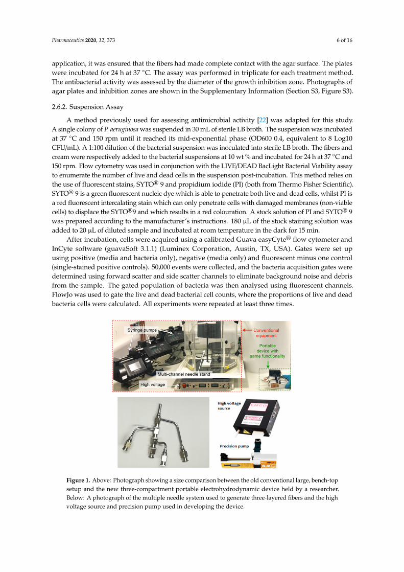
Figure 1. Above: Photograph showing a size comparison between the old conventional large, bench-top setup and the new three-compartment portable electrohydrodynamic device held by a researcher. Below: A photograph of the multiple needle system used to generate three-layered fibers and the high voltage source and precision pump used in developing the device.
The high precision micro-pumps (Micrel MP mlh+, Micrel Medical Devices SA, Koropi, Athens, Greece) are compact, lightweight and affordable (each at 0.15 m3 (volume), 0.2 kg (weight), when inclusive of 6 AAA batteries, and retailing at £1k). The battery-operation can sustain up to 600 h of continuous infusion at a high precision of 0.1 mL hr−1. These micro-pumps are as effective as conventional syringe pumps for electrospinning but are at least 50 times smaller, 10 times lighter and 4 times cheaper (e.g., conventional precision syringe pumps are ~7.5 m3, 2 kg and retail at £2–4k). The apparatus is easy to operate and can be hand-held for the direct generation of structures (both particles and fibers) at the point-of-use (Figure 1). Previously, we demonstrated how this miniaturised device could be used in the manufacture of a system containing silver nanoparticles as antibacterial agents to be applied as a wound dressing [17]. This study represents, however, the first time trilayered electrospinning has been achieved using a truly miniaturised setup.
3.2. Solution Compositions for Optimal Trilayered Fiber Formation
To generate trilayered fibers with distinct compartments containing MTZ and iodine under optimal processing conditions, we first characterised the physical properties of each solution used for the trilayered co-flow (Table 2). Trilayered fibers generated from 10 wt % of PVP-I solutions were found to be beaded, with an average diameter of 1.08 ± 0.54 µm (Figure 2a,c). The bead-on-string morphology is undesirable because it compromises the compartmental integrity of the trilayered fibers. It is well known that increasing the solution viscosity leads to a reduction in the undesirable bead-on-string morphology [23]; the viscosity of 10 wt % of PVP-I solutions at 4.1 ± 0.3 mPa.s was too low to enable a sufficient molecular chain entanglement for bead-free fiber formation (Table 2). By mixing the PVP-I complex with a high molecular weight PVP (Mw at 360,000 g/mol, Table 1), the resultant PVP-I/PVP solution had a higher viscosity of 78.6 ± 1.2 mPa.s and enabled bead-free fiber formation (Table 2, Figure 2b). The smooth trilayered fibers had an average diameter of 3.16 ± 1.05 µm (Figure 2d). The increased fiber diameter is attributed to both the increased viscosity as well as the lower electrical conductivity of the PVP-I/PVP solution in comparison to the pure PVP-I solution used in forming the beaded fibers [24]. The electrical conductivity of the pure PVP-I solution was 340 ± 4 µS m−1, which is nearly threefold higher than that of the PVP-I/PVP solutions at 121 ± 0.5 µS m−1. A decrease in solution electrical conductivity is known to generate larger fiber diameters in electrospinning [25]; hence, these results are compatible with the existing knowledge base.
Figure 1. Above: Photograph showing a size comparison between the old conventional large, bench-topsetup and the new three-compartment portable electrohydrodynamic device held by a researcher.Below: A photograph of the multiple needle system used to generate three-layered fibers and the highvoltage source and precision pump used in developing the device.
Pharmaceutics 2020, 12, 373 7 of 16
3. Results
3.1. Portable Multi-Layer Electrospinning Device
The core-shell concentric electrospinning device could be a useful innovation in and of itself, in thatthis portable assembly can be readily adapted to generate single-layer, core-shell layer, three-layer andfour-layer products, depending on the number of compartments required. The components for theportable multi-layer electrospinning device are off-the-shelf and simple to assemble; the assembly ofthe miniature high voltage (HV) supply and precision syringe micro-pumps are less expensive but aseffective as their heavy, voluminous and practically immobile bench-top counterparts. The portableHV supply (adapted from EMCO 4330+, XP Power) can produce 0–33 kV at 10 W, a wider range than abench-top HV supply which typically supplies 0–30 kV. The mini HV unit weighs less than 0.7 kg at avolume of 0.28 m3, and costs < £800. This is 95 times smaller, 10 times lighter and 3 times cheaper thana traditional HV unit (~7 kg, 27 m3, £2–5k).
The high precision micro-pumps (Micrel MP mlh+, Micrel Medical Devices SA, Koropi, Athens,Greece) are compact, lightweight and affordable (each at 0.15 m3 (volume), 0.2 kg (weight), when inclusiveof 6 AAA batteries, and retailing at £1k). The battery-operation can sustain up to 600 h of continuousinfusion at a high precision of 0.1 mL h−1. These micro-pumps are as effective as conventional syringepumps for electrospinning but are at least 50 times smaller, 10 times lighter and 4 times cheaper (e.g.,conventional precision syringe pumps are ~7.5 m3, 2 kg and retail at £2–4k). The apparatus is easy tooperate and can be hand-held for the direct generation of structures (both particles and fibers) at thepoint-of-use (Figure 1). Previously, we demonstrated how this miniaturised device could be used in themanufacture of a system containing silver nanoparticles as antibacterial agents to be applied as a wounddressing [17]. This study represents, however, the first time trilayered electrospinning has been achievedusing a truly miniaturised setup.
3.2. Solution Compositions for Optimal Trilayered Fiber Formation
To generate trilayered fibers with distinct compartments containing MTZ and iodine underoptimal processing conditions, we first characterised the physical properties of each solution usedfor the trilayered co-flow (Table 2). Trilayered fibers generated from 10 wt % of PVP-I solutions werefound to be beaded, with an average diameter of 1.08 ± 0.54 µm (Figure 2a,c). The bead-on-stringmorphology is undesirable because it compromises the compartmental integrity of the trilayeredfibers. It is well known that increasing the solution viscosity leads to a reduction in the undesirablebead-on-string morphology [23]; the viscosity of 10 wt % of PVP-I solutions at 4.1 ± 0.3 mPa·s wastoo low to enable a sufficient molecular chain entanglement for bead-free fiber formation (Table 2).By mixing the PVP-I complex with a high molecular weight PVP (Mw at 360,000 g/mol, Table 1),the resultant PVP-I/PVP solution had a higher viscosity of 78.6 ± 1.2 mPa·s and enabled bead-freefiber formation (Table 2, Figure 2b). The smooth trilayered fibers had an average diameter of 3.16± 1.05 µm (Figure 2d). The increased fiber diameter is attributed to both the increased viscosity aswell as the lower electrical conductivity of the PVP-I/PVP solution in comparison to the pure PVP-Isolution used in forming the beaded fibers [24]. The electrical conductivity of the pure PVP-I solutionwas 340 ± 4 µS m−1, which is nearly threefold higher than that of the PVP-I/PVP solutions at 121 ±0.5 µS m−1. A decrease in solution electrical conductivity is known to generate larger fiber diametersin electrospinning [25]; hence, these results are compatible with the existing knowledge base.
The bead-free, smooth continuous fibers comprising PCL + MTZ, PVP-I/PVP (1:1) and PEG,henceforth referred to as trilayered fibers, were used for the subsequent analysis in this study.

Pharmaceutics 2020, 12, 373 8 of 16
Table 2. Physical properties of the electrospinning solutions used to generate trilayered fibers.
Solution Concentration(wt %)
Viscosity(mPa·s)
Surface Tension(mNm−1)
Conductivity(µSm−1)
PCL + MTZ 12 + 4 1355.0 ± 6.4 40.2 ± 0.6 5.37 ± 0.04
PEG 25 1605.0 ± 5.0 39.0 ± 0.9 2.90 ± 0.02
PVP-I 10 4.1 ± 0.3 24.6 ± 0.3 340.00 ± 4.00
PVP + PVP-I1:1 w/w 10 78.6 ± 1.2 27.8 ± 0.3 121.00 ± 0.50
Pharmaceutics 2020, 12, x FOR PEER REVIEW 8 of 16
Table 2. Physical properties of the electrospinning solutions used to generate trilayered fibers.
Solution Concentration (wt %)
Viscosity (mPa.s)
Surface tension (mNm−1)
Conductivity (µSm−1)
PCL + MTZ 12 + 4 1355.0 ± 6.4 40.2 ± 0.6 5.37 ± 0.04 PEG 25 1605.0 ± 5.0 39.0 ± 0.9 2.90 ± 0.02
PVP-I 10 4.1 ± 0.3 24.6 ± 0.3 340.00 ± 4.00 PVP + PVP-I
1:1 w/w 10 78.6 ± 1.2 27.8 ± 0.3 121.00 ± 0.50
Figure 2. (a,b) SEM images of the fibers, and (c,d) the corresponding fiber diameter distribution profiles. Left panel: beaded trilayered fibers, generated with 10 wt % PVP-I in the middle layer. Right panel: smooth trilayered fibers, generated with 10 wt % PVP-I/PVP (1:1 w/w) in the middle layer.
The bead-free, smooth continuous fibers comprising PCL + MTZ, PVP-I/PVP (1:1) and PEG, henceforth referred to as trilayered fibers, were used for the subsequent analysis in this study.
3.3. Analyses of the Trilayered Fiber Compositions
3.3.1. Structural Features
TEM and SEM analyses were performed to confirm the multi-layered structure (Figure 3). The three distinct layers were clearly observed under TEM (Figure 3a), confirming the three-layer core-shell structure of the fibers. As shown in the TEM image, the core layer corresponds to the MTZ-PCL centre, the middle layer corresponds to the PVP-I complex, and the outermost layer corresponds to the PEG shell of the fiber. While an examination of the fibers by SEM can typically only reveal the surface morphology (Figure 3b), the images shown in Figure 3c–e nevertheless allow the three layers to be visualised for those fibers that demonstrate differential extensions of the layers.
Figure 2. (a,b) SEM images of the fibers, and (c,d) the corresponding fiber diameter distribution profiles.Left panel: beaded trilayered fibers, generated with 10 wt % PVP-I in the middle layer. Right panel:smooth trilayered fibers, generated with 10 wt % PVP-I/PVP (1:1 w/w) in the middle layer.
3.3. Analyses of the Trilayered Fiber Compositions
3.3.1. Structural Features
TEM and SEM analyses were performed to confirm the multi-layered structure (Figure 3). The threedistinct layers were clearly observed under TEM (Figure 3a), confirming the three-layer core-shellstructure of the fibers. As shown in the TEM image, the core layer corresponds to the MTZ-PCL centre,the middle layer corresponds to the PVP-I complex, and the outermost layer corresponds to the PEGshell of the fiber. While an examination of the fibers by SEM can typically only reveal the surfacemorphology (Figure 3b), the images shown in Figure 3c–e nevertheless allow the three layers to bevisualised for those fibers that demonstrate differential extensions of the layers.

Pharmaceutics 2020, 12, 373 9 of 16
Pharmaceutics 2020, 12, x FOR PEER REVIEW 9 of 16
Figure 3. (a) TEM and (b–e) SEM images showing the three-layer compartmental structure of trilayered fibers, generated with 10 wt % PVP-I/PVP (1:1 w/w) in the middle layer.
3.3.2. Compositional Features
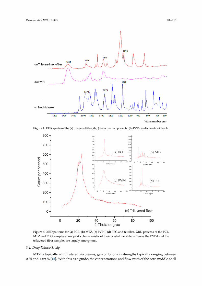
The FTIR spectra of MTZ and iodine-loaded trilayered fibers were compared to those of pure MTZ and PVP-I, to confirm the entrapment of the active ingredients in the fibers (Figure 4). Free iodine interacts with the negative end of the C=O dipole of PVP molecules, forming a complex with the amide functional group and is identified as a characteristic peak around 1654 cm−1 [26,27]. This characteristic peak was seen in the PVP-I spectrum as well as the fibers, confirming the presence of iodine. Peaks occurring at 1479, 1275, 1070 and 870 cm−1 representing the N=O, C–O, C–N and C–NO2 stretching are characteristic of MTZ [28,29]. These characteristic peaks were observed in both the pure MTZ compound and the generated fibers, confirming the entrapment of MTZ in the fibers.
Figure 3. (a) TEM and (b–e) SEM images showing the three-layer compartmental structure of trilayeredfibers, generated with 10 wt % PVP-I/PVP (1:1 w/w) in the middle layer.
3.3.2. Compositional Features
The FTIR spectra of MTZ and iodine-loaded trilayered fibers were compared to those of pureMTZ and PVP-I, to confirm the entrapment of the active ingredients in the fibers (Figure 4). Free iodineinteracts with the negative end of the C=O dipole of PVP molecules, forming a complex with the amidefunctional group and is identified as a characteristic peak around 1654 cm−1 [26,27]. This characteristicpeak was seen in the PVP-I spectrum as well as the fibers, confirming the presence of iodine. Peaksoccurring at 1479, 1275, 1070 and 870 cm−1 representing the N=O, C–O, C–N and C–NO2 stretchingare characteristic of MTZ [28,29]. These characteristic peaks were observed in both the pure MTZcompound and the generated fibers, confirming the entrapment of MTZ in the fibers.
Understanding the solid-state of the active ingredients in the drug-carrying fibers, whether itbe amorphous, crystalline or a mixture of both, can help inform the likely performance and storagebehaviour of such formulations [30]. The XRD spectra of the fibers were compared to those of theindividual active ingredients to determine the crystalline state of the drug-loaded fibers. The differencesbetween the spectra reflect the extent of change that the solid-state of the starting material has undergonepost-processing. Figure 5 shows that PCL, MTZ and PEG are in their crystalline state (Figure 5a–c),while the PVP-I is largely amorphous (Figure 5d).
The drug-loaded fibers containing the aforementioned materials were found to be part-crystallineand part-amorphous (Figure 5e), almost certainly reflecting the crystalline components of the polymers(PCL and PEG). However, the extent of crystallinity of the polymers in such a complex architectureis difficult to ascertain, given the known propensity of electrospinning to elicit polymer chainreorganisation during electrospinning [31]. While the presence of crystalline MTZ as a minorcomponent cannot be completely precluded on the basis of this data, the absence of any evidence formajor MTZ peaks at angles where the polymer response was low (and hence unlikely to hide the drugresponse) would support drug amorphisation. MTZ is sparingly soluble in aqueous systems, andhence molecular dispersion in the fiber would promote dissolution via the absence of a crystal lattice,in turn potentially aiding bioavailability.
Pharmaceutics 2020, 12, 373 10 of 16Pharmaceutics 2020, 12, x FOR PEER REVIEW 10 of 16
Figure 4. FTIR spectra of the (a) trilayered fiber; (b,c) the active components: (b) PVP-I and (c) metronidazole.
Understanding the solid-state of the active ingredients in the drug-carrying fibers, whether it be amorphous, crystalline or a mixture of both, can help inform the likely performance and storage behaviour of such formulations [30]. The XRD spectra of the fibers were compared to those of the individual active ingredients to determine the crystalline state of the drug-loaded fibers. The differences between the spectra reflect the extent of change that the solid-state of the starting material has undergone post-processing. Figure 5 shows that PCL, MTZ and PEG are in their crystalline state (Figure 5a–c), while the PVP-I is largely amorphous (Figure 5d).
The drug-loaded fibers containing the aforementioned materials were found to be part-crystalline and part-amorphous (Figure 5e), almost certainly reflecting the crystalline components of the polymers (PCL and PEG). However, the extent of crystallinity of the polymers in such a complex architecture is difficult to ascertain, given the known propensity of electrospinning to elicit polymer chain reorganisation during electrospinning [31]. While the presence of crystalline MTZ as a minor component cannot be completely precluded on the basis of this data, the absence of any evidence for major MTZ peaks at angles where the polymer response was low (and hence unlikely to hide the drug response) would support drug amorphisation. MTZ is sparingly soluble in aqueous systems, and hence molecular dispersion in the fiber would promote dissolution via the absence of a crystal lattice, in turn potentially aiding bioavailability.
Wavenumber cm−1
Figure 4. FTIR spectra of the (a) trilayered fiber; (b,c) the active components: (b) PVP-I and (c) metronidazole.Pharmaceutics 2020, 12, x FOR PEER REVIEW 11 of 16
Figure 5. XRD patterns for (a) PCL, (b) MTZ, (c) PVP-I, (d) PEG and (e) fiber. XRD patterns of the PCL, MTZ and PEG samples show peaks characteristic of their crystalline state, whereas the PVP-I and the trilayered fiber samples are largely amorphous.
3.4. Drug Release Study
MTZ is topically administered via creams, gels or lotions in strengths typically ranging between 0.75 and 1 wt % [15]. With this as a guide, the concentrations and flow rates of the core-middle-shell solutions during trilayered electrospinning were optimised (Table 1) to enable a final encapsulation concentration of 1 wt % MTZ within the total volume of the core-shell fiber.
Drug release from the trilayered fibers was compared to those from creams containing similar quantities of MTZ. Antibiotic creams are currently routinely applied as part of wound dressings, with the hope of releasing the active drug contained therein over the period during which the wound dressing is in place, usually over several days [32–34]. While the intended purpose of the fibers (treatment of wound infection and promotion of healing via tailored release of active drug, moisture control and provision of a physical barrier to prevent further contamination all in one) is distinct from that of antibacterial creams for wounds, there is merit in comparing the new formulation to an existing and established delivery system, particularly in terms of looking at the longer-term supply of adequate amounts of active drugs than would typically be required from a cream. The drug release study from the fibrous formulation and two cream formulations (MTZ with and without iodine) is shown in Figure 6. To compare the release of drug from the trilayered fibers and creams, a setup comprising the delivery system enclosed within a dialysis membrane in a volume of 5 mL and further immersed in a liquid with a volume of 45 mL (outer chamber) was used, from which sampling was made from the outer chamber to determine the amount of drug released. This was to ensure that the delivery systems being studied were totally immersed within the dissolution media, as both systems had tendencies to float and disrupt the continuous release of the drug into the media. In this two-chamber setup, sampling for analysing the drug release was possible from the outer chamber, but as the drug was released into the inner chamber enclosed by a dialysis membrane it was necessary to establish the relationship between the sampled drug and the actual amount of drug released into the dialysis chamber. For a more robust comparison between the drug released from creams and the trilayered fibers, it was necessary to establish that, regardless of the difference in the diffusion coefficient (which is certain to be the case for creams and fibers), the amount of drug sampled from
Figure 5. XRD patterns for (a) PCL, (b) MTZ, (c) PVP-I, (d) PEG and (e) fiber. XRD patterns of the PCL,MTZ and PEG samples show peaks characteristic of their crystalline state, whereas the PVP-I and thetrilayered fiber samples are largely amorphous.
3.4. Drug Release Study
MTZ is topically administered via creams, gels or lotions in strengths typically ranging between0.75 and 1 wt % [15]. With this as a guide, the concentrations and flow rates of the core-middle-shell

Pharmaceutics 2020, 12, 373 11 of 16
solutions during trilayered electrospinning were optimised (Table 1) to enable a final encapsulationconcentration of 1 wt % MTZ within the total volume of the core-shell fiber.
Drug release from the trilayered fibers was compared to those from creams containing similarquantities of MTZ. Antibiotic creams are currently routinely applied as part of wound dressings,with the hope of releasing the active drug contained therein over the period during which the wounddressing is in place, usually over several days [32–34]. While the intended purpose of the fibers(treatment of wound infection and promotion of healing via tailored release of active drug, moisturecontrol and provision of a physical barrier to prevent further contamination all in one) is distinctfrom that of antibacterial creams for wounds, there is merit in comparing the new formulation to anexisting and established delivery system, particularly in terms of looking at the longer-term supply ofadequate amounts of active drugs than would typically be required from a cream. The drug releasestudy from the fibrous formulation and two cream formulations (MTZ with and without iodine) isshown in Figure 6. To compare the release of drug from the trilayered fibers and creams, a setupcomprising the delivery system enclosed within a dialysis membrane in a volume of 5 mL and furtherimmersed in a liquid with a volume of 45 mL (outer chamber) was used, from which sampling wasmade from the outer chamber to determine the amount of drug released. This was to ensure thatthe delivery systems being studied were totally immersed within the dissolution media, as bothsystems had tendencies to float and disrupt the continuous release of the drug into the media. In thistwo-chamber setup, sampling for analysing the drug release was possible from the outer chamber,but as the drug was released into the inner chamber enclosed by a dialysis membrane it was necessaryto establish the relationship between the sampled drug and the actual amount of drug released intothe dialysis chamber. For a more robust comparison between the drug released from creams andthe trilayered fibers, it was necessary to establish that, regardless of the difference in the diffusioncoefficient (which is certain to be the case for creams and fibers), the amount of drug sampled from theouter chamber for analysis maintains its relationship with the actual amount of drug released withinthe dialysis membrane.
Pharmaceutics 2020, 12, x FOR PEER REVIEW 12 of 16
the outer chamber for analysis maintains its relationship with the actual amount of drug released within the dialysis membrane.
To determine this relationship, some assumptions and the modelling of the experimental data from the study were made. These were essential to establish an accurate comparison of drug release from the trilayered fibers and creams. The mathematical modelling of the experimental data is presented as supplementary information (Section 2, Figure S2).
Figure 6. Cumulative percentage of drug (MTZ and iodated MTZ) released from the trilayered fiber systems and cream formulations obtained during the study over a five-day period.
3.5. Release from the Trilayered Fiber System and Creams
As it has been established (in the Supplementary Information, Section 1) that the sampled drug profiles reflect the profiles of the actual drug released, regardless of the system being assessed, the amounts of drug sampled from the fiber and cream systems were compared. From the fiber system, 97.8 ± 2.3% of MTZ had been released after five days. In contrast, only 48.5 ± 2.2% and 49.5 ± 2.7% had been released from the MTZ and MTZ-iodine cream formulations, although all systems being compared were formulated to contain the same amount of MTZ. In addition, the inclusion of iodine in the cream formulations was not found to influence the MTZ release significantly. It has been reported for creams that only the fraction of the MTZ present in the external aqueous phase would be able to diffuse out for therapeutic activity [35,36]. On the other hand, MTZ as a solid dispersion in a polymer matrix in the trilayered fiber is most likely released via drug diffusion and matrix erosion [37], a process significantly facilitated by the increased surface area to volume ratio acquired by the compartmental, fiber delivery format. Suboptimal concentrations of antibiotics at topical infection sites have been confirmed to confer a survival advantage of bacteria leading to antimicrobial resistance and ultimately clinical failures [38]. Having a solid dispersion of MTZ in a polymeric matrix ensured the release of more antibiotics from the fibers, as seen in the release study, and this was confirmed to offer better antibacterial action against the test organisms used in our antimicrobial assay, discussed in Section 3.5. The examination of the first 8 h of release indicated that 69.9 ± 2.3% of MTZ in the compartmentalised fiber system had been released compared to 37.3 ± 3.2% and 35.7 ± 2.8% of the MTZ-I and MTZ cream formulations, respectively. Thus, the three-layered fiber system, unlike the cream formulations, appears to deliver more of the active ingredient within the specified time.
0
10
20
30
40
50
60
70
80
90
100
0 20 40 60 80 100 120 140
Cum
ulat
ive
rele
ase
(%)
Time (hours)
Fibre Iodated MTZ Cream MTZ Cream
Figure 6. Cumulative percentage of drug (MTZ and iodated MTZ) released from the trilayered fibersystems and cream formulations obtained during the study over a five-day period.
To determine this relationship, some assumptions and the modelling of the experimental data fromthe study were made. These were essential to establish an accurate comparison of drug release from

Pharmaceutics 2020, 12, 373 12 of 16
the trilayered fibers and creams. The mathematical modelling of the experimental data is presented asSupplementary Information (Section S2, Figure S2).
3.5. Release from the Trilayered Fiber System and Creams
As it has been established (in the Supplementary Information, Section S1) that the sampleddrug profiles reflect the profiles of the actual drug released, regardless of the system being assessed,the amounts of drug sampled from the fiber and cream systems were compared. From the fiber system,97.8 ± 2.3% of MTZ had been released after five days. In contrast, only 48.5 ± 2.2% and 49.5 ± 2.7%had been released from the MTZ and MTZ-iodine cream formulations, although all systems beingcompared were formulated to contain the same amount of MTZ. In addition, the inclusion of iodine inthe cream formulations was not found to influence the MTZ release significantly. It has been reportedfor creams that only the fraction of the MTZ present in the external aqueous phase would be able todiffuse out for therapeutic activity [35,36]. On the other hand, MTZ as a solid dispersion in a polymermatrix in the trilayered fiber is most likely released via drug diffusion and matrix erosion [37], a processsignificantly facilitated by the increased surface area to volume ratio acquired by the compartmental,fiber delivery format. Suboptimal concentrations of antibiotics at topical infection sites have beenconfirmed to confer a survival advantage of bacteria leading to antimicrobial resistance and ultimatelyclinical failures [38]. Having a solid dispersion of MTZ in a polymeric matrix ensured the release ofmore antibiotics from the fibers, as seen in the release study, and this was confirmed to offer betterantibacterial action against the test organisms used in our antimicrobial assay, discussed in Section 3.5.The examination of the first 8 h of release indicated that 69.9 ± 2.3% of MTZ in the compartmentalisedfiber system had been released compared to 37.3 ± 3.2% and 35.7 ± 2.8% of the MTZ-I and MTZcream formulations, respectively. Thus, the three-layered fiber system, unlike the cream formulations,appears to deliver more of the active ingredient within the specified time.
3.6. Antimicrobial Assays
P. aeruginosa, a common Gram-negative rod-shaped bacterium known for its resistance toantimicrobials [39], was selected as the test microorganism to assess the antibacterial activity of MTZ-loadedfibers compared with MTZ and MTZ-I creams. Furthermore, biofilms secreted by P. aeruginosa arecommonly associated with difficult-to-heal chronic wounds [40], and hence this organism provides ahighly relevant model for the novel fiber delivery system. Previous research has shown MTZ to be aneffective antibiotic against resistant strains of P. aeruginosa when used in combination therapies [41].
Two antimicrobial assays were employed: (1) an agar diffusion assay to investigate the performanceon a surface, thereby mimicking topical delivery (Supplementary Information, Section S3.1, Figure S3),and (2) a bacterial cell suspension approach to investigate the antimicrobial performance of the fiberswhen immersed in liquid media, hence mimicking anaerobic conditions.
Flow cytometry was used to give a rapid, comprehensive and quantifiable overview of thebacterial population post-treatment (Supplementary Information, Section S3.2, Figures S4 and S5).Previous literature has shown flow cytometry to provide a more accurate quantitative measurement ofcell viability when compared to plate count estimates [22].
In both assays, the trilayered fibrous formulation demonstrated a more potent elimination ofP. aeruginosa cells than the MTZ and MTZ-I creams did. The antimicrobial effect was due to thepresence of MTZ, as the drug-free fiber controls showed no antimicrobial action while the formulationscontaining iodine in addition to MTZ did not result in significantly higher cell death rates (Figure 7).More specifically, in the agar diffusion assay, the trilayered fibers inhibited a circular area with anaverage diameter of 35 ± 1.4 mm, whereas the MTZ and MTZ-I formulations resulted in growthinhibition zones with average diameters of 25 ± 1 mm and 25 ± 2 mm, respectively (Figure 7a showsthe corresponding zone areas).

Pharmaceutics 2020, 12, 373 13 of 16
Pharmaceutics 2020, 12, x FOR PEER REVIEW 13 of 16
3.6. Antimicrobial Assays
P. aeruginosa, a common Gram-negative rod-shaped bacterium known for its resistance to antimicrobials [39], was selected as the test microorganism to assess the antibacterial activity of MTZ-loaded fibers compared with MTZ and MTZ-I creams. Furthermore, biofilms secreted by P. aeruginosa are commonly associated with difficult-to-heal chronic wounds [40], and hence this organism provides a highly relevant model for the novel fiber delivery system. Previous research has shown MTZ to be an effective antibiotic against resistant strains of P. aeruginosa when used in combination therapies [41].
Two antimicrobial assays were employed: (1) an agar diffusion assay to investigate the performance on a surface, thereby mimicking topical delivery (Supplementary Information, Section 3.1, Figure S3), and (2) a bacterial cell suspension approach to investigate the antimicrobial performance of the fibers when immersed in liquid media, hence mimicking anaerobic conditions.
Flow cytometry was used to give a rapid, comprehensive and quantifiable overview of the bacterial population post-treatment (Supplementary Information, Section 3.2, Figures S4 and S5). Previous literature has shown flow cytometry to provide a more accurate quantitative measurement of cell viability when compared to plate count estimates [22].
In both assays, the trilayered fibrous formulation demonstrated a more potent elimination of P. aeruginosa cells than the MTZ and MTZ-I creams did. The antimicrobial effect was due to the presence of MTZ, as the drug-free fiber controls showed no antimicrobial action while the formulations containing iodine in addition to MTZ did not result in significantly higher cell death rates (Figure 7). More specifically, in the agar diffusion assay, the trilayered fibers inhibited a circular area with an average diameter of 35 ± 1.4 mm, whereas the MTZ and MTZ-I formulations resulted in growth inhibition zones with average diameters of 25 ± 1 mm and 25 ± 2 mm, respectively (Figure 7a shows the corresponding zone areas).
Figure 7. (a) Growth inhibition zone area of P. aeruginosa following 24-h incubation on LB agar plates with various drug formulations. (b) Dead/live cell count by flow cytometry of cell suspensions incubated for 24 h with drug-loaded fiber and cream samples.
Additionally, a 74% cell death was noted after exposure to the trilayered fiber samples in suspension, whereas a rate of 37% and 41% cell death, respectively, was noted after exposure to MTZ and MTZ-I cream formulations over the same period of incubation (Figure 7b). The more significant cell death rate found in samples with the fibers is attributed to the better release of MTZ from the trilayered compartmental system, which is also confirmed by the drug release study in Section 3.4. The enhanced release from the fiber system enabled a better availability and stronger bactericidal action, compared to the creams containing similar quantities of MTZ. The multi-layered fiber structure allowed the release of MTZ and iodine from different compartments and facilitated the antibacterial actions of the active ingredient. The inherent properties, mainly a high surface-to-
Figure 7. (a) Growth inhibition zone area of P. aeruginosa following 24-h incubation on LB agarplates with various drug formulations. (b) Dead/live cell count by flow cytometry of cell suspensionsincubated for 24 h with drug-loaded fiber and cream samples.
Additionally, a 74% cell death was noted after exposure to the trilayered fiber samples insuspension, whereas a rate of 37% and 41% cell death, respectively, was noted after exposure to MTZand MTZ-I cream formulations over the same period of incubation (Figure 7b). The more significantcell death rate found in samples with the fibers is attributed to the better release of MTZ from thetrilayered compartmental system, which is also confirmed by the drug release study in Section 3.4.The enhanced release from the fiber system enabled a better availability and stronger bactericidalaction, compared to the creams containing similar quantities of MTZ. The multi-layered fiber structureallowed the release of MTZ and iodine from different compartments and facilitated the antibacterialactions of the active ingredient. The inherent properties, mainly a high surface-to-volume ratio andsurface porosity of fibers, expedited the release of active ingredient for antibacterial activity, whereasthe limited activity in the cream formulations are attributed to the fact that the non-compartmentalsystem only makes a small fraction of the active ingredients available for antibacterial action.
4. Discussion
Re-engineering an electrospinning device to be more accessible and practically useful for deliveringmultiple active ingredients simultaneously is an intervention with a tremendous potential for improvingthe management of difficult chronic wounds. One such challenging clinical condition that standsto benefit from such an innovation is diabetic foot ulcers (DFU), given the need to deliver multipleactive agents to simultaneously target the various underlying causes of such wounds. Presently,the gold standard for treating difficult infected chronic wounds where vascularisation is impairedincludes debridement, treating the infections, the application of growth factors and modulators,revascularisation and off-loading the ulcer [42]. In treating infections and applying other interventionssuch as growth factors, agents are typically applied separately, and wound dressings capable ofdelivering multiple agents simultaneously are yet to be used. Electrospinning systems that couldpotentially deliver such interventions are currently in preparatory stages [43].
Therefore, developing a multi-layered system that clearly isolates active ingredients from eachother until required for activity shows tremendous potential for improving the healing process of suchdifficult-to-heal wounds. Having demonstrated that our portable multi-compartment fiber generatingsystem is capable of releasing more antibiotics in comparison to creams for a superior antibacterialactivity points to the possibility of further developing this portable electrospinning device into anefficient and cost-effective means of managing difficult wounds in the near future.
Pharmaceutics 2020, 12, 373 14 of 16
5. Conclusions
In summary, this work presents three novel findings: (1) the point-of-need generation of trilayeredfibers containing compartmentalised drugs for in situ wound treatment; (2) a simple-to-operate, miniaturisedportable device for the electrospinning of multi-layered core-shell structures at the point-of-use; (3) thedelivery of multiple active drugs for a possible combination therapy using a core-shell fiber configuration.The drug-loaded fibers were characterised by scanning electron microscopy, transmission electron microscopy,attenuated total reflection Fourier transform infrared spectroscopy and X-ray diffraction. In addition,the trilayered fibers demonstrated a superior antibacterial activity in comparison with the non-compartmentalcream systems. This work paves the way for applying multiple therapeutic agents simultaneously in acompartmental format at the point-of-need and strengthens the prospects of having more efficient andcost-effective means of managing difficult wounds in the foreseeable future.
Supplementary Materials: The following are available online at http://www.mdpi.com/1999-4923/12/4/373/s1.Figure S1: (a) Assembling various components of the portable electrospinning device; (b) the device in operation,producing yellowish metronidazole/iodine loaded fibers; and (c) a schematic representation of the device setup.Figure S2. (a) Fractional release from the delivery systems (blue) and into the outer liquid (orange) as a functionof time for scenarios with varying diffusion coefficients from the delivery system (Dp) and through the dialysismembrane (Dm). (b) Cumulative percentage of drug released from the trilayered fibersystems and creamformulations obtained during the study over a five-day period. Figure S3. Photographs of the results showingthe growth inhibition of P. aeruginosa following the 24-h incubation of the bacterial cells on LB agar plates with:(a) MTZ cream, (b) MTZ-I cream, and (c) triaxial fibers. Figure S4. A schematic representation of cell sorting usingflow cytometry [Image source: Sari Sabban©2011 / CC BY-SA (https://creativecommons.org/licenses/by-sa/3.0)].Figure S5. Dead/live cell count by flow cytometry of cell suspensions incubated for 24 h for (a) control (withoutcream or fiber) 4% dead/96% live, (b) plain fibers without drug 3% dead/97% live, (c) drug-loaded cream 59%dead/41% live and (d) drug-loaded fiber 76% dead/24% live.
Author Contributions: Conceptualization, F.B., C.L., M.E. and D.Q.M.C.; methodology, F.B. and C.L.; formal analysis,F.B. and C.L.; investigation, F.B., C.L., L.C. and R.K.M.; mathematical modelling, A.H.; writing—original draftpreparation, F.B.; writing—review and editing, C.L., M.E. and D.Q.M.C.; supervision, M.E. and D.Q.M.C.; fundingacquisition, F.B., C.L., M.E., D.C. All authors have read and agreed to the published version of the manuscript.
Funding: This research work was supported by the Engineering and Physical Sciences Research Council, UK [grantnumber EP/P022677/1].
Conflicts of Interest: The authors declare no conflict of interest. The funders had no role in the design of thestudy; in the collection, analyses, or interpretation of data; in the writing of the manuscript, or in the decision topublish the results.
References
1. Phillips, C.J.; Humphreys, I.; Fletcher, J.; Harding, K.; Chamberlain, G.; Macey, S. Estimating the costsassociated with the management of patients with chronic wounds using linked routine data. Int. Wound J.2016, 13, 1193–1197. [CrossRef]
2. Leaper, D.; Assadian, O.; Edmiston, C.E. Approach to chronic wound infections. Br. J. Dermatol. 2015, 173,351–358. [CrossRef]
3. Inoue, D.; Kabata, T.; Ohtani, K.; Kajino, Y.; Shirai, T.; Tsuchiya, H. Inhibition of biofilm formation oniodine-supported titanium implants. Int. Orthod. 2017, 41, 1093–1099. [CrossRef]
4. Daeschlein, G. Antimicrobial and antiseptic strategies in wound management. Int. Wound J. 2013, 10, 9–14.[CrossRef]
5. Fitzgerald, J.B.; Schoeberl, B.; Nielsen, U.B.; Sorger, P.K. Systems biology and combination therapy in thequest for clinical efficacy. Nat. Chem. Biol. 2006, 2, 458–466. [CrossRef] [PubMed]
6. Gerding, D.N. Foot Infections in Diabetic Patients: The Role of Anaerobes. Clin. Infect. Dis. 1995, 20,S283–S288. [CrossRef] [PubMed]
7. Bessa, L.J.; Fazii, P.; di Giulio, M.; Cellini, L. Bacterial isolates from infected wounds and their antibioticsusceptibility pattern: Some remarks about wound infection. Int. Wound J. 2015, 12, 47–52. [CrossRef][PubMed]
8. Delcea, M.; Yashchenok, A.; Videnova, K.; Kreft, O.; Möhwald, H.; Skirtach, A.G. Multicompartmentalmicro-and nanocapsules: Hierarchy and applications in biosciences. Macromol. Biosci. 2010, 10, 465–474.[CrossRef] [PubMed]
Pharmaceutics 2020, 12, 373 15 of 16
9. Qi, S.; Craig, D. Recent developments in micro-and nanofabrication techniques for the preparation ofamorphous pharmaceutical dosage forms. Adv. Drug Deliv. Rev. 2016, 100, 67–84. [CrossRef]
10. Davoodi, P.; Feng, F.; Xu, Q.; Yan, W.-C.; Tong, Y.W.; Srinivasan, M.; Sharma, V.K.; Wang, C.-H. Coaxialelectrohydrodynamic atomization: Microparticles for drug delivery applications. J. Control. Release 2015, 205,70–82. [CrossRef]
11. Maroni, A.; Melocchi, A.; Parietti, F.; Foppoli, A.; Zema, L.; Gazzaniga, A. 3d printed multi-compartmentcapsular devices for two-pulse oral drug delivery. J. Control. Release 2017, 268, 10–18. [CrossRef] [PubMed]
12. Lee, J.H.; Nan, A. Combination drug delivery approaches in metastatic breast cancer. J. Drug Deliv. 2012,2012, 1–17. [CrossRef] [PubMed]
13. Mouthuy, P.-A.; Groszkowski, L.; Ye, H. Performances of a portable electrospinning apparatus. Biotechnol. Lett.2015, 37, 1107–1116. [CrossRef] [PubMed]
14. Xu, S.-C.; Qin, C.-C.; Yu, M.; Dong, R.-H.; Yan, X.; Zhao, H.; Han, W.-P.; Zhang, H.-D.; Long, Y.-Z. Abattery-operated portable handheld electrospinning apparatus. Nanoscale 2015, 7, 12351–12355. [CrossRef]
15. Yan, X.; Yu, M.; Zhang, L.-H.; Jia, X.-S.; Li, J.-T.; Duan, X.-P.; Qin, C.-C.; Dong, R.-H.; Long, Y.-Z. A portableelectrospinning apparatus based on a small solar cell and a hand generator: Design, performance andapplication. Nanoscale 2016, 8, 209–213. [CrossRef]
16. Mody, S.B.; Doshi, M.M.; Joshi, M. Novel topical microbicidal compositions. U.S. Patent 20030228376A1,11 December 2003.
17. Brayfield, A. Martindale: The Complete Drug Reference; Pharmaceutical Press: London, UK, 2014.18. Summa, M.; Russo, D.; Penna, I.; Margaroli, N.; Bayer, I.S.; Bandiera, T.; Athanassiou, A.; Bertorelli, R.
A biocompatible sodium alginate/povidone iodine film enhances wound healing. Eur. J. Pharm. Biopharm.2018, 122, 17–24. [CrossRef]
19. Brako, F.; Luo, C.; Craig, D.Q.; Edirisinghe, M. An Inexpensive, Portable Device for Point-of-NeedGeneration of Silver-Nanoparticle Doped Cellulose Acetate Nanofibersfor Advanced Wound Dressing.Macromol. Mater. Eng. 2018, 303, 1700586. [CrossRef]
20. Serra, R.; Grande, R.; Butrico, L.; Rossi, A.; Settimio, U.F.; Caroleo, B.; Amato, B.; Gallelli, L.; de Franciscis, S.Chronic wound infections: The role of Pseudomonas aeruginosa and Staphylococcus aureus. Expert Rev.Anti-Infect. Ther. 2015, 13, 605–613. [CrossRef]
21. Schmidtchen, A.; Wolff, H.; Hansson, C. Differential proteinase expression by Pseudomonas aeruginosaderived from chronic leg ulcers. Acta Derm. Venereol. Stockh. 2001, 81, 406–409. [CrossRef]
22. Bankier, C.; Cheong, Y.; Mahalingam, S.; Edirisinghe, M.; Ren, G.; Cloutman-Green, E.; Ciric, L. A comparisonof methods to assess the antimicrobial activity of nanoparticle combinations on bacterial cells. PLoS ONE2018, 13, e0192093. [CrossRef]
23. Fong, H.; Chun, I.; Reneker, D. Beaded nanofibersformed during electrospinning. Polymer 1999, 40, 4585–4592.[CrossRef]
24. Sill, T.J.; von Recum, H.A. Electrospinning: Applications in drug delivery and tissue engineering. Biomaterials2008, 29, 1989–2006. [CrossRef] [PubMed]
25. Angammana, C.J.; Jayaram, S.H. Analysis of the effects of solution conductivity on electrospinning processand fibermorphology. IEEE Trans. Ind. Appl. 2011, 41, 1109–1117. [CrossRef]
26. Xu, N.; Ding, D. Preparation and antibacterial activity of chitosan derivative membrane complexation withiodine. RSC Adv. 2015, 5, 79820–79828. [CrossRef]
27. Kirsh, I.U.E.; Kirsh, Y.E. Water Soluble Poly-N-Vinylamides: Synthesis and Physicochemical Properties; John Wiley& Sons: Hoboken, NJ, USA, 1998.
28. Herculano, R.D.; de Queiroz, A.A.A.; Kinoshita, A.; Oliveira, O.N.; Graeff, C.F.O. On the release ofmetronidazole from natural rubber latex membranes. Mater. Sci. Eng. C 2011, 31, 272–275. [CrossRef]
29. Trivedi, M.K.; Patil, S.; Shettigar, H.; Bairwa, K.; Jana, S. Spectroscopic characterization of biofield treatedmetronidazole and tinidazole. Med. Chem. 2015, 5, 340–344.
30. Grohganz, H.; Priemel, P.A.; Löbmann, K.; Nielsen, L.H.; Laitinen, R.; Mullertz, A.; van den Mooter, G.;Rades, T. Refining stability and dissolution rate of amorphous drug formulations. Expert Opin. Drug Deliv.2014, 11, 977–989. [CrossRef]
31. Yu, D.-G.; Gao, L.-D.; White, K.; Branford-White, C.; Lu, W.-Y.; Zhu, L.-M. Multicomponent amorphousnanofiberselectrospun from hot aqueous solutions of a poorly soluble drug. Pharm. Res. 2010, 27, 2466–2477.[CrossRef]
Pharmaceutics 2020, 12, 373 16 of 16
32. Banerjee, S.; Argáez, C. Topical Antibiotics for Infection Prevention: A Review of the Clinical Effectivenessand Guidelines. In Review from Canadian Agency for Drugs and Technologies in Health; CADTH: Ottawa, ON,Canada, 2017.
33. Rai, M.; Yadav, A.; Gade, A. Silver nanoparticles as a new generation of antimicrobials. Biotechnol. Adv. 2009,27, 73–86. [CrossRef]
34. Tiwari, V.K. Burn wound: How it differs from other wounds. Indian J. Plast. Surg. 2012, 45, 364–373.[CrossRef]
35. Larsen, D.B.; Parshad, H.; Fredholt, K.; Larsen, C. Characteristics of drug substances in oily solutions. Drugrelease rate, partitioning and solubility. Int. J. Pharm. 2002, 232, 107–117. [CrossRef]
36. Ferreira, L.; Seiller, M.; Grossiord, J.; Marty, J.; Wepierre, J. Vehicle influence on in vitro release ofmetronidazole: Role of w/o/w multiple emulsion. Int. J. Pharm. 1994, 194, 251–259. [CrossRef]
37. Chou, S.-F.; Carson, D.; Woodrow, K.A. Current strategies for sustaining drug release from electrospunnanofibers. J. Control. Release 2015, 220, 584–591. [CrossRef] [PubMed]
38. Gould, I.M.; MacKenzie, F.M. Antibiotic exposure as a risk factor for emergence of resistance: The influenceof concentration. Proc. J. Appl. Microbiol. Symp. Suppl. 2002, 92, 72–84. [CrossRef]
39. Poole, K. Efflux-mediated multiresistance in Gram-negative bacteria. Clin. Microbiol. Infect. 2004, 10, 12–26.[CrossRef] [PubMed]
40. Bjarnsholt, T.; Kirketerp-Møller, K.; Jensen, P.Ø.; Madsen, K.G.; Phipps, R.; Krogfelt, K.; Høiby, N.; Givskov, M.Why chronic wounds will not heal: A novel hypothesis. Wound Repair Regen. 2008, 16, 2–10. [CrossRef][PubMed]
41. Miller, B.; Popejoy, M.W.; Hershberger, E.; Steenbergen, J.N.; Alverdy, J. Characteristics and outcomes ofcomplicated intra-abdominal infections involving Pseudomonas aeruginosa from a randomized, double-blind,phase 3 ceftolozane-tazobactam study. Antimicrob. Agents Chemother. 2016, 60, 4387–4390. [CrossRef]
42. Alexiadou, K.; Doupis, J. Management of diabetic foot ulcers. Diabetes Ther. 2012, 3, 1–15. [CrossRef]43. Chen, S.; Liu, B.; Carlson, M.A.; Gombart, A.F.; Reilly, D.A.; Xie, J. Recent advances in electrospun
nanofibersfor wound healing. Nanomedicine 2017, 12, 1335–1352. [CrossRef]
© 2020 by the authors. Licensee MDPI, Basel, Switzerland. This article is an open accessarticle distributed under the terms and conditions of the Creative Commons Attribution(CC BY) license (http://creativecommons.org/licenses/by/4.0/).