a pseudouridine synthase module is essential for...
TRANSCRIPT

Scientific Report
A pseudouridine synthase module is essential formitochondrial protein synthesis and cell viabilityHana Antonicka1, Karine Choquet1, Zhen-Yuan Lin2, Anne-Claude Gingras2,3, Claudia L Kleinman4 &
Eric A Shoubridge1,*
Abstract
Pseudouridylation is a common post-transcriptional modificationin RNA, but its functional consequences at the cellular level remainlargely unknown. Using a proximity-biotinylation assay, we identi-fied a protein module in mitochondrial RNA granules, platforms forpost-transcriptional RNA modification and ribosome assembly,containing several proteins of unknown function including threeuncharacterized pseudouridine synthases, TRUB2, RPUSD3, andRPUSD4. TRUB2 and RPUSD4 were previously identified as coreessential genes in CRISPR/Cas9 screens. Depletion of the individualenzymes produced specific mitochondrial protein synthesis andoxidative phosphorylation assembly defects without affectingmitochondrial mRNA levels. Investigation of the molecular targetsin mitochondrial RNA by pseudouridine-Seq showed that RPUSD4plays a role in the pseudouridylation of a single residue in the 16SrRNA, a modification that is essential for its stability and assemblyinto the mitochondrial ribosome, while TRUB2/RPUSD3 were simi-larly involved in pseudouridylating specific residues in mitochon-drial mRNAs. These results establish essential roles forepitranscriptomic modification of mitochondrial RNA in mitochon-drial protein synthesis, oxidative phosphorylation, and cell survival.
Keywords epitranscriptomic modification; mitochondrial protein synthesis;
oxidative phosphorylation; pseudouridine synthase; ribosome assembly
Subject Categories Membrane & Intracellular Transport; Protein Biosynthe-
sis & Quality Control; RNA Biology
DOI 10.15252/embr.201643391 | Received 22 September 2016 | Revised 21
November 2016 | Accepted 22 November 2016 | Published online 14 December
2016
EMBO Reports (2017) 18: 28–38
Introduction
Genomewide mapping studies of the epitranscriptome in human
cells have revealed widespread post-transcriptional modifications in
both non-coding and coding RNA [1,2]. Pseudouridine (w), the most
common RNA modification, has been suggested to play a role in
RNA stability, RNA folding/secondary structure, and translation effi-
ciency and fidelity; however, the functional consequences of this
modification at the cellular level, especially in mRNA, generally
remain obscure [3]. Human cells contain 13 pseudouridine
synthases, but the majority remain uncharacterized, and their
substrates and subcellular distribution are unknown [3]. The mito-
chondrial 16S rRNA contains a single known pseudouridine site [4],
and mitochondrial tRNAs are extensively pseudouridylated by
PUS1, an enzyme that also modifies cytosolic tRNAs, and when
mutated causes mitochondrial myopathy and sideroblastic anemia
[5,6]. Genomewide pseudouridine profiling suggested that in addi-
tion to these known pseudouridine sites in mitochondrial RNA,
specific mRNAs, and the 12S rRNA might be pseudouridylated in
human cells [7], but these results have not been confirmed, nor has
the enzymatic machinery been identified.
Mammalian mitochondrial RNA (mtRNA) is transcribed as two
polycistronic transcripts, which are further processed to produce 2
rRNAs, 11 mRNAs (including two bicistronic transcripts), and 22
tRNAs. Post-transcriptional handling of mitochondrial transcripts, as
well as the assembly of the mitochondrial ribosome, occurs in non-
membrane delimited structures called mitochondrial RNA granules [8–
11]. These structures contain newly synthetized mtRNA and a number
of RNA-binding proteins responsible for stabilization, processing, modi-
fication, folding, and degradation of mtRNA. Here we have uncovered a
protein module containing three uncharacterized pseudouridine
synthases that localizes to these granules. We identify the pseudouridy-
lated sites in mitochondrial RNA that are the targets of these enzymes
and investigate the functional consequences of these epitranscriptomic
modifications for mitochondrial ribosome biogenesis, mitochondrial
translation, and assembly of the oxidative phosphorylation complexes.
Results and Discussion
Identification of a mitochondrial pseudouridine synthase proteinmodule using BioID
Several RNA-binding proteins that localize to the RNA granule have
been linked to human disease [12], and loss-of-function variants in
1 Department of Human Genetics, Montreal Neurological Institute, McGill University, Montreal, QC, Canada2 Lunenfeld-Tanenbaum Research Institute, Mount Sinai Hospital, Toronto, ON, Canada3 Department of Molecular Genetics, University of Toronto, Toronto, ON, Canada4 Department of Human Genetics, Segal Cancer Centre and Lady Davis Institute, Jewish General Hospital, McGill University, Montréal, QC, Canada
*Corresponding author. Tel: +1 514 398 1997; Fax: +1 514 398 1509; E-mail: [email protected]
EMBO reports Vol 18 | No 1 | 2017 ª 2016 The Authors28
Published online: December 14, 2016
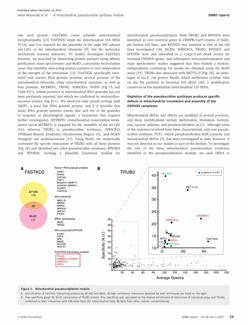
one such protein, FASTKD2, cause infantile mitochondrial
encephalopathy [13]. FASTKD2 binds the mitochondrial 16S rRNA
[9,14], and it is required for the assembly of the large 39S subunit
(mt-LSU) of the mitochondrial ribosome [9], but the molecular
mechanism remains unknown. To further investigate FASTKD2
function, we searched for interacting protein partners using affinity
purification–mass spectrometry and BioID, a proximity-biotinylation
assay that identifies interacting protein partners in vivo independent
of the strength of the interaction [15]. FASTKD2 specifically inter-
acted with known RNA granule proteins, several proteins of the
mitochondrial ribosome, other mitochondrial enzymes, as well as
four proteins, MTERFD1, TRUB2, WBSCR16, NGRN (Fig 1A and
Table EV1), whose presence in mitochondrial RNA granules has not
been previously reported, and which we confirmed by immunofluo-
rescence studies (Fig EV1). We observed only partial overlap with
GRSF1, a bone fide RNA granule protein, and it is possible that
many RNA granule proteins transit into and out of the granules
in response to physiological signals, a hypothesis that requires
further investigation. MTERFD1 (mitochondrial transcription termi-
nation factor MTERF3) is required for the assembly of the mt-LSU
[16], whereas TRUB2 (a pseudouridine synthase), WBSCR16
(Williams-Beuren Syndrome Chromosome Region 16), and NGRN
(Neugrin) are uncharacterized [17]. Using BioID, we reciprocally
confirmed the specific interaction of TRUB2 with all these proteins
(Fig 1B) and identified two other pseudouridine synthases, RPUSD3
and RPUSD4, forming a plausible functional module for
mitochondrial pseudouridylation. Both TRUB2 and RPUSD4 were
identified as core essential genes in CRISPR/Cas9 screens of multi-
ple human cell lines, and RPUSD3 was essential in two of the cell
lines investigated [18]. NGRN, WBSCR16, TRUB2, RPUSD3, and
RPUSD4 were also identified in a Crispr/Cas9 death screen for
essential OXPHOS genes, and subsequent immunoprecipitation and
mass spectrometry studies suggested that they formed a module,
independently confirming the results we obtained using the BioID
assay [19]. TRUB2 also interacted with METTL15 (Fig 1B), an ortho-
logue of the E. coli protein RsmH, which methylates cytidine 1402
(at the N4 position) in bacterial 16S rRNA [20], a modification
conserved in the mammalian mitochondrial 12S rRNA.
Depletion of the pseudouridine synthases produces specificdefects in mitochondrial translation and assembly of theOXPHOS complexes
Mitochondrial tRNAs and rRNAs are modified at several positions,
and these modifications include methylation, thiolation, formyla-
tion, taurine addition, and pseudouridylation [6,21]. Although some
of the enzymes involved have been characterized, only one pseudo-
uridine synthase, PUS1, which pseudouridylates both cytosolic and
mitochondrial tRNAs [5], has been investigated to date; however, it
was not detected in our studies as part of the module. To investigate
the role of the three mitochondrial pseudouridine synthases
identified in the pseudouridylation module, we used siRNA to
A B
Figure 1. Mitochondrial pseudouridylation module.
A Identification of FASTKD2 interacting proteins by AP–MS and BioID. 26 high-confidence interactors detected by both techniques are listed on the right.B Prey specificity graph for BioID interactome of TRUB2 protein. Prey specificity was calculated as the relative enrichment of interaction of individual preys and TRUB2,
compared to their interaction with 138 other baits (42 mitochondrial baits, 96 baits from other cellular compartments).
ª 2016 The Authors EMBO reports Vol 18 | No 1 | 2017
Hana Antonicka et al A mitochondrial pseudouridine synthase module EMBO reports
29
Published online: December 14, 2016

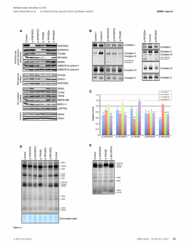
deplete each of them individually in human 143B cells,
a cell line that we used to characterize the composition of mitochon-
drial RNA granules [8] (Figs 2A and EV2A). The efficiency of
suppression at the protein level was between 77 and 95%. No
commercially available anti-RPUSD3 antibody could detect a specific
band on an immunoblot, so the efficiency of the knockdown (95%)
was confirmed by qRT–PCR (Table EV2). Interestingly, depletion of
MTERFD1, used as a positive control, led to a significant decrease in
the levels of FASTKD2, TRUB2, RPUSD4, and NGRN (Fig EV2A),
suggesting that it plays a role as a regulator of the stability of this
protein module. Depletion of TRUB2 resulted in a 50% decrease in
WBSCR16 isoform 1, and a small, but significant, decrease in
FASTKD2. Depletion of each of the three pseudouridine synthases
led to a decrease in the levels of MRPL11, a protein of the mt-LSU,
implicating the pseudouridine synthase module in mitochondrial
translation and the biogenesis of the mitochondrial ribosome.
Depletion of each of the pseudouridine synthases also resulted in
combined oxidative phosphorylation (OXPHOS) assembly defects
(Fig 2B and C) due to the decreased synthesis of the mtDNA-encoded
polypeptides (Figs 2D and EV2B). Mitochondrial protein synthesis, as
measured by a pulse-translation assay, was decreased to 76, 20, and
53% of control in cells in which TRUB2, RPUSD4, and RPUSD3 were
depleted, but the nature of the defect differed in all three cases. The
decrease in mitochondrial protein synthesis in TRUB2-, RPUSD3-, or
RPUSD4-depleted cells was not caused by a decrease in the levels
of individual mRNAs (Table EV2); however, the level of the 16S
rRNA was decreased to 22% of control in RPUSD4-depleted cells
(Table EV2), consistent with the severe global decrease in mitochon-
drial protein synthesis. In 143B cells, only depletion of RPUSD4 and
FASTKD2 resulted in a significant decrease in 16S rRNA (Fig EV2C),
in contrast to the results reported after Crispr/Cas9 knockdown in
K562 cells [19]. It is possible that the differences between the two
studies simply reflect the fact that the studies were carried out in
different cell types, but this will require further investigation.
Remarkably, in TRUB2 knockdown cells, the synthesis of ATP6 and
ATP8 proteins (encoded on a bicistronic transcript) was severely
reduced (Figs 2E and EV2B), a pattern not seen previously in cells in
which any component of the mitochondrial RNA metabolism or
translation machinery was depleted or mutated.
Depletion of TRUB2 and RPUSD4 leads to defects inmitochondrial ribosome assembly
To investigate the effects of depleting the individual pseudouridine
synthases on the assembly of the mitochondrial ribosome, we
performed a sucrose gradient sedimentation analysis in both control
and depleted cells. Nearly all immunodetectable TRUB2 and
RPUSD4 co-sedimented with the mt-LSU (Fig 3A). Depletion of both
TRUB2 and RPUSD4 led to a decrease in the assembly mt-LSU, as
well as the 55S monosome (Figs 3B and C, and EV3), with the most
severe defect appearing in cells lacking RPUSD4. The assembly of
the 28S small ribosomal subunit (mt-SSU) was either not affected
(siRPUSD4) or slightly increased (siTRUB2). In contrast, knockdown
of RPUSD3 did not affect the assembly of the mitochondrial ribo-
some, suggesting that these enzymes have non-overlapping func-
tions in mitochondrial protein synthesis.
To assess whether any of the pseudouridylation module proteins
formed stable high molecular weight complexes, we performed a
2D-gel analysis (Fig 3D). TRUB2, FASTKD2, MTERFD1, and NGRN
migrated largely at their predicted monomeric molecular weights,
while RPUSD4 and WBSCR16 were present predominantly in higher
molecular weight complexes of different sizes. Consistent with our
observation that UV cross-linking was necessary to immunoprecipi-
tate the proteins interacting with FASTKD2, these data demonstrate
that the proteins in the pseudouridine synthase module do not form
a stable multimeric complex, suggesting that their interactions are
transient and facilitated by mitochondrial RNA.
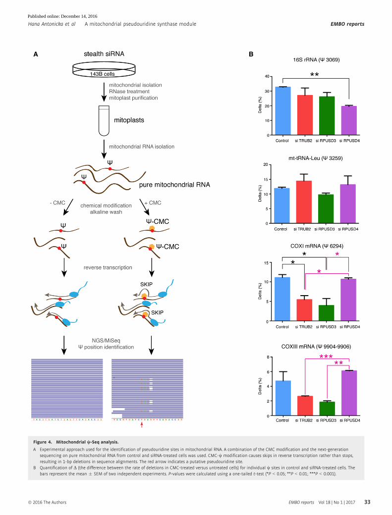
Pseudouridine-Seq (w-Seq) identifies modified sites inmitochondrial RNA
To determine the specific sites modified by TRUB2, RPUSD3, and
RPUSD4, we employed pseudouridine-Seq (w-Seq) on purified mito-
chondrial RNA (Fig 4A). This method utilizes an alkaline-resistant
modification of pseudouridine by N-cyclohexyl-N0-(2-morpholi-
noethyl)carbodiimide metho-p-toluenesulfonate (CMC), reported to
cause a termination in reverse transcription at a position 30 to
w-CMC [7,22]. In our experiments, CMC treatment caused pseu-
douridylated base skipping during reverse transcription, rather than
a complete stop (Fig 4A), leading to a deletion of the base in the
resulting sequencing reads (Fig EV4B). We computed the difference
between the deletion rate in treated versus untreated samples (D)for all nucleotides in the mitochondrial genome. Pseudouridylated
sites were determined using a cutoff of D > 2.5% in duplicate
control samples (seven sites, Table EV3), which were further filtered
to remove sequencing artifacts (Table EV3). We detected four high-
confidence sites (Figs 4B and EV4B): 16S rRNA (w3069), mt-tRNA-
Leu (w3259), COXI (w6294), and COXIII (w9904–9906). The precise
position for w in COXIII mRNA could not be determined due to the
alignment ambiguity of three consecutive U’s. Analysis of the motifs
surrounding the w site in both the 16S rRNA and the mRNAs
showed a similar consensus sequence (Fig EV5A), and in silico
Figure 2. Mitochondrial pseudouridine synthases TRUB2, RPUSD3, and RPUSD4 are necessary for OXPHOS biogenesis.
A Immunoblot analysis of indicated proteins in control and siRNA-treated cells.B BN–PAGE analysis of siRNA-mediated depletion shows an OXPHOS defect as revealed by subunit-specific antibodies against individual OXPHOS complexes.C Quantification of the levels of individual OXPHOS complexes normalized to complex II levels. The graph represents the relative abundance of individual complexes
in cells treated with the specified siRNA versus controls. The bars represent the mean � SEM of 3–7 independent experiments. P-values were calculated usingpaired two-tailed t-test (*P < 0.05; **P < 0.01).
D, E Pulse-labeling mitochondrial translation experiment of the 13 mitochondria-encoded polypeptides (seven subunits of complex I [ND], three subunits of complex IV[COX], two subunits of complex V [ATP], and one subunit of complex III [cyt b]) in control and siRNA-treated cells. (E) Detail of a mitochondrial pulse-labelingexperiment showing a severe decrease in the translation of ATP6 and ATP8 in TRUB2-depleted cells.
Source data are available online for this figure.
▸
EMBO reports Vol 18 | No 1 | 2017 ª 2016 The Authors
EMBO reports A mitochondrial pseudouridine synthase module Hana Antonicka et al
30
Published online: December 14, 2016
A B
C
ED
Figure 2.
ª 2016 The Authors EMBO reports Vol 18 | No 1 | 2017
Hana Antonicka et al A mitochondrial pseudouridine synthase module EMBO reports
31
Published online: December 14, 2016

predictions of the secondary structure of the RNAs showed that all
w sites are either in a loop, or in a stem adjacent to a loop, and thus
likely to be accessible (Fig EV5B). We did not observe the presence
of the previously suggested w in 12S rRNA or COXII mRNA [7].
Neither the w11155 in ND4 mRNA (D = 1.9%) nor the w7658 in
COXII mRNA (D = 2.2%) passed our cutoff; however, they remain
potential, low-level pseudouridine sites. We only detected one high-
confidence modification in mt-tRNA, as our library preparation
A
C D
B
Figure 3. Interactions between the pseudouridine synthase module and the mitochondrial ribosome.
A, B Identification of mitochondrial ribosomal proteins and pseudouridine synthase interacting proteins by sucrose gradient centrifugation in control cells (A) and cellstreated with siRNA (B). Individual fractions were separated by SDS–PAGE and immunoblotted with the indicated antibodies. Panel control 2 in (B) is identical to theone shown in (A). The migration of the mt-SSU (28S), the mt-LSU (39S), and the mitochondrial monosome (55S) is shown.
C Quantification of the levels of the mt-SSU (28S), the mt-LSU (39S), and the mitochondrial monosome (55S) normalized to the SDHA levels. The graph represents therelative abundance of individual subunits in cells treated with specified siRNA versus controls. Quantification of the mt-SSU, mt-LSU, and monosome was done byaveraging the intensity of the signals from three different antibodies directed against specific structural subunits of the ribosome. The bars represent the mean � SEM.
D 2D-immunoblot analysis (BN–PAGE/SDS–PAGE) of pseudouridine synthase interacting proteins. The migration (sizes in kDa) of known protein complexes in the firstdimension is indicated on the top of the blot.
Source data are available online for this figure.
EMBO reports Vol 18 | No 1 | 2017 ª 2016 The Authors
EMBO reports A mitochondrial pseudouridine synthase module Hana Antonicka et al
32
Published online: December 14, 2016
A B
Figure 4. Mitochondrial w-Seq analysis.
A Experimental approach used for the identification of pseudouridine sites in mitochondrial RNA. A combination of the CMC modification and the next-generationsequencing on pure mitochondrial RNA from control and siRNA-treated cells was used. CMC-w modification causes skips in reverse transcription rather than stops,resulting in 1-bp deletions in sequence alignments. The red arrow indicates a putative pseudouridine site.
B Quantification of D (the difference between the rate of deletions in CMC-treated versus untreated cells) for individual w sites in control and siRNA-treated cells. Thebars represent the mean � SEM of two independent experiments. P-values were calculated using a one-tailed t-test (*P < 0.05; **P < 0.01; ***P < 0.001).
ª 2016 The Authors EMBO reports Vol 18 | No 1 | 2017
Hana Antonicka et al A mitochondrial pseudouridine synthase module EMBO reports
33
Published online: December 14, 2016

selected for products > 200 base pairs (mt-tRNAs are mostly ~70 nt
long). Nonetheless, another plausible w3286 site in mt-tRNA-Leu
was detected, corresponding to a mt-tRNA position 55, a site known
to be pseudouridylated in mt-tRNA-Glu, mt-tRNA-Gln, mt-tRNA-Ser
(UCN), and mt-tRNA-Tyr [6]. Additional studies will be needed to
verify the existence of this modification.
w-Seq analysis is not able to determine the absolute levels of
pseudouridine in a single RNA. When a fully pseudouridylated
probe was used in a w-Seq experiment, only 43% of reads termi-
nated 30 to the w site [23]. In our experiments, 32% of reads at the
w3069 site in 16S rRNA contained a deletion, suggesting that a high
percentage (likely all) of the mitochondrial 16S rRNA is pseudo-
uridylated. The percentage of deletion-containing reads was lower
for the other identified w sites, implying the co-existence of both the
w- and U-containing transcripts, and suggesting that the modifi-
cation is likely to be dynamic.
Identification of the enzymes responsible for pseudouridylatingmitochondrial RNAs
Pseudouridylation analysis in TRUB2-, RPUSD3-, and RPUSD4-
depleted cells showed the presence of pseudouridine modifications
at the same positions as in control cells, but we detected clear
differences in the level of pseudouridylation at some of these sites
(Fig 4B). The levels of w3259 modification in mt-tRNA-Leu, which is
modified by PUS1 [5], were not different among the investigated
samples, indicating that, as expected, neither TRUB2, RPUSD3, or
RPUSD4 are involved in this modification. In RPUSD4-depleted cells,
the level of the 16S rRNA modification (w3069) was specifically
reduced to 60% of control, while the levels of w modification at the
other two sites in mRNAs were comparable to control. The w3069modification was not affected in TRUB2 or RPUSD3 knockdown
cells. These results strongly suggest that RPUSD4 is responsible for
the pseudouridylation of the 16S rRNA, and that this modification is
essential for the stability of the transcript, as the depletion of RPUSD4
causes a fourfold decrease in 16S rRNA levels, resulting in a severely
reduced capacity to assemble the mt-LSU, diminished synthesis of
mtDNA-polypeptides, and an OXPHOS assembly defect.
Depletion of both TRUB2 and RPUSD3 resulted in decreased
pseudouridylation in the COXI (w6294) and COXIII (w9904–9906)mRNAs. Although the decrease in COXIII pseudouridylation was not
significantly different from the control samples, the level of modifi-
cation at this position was significantly reduced compared to
RPUSD4-depleted cells. We cannot exclude the possibility that both
TRUB2 and RPUSD3 enzymes are modifying these mRNAs;
however, three lines of evidence suggest that RPUSD3 is the major
modifying enzyme for COXI and COXIII mRNA and that the effect of
TRUB2 is secondary. First, depletion of RPUSD3 leads to a signifi-
cant decrease in the synthesis of COXI and COXIII polypeptides
(Figs 2D and EV2B), resulting in a substantial decrease in the
assembly of complex IV (Fig 2B and C), demonstrating that
mitochondrial mRNA pseudouridylation of these mRNAs is
important for the efficient synthesis of these polypeptides (without
affecting the stability of the mRNA). Second, depletion of TRUB2
resulted in a 50% decrease in RPUSD3 mRNA (Table EV2). Until an
appropriate anti-RPUSD3 antibody becomes available, RPUSD3
protein levels and the interdependence of these two proteins cannot
be directly interrogated. Finally, depletion of TRUB2 leads primarily
to a defect in the synthesis of ATP6 and ATP8 subunits, while
complex IV is relatively spared (Fig 2D and E). The mechanism
underlying translation suppression of the bicistronic ATP8/6
transcript by TRUB2 depletion will require further investigation.
In summary, our results show that the pseudouridine synthase
module we identify is in fact more complex than the suggested 16S
rRNA regulatory module [19]. The BioID assay also identified addi-
tional components of the protein module (MTERFD1 and METTL15)
that play a role in ribosome biogenesis and thus the control of mito-
chondrial protein synthesis. Although we have not uncovered the
exact molecular function of FASTKD2, it is clearly involved in the
post-transcriptional handling of the 16S rRNA, and we have now
placed this function in the much larger context of a post-transcrip-
tional regulatory module. Our data strongly suggest that RPUSD4, a
core essential enzyme, pseudouridylates the mitochondrial 16S
rRNA, and that this is required for its assembly into a functional
mitochondrial ribosome. mRNA pseudouridine modifications, cata-
lyzed by TRUB2/RPUSD3, modulate the efficiency of mitochondrial
protein synthesis without apparent changes in transcript abundance
or stability. These results establish clear roles for pseudouridine
modifications in mitochondrial non-coding and coding RNA that are
essential for expression of the mitochondrial transcriptome and for
cell viability.
Materials and Methods
Cell lines
Flp-In T-REx 293 and 143B cell lines were grown in high-glucose
Dulbecco’s modified Eagle’s medium (DMEM) supplemented with
10% fetal bovine serum, at 37°C in an atmosphere of 5% CO2.
Antibodies
The following antibodies were used in this study: anti-FASTKD2
(Proteintech Group 17464-1-AP), anti-MTERFD1 (Sigma HPA00
2966), anti-TRUB2 (Proteintech Group 19891-1-AP), anti-RPUSD4
(Sigma HPA039689), anti-NGRN (Proteintech Group 14885-1-AP),
anti-WBSCR16 (Proteintech Group 13796-1-AP), anti-FLAG (Sigma
F1804), anti-GRSF1 (Sigma HPA036985), anti-DHX30 (Abcam ab85687),
anti-FASTKD5 (Sigma SAB2700438), anti-GFM1 (in house), anti-
TUFM/TSFM (a kind gift of Linda Spremulli), anti-MRPS18B
(Proteintech Group 16139-1-AP), anti-MRPL11 (Cell Signalling
#2066S), anti-LRPPRC (in house), anti-SDHA (Abcam ab14715),
anti-Porin (Abcam ab14734), anti-NDUFA9 (Abcam ab14713),
anti-COX IV (Abcam ab110261), anti-ATP5A1 (Abcam ab14748),
anti-UQCRC2 (Abcam ab14745), anti-MRPS27 (Proteintech Group
17280-1-AP), anti-MRPS22 (Proteintech Group 10984-1-AP), anti-
MRPL44 (Proteintech Group 16394-1-AP), anti-MRPL14 (Sigma
SAB4502786).
Affinity purification–mass spectroscopy (AP–MS) analysis
143B cells grown on plates (diameter 15 cm2) were washed in PBS
and UV cross-linked in a UV Stratalinker 1800 (Stratagene) for
10 min on ice. Mitochondria (400 lg) isolated from UV cross-linked
143B cells were extracted in 200 ll of extraction buffer [50 mM
EMBO reports Vol 18 | No 1 | 2017 ª 2016 The Authors
EMBO reports A mitochondrial pseudouridine synthase module Hana Antonicka et al
34
Published online: December 14, 2016

Tris–HCl, pH 7.5, 150 mM NaCl, 1% taurodeoxycholate, and
Complete protease inhibitors without EDTA (Roche)] on ice for
40 min, followed by a centrifugation at 9,000 g at 4°C for 20 min,
and the supernatant was pre-cleared overnight with non-coated
Dynabeads protein A (Invitrogen) to reduce non-specific protein
binding. Binding of FASTKD2 antibody to Dynabeads protein A
(Invitrogen) was performed according to the manufacturer’s instruc-
tions (version no. 004), with the exception that the incubation of
the antibody with the beads was carried out overnight. The pre-
cleared extracts were used in the immunoprecipitation experiment
with antibody cross-linked beads and non-coated beads (control-IP).
The immunoprecipitation reaction was performed overnight at 4°C.
Samples were eluted using 0.1 M glycine (pH 2.5) supplemented
with 1% dodecyl maltoside, TCA precipitated and analyzed on an
Orbitrap (Thermo Scientific, Watlham, MA) at the Institute de
Recherches Cliniques de Montreal.
Mitochondrial isolation
143B cells were resuspended in ice-cold 250 mM sucrose/10 mM
Tris–HCl (pH 7.4) and homogenized with seven passes in a pre-
chilled zero-clearance homogenizer (Kimble/Kontes, Vineland, NJ).
A post-nuclear supernatant was obtained by centrifugation of the
samples twice for 10 min at 600 g. Mitochondria were pelleted by
centrifugation for 10 min at 10,000 g and washed once in the same
buffer. Protein concentration was determined by Bradford assay.
BioID
BirA*-FLAG constructs were generated using Gateway cloning into
the pDEST5-BirA*-FLAG-C-ter vector. As starting clones, clones
number BC001457 and BC001544 were used for cloning of TRUB2
and FASTKD2, respectively. Human cells [Flp-In T-REx 293 cells]
were transfected using Lipofectamine 2000 (Invitrogen). Cells were
seeded at 250,000 cells per well in a 6-well plate in 2 ml DMEM
supplemented with 10% FBS and 1% pen/strep (100 U/ml). The
next day, cells were transfected with 200 ng pDEST5-ProteinX-
BirA*-FLAG, and 2 lg of pOG44 in 250 ll of 1× Opti-MEM (Invitro-
gen) mixed with 5 ll of Lipofectamine 2000 reagent in 250 ll of 1×Opti-Mem. The Opti-MEM/Lipofectamine solution was added to the
Opti-MEM/plasmid solution and incubated 20 min before addition
to the cells (in medium without antibiotics). The medium was
changed 4 h after transfection. The next day, transfected cells were
passaged into 10 cm2 plates, and the following day selected by the
addition of hygromycin (Calbiochem) at a final concentration of
200 lg/ml. This selection media was changed every 2–3 days until
clear visible colonies were present. Up to six colonies per construct
were picked and expanded, and the localization and the expression
level of the construct were assessed by immunofluorescence using
an anti-FLAG antibody. The selected clones were scaled up to six
15 cm2 plates for treatment and harvesting. Cells were grown to
70% confluency before induction of protein expression using
1 lg/ml tetracycline (Sigma), and media supplementation with
50 lM biotin for protein labeling. Cells were harvested 24 h later as
follows: Cell medium was decanted, cells were washed twice with
5 ml of PBS per 15 cm2 plate and then harvested by scraping in
5 ml of PBS. Cells from 3 × 15 cm2 plates were pelleted at 800 rpm
for 3 min, PBS aspirated and pellets transferred to a �80°C freezer.
Purification of biotinylated proteins followed by their identification
by mass spectrometry was performed as described previously [24],
and data were compared to negative controls expressing the BirA*
tag alone, the BirA* tag fused to GFP, and untransfected cells. The
prey specificity module of the ProHits-viz software (http://prohits-
viz.lunenfeld.ca) was used to analyze the TRUB2 BioID data, using
42 mitochondrial baits and 96 baits from other cellular compart-
ments as controls.
Immunocytochemistry
Flp-In T-REx 293 cells expressing ProteinX-BirA-FLAG constructs
grown on coverslips were induced using 1 lg/ml tetracycline
(Sigma) for 24 h, fixed with 4% formaldehyde solution, solubilized
by Triton X-100, and incubated with the anti-FLAG and anti-GRSF1
antibodies. The appropriate anti-species secondary antibodies
coupled with Alexa fluorochromes (Invitrogen) were subsequently
used at a dilution of 1:1,000. Cells were imaged with an Olympus
IX83 microscope connected to a Yokogawa CSU-X confocal scanning
unit. Images were analyzed using ImageJ software Fiji [25].
siRNA transfection
Stealth RNA interference duplex constructs (Invitrogen) were used for
transient knockdown of TRUB2 (HSS120266), RPUSD3 (HSS138721+
HSS138722), RPUSD4 (HSS131380), MTERFD1 (HSS121476), and
FASTKD2 (HSS176985) in 143B cells. Stealth siRNA duplexes were
transiently transfected into cells using Lipofectamine RNAiMAX
(Invitrogen), according to the manufacturer’s specifications. The
transfection was repeated on day 3, and the cells were harvested on
day 6 for analysis.
Denaturing, native, and two-dimensional PAGE
SDS–PAGE was used to separate isolated mitochondrial extracts and
fractions from sucrose gradient sedimentation. In general, isolated
mitochondria were extracted with 1.5% dodecyl maltoside/PBS after
which 20 lg of protein were run on either 10 or 12.5% polyacry-
lamide gels.
Blue-native PAGE (BN–PAGE) was used to separate individual
OXPHOS complexes. Isolated mitochondria were solubilized with
1% dodecyl maltoside and centrifuged for 20 min at 20,000 g.
10–20 lg of supernatant was run in the first dimension on 6–15%
polyacrylamide gradient gels as previously described [26]. For the
second dimension analysis, BN–PAGE/SDS–PAGE was carried out
as detailed previously [27]. Separated proteins were transferred to a
nitrocellulose membrane using a semi-dry system (GE Healthcare
Life Sciences), and immunoblot analysis was performed with the
indicated antibodies.
Mitochondrial translation assay
Pulse labeling of mitochondrial translation products in 143B control
and siRNA-treated cells was performed with 200 lCi/ml of a [35S]-
methionine/cysteine mix (Perkin Elmer Health Sciences), in DMEM
lacking methionine and cysteine and containing 100 lg/ml of the
cytoplasmic translation inhibitor emetine, for 60 min as described
in detail elsewhere [28]. The gel was stained with Bio-Safe
ª 2016 The Authors EMBO reports Vol 18 | No 1 | 2017
Hana Antonicka et al A mitochondrial pseudouridine synthase module EMBO reports
35
Published online: December 14, 2016

Coomassie stain (Bio-Rad), dried, and exposed to a phosphorimager
cassette.
qRT–PCR
Total RNA was isolated from control and siRNA-treated cells using
miRNeasy kit (Qiagen). qRT–PCR analysis for TRUB2, RPUSD3, and
RPUSD4mRNAs, as well as all mitochondrial transcripts, was performed
at the Institute for Research In Immunology and Cancer (IRIC).
Sucrose density gradient sedimentation
Mitochondria from control and siRNA-treated cells (400 lg) were
lysed in lysis buffer (260 mM sucrose, 100 mM KCl, 20 mM
MgCl2, 10 mM Tris–Cl pH 7.5, 1% Triton X-100, 5 mM b-mercap-
toethanol, protease inhibitor cocktail without EDTA (Roche)) on
ice for 20 min, centrifuged at 9,400 g for 45 min at 4°C and subse-
quently loaded on a 1 ml 10–30% discontinuous sucrose gradient
(50 mM Tris–Cl, 100 mM KCl, 10 mM MgCl2), and centrifuged at
32,000 rpm (130,000 g) for 130 min at 4°C in a Beckman SW60-Ti
rotor. After centrifugation, 13 fractions were collected from the top
and used for SDS–PAGE analysis. Quantification of the mt-SSU,
mt-LSU, and monosome was done by averaging the intensity of
the signals from three different antibodies directed against specific
structural subunits of the ribosome.
Mitoplast preparation and isolation of pure mitochondrial RNA
Pure mitochondrial RNA was prepared as described previously [29].
Freshly prepared mitochondria from control and siRNA-treated cells
(from ten 15 cm2 plates per sample) were pelleted at 15,000 g for
10 min at 4°C and resuspended in 100 ll STE buffer (250 mM
sucrose, 10 mM Tris–HCl (pH 7.4), 1 mM EDTA, 1× protein inhibitors
(Roche)). RNase A (10 mg/ml) was added to a final concentration of
100 lg/ml and incubated on ice for 30 min to digest the contaminat-
ing cytoplasmic RNA. The mitochondrial suspension was layered on
a discontinuous sucrose gradient (1.7 M and 1 M in 10 mM Tris–HCl
(pH 7.6), 1 mM EDTA) and centrifuged at 70,000 g for 40 min at 4°C
to isolate pure mitochondria. The interphase between the two sucrose
cushions was collected by a Pasteur pipette (~400 ll) and washed
twice in 1 ml STE and centrifuged at 15,000 g for 10 min at 4°C. To
prepare mitoplasts, the pure mitochondrial fraction was resuspended
in 25 ll of STE/BSA (STE supplemented with 0.1% (w/v) fat-free
BSA) and stirred on ice. 250 ll of digitonin (6.25 mg/ml in STE/BSA)
was added dropwise over ~1–2 min, and the solution was stirred on
ice for total 15 min. 2 ml STE/BSA was added, and the suspension
was homogenized by four strokes of a zero-clearance homogenizer,
centrifuged at 15,000 g for 10 min at 4°C. Mitoplasts were washed in
STE and centrifuged at 15,000 g for 10 min at 4°C. Pure mitochon-
drial RNA was isolated from mitoplasts using the miRNeasy kit (Qia-
gen). RNA quality and quantity were determined using a Bioanalyzer
2100 (Agilent) and NanoDrop (Thermoscientific) at the McGill
University and Genome Quebec Innovation Centre.
CMC modification of pure mitochondrial RNA
The CMC (N-cyclohexyl-N0-(2-morpholinoethyl)carbodiimide metho-
p-toluenesulfonate; Santa Cruz Biotechnology) modification was
performed essentially as described previously [30] with minor
adjustments. 1.5 lg (in 30 ll) of pure mitochondrial RNA was
divided into two samples: 18 ll (+CMC sample) and 12 ll (�CMC
sample), and the volume of both samples was brought up to 20 ll.2.9 ll of 40 mM EDTA (pH 8.0) was added, and the samples were
denatured at 80°C for 3 min and placed on ice. Fresh CMC (0.4 M)
was prepared in BEU buffer (50 mM bicine pH 8.5, 4 mM EDTA,
7 M urea). 100 ll of CMC or 100 ll BEU buffer was added to +CMC
and �CMC samples, respectively. Samples were incubated at 40°C
for 45 min in Thermomixer (Eppendorf) at 1,000 rpm. RNA was
precipitated by the addition of 2 ll GlycoBlue (Invitrogen), 50 ll3 M sodium acetate (pH 5.2), and 1 ml ice-cold 100% ethanol at
�80°C for a minimum of 30 min and centrifuged at 20,000 g for
30 min at 4°C. Precipitated RNA was washed twice with 500 ll ice-cold 70% ethanol, centrifuged at 20,000 g for 10 min at 4°C, and
air-dried for ~2 min. To reverse the binding of CMC to U and G resi-
dues, RNA was resuspended in 30 ll sodium carbonate buffer
(50 mM sodium carbonate (pH 10.4), 2 mM EDTA) and incubated
at 50°C for 2 h in a Thermomixer at 1,000 rpm. RNA was precipi-
tated by addition of 2 ll GlycoBlue (Invitrogen), 3.5 ll 3 M sodium
acetate (pH 5.2), and 90 ll ice-cold 100% ethanol at �80°C for
minimum 30 min and centrifuged at 20,000 g for 30 min at 4°C.
Precipitated RNA was washed twice with 500 ll ice-cold 70%
ethanol, centrifuged at 20,000 g for 10 min at 4°C, air-dried for
~2 min, and resuspended in 12 ll of water. RNA quality and quan-
tity were determined using the Bioanalyzer 2100 and NanoDrop.
RNASeq
RNA quality assessment, RNASeq library preparation (Kapa
Stranded RNA-Seq; Illumina), and MiSeq 150 nt pair-end sequenc-
ing were performed at the McGill University and Genome Quebec
Innovation Centre.
RNA-sequencing analysis
Sequencing runs were processed with Illumina CASAVA software.
Trimmomatic v0.32 [31] was used to trim reads, including removal
of low-quality bases at the end of reads (phred33<30), clipping of
the first three bases and clipping of Illumina adaptor sequences
using the palindrome mode. We performed quality trimming with a
sliding window, cutting once the average quality of a window of
four bases fell below 30. We discarded reads shorter than 30 base
pairs after trimming. Trimmed reads were aligned to the reference
genome hg19 using STAR v2.3.0e [32]. Quality control was
performed using metrics obtained with FASTQC v0.11.4, SAMtools
[33], BEDtools [34], and custom scripts. Bigwig tracks were
produced with custom scripts, using BEDtools [34] and UCSC tools.
Data were visualized using the Integrative Genomics Viewer (IGV)
[35].
Pseudouridylation quantification
CMC treatment caused skips of pseudouridylated bases during
reverse transcription (RT), leading to a deletion of the base in the
resulting sequencing reads (Fig EV4B), rather than complete stops
of the RT described in previous studies [7,22]. In order to identify
putative pseudouridylation sites, we used SAMtools [33] to
EMBO reports Vol 18 | No 1 | 2017 ª 2016 The Authors
EMBO reports A mitochondrial pseudouridine synthase module Hana Antonicka et al
36
Published online: December 14, 2016
summarize base-calls of aligned reads to each position of the mito-
chondrial genome. Number of deletions at each position normalized
by total read depth was obtained for each site using custom scripts,
and the final score computed was defined as
Di ¼ dCMCþi
TCMCþi
� dCMC�i
TCMC�i
� �� 100;
where dCMCþ=�i and TCMCþ=�
i are the number of deletions and total
number of reads observed at position i, respectively, with or with-
out treatment. The score was computed for all sites in the genome
(not limited to U’s), since ambiguities in the alignment step can
result in deletions observed a few base pairs away from the actual
pseudouridylation site (Fig EV4B). A cutoff threshold of D > 2.5%
in both control samples was used to identify pseudouridylated
sites. Resulting potential candidate sites were individually exam-
ined using IGV to resolve alignment ambiguities.
Sequence motif and secondary structure analyses
Sequence motifs surrounding the pseudouridylated sites were
obtained using WebLogo (weblogo.berkeley.edu). Secondary struc-
tures of the regions surrounding the w sites in COXI and COXIII
mRNAs were predicted using the Mfold web server [36]. Regions of
100, 200 nucleotides surrounding each of the w sites or the complete
mRNA molecule were used for predictions.
Statistical analysis
Data are presented as the mean � SEM. Statistical analysis was
performed using either Graph Pad or Microsoft Excel software.
P-values were calculated using a t-test as indicated in figure legends.
Expanded View for this article is available online.
AcknowledgementsThis work was supported by grants from the CIHR (MT-15460) and UMDF to
EAS, and a grant from the CIHR (FDN 143301) to A-CG. CLK is a Chercheur
Boursier of the FRQS (Fonds de Québec-Santé), and KC is supported by a
doctoral fellowship from the FRQS. We thank Kathleen Daigneault for techni-
cal assistance, James D.R. Knight and Christopher Go for assistance with analy-
sis of the BioID data.
Author contributionsHA designed and performed the experiments and co-wrote the manuscript, KC
and CLK analyzed and interpreted the pseudouridine-Seq data, and Z-YL and
A-CG performed the BioID experiments and helped analyze the data. EAS
helped with experimental design, supervised the project, and co-wrote the
manuscript.
Conflict of interestThe authors declare that they have no conflict of interest.
References
1. Gilbert WV, Bell TA, Schaening C (2016) Messenger RNA modifications:
form, distribution, and function. Science 352: 1408 – 1412
2. Liu N, Pan T (2016) N6-methyladenosine-encoded epitranscriptomics.
Nat Struct Mol Biol 23: 98 – 102
3. Li X, Ma S, Yi C (2016) Pseudouridine: the fifth RNA nucleotide with
renewed interests. Curr Opin Chem Biol 33: 108 – 116
4. Ofengand J, Bakin A (1997) Mapping to nucleotide resolution of pseu-
douridine residues in large subunit ribosomal RNAs from representative
eukaryotes, prokaryotes, archaebacteria, mitochondria and chloroplasts.
J Mol Biol 266: 246 – 268
5. Patton JR, Bykhovskaya Y, Mengesha E, Bertolotto C, Fischel-Ghodsian N
(2005) Mitochondrial myopathy and sideroblastic anemia (MLASA):
missense mutation in the pseudouridine synthase 1 (PUS1) gene is asso-
ciated with the loss of tRNA pseudouridylation. J Biol Chem 280:
19823 – 19828
6. Suzuki T, Suzuki T (2014) A complete landscape of post-transcriptional
modifications in mammalian mitochondrial tRNAs. Nucleic Acids Res 42:
7346 – 7357
7. Carlile TM, Rojas-Duran MF, Zinshteyn B, Shin H, Bartoli KM, Gilbert WV
(2014) Pseudouridine profiling reveals regulated mRNA pseudouridyla-
tion in yeast and human cells. Nature 515: 143 – 146
8. Antonicka H, Sasarman F, Nishimura T, Paupe V, Shoubridge EA (2013)
The mitochondrial RNA-binding protein GRSF1 localizes to RNA granules
and is required for posttranscriptional mitochondrial gene expression.
Cell Metab 17: 386 – 398
9. Antonicka H, Shoubridge EA (2015) Mitochondrial RNA granules are
centers for posttranscriptional RNA processing and ribosome biogenesis.
Cell Rep 10: 920 – 932
10. Jourdain AA, Koppen M, Wydro M, Rodley CD, Lightowlers RN, Chrza-
nowska-Lightowlers ZM, Martinou JC (2013) GRSF1 regulates RNA
processing in mitochondrial RNA granules. Cell Metab 17: 399 – 410
11. Tu YT, Barrientos A (2015) The human mitochondrial DEAD-Box protein
DDX28 resides in RNA granules and functions in mitoribosome assem-
bly. Cell Rep 10: 854 – 864
12. Powell CA, Nicholls TJ, Minczuk M (2015) Nuclear-encoded factors
involved in post-transcriptional processing and modification of mito-
chondrial tRNAs in human disease. Front Genet 6: 79
13. Ghezzi D, Saada A, D’Adamo P, Fernandez-Vizarra E, Gasparini P, Tiranti
V, Elpeleg O, Zeviani M (2008) FASTKD2 nonsense mutation in an infan-
tile mitochondrial encephalomyopathy associated with cytochrome c
oxidase deficiency. Am J Hum Genet 83: 415 – 423
14. Popow J, Alleaume AM, Curk T, Schwarzl T, Sauer S, Hentze MW (2015)
FASTKD2 is an RNA-binding protein required for mitochondrial RNA
processing and translation. RNA 21: 1873 – 1884
15. Roux KJ, Kim DI, Raida M, Burke B (2012) A promiscuous biotin ligase
fusion protein identifies proximal and interacting proteins in mamma-
lian cells. J Cell Biol 196: 801 – 810
16. Wredenberg A, Lagouge M, Bratic A, Metodiev MD, Spahr H, Mourier A,
Freyer C, Ruzzenente B, Tain L, Gronke S et al (2012) MTERF3 regulates
mitochondrial ribosome biogenesis in invertebrates and mammals. PLoS
Genet 9: e1003178
17. Calvo SE, Clauser KR, Mootha VK (2016) MitoCarta2.0: an updated inven-
tory of mammalian mitochondrial proteins. Nucleic Acids Res 44:
D1251 –D1257
18. Hart T, Chandrashekhar M, Aregger M, Steinhart Z, Brown KR, MacLeod
G, Mis M, Zimmermann M, Fradet-Turcotte A, Sun S et al (2015) High-
resolution CRISPR screens reveal fitness genes and genotype-specific
cancer liabilities. Cell 163: 1515 – 1526
19. Arroyo JD, Jourdain AA, Calvo SE, Ballarano CA, Doench JG, Root DE,
Mootha VK (2016) A genome-wide CRISPR death screen identifies
ª 2016 The Authors EMBO reports Vol 18 | No 1 | 2017
Hana Antonicka et al A mitochondrial pseudouridine synthase module EMBO reports
37
Published online: December 14, 2016
genes essential for oxidative phosphorylation. Cell Metab 24:
875 – 885
20. Kimura S, Suzuki T (2010) Fine-tuning of the ribosomal decoding center
by conserved methyl-modifications in the Escherichia coli 16S rRNA.
Nucleic Acids Res 38: 1341 – 1352
21. Rorbach J, Minczuk M (2012) The post-transcriptional life of mammalian
mitochondrial RNA. Biochem J 444: 357 – 373
22. Bakin A, Ofengand J (1993) Four newly located pseudouridylate residues
in Escherichia coli 23S ribosomal RNA are all at the peptidyltransferase
center: analysis by the application of a new sequencing technique.
Biochemistry 32: 9754 – 9762
23. Schwartz S, Bernstein DA, Mumbach MR, Jovanovic M, Herbst RH, Leon-
Ricardo BX, Engreitz JM, Guttman M, Satija R, Lander ES et al (2014)
Transcriptome-wide mapping reveals widespread dynamic-regulated
pseudouridylation of ncRNA and mRNA. Cell 159: 148 – 162
24. Couzens AL, Knight JD, Kean MJ, Teo G, Weiss A, Dunham WH, Lin ZY,
Bagshaw RD, Sicheri F, Pawson T et al (2013) Protein interaction
network of the mammalian Hippo pathway reveals mechanisms of
kinase-phosphatase interactions. Sci Signal 6: rs15
25. Schindelin J, Arganda-Carreras I, Frise E, Kaynig V, Longair M, Pietzsch
T, Preibisch S, Rueden C, Saalfeld S, Schmid B et al (2012) Fiji: an
open-source platform for biological-image analysis. Nat Methods 9:
676 – 682
26. Leary SC, Sasarman F (2009) Oxidative phosphorylation: synthesis of
mitochondrially encoded proteins and assembly of individual structural
subunits into functional holoenzyme complexes. Methods Mol Biol 554:
143 – 162
27. Antonicka H, Ogilvie I, Taivassalo T, Anitori RP, Haller RG, Vissing J,
Kennaway NG, Shoubridge EA (2003) Identification and characteriza-
tion of a common set of complex I assembly intermediates in
mitochondria from patients with complex I deficiency. J Biol Chem 278:
43081 – 43088
28. Sasarman F, Shoubridge EA (2012) Radioactive labeling of mitochondrial
translation products in cultured cells. Methods Mol Biol 837: 207 – 217
29. Rackham O, Filipovska A (2014) Analysis of the human mitochondrial
transcriptome using directional deep sequencing and parallel analysis of
RNA ends. Methods Mol Biol 1125: 263 – 275
30. Carlile TM, Rojas-Duran MF, Gilbert WV (2015) Pseudo-seq: genome-
wide detection of pseudouridine modifications in RNA. Methods Enzymol
560: 219 – 245
31. Bolger AM, Lohse M, Usadel B (2014) Trimmomatic: a flexible trimmer
for Illumina sequence data. Bioinformatics 30: 2114 – 2120
32. Dobin A, Davis CA, Schlesinger F, Drenkow J, Zaleski C, Jha S, Batut P,
Chaisson M, Gingeras TR (2013) STAR: ultrafast universal RNA-seq
aligner. Bioinformatics 29: 15 – 21
33. Li H, Handsaker B, Wysoker A, Fennell T, Ruan J, Homer N, Marth G,
Abecasis G, Durbin R (2009) The sequence alignment/map format and
SAMtools. Bioinformatics 25: 2078 – 2079
34. Quinlan AR, Hall IM (2010) BEDTools: a flexible suite of utilities for
comparing genomic features. Bioinformatics 26: 841 – 842
35. Thorvaldsdottir H, Robinson JT, Mesirov JP (2013) Integrative Genomics
Viewer (IGV): high-performance genomics data visualization and explo-
ration. Brief Bioinform 14: 178 – 192
36. Zuker M (2003) Mfold web server for nucleic acid folding and hybridiza-
tion prediction. Nucleic Acids Res 31: 3406 – 3415
37. Cannone JJ, Subramanian S, Schnare MN, Collett JR, D’Souza LM, Du Y,
Feng B, Lin N, Madabusi LV, Muller KM et al (2002) The comparative
RNA web (CRW) site: an online database of comparative sequence and
structure information for ribosomal, intron, and other RNAs. BMC Bioin-
formatics 3: 2
EMBO reports Vol 18 | No 1 | 2017 ª 2016 The Authors
EMBO reports A mitochondrial pseudouridine synthase module Hana Antonicka et al
38
Published online: December 14, 2016