a quantitative model for the cooperative mechanismof … modelfor the cooperative mechanismof...
TRANSCRIPT
Proc. Natl. Acad. Sci. USAVol. 81, pp. 1093-1097, February 1984Biophysics
A quantitative model for the cooperative mechanism ofhuman hemoglobin
(cooperativity/free energy coupling/protein interactions)
MICHAEL L. JOHNSON*, BENJAMIN W. TURNER+, AND GARY K. ACKERSt
*Department of Pharmacology, University of Virginia, Charlottesville, VA 22908; and tDepartment of Biology, The Johns Hopkins University,Baltimore, MD 21218
Communicated by Saul Roseman, October 13, 1983
ABSTRACT A quantitative model has been developed forthe cooperative oxygenation of human hemoglobin. The modelcorrelates the structural and energetic features of ligand-linked subunit interactions within the tetrameric molecule andthe coupling of these interactions to the binding of oxygen andBohr protons. Recent findings are incorporated regarding (i)the sites of regulatory energy change within the tetramericmolecule, (it) the nature of the Bohr effect for tetramers anddimers, (iii) the fractional Bohr proton release at each stage ofoxygenation, (iv) relative probabilities of binding to the a andP chains within the tetramer, and (v) an extensive data baserecently obtained on the linked processes of oxygenation, pro-ton binding, and subunit interactions [Chu, A. H., Turner,B. W. & Ackers, G. K. (1984) Biochemistry 23, 604-617].Least squares minimization was used to evaluate from thesedata the free energies for the various processes. A special fea-ture of the model lies in the synchronization of Bohr protonrelease with changes in quaternary structure. This leads to thestriking prediction that a major fraction (as much as 30%) oftetramers are in the oxy quaternary structure after the firstoxygen is bound. The model provides a rationale for the essen-tial features of regulatory energy control, and it defines severalkinds of additional information that are needed for a morecomplete understanding of the hemoglobin mechanism.
Cooperative oxygen binding in human hemoglobin is a clas-sic example of the problem of relating structure to functionin biological macromolecules. The appeal of hemoglobin as asystem in which to study this problem stems in part from thefacts that (i) the tetrameric molecule exhibits self-regulationby changing its affinity for oxygen at the four successivebinding steps, (ii) structurally the hemoglobin molecule isrelatively simple compared with other self-regulating macro-molecular assemblies, and (iii) hemoglobin operates essen-tially as an equilibrium thermodynamic system in vivo, sothat the biological processes of interest are purely thermody-namic in character-e.g., the changes in Gibbs free energiesof the stepwise binding reactions. The structure-functionproblem is thus one of relating structural changes to thermo-dynamic changes and of understanding how these processesare influenced by interactions of the protein with small "reg-ulatory" molecules such as protons, Cl-, C02, and organicphosphates.Much of the necessary structural information regarding
the tertiary and quaternary changes that accompany oxygen-ation has been provided by extensive x-ray crystallographicstudies, beginning with the classic work of Perutz and col-leagues (1). The crystallographic results (cf. ref. 2) have beensupplemented by structural studies in solution by extended
x-ray absorption fine structure (3), resonance Raman (4),and NMR (5) spectroscopy.
Equally important to an understanding of the cooperativemechanism is a knowledge of the sources and manifestationsoffree energy change that accompany the functional cycle ofoxygenation-deoxygenation. We have carried out an exten-sive series of studies over the last 10 years aimed at resolvingenergetic aspects of the hemoglobin mechanism and of cor-relating the thermodynamic and structural information (cf.refs. 6-15). These studies, and work from other laboratories,have resulted in findings that impose stringent constraints onthe nature of interactions responsible for cooperativity. Inthis paper we present a statistical thermodynamic model thatincorporates these recent findings. (For other models of he-moglobin see refs. 11 and 16-23.)An important requirement of structure-energy correla-
tions in macromolecules is that comparably detailed infor-mation must exist in both areas for a meaningful correlationto be established. In the past, the structural analyses haveprovided an admirable wealth of detailed (atomic-level) in-formation, whereas the corresponding development of ener-getic information has been severely lagging. Thus modelsbased upon detailed structural assumptions-e.g., the Perutzmechanism (21, 22)-have only recently become amenableto critical tests (3-5, 12-14). An example is the detailed mod-el of Szabo and Karplus (23), which we have now testedagainst the extensive sets of highly accurate pH-dependentoxygenation data recently obtained (15). We could find noreasonable set of values for that model's parameters capableof fitting the entire range of experimental data now available(see Addendum).An appropriate base of information on hemoglobin does
not presently exist for an adequate correlation betweenstructural and energetic properties at the atomic level. As aresult of recent studies, however, there is now a base of ther-modynamic information that is sufficient to define the rolesof pairwise intersubunit interactions in mediating cooper-ativity. The model we have developed has been formulatedat this level, without any attempt to account for the detailedroles of the numerous pairwise atomic-level interactions thatchange throughout the molecule upon oxygenation. Ourstrategy has been to devise the simplest model that incorpo-rates the presently available information with a minimum ofad hoc assumptions.
DEFINITIONS AND MODEL ASSUMPTIONSRationale. The major assumptions used in the model are all
derived from, or supported by, experimental findings, brieflysummarized here.
Structural and thermodynamic studies have shown thatthe hemoglobin tetramer is fundamentally a system of inter-acting dimers (designated a1,81 and a2,f2) in which three pair-wise intersubunit contacts are altered upon oxygenation:
1093
The publication costs of this article were defrayed in part by page chargepayment. This article must therefore be hereby marked "advertisement"in accordance with 18 U.S.C. §1734 solely to indicate this fact.

Proc. NatL. Acad Sci. USA 81 (1984)
a1(2, a281, and a'a2 (cf. refs. 2, 6, and 7). Topographicallythese may be represented by the diagram:
in which each line connecting a subunit pair represents agroup of noncovalent interactions of various types-e.g., hy-drogen bonds, ionic bonds (salt bridges), van der Waals in-teractions, and hydrophobic interactions. The f8 chains shareno intersubunit contact in either the unligated or ligatedstates. The dissociated dimers bind oxygen noncooperative-ly (7, 15) but have a Bohr effect similar to that of isolated aand 3chains (24). Upon binding of oxygen to a given subunitwithin the tetramer small changes in tertiary structure occur(2), and these are propagated into the a1'132 intersubunit con-tact region of the molecule. The resulting local structurechanges within this intersubunit contact region give rise tothe free energies that constitute cooperativity (14). This reg-ulatory energy is controlled by binding events at the hemesites. X-ray studies show that within each of the contactsal/32 or a2f31 the ligation-induced structure changes occurprimarily in two "subcontact" regions-the aFG-(8c helix andthe ,FG-ac helix (2). Evidence from NMR studies (25) andthermodynamic work (26) indicates that oxygenation is ac-companied by transitions involving more than two energeti-cally significant structural forms; thus the changes in inter-subunit contacts are not concerted [i.e., as in the Monod-Wyman-Changeux model (16)].The model described in this paper does not distinguish be-
tween opposing views as to just which residues are responsi-ble for the Bohr effect (cf. refs. 5, 21, 22, and 27-30). Ratherit provides a unifying framework for understanding the rela-tionships between Bohr proton release and the other molecu-lar processes regardless of the exact source of the protons.Whereas assembly of two deoxygenated al,81 dimers into
a tetramer leads to a decrease in oxygen binding affinity, theassembly of a triliganded tetramer (from a doubly ligandeddimer and a single liganded dimer) leads to an increased af-finity for binding oxygen at the vacant site. This phenome-non is called quaternary enhancement (8, 9, 15).
Specific Assumptions. (i) A subunit may assume either oftwo tertiary forms. When oxygen is bound the subunit as-sumes the "ligated tertiary structure"; otherwise it has the"unligated tertiary structure." The free energy of binding anoxygen to a subunit (a or (3) is defined to include the freeenergy of tertiary change for that subunit, thus incorporatingthe energetic contribution of the tertiary Bohr effect (as-sumption vi-b below).
(ii) Hemoglobin tetramers exist in two quaternary forms,which we designate the "deoxy quaternary state" and the"oxy quaternary state." The free energies of these states aredefined relative to the free energy of oxygenated dimers,which are used as a reference species. The free energy differ-ence between the oxy and deoxy states includes the free en-ergy of the quaternary Bohr effect (assumption vi-a below).
(iii) When a tetramer is in the deoxy quaternary state, aligated subunit suffers an unfavorable free energy ofquater-nary enhancement.
(iv) When a tetramer is in the oxy quaternary state, eachunligated subunit suffers an unfavorable free energy ofqua-ternary enhancement.
(v) Ligation-sensitive noncovalent interactions betweenpairwise subunit contacts al132, a2(31, and a1a2 are alteredaccording to the following rules: (a) When either subunit of apair is ligated, the intersubunit free energy assumes a new
value. (b) When both members of the pair are ligated, theligation-sensitive interaction free energy vanishes. Values ofthese interaction energies may be different for the two qua-ternary forms.
(vi) The alkaline Bohr effect for tetramers is the sum oftwo components: (a) The quaternary Bohr effect is thechange in protons bound that results from alteration of thestructure upon going from deoxy to oxy quaternary states.(b) The tertiary Bohr effect is the change in protons boundthat arises from structure changes within a subunit when li-gated.The assumptions incorporated into the model prescribe
the 20 microscopic states of tetrameric hemoglobin as de-picted in Fig. 1.
Definitions of Model Parameters.3, Sp Intrinsic free energy for oxygenation of a
and 83 subunits in dimers and tetramers.5deoxyg 8oxy Free energy to assemble oxygenated dimers
into oxygenated tetramers in the deoxy andoxy quaternary states.
deoxy9 boxy Oxygenation-sensitive free energy of inter-action between a1 and a2 in the deoxy or oxyquaternary state when neither chain is oxy-genated.
de'poxy oaox'y Oxygenation-sensitive free energy of inter-action between al and (2 or a2 and (31 sub-units when neither chain is oxygenated.
adeo~xy oxyK Analogous to the previous four parametersd~eoXY, 8'ap except that they pertain to interactions
where one of the two subunits is oxygenat-
SQEed.Free energy of quaternary enhancement.
QUANTITATIVE FORMULATION
Here we list the principal relationships used for "transla-tion" of the model parameters into the model-independentthermodynamic parameters. The statistical thermodynamicmethods we used have been described (12).The equilibrium binding constants for i ligands reacting
with unliganded tetramer are given by
K4i 4"is40
[1]
where 64i is the subsystem partition function for tetramerswith i ligands bound
64i = I gi exp(-Gij/RT).i
[2]
In Eq. 2 Gij is the free energy of configuration] with i ligandsbound relative to the reference species-unligated dimers.Gij is the sum of all energetic contributions to a given state-i.e., the number of interactions of a given type times the freeenergy of that interaction, summed over each of the types ofinteractions. gij is the degeneracy of energy Gj. R is the gasconstant and T is the absolute temperature.The probabilities of microscopic states among tetramers
with i ligands bound are:
gij exp(-Gij/RT)f4i
[3]
Relationships similar to those of Eqs. 1-3 hold for dimers.The equilibrium constant for dimer-tetramer assembly in theunligated species is given by:
-K2= 40 2 ' [4]
1094 Biophysics: Johnson et aL

Proc. Natl. Acad. Sci. USA 81 (1984) 1095
8s8Bs-mi
Table 1. Bohr protons released (AT4i) upon stepwise oxygenationpH AV41 AV42 + AL)43 Ai3m Az32
7.40 0.64 ± 0.07 1.62 ± 0.27 0.05 ± 0.06 0.22 ± 0.128.00 0.38 ± 0.03 1.16 ± 0.12 0.05 + 0.06 0.07 ± 0.058.50 0.16 ± 0.03 0.78 ± 0.11 0.05 ± 0.05 -0.05 ± 0.048.95 -0.03 ± 0.06 0.44 ± 0.23 0.05 + 0.06 -0.16 ± 0.09
Errors are 65% confidence limits.
SXg [R
FIG. 1. Allowed tetrameric states. Each state is represented by apair of dimeric subunits (a',l' and a2,82). Rectangular subunits de-note the deoxy quaternary state and rounded subunits denote theoxy quaternary state. Ligation is represented by the presence of anX for each oxygen molecule bound. Horizontal lines represent sub-unit interactions a'f32 and a2,31. Diagonal lines correspond to a'a2interaction. Solid lines denote interaction when neither subunit isligated and the broken lines represent the altered interaction whenone member of the pair is ligated.
Eq. 1-4 provide the necessary connection between the rawdata and energetic parameters of the model since the formerare described (6) by:
Z2' + Z4' [(Z2 + 40K2Z4[Pt])½ Z2- Z4Z2 + (V2 + 4OK2Z4[PtJ)½
where
Z2 = 1 + K21[X] + K22[X]2Z2 = K21[X] + 2K22AX]2
Z4= 1 + K41[X] + K42[X]2 + K43[X]3 + K44[X]4Z4'= K41[X] + 2K42[X]2 + 3K43[X]3 + 4K44[Xj4.
[Pt] is the total protein concentration in terms of moles ofheme. [X] is the molar oxygen concentration, and K2j and K4jare the product Adair constants for oxygen binding to dimersand tetramers, respectively (cf. ref. 6 for detailed definitionand derivation of Eq. 5).
ANALYSIS OF EXPERIMENTAL DATAThe model was tested against the recent experimental datasets of Chu et al. (15) and of Mills and Ackers (9) consistingof oxygen binding isotherms measured as a function of he-moglobin concentration. Each data set was taken at a fixedpH (i.e., pH 7.4, 8.0, 8.5, 8.95) in 0.1 M Tris.HCI/0.1 MNaCl/1 mM Na2EDTA at 21.50C. For each pH a corre-sponding study had been conducted on the dimer-tetramerassembly in the unligated and fully oxygenated states (24).These data, in combination with the oxygen binding iso-therms, were analyzed (15) to obtain the pH dependence forthe various steps of the linkage between subunit assemblyand oxygen binding by dimers and tetramers. From theseresults the numbers of protons released at each stage of oxy-genation were determined. These are given in Table 1. Thetetramer Bohr effect (over all four steps) was in close agree-
ment with the differential titration data of Antonini et al.(31).We fit the raw experimental data points to the mathemati-
cal functions of the model by a nonlinear least squares proce-dure, described elsewhere (12, 32, 33). In carrying out theanalyses we imposed the following constraints on the fittingprocess.
(i) For each pH, we assumed the experimentally deter-mined dimer Bohr effect to be a measure of the tertiary Bohreffect. We therefore calculated the quaternary Bohr effectfor tetramers at each oxygen binding step as the differencebetween numbers of protons released by the tetrameric spe-cies and the numbers released by the dimeric species. Thenthe fractional change in the resulting quaternary Bohr effectwas taken as a measure of the fractional change in quaterna-ry structure at each oxygenation step.
(ii) We constrained the ratio of probabilities of binding ox-ygen by the a and 8 chains to be 1.0 ± 0.05 in the rangebetween zero and half-saturation of tetramers, in accordwith the results of NMR determinations (34, 35).
xiii) At pH 7.4 the change in quaternary structure withfractional saturation of tetramers Y4 was constrained to belinear to within 5% between Y4 = 0 and Y4 = 0.5, in accordwith results of NMR determinations (34).
(iv) We constrained the relative values of pairwise subunitinteraction energies. For each quaternary state the ratio ofthe pairwise interaction energy when neither member of thepair is ligated to that when a single member is ligated is thesame for both kinds of interaction ca and 8",.
(v) The value chosen for QE was based on experimentaldeterminations (8, 9, 15).
RESULTS AND DISCUSSIONWe found that the new model was capable of fitting the ex-perimental data over the entire pH range on the basis of (i)comparison of the variance of fit with the variance found indetermining the thermodynamic constant at each pH (15), (ii)randomness in the distribution of residuals to the best fit, (iii)cross-correlation between the fitted parameters, and (iv)comparison between values of the thermodynamic constantscalculated by using the best-fit model parameters and the er-ror limits of their values obtained from fitting the same datafor the thermodynamic constants alone.For all of the data sets analyzed it was possible to obtain
fits in which the model parameters had physically reasonablevalues. Representative values are given in Table 2. The ob-served shifts by several kilocalories in quaternary interac-tion parameters (i.e., 8deoxy) over a narrow pH range reflectthe highly cooperative nature of the molecular transitions.These net energetic terms are undoubtedly the resultant ofmany larger interaction energies that mostly cancel.On the basis of these analyses, we used the model to pre-
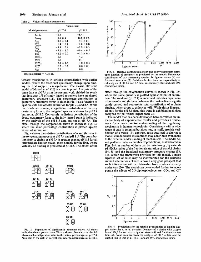
dict the populations of states for the various microscopicforms of the tetramer. Representative examples of the domi-nant states are given in Fig. 2 for the distributions at pH 7.4and 8.5. We note that this model prescribes a major fraction(as much as 30%) of quaternary structure change upon bind-ing of the first oxygen molecule. This distribution is a neces-sary correlate of the synchronization of Bohr proton releasewith quaternary structure change. Such a distribution of qua-
Biophysics: Johnson et aL
Proc. Natl. Acad. Sci. USA 81 (1984)
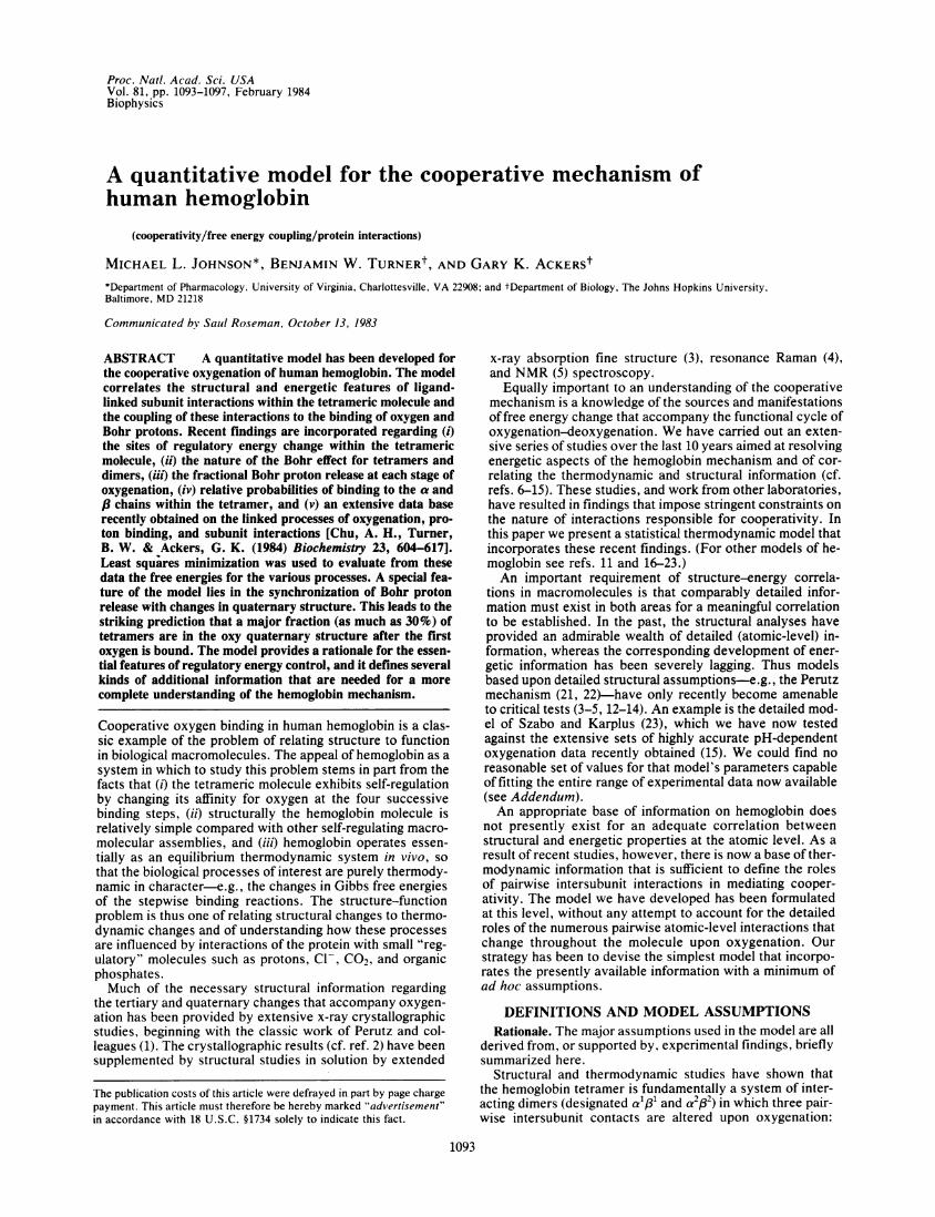
Table 2. Values of model parametersValue, kcal
Model parameter
8deoxy
boxycaaSdeoxy
°oxy
uOxy
tOxy
5aaF KUdeoxy
6oxy
8QE
One kilocalorie = 4.18 kJ.
pH 7.4
-8.3-4.1 ± 2-8.0 ± 0.11.4 ± 0.4
-3.4 ± 0.4-5.6 ± 1.5-2.2 ± 0.20.70.3
-3.1 ± 1.50.2 ± 0.10.8
pH 8.5
-8.47-10.6 ± 0.6-9.3 ± 0.1-2.0 ± 0.8-1.9 ± 0.3-0.4 ± 0.5-1.3 ± 0.34.2
-0.11.0 ± 0.30.0 + 0.10.8
ternary transitions is in striking contradiction with earliermodels, where the fractional quaternary change upon bind-ing the first oxygen is insignificant. The classic allostericmodel of Monod et al. (16) is a case in point. Analysis of thesame data at pH 7.4 as in the present work yielded the resultthat less than 1% of singly ligated tetramers have an alteredquaternary structure (11). The percentage contribution ofquaternary structural forms is given in Fig. 3 as a function ofligation state and of total saturation for pH 7.4 and 8.5. Whilethe trends are similar, a significant contribution of the oxyquaternary form to the unligated species is shown for pH 7.4but not at pH 8.5. Conversely, a distinct contribution of thedeoxy quaternary form to the fully ligated state is indicatedby the analysis of the pH 8.5 data but not at pH 7.4. Theeffect through the oxygenation curve is shown in Fig. 3Bwhere the same percentage contribution is plotted againstextent of saturation.
Fig. 4 shows the relative contributions of a and 3chains inthe oxygenation process at pH 7.4 and pH 8.5. The contribu-tion from a chains at pH 7.4 is greater than at pH 8.5 for allintermediate ligation states, most notably for the first, wherevirtually no binding is predicted at pH 8.5. The extent of the
93 (98)
19(84) 54( 0)_I
27 (15)anx~0W~
100
80 A p-B
~-60
40
0'20 t
0 1 2 3 4 0.25 0.50 0.75 1.00Ligation state Y
FIG. 3. Relative contribution of oxy and deoxy quaternary formsupon ligation of tetramers as predicted by the model: Percentagecontribution of oxy quaternary species for ligation states (A) andfractional saturation (B). Solid and broken lines correspond to typi-cal analyses of pH 7.4 and 8.5 data, respectively. Bars indicate 65%confidence limits.
effect through the oxygenation curves is shown in Fig. 3B,where the same quantity is plotted against extent of satura-tion. The solid line (pH 7.4) is linear and indicates equal con-tribution of a and 3chains, whereas the broken line is signifi-cantly curved and represents total contribution of a chainbinding, which drops to as low as 40%. While this is illustrat-ed only for the pH 8.5 data, this trend is exhibited in all dataanalyzed for pH values higher than 7.4.The model that has been developed here correlates an im-
mense body of experimental results and provides a frame-work for a more precise understanding of the regulatorymechanism in human hemoglobin. Consistency with a widerange of data is essential but does not, in itself, provide veri-fication of a model. By contrast, tests that lead to altering amodel's fundamental assumptions may contribute even moreto the eventual understanding of mechanisms. The model de-veloped here makes a number of predictions, as illustrated inFigs. 1-4. A number of these can be tested-e.g., by extend-ed NMR studies of the fractional saturation of a and (3chains(34, 35) and the fractional quaternary structure change (33,34). Within the framework provided by this model, a morerigorous set of rules may be incorporated for the pairwisesubunit interactions. There is now a very good prospect thatsuch information will be obtainable from studies currentlyunder way (26). The model can be extended further to incor-porate the effects of 2,3-diphosphoglycerate, C02, and Cl-
18( 0) 10( 0)
mzin mflMU BE
2(16) 4(11) 48(29) 18(37)
35(28) 63(66)
100(95)
FIG. 2. Population of significantly abundant states. All stateswith abundance greater than 5% are shown. Numbers on the leftabove each configuration refer to the actual percentages at pH 7.4.Numbers to the right in parentheses refer to percentages at pH 8.5.
N., ' i :
0.5
0.0 -0 1 2 3 4 0.25 0.50 0.75 1.00
Ligation state Y
FIG. 4. Predictions for the relative probabilities of binding oxy-gen molecules to a vs. 8 chains: Number of a chains with oxygenbound (N,) for successive ligation states (A) and fractional satura-tion (B). Solid lines are from the analysis of pH 7.4 data and thedashed line to that of pH 8.5. Bars are 65% confidence limits.
1096 Biophysics: Johnson et aL

Proc. NatL. Acad. Sci. USA 81 (1984) 1097
Table 3. Multiple minima of the Szabo-Karplus Model
True-RT RT RT RT variance
Case In Q In S In K" In Kit pKa pK 3 x1051 6.29 2.62 9.60 9.45 7.5 6.2 2.562 8.52 2.82 8.99 9.47 8.1 5.7 2.513 6.99 2.66 9.28 9.34 7.5 6.0 2.52
Model-independent experimental variance 2.52
Data of Mills et al. (7, 12) analyzed by methods described previ-ously (12) to yield model parameter free energies in kcal: Q, for qua-ternary structure change; S for salt bridge formation; K'and Kit, theintrinsic subunit oxygenation constants; pK' and pK'3, the acid pKvalues for Bohr groups on a and /3 subunits, respectively. Model-independent parameters used in the fits were: 0AG2 = -14.43 ande = Sp = -8.405 kcal (7).
binding, as well as mutant and chemically modified hemoglo-bins. These further developments and tests of the model willbe described elsewhere.
ADDENDUMWe cannot be certain that no set of parameters could befound that would bring the Szabo-Karplus model into corre-spondence with the experimental data. The model is plaguedby innumerable local minima, precluding a unique fit (12,23). The best parameter set reported appears to be that pre-sented very recently by Lee and Karplus (36) given in Table3 (case 1). We have analyzed this parameter set by compar-ing it to the actual published data of Mills et al. (7, 12) ratherthan the simulated data used by Lee and Karplus (36). Theresulting true sample variance is given in Table 3. We furthertested the ability of this parameter set to predict the tetramerBohr effect, and the results are shown as the solid line of Fig.5. For comparison we show the experimental data points ob-tained by us (15) under identical buffer conditions and tem-perature as the pH 7.4 data (7). Differential titration data ofAntonini et al. (31) are also shown. The two sets of experi-mental values, which virtually coincide, are in sharp dis-agreement with the prediction of the Szabo-Karplus model.Thus the analysis of the minimum recently reported by Leeand Karplus (36) reinforces the earlier conclusion (12) thatthe Szabo-Karplus model is incapable of accounting for thecomposite body of highly reliable experimental informationpresently available. To illustrate the problem of multipleminima we present in Table 3 two additional parameter setsthat also provide excellent fits to the oxygenation data ofMills et al. (7) with physically reasonable constants. Like the
_- 2C.)
a)
I
m
7 8 9pH
FIG. 5. Tetramer Bohr effect (number of protons released uponbinding four oxygen molecules). o, Data from differential titrations(31); m, data from pH dependence of oxygenation data (15) deter-mined under buffer and temperature conditions identical to those ofMills et al. (7). Error bars are 65% confidence limits. Solid line is theBohr effect predicted by the Szabo-Karplus model with parametersreported by Lee and Karplus (ref. 36; see case 1 in Table 3).
Lee-Karplus result, none of these parameter sets, nor otherswe have found, satisfy the criteria offitting oxygenation datawith physically reasonable model parameters and also cor-rectly predicting the Bohr effect.
This work has been supported by Grants GM-28928 and GM-24486 from the National Institutes of Health and by Grant PCM 80-14533 from the National Science Foundation.
1. Perutz, M. F., Muirhead, H., Mazzarella, L., Crowther,R. A., Greer, J. & Kilmartin, J. V. (1969) Nature (London)222, 1240-1243.
2. Baldwin, J. & Chothia, C. (1979) J. Mol. Biol. 129, 175-220.3. Eisenberger, P., Shulman, R. G., Kincaid, B. M., Brown,
G. S. & Ogawa, S. (1978) Nature (London) 274, 30-34.4. Asher, S. A., Adams, M. L. & Schuster, T. M. (1981) Bio-
chemistry 20, 3339-3346.5. Russu, I. N., Ho, N. T. & Ho, C. (1983) Biochemistry 22,
5031-5043.6. Ackers, G. K. & Halvorson, H. R. (1974) Proc. Nati. Acad.
Sci. USA 71, 4312-4316.7. Mills, F. C., Johnson, M. L. & Ackers, G. K. (1976) Biochem-
istry 15, 5350-5362.8. Valdes, R. & Ackers, G. K. (1978) Proc. Nati. Acad. Sci. USA
75, 311-314.9. Mills, F. C. & Ackers, G. K. (1979) Proc. Nati. Acad. Sci.
USA 76, 273-277.10. Ackers, G. K. (1980) Biophys. J. 32, 331-346.11. Ackers, G. K. & Johnson, M. L. (1981) J. Mol. Biol. 147, 559-
582.12. Johnson, M. L. & Ackers, G. K. (1982) Biochemistry 21, 201-
211.13. Flanagan, M. A., Ackers, G. K., Hanania, G. I. H. & Gurd,
F. R. N. (1981) Biochemistry 20, 7439-7449.14. Pettigrew, D. W., Romeo, P. H., Tsapis, A., Thillet, J., Smith,
M. L., Turner, B. W. & Ackers, G. K. (1982) Proc. Nati.Acad. Sci. USA 79, 1849-1853.
15. Chu, A. H., Turner, B. W. & Ackers, G. K. (1984) Biochemis-try 23, 604-617.
16. Monod, J., Wyman, J. & Changeux, J.-P. (1965) J. Mol. Biol.12, 88-118.
17. Koshland, D. E., Nemethy, G. & Filmer, D. (1966) Biochemis-try 5, 364-385.
18. Weber, G. (1972) Biochemistry 11, 864-878.19. Weber, G. (1982) Nature (London) 300, 603-607.20. Herzfeld, J. & Stanley, E. (1974) J. Mol. Biol. 82, 231-265.21. Perutz, M. F. (1970) Nature (London) 228, 726-734.22. Perutz, M. F. (1970) Nature (London) 228, 734-739.23. Szabo, A. & Karplus, M. (1972) J. Mol. Biol. 72, 163-197.24. Chu, A. H. & Ackers, G. K. (1981) J. Biol. Chem. 256, 1199-
1205.25. Miura, S. & Ho, C. (1982) Biochemistry 21, 6280-6287.26. Smith, F. R. & Ackers, G. K. (1983) Biophys. J. 41, 415.27. Kilmartin, J. V., Fogg, J. H. & Perutz, M. F. (1980) Biochem-
istry 19, 3189-3193.28. Perutz, M. F., Kilmartin, J. V., Nishikura, K., Fogg, J. H.,
Butler, P. J. G. & Rollema, H. J. (1980) J. Mol. Biol. 138, 649-670.
29. Matthew, J. B., Hanania, G. I. H. & Gurd, F. R. N. (1979)Biochemistry 18, 1919-1928.
30. Matthew, J. B., Hanania, G. I. H. & Gurd, F. R. N. (1979)Biochemistry 18, 1928-1936.
31. Antonini, E., Wyman, J., Brunori, M., Fronticelli, C., Bucci,E. & Rossi-Fanelli, A. (1965) J. Biol. Chem. 240, 1096-1103.
32. Johnson, M. L., Halvorson, H. R. & Ackers, G. K. (1976)Biochemistry 15, 5363-5371.
33. Johnson, M. L. (1984) Methods Enzymol., in press.34. Viggiano, G. & Ho, C. (1979) Proc. Nati. Acad. Sci. USA 76,
3673-3677.35. Viggiano, G., Ho, N. T. & Ho, C. (1979) Biochemistry 18,
5238-5247.36. Lee, A. & Karplus, M. (1983) Proc. Natl. Acad. Sci. USA 80,
7055-7059.
*000
I . . . I . . . . I . -
'0
a00 I:00
Biophysics: Johnson et aL
-A