a rapid screening method to evaluate the impact of ...€¦a rapid screening method to evaluate the...
TRANSCRIPT

A rapid screening method to evaluate the impactof nanoparticles on macrophages†Inès Mottas,a,b Ana Milosevic,c Alke Petri-Fink,c,d Barbara Rothen-Rutishauserc andCarole Bourquin*a,b
Nanotechnology is an emerging and highly promising field to develop new approaches for biomedical
applications. There is however at present an unmet need for a rapid and universal method to screen nano-
particles (NP) for immunocompatibility at early stages of their development. Indeed, although many types
of highly diverse NP are currently under investigation, their interaction with immune cells remains fairly
unpredictable. Macrophages which are professional phagocytic cells are believed to be among the first
cell types that take up NP, mediating inflammation and thus immunological responses. The present work
describes a highly reproducible screening method to study the NP interaction with macrophages. Three
essential questions are answered in parallel, in a single multiwell plate: Are the NP taken up by macro-
phages? Do the NP cause macrophage cell death? Do the NP induce inflammatory reactions? This assay
is proposed as a standardized screening protocol to obtain a rapid overview of the impact of different
types of NP on macrophages. Due to high reproducibility, this method also allows quality control assess-
ment for such aspects as immune-activating contaminants and batch-to-batch variability.
Introduction
Nanomedicine represents a rapidly developing field whichbears high promise for many clinical applications.1
Nanoparticles (NP) are defined as particulate objects from 1 to100 nm in diameter with specific properties that cannot befound in bulk materials.2 NP can be designed to possessunique optical properties allowing novel imaging and diagno-stic applications,3 or to serve as highly modular drug deliverysystems that target specific organs or cell populations withinthe body, thus improving drug efficacy and at the same timereducing unwanted side effects.4–7 It is even becoming poss-ible to combine diagnostic and therapeutic characteristics inengineered NP to yield promising theranostic tools.8 BecauseNP themselves can have an impact on cell viability and func-tion, it is critical to assess their biocompatibility early in the
development process. Indeed, even if NP are individually wellcharacterized with respect to many factors such as shape, size,zeta potential, and colloidal stability, the outcome of theirinteraction with cells (which also differ in type and function)is difficult to predict.9 To rapidly and efficiently select appro-priate NP for clinical applications, it is essential to developefficient screening methods to test their interactions with thecell types that they will encounter in the organism.
Macrophages can be found in all tissues of the body, andthey are particularly abundant at sites that are in direct contactwith the environment such as lungs, skin, and gastrointestinalmucosa, or in organs with important blood supply such as thespleen, kidney and liver.10 The prime ability of macrophages isthe engulfment of microorganisms, damaged cells and otherparticulate materials by phagocytosis.11,12 For these reasons,macrophages are a critical cell type with respect to biomedicalNP applications, as they are considered to be one of the firstcell populations that are encountered by NP entering thebody.12 Through their phagocyte function, these cells mediateclearance of NP from the body and thus modulate NP bioavail-ability.13 In addition to their scavenger role, macrophages areamong the first cells to initiate an inflammatory immuneresponse. When they recognize specific microbial componentsas a danger signal, macrophages rapidly produce proinflam-matory cytokines such as interleukin 6 (IL-6) and tumor necro-sis factor-α (TNF-α) in order to initiate a down-stream immuneresponse.10 If NP themselves are recognized as a dangersignal, or if traces of immune-activating substances are
aChair of Pharmacology, Department of Medicine, Faculty of Science, University of Fribourg, Chemin du Musée 5, 1700 Fribourg, SwitzerlandbSchool of Pharmaceutical Sciences, University of Geneva, University of Lausanne, and Department of Anesthesiology, Pharmacology, and Intensive Care,Rue Michel-Servet 1, 1211 Geneva, Switzerland. E-mail: [email protected]; Tel: +41 22 37 90701cAdolphe-Merkle Institute, University of Fribourg, Chemin des Verdiers 4,1700 Fribourg, SwitzerlanddChemistry Department, University of Fribourg, Chemin Du Musée 9, 1700 Fribourg, Switzerland
† Electronic supplementary information (ESI) available.
1
http://doc.rero.ch
Published in "Nanoscale 9(7): 2492–2504, 2017"which should be cited to refer to this work.

present in the NP preparation, macrophages may trigger animmune response against the material.14 In addition, someNP have been reported to affect viability of macrophages.15
This immunotoxicity may negatively affect the maintenance ofimmunity against pathogens.
It is thus essential to characterize the modes of interactionof macrophages with NP designed for biomedical purposes atthe early stages of their development. Indeed, such assaysshould be performed for every NP batch before further bio-logical testing, as there can be considerable variation betweenindividual NP preparations. Several studies have reportedinvestigation of NP–macrophage interactions,12,16–18 but theexperiments were generally performed in a serial manner,requiring a high concentration of particles to assess all essen-tial parameters in order to test the NP. One report described ascreening method to assess liposome interactions with macro-phages using fluorescence microscopy to quantify uptake andcell viability.19 However microscopy represents a challenge toacquire and analyze all samples equally, and the assessmentof more than 3 parameters at the same time is unusual.20
Another group described how to screen NP cytotoxicity onfibroblasts and alveolar epithelial cells.21 This method couldhowever not easily be applied to macrophages, as NP internal-ization and immune activation were not considered. Takentogether, there is an unmet need for a standardized method toassess the essential parameters of NP–macrophage inter-actions rapidly and efficiently.
Here we present an automated screening assay by which NPcytotoxicity, cellular uptake, and immune activating effects onmacrophages in vitro are characterized in a standardizedmanner. These three essential aspects of NP–macrophageinteractions can be assessed in a single culture plate within a24 h experiment. Since NP themselves may interfere withdifferent readouts,22,23 we proposed adequate controls in orderto ensure that NP interferences do not lead to an over- orunderestimation of the NP effects. Cost effectivity and highreproducibility of the protocol make the screening methodattractive for identifying a lead candidate for biomedical pur-poses, for quality control of each NP preparation and for thedetection of immune-activating contaminants.
Materials and methodsNP synthesis
Citrate-stabilized gold NP were synthesized as previouslyreported.24 To enhance NP colloidal stability,25 the gold corewas coated with a polymer mixture comprising of polyvinylalcohol (PVA; Mowiol 3–85, Mw = 15 000 Da) (Calbiochem,EMD bioscience, Inc. La Jolla, USA) and a vinylalcohol–vinyl-amine copolymer (NH2PVA; M12, Mw = 240 000 Da) (Erkol S.A.,Tarragona, Spain) as previously described for hetero-functiona-lized AuNP.24 For this study, polymers were fluorescentlylabeled with a Cyanine5 NHS dye (Cy5; Lumiprobe, Hannover,Germany) via ester bond formation for the polyvinyl alcoholpolymer and the amide bond for the vinylalcohol–vinylamine
copolymer. For fluorescently-labeled AuNP the polymer mixturewas composed of 0.5 mg ml−1 of NH2PVA, 0.375 mg ml−1 ofNH2PVA-noCy5, 1.612 mg ml−1 PVA and 4.7 mg ml−1 PVA-noCy5. For unlabeled AuNP the polymer mixture was com-posed of 0.875 mg ml−1 NH2PVA-noCy5 and 6.3 mg ml−1 PVA-noCy5. The following NP were analyzed: citrate-stabilized goldNP (citrate AuNP) and polymer-coated NP with Cy5 labeling(NH2PVA-AuNP). Polymer-coated NP without Cy5 labeling(NH2PVA-AuNP-noCy5) were used only as control in the uptakeexperiment (Fig. 2). Additional AuNP formulations were usedin order to validate the method (see the ESI†).
NP characterization and stability
All NP were characterized in terms of their optical properties,size and zeta potential. Optical properties were characterizedusing a UV–Vis spectrometer (Jasco Europe S.R.L., Milano,Italy), where the spectra of NP were measured in water forcitrate AuNP and PBS for NH2PVA-AuNP and NH2PVA-AuNP-noCy5. The size distribution of the Au core was determined bytransmission electron microscopy (TEM) using a FEI TecnaiSpirit (FTI) transmission electron microscope. The hydro-dynamic radius was determined by dynamic light scattering(DLS) (LS Instruments A.G., Fribourg, Switzerland) in de-polarized DLS (DDLS) mode. DDLS measurements wereperformed at an angle of 90° in water for citrate AuNP and inPBS for NH2PVA-AuNP and NH2PVA-AuNP-noCy5. The zetapotential was measured using a ZetaSizer (BrookhavenInstruments Corporation, Holtsville, USA) in water for AuNPand PBS for NH2PVA-AuNP and NH2PVA-AuNP-noCy5.
NP colloidal stability was assessed using UV-Vis and DDLSfor time resolved measurements over 24 h at 37 °C. The stabi-lity of all three NP was tested in PBS and complete cell culturemedium (cDMEM) consisting of high glucose (4.5 g l−1)Dulbecco’s modified Eagle’s medium (DMEM) (Biowest,Nuaillé, France) was supplemented with 1% glutamine (PAAlaboratories, Pashing, Austria), 1 nM sodium pyruvate, 10%fetal bovine serum (FBS) (Biological Industries, Beit Ha’Emeq,Israel) and 0.5% ciproxine (Bayer, Zürich, Switzerland).
Macrophage cell line and NP exposure
J774.1 macrophages (ATCC, Manassas, USA) were cultivated incDMEM at 37 °C and 5% CO2. Before each experiment, cellswere concentrated to 5 × 10⁵ cells per mL before adding100 μl per well (5 × 10⁴ cells per well) in flat-bottom 96-wellplates (Corning, New York, USA). After overnight incubation,medium was replaced by 50 μl fresh cDMEM. NP, polymersand other test substances were added as indicated at 2× con-centration in 50 μl cDMEM. For all NP, the final working con-centration was 10 μg ml−1 gold (determined by UV-Vis at400 nm), corresponding to approximately 1.8 × 10¹⁰ 17 nmgold particles per mL. For polymer controls (NH2PVA andNH2PVA-noCy5), the working concentration corresponds to theconcentration of the polymer on the NP. As a positive controlfor immune activation, J774.1 macrophages were cultured with100 ng ml−1 lipopolysaccharide (LPS; Invivogen, Toulouse,France). Staurosporine was used to induce apoptotic cell death
2
http://doc.rero.ch

(1 nM; Sigma-Aldrich, Saint-Louis, USA). To control NP inter-ferences, the positive controls were tested in combination withNP added after 23 h. After a 24 h incubation, supernatantswere stored at −20 °C for cytokine quantification and100 μl per well PBS were added to the cells for light microscopyand flow cytometry analysis. Please refer to the ESI† for a listof required materials and instruments and to Fig. S1† forfurther details on the 96-well plate organization.
Flow cytometry
For flow cytometry, J774.1 cells in PBS were gently detached bypipetting and transferring to U-bottom 96-well plates for stain-ing as described below. All parameters were assessed in one96-well plate in a single measurement (Fig. S1†). Analysis wasperformed with a MACSquant analyzer 10 using the chill96-rack (both from Miltenyi Biotec, Bergisch Gladbach,Germany). Every well was gently mixed automatically beforeacquisition of 50 μl. Data in the FCS 3.1 format were analyzedusing the FlowJo software version 10.0.8r1 (FlowJo Analysissoftware, Ashland, USA).
To measure viability, the cells were incubated with PacificBlue-labeled annexin V diluted, 1 : 100, in annexin V bindingbuffer (both from Biolegend, San Diego, USA). After 30 minincubation at room temperature protected from light, onevolume of annexin V binding buffer was added. Propidiumiodide (PI; Sigma-Aldrich, Saint-Louis, USA) diluted, 1 : 200,in PBS was automatically added by using the MACSquant ana-lyzer 10 just before mixing and acquisition. For quantifi-cation of NP uptake, the proportion of Cy5-positive cellswithin live cells (annexin V/PI double-negative population)was determined.
To assess macrophage activation, the J774.1 cells werestained with a Zombie Green fixable viability dye (Biolegend,San Diego, USA) according to the manufacturer’s protocol.After washing with flow cytometry buffer consisting of PBSsupplemented with 2 mM EDTA (Calbiochem, Darmstadt,Germany) and 0.5% BSA (PAA laboratories, Pashing, Austria),the cells were incubated with unlabeled anti-CD16/32 to blockFc receptors (Biolegend, San Diego, USA). After 10 min at 4 °C,specific antibodies for surface activation markers or theirrespective isotype controls in flow cytometry buffer wereadded: Pacific Blue-CD80, PE-CD86, PE/Cy7-CD11b andAPC/Cy7-MHCII (all from Biolegend, San Diego, USA). Afterfurther 30 min at 4 °C, the cells were washed once with flowcytometry buffer before acquisition.
Bright field imaging
The live cell morphology and density were pictured by bright fieldmicroscopy (CKX53, Olympus, Tokyo, Japan) at 10× magnification.
ELISA
To quantify cytokine secretion, ELISA Max Deluxe sets formouse interleukin 6 (IL-6) and tumor necrosis factor-α (TNF-α)(both from Biolegend, San Diego, USA) were used according tothe manufacturer’s protocol in half-well plates. Absorbancewas measured at 450 nm with an Infinite 200 PRO plate-reader
(TECAN, Männedorf, Switzerland) where the reference absor-bance at 570 nm was subtracted. Cytokine concentrations werecalculated according to the standard curve performed induplicate.
Statistical analysis
Graphs were plotted with the GraphPad Prism softwareversion 6.0e (GraphPad Software, San Diego, USA). The stat-istical significance of multiple groups in comparison to thecontrol group (untreated sample) was assessed using one-wayANOVA followed by Dunnett’s multiple comparison test. Anunpaired t-test was used in Fig. 4 to compare two groups. For allexperiments with the J774.1 cells, graphs show the mean anderror bars indicate the standard error of the mean (SEM) of fouror five independent experiments performed on different days.
Results
This rapid method presented herein to test NP–macrophageinteractions and cellular responses is based on a multi-para-meter analysis using a macrophage cell line. After exposure ofmacrophages to NP in a 96-well cell culture plate, the cellswere analyzed by flow cytometry and bright field microscopy,whereas supernatants were collected for ELISA quantificationof cytokines. This strategy allows to determine several crucialaspects of interactions between NP and macrophages in paral-lel from a single culture plate (Scheme 1): NP-related cyto-toxicity (1), cellular uptake of NP (2), and proinflammatoryactivation (3).
NP characterization and stability
To illustrate the multi-parameter screening method for NP–macrophage interactions, we chose to use polymer-coated goldparticles (AuNP) as model NP. AuNP possess several advan-tages which make them promising candidates for biomedicalapplications, including tunable sizes, the possibility of surfacefunctionalization, optical properties that enable their detec-tion, and low toxicity.26 We selected hetero-functionalizedNH2PVA-AuNP as a model for the assay, as we have previouslyshown that these NP are best taken up among the formu-lations tested.24 Citrate-stabilized AuNP were coated with thevinylalcohol–vinylamine copolymer/polyvinyl alcohol polymermixture through ligand exchange as described.24 In order toassess NP uptake by macrophages, the first polymer layer waslabeled with the Cy5 fluorescent dye (NH2PVA-AuNP). NP withunlabeled polymers were used as a control (NH2PVA-AuNP-noCy5). Cy5 (650/670), a far red dye, was selected to limitpotential fluorescence interferences with the gold core(Fig. 1B). All AuNP were thoroughly characterized by conven-tional methods: TEM (gold core size distribution), UV-Vis (par-ticle size and concentration), DDLS (hydrodynamic size), andzeta potential (Fig. 1). DDLS was preferred to characterize theparticle size (hydrodynamic radius), as it was previously shownto have an advantage over conventional dynamic light scatter-ing (DLS) for measurements in a complex fluid such as cell
3
http://doc.rero.ch

culture medium.27 NP stability in complete macrophageculture medium (cDMEM) was determined after 24 hincubation at 37 °C by start/end point UV-Vis measurementsand time resolved DDLS (Fig. 2). Although citrate AuNP do notpreserve colloidal stability in PBS (data not shown),27 theseparticles were stable in cDMEM, probably due to protein
corona formation.28,29 Both NH2PVA-AuNP-noCy5 andNH2PVA-AuNP were stable in cDMEM over 24 h.
Impact of NP on macrophage viability
As a cellular model, we used the well-described J774.1 murinemacrophage cell line, which possesses many of the defining
Scheme 1 Parameters measured by NP screening assay. J774.1 macrophages are exposed to NP for 24 h. Three effects of NP–macrophage inter-actions are measured in parallel. (1) Cytotoxicity is assessed by bright field microscopy and measured by flow cytometry with annexin V andpropidium iodide (PI) staining. (2) Uptake of fluorescently-labeled NP by macrophages is determined by flow cytometry. (3) The immune activationphenotype is determined by flow cytometry measurement of cell surface activation markers (MHC II, CD80, CD86, CD11b) and by quantification ofproinflammatory cytokines in the supernatant (IL-6, TNF-α).
Fig. 1 Characterization of AuNP. (A) TEM image of AuNP with the corresponding diameter distribution histogram. (B) UV-Vis spectra of NP.Measurements were performed in the media used for synthesis: water (citrate AuNP) or PBS (NH2PVA-AuNP-noCy5 and NH2PVA-AuNP). Data arenormalized according to absorbance at 400 nm. a.u.: area unit. (C) Table shows NP core diameter obtained via TEM, NP hydrodynamic diameterobtained via depolarized dynamic light scattering (DDLS) measurements and zeta potential measurements (ameasurements performed in water;bmeasurements performed in PBS, both without incubation) at 40 μg AuNP per ml. Mean ± SD of 3–5 measurements for DDLS and 10 measurementsfor zeta-potential are shown.
4
http://doc.rero.ch

characteristics of macrophages. To examine macrophage viabi-lity, morphology and confluency, the J774.1 cells were visuallyassessed by bright field microscopy after a 24 h culture(Fig. 3A). Staurosporine, which induces apoptotic cell death,strongly reduced the proportion of round live cells.Lipopolysaccharide (LPS), which is a bacterially derived endo-toxin used as a positive proinflammatory control, promotedadhesion and spreading of cells, inducing the typical mor-phology of immune-activated macrophages (arrows).30 In con-trast, exposure to citrate AuNP, the NH2PVA polymer orNH2PVA-AuNP did not induce any change in morphology, withcells remaining small and round. Some material aggregates(arrows) were seen with NH2PVA polymer exposure.
Two major mechanisms of cell death can be distinguished,apoptosis and necrosis. Whereas apoptotic cell death does notgenerally promote inflammation, necrotic cell death leads tothe disruption of the cell membrane and release of pro-inflammatory substances. To investigate whether AuNP inducedcell death is by one of these mechanisms, macrophages wereassessed by flow cytometry after staining with annexin V andpropidium iodide (PI). Annexin V detects the presence of phos-phatidylserine on the outer layer of the membrane bilayer, anearly sign of apoptosis, whereas PI intercalates into DNA whenthe cell membrane is disrupted.31 This staining allows easydiscrimination between live, apoptotic and dead cells (Fig. 3B,top left). The untreated J774.1 cells show approximately 60%live cells under these conditions, as this is a cell line with ahigh proliferation rate and turnover. Our results showed thatcitrate AuNP, the NH2PVA polymer and NH2PVA-AuNP did notdecrease the proportion of live cells after a 24 h exposure, incontrast to staurosporine (Fig. 3B and C). The NH2PVApolymer and NH2PVA-AuNP did increase the proportionof AnnexinV−/PI+ dead cells, although not significantly,suggesting that the polymer may promote necrotic cell death
at higher concentrations (Fig. S2†). Since the NP themselvescould in principle interfere with the measurements (the goldcore could for instance affect the PI fluorescence signal with amaximal emission at 538 nm), it was necessary to verify thatthe materials did not affect the fluorescence signals of annexinV or PI. To verify this point, macrophages were incubated for24 h with staurosporine. For the last hour before assessmentby flow cytometry, AuNP, the NH2PVA polymer orNH2PVA-AuNP were added to the staurosporine-treated cells.Our results showed that none of these conditions interferewith the annexin V/PI staining, as there was no difference inmacrophage viability compared to the cells incubated onlywith staurosporine (Fig. 3C, right panel). We chose to use flowcytometry rather than a metabolic assay such as the 3-(4,5-di-methylthiazol-2-yl)-2,5 diphenyltetrazolium (MTT) test toassess cell viability, because this technique allows a multi-parametric readout. In addition, some NP have been describedto absorb the MTT reagent32 or to interfere directly with colori-metric reading,33 and the MTT assay does not directly measurecell counts but rather depends on mitochondrial enzymaticactivity. In the case where NP could affect mitochondrialactivity, the MTT reading would also be affected.34 Thus, weconfirmed by using two independent methods thatNH2PVA-AuNP do not affect macrophage viability.
Uptake of fluorescently labeled NP by macrophages
Macrophages are specialized in the uptake of particulatematerials such as tissue debris and certain pathogens.12 Sinceuptake by macrophages critically modifies the pharmaco-kinetics and biodistribution of the NP in the body,35 it isessential to assess NP uptake by macrophages early in thedevelopment process of any particles designed for biomedicalapplications.
Fig. 2 NP stability in cell culture medium. NP were incubated for 24 h in complete cell culture medium (cDMEM) at 37 °C. (A) UV-Vis assessment ofNP stability. UV-Vis spectra were measured at the beginning of incubation (0 h) and after 24 h. (B) Timeline of DDLS measurements. During incu-bation DDLS measurements at 90° angle were performed in a time resolved fashion (every 300 seconds).
5
http://doc.rero.ch

Flow cytometry analysis detects morphological changesproportionally to the size and granularity of the cell by forwardscatter (FSC) and side scatter (SSC), respectively. In conse-
quence, if NP are taken up, the SSC intensity may increase.36–38
However our data show no significant SSC shift when com-paring NP-treated cells with untreated cells (Fig. 4A). As thecell size and granularity change when cells are dying,39 thistechnique requires the exclusion of dying cells before theuptake analysis. Therefore, we first gated on live (annexin V/PIdouble-negative) cells.
A more sensitive strategy to determine whetherNH2PVA-AuNP are taken up by macrophages is to use the fluo-rescently labeled NP. The J774.1 cells were exposed for 24 h at37 °C to NH2PVA-AuNP labeled with Cy5 and fluorescenceintensity was then quantified by flow cytometry. Macrophagesincubated with Cy5-labeled NH2PVA-AuNP or with the Cy5-labeled NH2PVA polymer alone showed higher fluorescencethan non-exposed macrophages (Fig. 4B, left panel). Indeed, atleast 80% of the macrophages exposed to NH2PVA-AuNP wereCy5-positive (Fig. 4C, left panel). To determine whether the NPwere actively taken up by the cells, incubation was also per-formed at 4 °C, a temperature at which many active processesare inhibited. At 4 °C, the percentage of Cy5-positive cellsdropped significantly to approximately 40% for cells exposedto NH2PVA-AuNP (Fig. 4C), suggesting that the NP are taken upat least in part by an active process. The Cy5-positive macro-phages exposed to NP at 4 °C may be decorated with NP ontheir surface, as this would not require an active uptake.
To verify that the NH2PVA polymer and the NP themselvesdo not affect the detection of the Cy5 fluorescent signal, weincubated macrophages with the NH2PVA polymer orNH2PVA-AuNP without Cy5 at 37 °C and 4 °C (Fig. 4A and B,right panels). No increase in fluorescence was seen, indicatingthat the NH2PVA polymer and NH2PVA-AuNP do not affect Cy5detection by flow cytometry.
Production of proinflammatory cytokines by macrophagesafter NP exposure
In addition to cytotoxicity and cellular uptake, it is essential toassess NP-induced immune activation on macrophages.Indeed, compounds used during NP synthesis often containtrace contaminations of immune-activating substances thatcan lead to important inflammatory reactions. LPS, an endo-toxin present on Gram-negative bacteria, is a frequent con-taminant that can induce strong proinflammatory responseseven at picogram concentrations in humans.40
As J774.1 macrophages show high sensitivity to LPS andother pathogen contaminants,41 we propose to use these cellsas a highly reactive and cost-limiting strategy to detectimmune-activating contaminations. The activation of macro-phages leads to the secretion of several proinflammatory cyto-kines such as interleukin 6 (IL-6) and tumor necrosis factor-α(TNF-α), which represent the first step towards a generalizedinflammatory response. We quantified cytokine concentrationby ELISA in the culture supernatant from the same wells ofNP-exposed macrophages used for flow cytometry. After 24 hof exposure to citrate AuNP, the NH2PVA polymer orNH2PVA-AuNP, none of these conditions induced either IL-6 orTNF-α (Fig. 5, left panels). LPS was used as a positive control
Fig. 3 Impact of NP on macrophage viability. J774.1 macrophages wereincubated for 24 h with NP, the NH2PVA polymer or staurosporine.(A) Representative bright field images at 10× magnification. Scale bar:100 μm. (B and C) Flow cytometry analysis with annexin V and PI.(B) One representative dot plot per condition is shown. (C) Percentageof live (annexin V/PI double-negative) cells. Interference controls (rightpanel): macrophages were incubated with staurosporine for 24 h. After23 h, the NP or NH2PVA polymer were added for the last hour beforeanalysis. Each bar represents mean ± SEM of 5 independent experi-ments. Asterisks (***, P < 0.001) indicate significant differences with theuntreated group using one-way ANOVA followed by Dunnett’s multiplecomparison test.
6
http://doc.rero.ch

and for all cytokines a significant increase compared to theuntreated control was observed.
To verify that the NP themselves did not interfere with theELISA assay, the J774.1 cells were exposed to LPS for 24 h toinduce proinflammatory cytokines and citrate AuNP, theNH2PVA polymer or NH2PVA-AuNP were added only for the lasthour of incubation, a time frame which was sufficient for NPuptake (Fig. S3†). Although the cytokine concentration wasexpected to be the same under all conditions, a lower absor-bance was measured by ELISA under the conditions with NPor the polymer (Fig. 5, right panels), suggesting that bothmaterials interfere with the assay at the concentrations used.The lower signal intensity could be due to cytokine adsorptionto the material surface.
Immune activation phenotype of macrophages after NPexposure
To verify the NP-induced activation of macrophages by asecond readout, we examined whether NH2PVA-AuNP caused aswitch from a naïve to an activated phenotype of the J774.1
cells. Upon immune activation, the cell surface markers ofmacrophages change to enhance their interactions with otherimmune cells. We therefore analyzed the up-regulation of keysurface activation markers such as major histocompatibilitycomplex class II (MHC II) molecules, that mediate antigenpresentation to T cells; CD80 and CD86, two co-stimulatorymolecules that support efficient T-cell activation; CD11b aleukocyte integrin associated with migration and adhesionthat is essential in inflammation.42 Citrate AuNP, the NH2PVApolymer and NH2PVA-AuNP did not induce up-regulation ofany of the tested activation markers on the J774.1 cells, in con-trast to the immune activator LPS (Fig. 6, left panels). Tocontrol NP interference, we incubated the macrophages withLPS for 24 h to induce the up-regulation of these typical acti-vation markers. Citrate AuNP, the NH2PVA polymer orNH2PVA-AuNP were added for the last hour before assessmentby flow cytometry, in order to determine if the materialsreduce the fluorescence signal of the labeled antibodies. Ourresults showed that neither the NP nor the polymer signifi-cantly modified the fluorescence signal compared to LPS alone
Fig. 4 Uptake of NP by macrophages. J774.1 macrophages were incubated for 24 h at 37 °C (A–C) or 4 °C (C, as indicated) with the NP or NH2PVApolymer. Uptake was analyzed by flow cytometry after gating on live (annexin V/PI double-negative) cells. (A) Representative modal histograms ofthe SSC signal. (B and C) Representative modal histograms of Cy5 (B) and percentage of Cy5+ cells (C) for macrophages incubated with Cy5-labeledNP and the polymer (test samples, left panel) or with unlabeled NP and the polymer (interference controls, right panel). Each bar represents mean ±SEM of 4 independent experiments (unlabeled NP and the polymer: 2 independent experiments). Asterisks (*, P < 0.05; **, P < 0.01) without bracketsindicate comparison to the untreated group using one-way ANOVA followed by Dunnett’s multiple comparison test. Asterisk (*, P < 0.05) with brack-ets indicates comparison using unpaired t-test. ns: not significant.
7
http://doc.rero.ch

(Fig. 6, middle panels). Because immune activation mayincrease cell autofluorescence and unspecific antibodybinding, antibody isotype controls are necessary to confirmthe specificity of the signal increase in LPS-treated macro-phages. The median fluorescence intensity (MFI) for isotypecontrol staining was not increased under any of the cultureconditions, demonstrating that the signal measured for acti-vation markers is specific (Fig. 6, right panels).
Discussion
Interest in NP use for biomedical applications is growing andthere is an urgent need for reliable, rapid and universal
methods to characterize NP interactions with immune cells.Herein we propose a rapid multi-parameter test to screen theNP impact on macrophages within one experimental day. Wesuggest flow cytometry as a standard method to investigate theproinflammatory potential of NP using a macrophage cell line,since it is an automated technique to quantify several para-meters simultaneously, including cytotoxicity, NP uptake andproinflammatory-activating effects. The samples are measuredon only one culture plate, thus allowing economical use of NPand reagents. We have shown exemplary results with polymer-coated heterofunctionalized AuNP as a model, but this assaycan be run with many different types of engineered nano-materials, as shown in Fig. S4† with differently functionalizedAuNP. In our laboratory we have also implemented this test forthe screening of protein-based NP, poly-lactic-co-glycolic acid(PLGA) NP, silica NP and micelles in order to select leadingcandidates between different synthesis strategies.
As a macrophage model, we used the widely investigatedJ774.1 murine cell line, which possesses characteristic func-tions of macrophages such as phagocytosis, antigen proces-sing, cytokine production, and many of the surface markers.43
We found the data obtained with this cell line to be highlyreproducible, allowing direct comparison of results even whentested on different days. MSC-2, a myeloid suppressor cell line,can also be used to run the screening assay (Fig. S4†), as canprimary cells generated from animal or human samples.Indeed, the results obtained with this screening protocol forNH2PVA-AuNP in J774.1 macrophages mirror data obtainedpreviously for these NP in primary human immune cells.44 Inany case, the first validation of NP using a cell line limits thenumber of experimental animals needed for further character-ization and avoids the loss of human samples.
For a step-by-step overview of the interpretation of the datafrom the screening assay, we propose a flow chart that sum-marizes the key points of this process (Fig. 7). Because NP–macrophage interactions are dependent on particle pro-perties,45 the physico-chemical characterization of the panel ofNP, including size, shape, charge, and colloidal stability alsoin the cell culture medium in which the cells are grown, isrequired before exposure. The working concentration for thescreening protocol should be selected according to theplanned biomedical application and based on the expectedconcentration in the organism.46,47 In addition, the workingconcentration should not lead to NP agglomeration.22 ForNH2PVA-AuNP, a potential application is the delivery ofimmunoactive drugs. We thus selected a working concen-tration (10 μg ml−1 of AuNP) that would be higher than theconcentration needed to deliver an effective dose of the drug.At this dose, the NP demonstrated colloidal stability in media.
As a first step, the screening protocol rapidly assessespotential cytotoxicity. This step allows the selection of the can-didates inducing the least cell death within a panel of NP. Italso allows rapid screening of NP during the developmentprocess and may alert the necessity of design modifications ifevident cytotoxicity is detected. As a second step, cellularuptake by macrophages is measured. In general, because
Fig. 5 Production of proinflammatory cytokines by macrophages afterNP exposure. Test samples (left panels): J774.1 macrophages were incu-bated for 24 h at 37 °C with NP, the NH2PVA polymer or LPS.Interference controls (right panels): macrophages were incubated for24 h at 37 °C with LPS (100 ng ml−1). NP or the NH2PVA polymer wereadded for the last hour of incubation. IL-6 and TNF-α were quantified inculture supernatants by ELISA. Each bar represents mean ± SEMof 5 independent experiments performed at least in duplicate. Asterisks(*, P < 0.05; **, P < 0.01; ***, P < 0.001) indicate significant differenceswith the untreated group using one-way ANOVA followed by Dunnett’smultiple comparison test.
8
http://doc.rero.ch

macrophages lead to NP clearance, a low uptake is desirableand the NP candidates should be selected accordingly. Lowcapture by macrophages will result in low NP clearance, allow-ing them more time to reach their target cells in the body.43
One widely used coating for NP which considerably reducestheir uptake by macrophages is polyethylene glycol (PEG).48 In
the previously established AuNP library in our laboratory,24 thecoating strategies strongly modify the uptake of 17 nm gold coreNP by macrophages.19 For instance PEG-coated AuNP showed alower uptake than PVA-coated NP, and carboxyl-functionali-zation resulted in a lower uptake than amino-functionalization.For specific applications with the aim to stimulate or to inhibit
Fig. 6 Changes in surface activation markers on macrophages after NP exposure. Median fluorescence intensity (MFI; fold change compared tountreated sample) of activation markers on J774.1 macrophages after incubation is shown. Test samples, left panels: macrophages were incubatedfor 24 h at 37 °C with NP, the NH2PVA polymer or LPS as shown in Fig. 5. Interference controls, middle panels: macrophages were incubated withLPS for 24 h as in Fig. 5. NP or the NH2PVA polymer were added for the last hour of incubation. Isotype controls, right panels: median fluorescenceintensity after staining of test samples and interference controls with unspecific isotype control antibodies is shown. Each bar represents mean ±SEM of at least 3 independent experiments. Asterisks (*, P < 0.05; ***, P < 0.001; ****, P < 0.0001) indicate significant differences with the untreatedgroup using one-way ANOVA followed by Dunnett’s multiple comparison test.
9
http://doc.rero.ch

an immune response, macrophages may be themselves thetarget of the NP, in which case the selection of NP with highcellular uptake may be appropriate.49,50
As third step of the screening assay, the NP immune effectsare determined by two parameters: the concentration of cyto-kines in the culture supernatant and the expression of typicalmarkers on the surface of activated macrophages. Cytokineswill diffuse rapidly in the body to initiate a generalizedimmune response, whereas surface markers are important forlocal cell-to-cell interactions. Except in specific applicationswhere immune activation is desired, such as in vaccinationstrategies, the NP that do not induce immunostimulationshould be selected in order to avoid unwanted proinflamma-tory effects in the organism.49 Any detected immunostimulat-ing effect may be caused by the NP themselves, in which casethe design of the particle may need to be adjusted. On theother hand, the observed immunoactivation may originatefrom immuno-contaminants introduced during synthesis.This may be verified using a further test, the limulus amebo-cyte lysate (LAL) assay,51,52 which is used as a gold standard totest for endotoxin contaminants. If contaminations aredetected, the conventional depyrogenation processes are oftennot suitable for NP,53 so that the NP synthesis process mayneed to be revisited.
Finally, because NP are well known to interfere withdifferent aspects of biological assays33,34 and because this maylead to incorrect interpretation of the NP impact on macro-phages, the screening protocol includes essential interferencecontrols at all steps. If interferences are detected during one of
the steps of the test, a complementary assay relying on adifferent detection method should be considered. In thisreport, we noted that both the AuNP and the polymer decreasethe ELISA absorbance reading at 450 nm in the interferencecontrol for IL-6 and TNF-α (Fig. 5). However, other types of NPwill not necessarily cause these interferences. We found it un-likely that the gold core directly interferes with the absorbancereading or the enzyme activity,26 because the polymer alonealso reduced the signal. The lower signal intensity is thusprobably due to cytokine adsorption to the material surface.To verify IL-6 and TNF-α production by alternative methods,intracellular cytokines could be assessed with flow cytometry,or cytokine mRNA levels could be measured by quantitativepolymerase chain reaction (qPCR).54
The one-plate screening assay presented here is designed togive a rapid overview of the different aspects of NP–macrophageinteractions. For a more detailed investigation of specific find-ings, we present a list of possible assays (Table 1). For instance,several methods are available to further characterize the mecha-nisms of toxicity (reviewed in ref. 15, 22, 47 and 55). For theassessment of NP uptake, the flow cytometry technique used inthe screening protocol requires fluorescent labeling of the NP.In general, it cannot be excluded that the dye may affect NPuptake,24 or that the fluorophore could detach from the core, orthat fluorescence may be quenched by the NP, as has beenshown for AuNP.56 After the initial screening, we thereforesuggest to confirm the uptake for selected NP candidates byusing a second method adapted to the type of NP used (forinstance, inductively coupled optical emission spectrometry
Fig. 7 Flow chart guide to data interpretation. Flow chart describing the different steps that a panel of NP must pass through in order to be vali-dated for deeper investigations.
10
http://doc.rero.ch
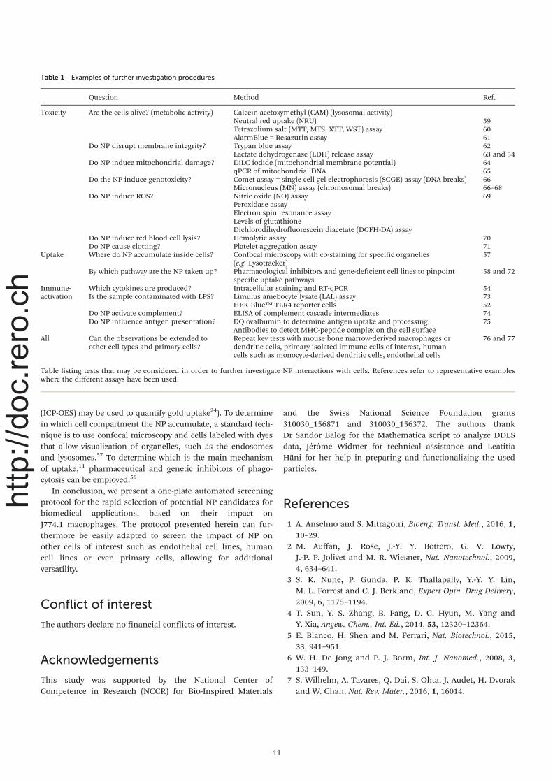
(ICP-OES) may be used to quantify gold uptake24). To determinein which cell compartment the NP accumulate, a standard tech-nique is to use confocal microscopy and cells labeled with dyesthat allow visualization of organelles, such as the endosomesand lysosomes.57 To determine which is the main mechanismof uptake,11 pharmaceutical and genetic inhibitors of phago-cytosis can be employed.58
In conclusion, we present a one-plate automated screeningprotocol for the rapid selection of potential NP candidates forbiomedical applications, based on their impact onJ774.1 macrophages. The protocol presented herein can fur-thermore be easily adapted to screen the impact of NP onother cells of interest such as endothelial cell lines, humancell lines or even primary cells, allowing for additionalversatility.
Conflict of interest
The authors declare no financial conflicts of interest.
Acknowledgements
This study was supported by the National Center ofCompetence in Research (NCCR) for Bio-Inspired Materials
and the Swiss National Science Foundation grants310030_156871 and 310030_156372. The authors thankDr Sandor Balog for the Mathematica script to analyze DDLSdata, Jérôme Widmer for technical assistance and LeatitiaHäni for her help in preparing and functionalizing the usedparticles.
References
1 A. Anselmo and S. Mitragotri, Bioeng. Transl. Med., 2016, 1,10–29.
2 M. Auffan, J. Rose, J.-Y. Y. Bottero, G. V. Lowry,J.-P. P. Jolivet and M. R. Wiesner, Nat. Nanotechnol., 2009,4, 634–641.
3 S. K. Nune, P. Gunda, P. K. Thallapally, Y.-Y. Y. Lin,M. L. Forrest and C. J. Berkland, Expert Opin. Drug Delivery,2009, 6, 1175–1194.
4 T. Sun, Y. S. Zhang, B. Pang, D. C. Hyun, M. Yang andY. Xia, Angew. Chem., Int. Ed., 2014, 53, 12320–12364.
5 E. Blanco, H. Shen and M. Ferrari, Nat. Biotechnol., 2015,33, 941–951.
6 W. H. De Jong and P. J. Borm, Int. J. Nanomed., 2008, 3,133–149.
7 S. Wilhelm, A. Tavares, Q. Dai, S. Ohta, J. Audet, H. Dvorakand W. Chan, Nat. Rev. Mater., 2016, 1, 16014.
Table 1 Examples of further investigation procedures
Question Method Ref.
Toxicity Are the cells alive? (metabolic activity) Calcein acetoxymethyl (CAM) (lysosomal activity)Neutral red uptake (NRU) 59Tetrazolium salt (MTT, MTS, XTT, WST) assay 60AlarmBlue = Resazurin assay 61
Do NP disrupt membrane integrity? Trypan blue assay 62Lactate dehydrogenase (LDH) release assay 63 and 34
Do NP induce mitochondrial damage? DiLC iodide (mitochondrial membrane potential) 64qPCR of mitochondrial DNA 65
Do the NP induce genotoxicity? Comet assay = single cell gel electrophoresis (SCGE) assay (DNA breaks) 66Micronucleus (MN) assay (chromosomal breaks) 66–68
Do NP induce ROS? Nitric oxide (NO) assay 69Peroxidase assayElectron spin resonance assayLevels of glutathioneDichlorodihydrofluorescein diacetate (DCFH-DA) assay
Do NP induce red blood cell lysis? Hemolytic assay 70Do NP cause clotting? Platelet aggregation assay 71
Uptake Where do NP accumulate inside cells? Confocal microscopy with co-staining for specific organelles(e.g. Lysotracker)
57
By which pathway are the NP taken up? Pharmacological inhibitors and gene-deficient cell lines to pinpointspecific uptake pathways
58 and 72
Immune-activation
Which cytokines are produced? Intracellular staining and RT-qPCR 54Is the sample contaminated with LPS? Limulus amebocyte lysate (LAL) assay 73
HEK-Blue™ TLR4 reporter cells 52Do NP activate complement? ELISA of complement cascade intermediates 74Do NP influence antigen presentation? DQ ovalbumin to determine antigen uptake and processing 75
Antibodies to detect MHC-peptide complex on the cell surfaceAll Can the observations be extended to
other cell types and primary cells?Repeat key tests with mouse bone marrow-derived macrophages ordendritic cells, primary isolated immune cells of interest, humancells such as monocyte-derived dendritic cells, endothelial cells
76 and 77
Table listing tests that may be considered in order to further investigate NP interactions with cells. References refer to representative exampleswhere the different assays have been used.
11
http://doc.rero.ch

8 J. Xie, S. Lee and X. Chen, Adv. Drug Delivery Rev., 2010, 62,1064–1079.
9 I. Lynch, A. Ahluwalia, D. Boraschi, H. Byrne, B. Fadeel,P. Gehr, A. Gutleb, M. Kendall and M. Papadopoulos,Bionanomaterials, 2013, 14, 195–216.
10 P. Murray and T. Wynn, Nat. Rev. Immunol., 2011, 11,723–737.
11 A. Aderem and D. Underhill, Annu. Rev. Immunol., 1999, 17,593–623.
12 R. Weissleder, M. Nahrendorf and M. Pittet, Nat. Mater.,2014, 13, 125–138.
13 M. Longmire, P. L. Choyke and H. Kobayashi,Nanomedicine, 2008, 3, 703–717.
14 T. Wynn, A. Chawla and J. Pollard, Nature, 2013, 496,445–455.
15 L. Yildirimer, N. T. Thanh, M. Loizidou and A. M. Seifalian,Nano Today, 2011, 6, 585–607.
16 T. Kusaka, M. Nakayama, K. Nakamura, M. Ishimiya,E. Furusawa and K. Ogasawara, PLoS One, 2014, 9, e92634.
17 H. Chang, J. Y. Yhee, G. H. Jang, D. G. You, J. H. Ryu,Y. Choi, J. H. Na, J. H. Park, K. H. Lee, K. Choi, K. Kim andI. C. Kwon, J. Controlled Release, 2016, 244, 205–213.
18 W.-K. Oh, S. Kim, M. Choi, C. Kim, Y. Jeong, B.-R. Cho,J.-S. Hahn and J. Jang, ACS Nano, 2010, 4, 5301–5313.
19 C. Kelly, C. Lawlor, C. Burke, J. Barlow, J. Ramsey,C. Jefferies and S.-A. Cryan, J. Liposome Res., 2014, 25,211–221.
20 D. Vanhecke, L. Rodriguez-Lorenzo, M. J. Clift, F. Blank,A. Petri-Fink and B. Rothen-Rutishauser, Nanomedicine,2014, 9, 1885–1900.
21 F. Sambale, F. Stahl, F. Rüdinger, D. Seliktar, C. Kasper,D. Bahnemann and T. Scheper, J. Nanomater., 2015, 2015,1–16.
22 A. Dhawan and V. Sharma, Anal. Bioanal. Chem., 2010, 398,589–605.
23 R. Guadagnini, B. Halamoda Kenzaoui, L. Walker,G. Pojana, Z. Magdolenova, D. Bilanicova, M. Saunders,L. Juillerat-Jeanneret, A. Marcomini, A. Huk, M. Dusinska,L. M. Fjellsbø, F. Marano and S. Boland, Nanotoxicology,2015, 9(Suppl 1), 13–24.
24 L. Rodriguez-Lorenzo, K. Fytianos, F. Blank, C. von Garnier,B. Rothen-Rutishauser and A. Petri-Fink, Small, 2014, 10,1341–1350.
25 T. Moore, L. Rodriguez-Lorenzo, V. Hirsch, S. Balog,D. Urban, C. Jud, B. Rothen-Rutishauser, M. Lattuada andA. Petri-Fink, Chem. Soc. Rev., 2015, 44, 6287–6305.
26 E. Boisselier and D. Astruc, Chem. Soc. Rev., 2009, 38, 1759–1782.
27 S. Balog, L. Rodriguez-Lorenzo, C. A. Monnier, M. Obiols-Rabasa, B. Rothen-Rutishauser, P. Schurtenberger andA. Petri-Fink, Nanoscale, 2015, 7, 5991–5997.
28 J. Gebauer, M. Malissek, S. Simon, S. Knauer, M. Maskos,R. Stauber, W. Peukert and L. Treuel, Langmuir, 2012, 28,9673–9679.
29 R. Murdock, L. Braydich-Stolle, A. Schrand, J. Schlager andS. Hussain, Toxicol. Sci., 2008, 101, 239–253.
30 G. Kleveta, K. Borzęcka, M. Zdioruk, M. Czerkies,H. Kuberczyk, N. Sybirna, A. Sobota and K. Kwiatkowska,J. Cell. Biochem., 2012, 113, 80–92.
31 S. Elmore, Toxicol. Pathol., 2007, 35, 495–516.32 J. M. Wörle-Knirsch, K. Pulskamp and H. F. Krug, Nano
Lett., 2006, 6, 1261–1268.33 A. Kroll, M. H. Pillukat, D. Hahn and J. Schnekenburger,
Arch. Toxicol., 2012, 86, 1123–1136.34 A. Kroll, M. Pillukat, D. Hahn and J. Schnekenburger,
Eur. J. Pharm. Biopharm., 2009, 72, 370–377.35 H. Gustafson, D. Holt-Casper, D. Grainger and
H. Ghandehari, Nano Today, 2015, 10, 487–510.36 R. M. Zucker, E. J. Massaro, K. M. Sanders, L. L. Degn and
W. K. Boyes, Cytometry, Part A, 2010, 77A, 677–685.37 R. Friedrich, C. Janko, M. Liebl, L. Trahms, M. Stapf,
I. Hilger, S. Lyer, C. Alexiou, M. Poettler, P. Tripal,J. Zaloga, I. Cicha, S. Dürr, J. Nowak, S. Odenbach andI. Slabu, Int. J. Nanomed., 2015, 10, 4185–4201.
38 H. Suzuki, T. Toyooka and Y. Ibuki, Environ. Sci. Technol.,2007, 41, 3018–3024.
39 L. Jiang, R. Tixeira, S. Caruso, G. Atkin-Smith, A. Baxter,S. Paone, M. Hulett and I. Poon, Nat. Protoc., 2016, 11,655–663.
40 S. Opal, J. Endotoxin Res., 2002, 8, 473–476.41 Z. Chang, M. Novotney and T. Suzuki, Cell Res., 1992, 2, 53–
66.42 D. Mosser and J. Edwards, Nat. Rev. Immunol., 2008, 8,
958–969.43 P. RAL and I. NAKOINZ, Nature, 1975, 257, 393–394.44 K. Fytianos, L. Rodriguez-Lorenzo, M. J. Clift, F. Blank,
D. Vanhecke, C. von Garnier, A. Petri-Fink and B. Rothen-Rutishauser, Nanomedicine, 2015, 11, 633–644.
45 P. Rivera-Gil, D. Aberasturi, V. Wulf, B. Pelaz, P. Pino,Y. Zhao, J. Fuente, I. Larramendi, T. Rojo, X.-J. Liang andW. Parak, Acc. Chem. Res., 2013, 46, 743–749.
46 H. Krug and P. Wick, Angew. Chem., Int. Ed., 2011, 50,1260–1278.
47 B. Kong, J. H. Seog, L. M. Graham and S. B. Lee,Nanomedicine, 2011, 6, 929–941.
48 J. Jokerst, T. Lobovkina, R. Zare and S. Gambhir,Nanomedicine, 2011, 6, 715–728.
49 D. Smith, J. Simon and J. Baker, Nat. Rev. Immunol., 2013,13, 592–605.
50 D. Getts, L. Shea, S. Miller and N. King, Trends Immunol.,2015, 36, 419–427.
51 Y. Li and D. Boraschi, Nanomedicine, 2016, 11, 269–287.52 S. Smulders, J.-P. Kaiser, S. Zuin, K. Landuyt, L. Golanski,
J. Vanoirbeek, P. Wick and P. Hoet, Part. Fibre Toxicol.,2012, 9, 1–11.
53 M. A. Dobrovolskaia and S. E. McNeil, J. Controlled Release,2013, 172, 456–466.
54 K. Sullivan, J. Cutilli, L. Piliero, D. Ghavimi-Alagha,S. Starr, D. Campbell and S. Douglas, Clin. Diagn. Lab.Immunol., 2000, 7, 920–924.
55 S. Arora, J. Rajwade and K. Paknikar, Toxicol. Appl.Pharmacol., 2011, 258.
12
http://doc.rero.ch

56 E. Dulkeith, A. C. Morteani, T. Niedereichholz, T. A. Klar,J. Feldmann, S. A. Levi, F. C. van Veggel, D. N. Reinhoudt,M. Möller and D. I. Gittins, Phys. Rev. Lett., 2002, 89, 203002.
57 B. Rothen-Rutishauser, D. A. Kuhn, Z. Ali, M. Gasser,F. Amin, W. J. Parak, D. Vanhecke, A. Fink, P. Gehr andC. Brandenberger, Nanomedicine, 2014, 9, 607–621.
58 D. Dutta and J. Donaldson, Cell. Logist., 2012, 2, 203–208.59 G. Repetto, A. del Peso and J. Zurita, Nat. Protoc., 2008, 3,
1125–1131.60 M. Rösslein, J. Elliott, M. Salit, E. Petersen, C. Hirsch,
H. Krug and P. Wick, Chem. Res. Toxicol., 2015, 28, 21–30.61 D. Breznan, D. Das, C. MacKinnon-Roy, B. Simard,
P. Kumarathasan and R. Vincent, Toxicol. in Vitro, 2015, 29,142–147.
62 W. Strober, Current Protocols in Immunology, wiley, 2015,vol. 111.
63 X. Han, R. Gelein, N. Corson, P. Wade-Mercer, J. Jiang,P. Biswas, J. Finkelstein, A. Elder and G. Oberdörster,Toxicology, 2011, 287, 99–104.
64 H. Rottenberg and S. Wu, Biochim. Biophys. Acta, Mol. CellRes., 1998, 1404, 393–404.
65 S. Hunter, D. Jung, R. Giulio and J. Meyer, Methods, 2010,51, 444–451.
66 L. Bowman, V. Castranova and M. Ding, Single Cell GelElectrophoresis Assay (Comet Assay) for Evaluating
Nanoparticles-Induced DNA Damage in Cells, springer, 2012,vol. 906.
67 H. Karlsson, Anal. Bioanal. Chem., 2010, 398, 651–666.68 Z. Magdolenova, Y. Lorenzo, A. Collins and M. Dusinska,
J. Toxicol. Environ. Health, Part A, 2012, 75, 800–806.69 M. Roesslein, C. Hirsch, J.-P. Kaiser, H. Krug and P. Wick,
Int. J. Mol. Sci., 2013, 14, 24320–24337.70 M. Dobrovolskaia, J. Clogston, B. Neun, J. Hall, A. Patri and
S. McNeil, Nano Lett., 2008, 8, 2180–2187.71 V. Miller, L. Hunter, K. Chu, V. Kaul, P. Squillace, J. Lieske
and M. Jayachandran, Nanomedicine, 2009, 4, 725–733.72 A. Ivanov, Pharmacological Inhibition of Endocytic Pathways:
Is It Specific Enough to Be Useful?, springer, 2008, vol. 440.73 B. Neun and M. Dobrovolskaia, Detection and Quantitative
Evaluation of Endotoxin Contamination in NanoparticleFormulations by LAL-Based Assays, 2011, vol. 697.
74 T. Mollnes, S. Jokiranta, L. Truedsson, B. Nilsson, S. deCordoba and M. Kirschfink, Mol. Immunol., 2007, 44, 3838–3849.
75 A. Mant, F. Chinnery, T. Elliott and A. Williams,Immunology, 2012, 136, 163–175.
76 O. Gamucci, A. Bertero, M. Malvindi, S. Sabella, P. Pompa,B. Mazzolai and G. Bardi, J. Vis. Exp., 2014.
77 N. Monteiro-Riviere and J. Riviere, Nanotoxicology, 2009, 3,188–193.
13
http://doc.rero.ch