a rare association of autoimmune hemolytic anemia with...
TRANSCRIPT
Case ReportA Rare Association of Autoimmune Hemolytic Anemia withGastric Adenocarcinoma
Kavita Agrawal1 and Flores Alfonso2
1Department of Internal Medicine, Overlook Medical Center, Summit, NJ 07901, USA2Department of Pathology, Overlook Medical Center, Summit, NJ 07901, USA
Correspondence should be addressed to Kavita Agrawal; [email protected]
Received 5 July 2017; Accepted 30 August 2017; Published 9 October 2017
Academic Editor: Josep M. Ribera
Copyright © 2017 Kavita Agrawal and Flores Alfonso. This is an open access article distributed under the Creative CommonsAttribution License, which permits unrestricted use, distribution, and reproduction in any medium, provided the original work isproperly cited.
An 80-year-old male presented with dyspnea on exertion for at least two months. He also complained of progressive dysphagia andweight loss of 35 pounds over the last eight months. Initial blood tests showed hemoglobin of 6.1 g/dl, reticulocytes count of 19.7%,total bilirubin of 3.2mg/dl, lactate dehydrogenase of 600U/L, and haptoglobin of less than 8mg/dl, and direct Coombs test waspositive for warm immunoglobulin G. The impression was autoimmune hemolytic anemia (AIHA). The evaluation of dysphagiawith esophagogastroduodenoscopy revealed a single irregular 4 cmmalignant appearing ulcerated mass at the incisura angularis ofthe stomach. The mass was confirmed as adenocarcinoma on biopsy. Diagnostic laparoscopy was positive for malignant cells andhe was diagnosed with stage IV adenocarcinoma of the stomach. Other extensive workup to determine the etiology of AIHA wasnegative (described in detail below). Surgery was deferred primarily due tometastasis of cancer. Initially, hemoglobin was stabilizedby intravenous methylprednisolone, high dose immunoglobulins, and packed red blood cell transfusions. After a few weeks,hemoglobin started trending down again. The patient was weaned off steroids and paradoxically IgG-mediated autohemolysis wascontrolled with the initiation of palliative chemotherapy. Our case highlights a rare occurrence of AIHA in association with gastricadenocarcinoma.
1. Case Report
An 80-year-old African American male presented with aninsidious onset of dyspnea on exertion for at least twomonthswith progressive worsening over two to three weeks. It wasalso associated with orthopnea and lower extremity swelling.Prior to this presentation, he used to walk one block orone flight of stairs without getting short of breath. Presently,however, he had difficulty walking even 30 feet on levelground or climbing few steps of a stair.
He also complained of difficulty swallowing for eightmonths. Initially noticed with solid foods, it had progressedsuch that, now, even liquids had to be swallowed slowly. Henoted that he was unable to swallow pills; this made himfeel like a pill is stuck in the middle of the chest and sohe stopped taking his medications. He also reported a 35-pound weight loss over the last eight months. He deniedodynophagia, nausea, vomiting, constipation, or abdominal
pain. He denied rash, arthralgias, photosensitivity, dry eyes,dry mouth, joint swelling, or family history of an autoim-mune or rheumatologic disease.
He had past medical history of hypertension. He denieda prior history of anemia or blood transfusions. He had nopast surgical history. He never had an upper endoscopy orcolonoscopy.He had no known allergies. His onlymedicationwas amlodipine, which he stopped taking eight monthsearlier due to dysphagia. He had a smoking history of 5pack-years but had stopped smoking 30 years ago, he hadoccasional alcohol use of 1-2 glasses of wine during weekends,and he denied illicit drugs use. He had no significant familyhistory. He had not seen his primary care doctor in at least ayear.He lived alone at home andwas independent in activitiesof his daily living.
Physical examination revealed a thin cachectic male withno apparent distress. His pulse was 76 beats per minute,blood pressure 159/80mmHg, respiratory rate 19 breaths per
HindawiCase Reports in Oncological MedicineVolume 2017, Article ID 8414602, 5 pageshttps://doi.org/10.1155/2017/8414602
2 Case Reports in Oncological Medicine
minute, and oxygen saturation 100% on two-liter nasal can-nula. His body mass index was 19.9 kg/m2. Pale conjunctivaand icteric sclerawere noted.Therewas no lymphadenopathy.Minimal bibasilar crackles were auscultated on lung exam.Heart sounds were normal and rhythm was regular. Nomurmurs were heard. The abdomen was soft, nontender,and nondistended with no hepatosplenomegaly. On bilaterallower extremities, 1+ pitting ankle edema was present. Norash or joint swelling was present.
Investigations (refer to Table 1) revealed a hemoglobinlevel of 6.1 g/dl which dropped to 5.1 g/dl in the next 12hours with no fluids, white blood cell count of 6160/𝜇l,platelet count of 348 × 103/𝜇l, mean corpuscular volumeof 113.8 fl, and a reticulocyte count of 19.1%. Peripheralsmear showed moderate red cell anisopoikilocytosis andpolychromasia. Neutrophils, lymphocytes, and platelets weremorphologically normal.
Further workup showed total bilirubin of 3.2mg/dl, con-jugated bilirubin of 1.2mg/dl, serum lactate dehydrogenaseof 600U/L, and haptoglobin of less than 8mg/dl, and directCoombs test was positive for warm immunoglobulin G.Serum protein electrophoresis with immunofixation revealedno abnormal monoclonal protein. Rapid human immunode-ficiency virus test was negative. Anti-nuclear antibodies werepositive, but other antibodies as shown in Table 1 were allnegative. As mentioned above, there was no history or examfindings suggestive of rheumatologic diseases. Therefore, thepositive ANA titer was considered incidental. Mycoplasmaantibodies were also negative. Other test results are shownin Table 1. A diagnosis of warm IgG-mediated autoimmunehemolytic anemia (AIHA) was made.
On day two of hospitalization, further tests were done torule out underlying lymphoproliferative disorders likely con-tributing to AIHA. Computed tomography of the abdomenand pelvis with oral and intravenous contrast showed nofrank evidence of lymphoproliferative disease. Computedtomography of the chest with intravenous contrast revealedan anterior mediastinal soft tissue mass with dystrophic cal-cifications, bilateral pleural effusions, and mediastinal lym-phadenopathy. Considerations for soft tissue mass includedthymic neoplasm and lymphadenopathy.
Bronchoscopy was performed on day six of hospitaliza-tion. Endobronchial ultrasound was used to perform biopsyof the anterior mediastinal soft tissue mass and subcarinaland mediastinal lymph nodes. Histological review showedcells consistent with lymphnode sampling but nomalignancywas identified. Flow cytometry from the biopsy showed noevidence of malignancy.
Dysphagia was worked up on day eight of hospitaliza-tion with an esophagogastroduodenoscopy. At 25 cm fromthe incisors, a tight benign appearing esophageal stricturecausing severe obstruction was encountered.This was dilatedusing 8, 10, and 11mm balloon dilators. The obstruction wasthen able to be traversed by scope. At the incisura angularisof the stomach, a single 4 cm mass was encountered. Ithad irregular margins and an ulcerated surface and did nothave any bleeding. The gastroesophageal junction, pylorus,duodenal bulb, and second part of the duodenum appearedto be normal. A biopsy from an ulcerated mass was taken.
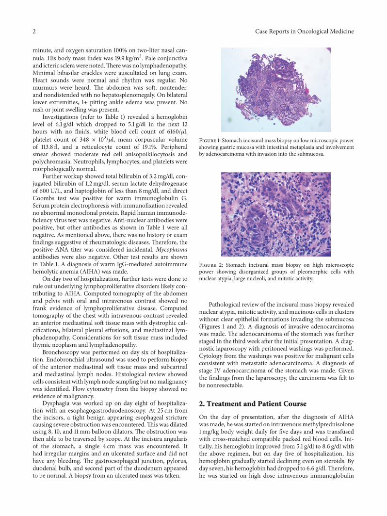
Figure 1: Stomach incisural mass biopsy on lowmicroscopic powershowing gastric mucosa with intestinal metaplasia and involvementby adenocarcinoma with invasion into the submucosa.
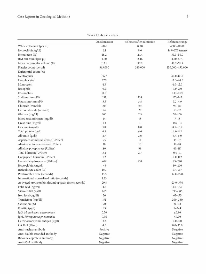
Figure 2: Stomach incisural mass biopsy on high microscopicpower showing disorganized groups of pleomorphic cells withnuclear atypia, large nucleoli, and mitotic activity.
Pathological review of the incisural mass biopsy revealednuclear atypia, mitotic activity, andmucinous cells in clusterswithout clear epithelial formations invading the submucosa(Figures 1 and 2). A diagnosis of invasive adenocarcinomawas made. The adenocarcinoma of the stomach was furtherstaged in the third week after the initial presentation. A diag-nostic laparoscopy with peritoneal washings was performed.Cytology from the washings was positive for malignant cellsconsistent with metastatic adenocarcinoma. A diagnosis ofstage IV adenocarcinoma of the stomach was made. Giventhe findings from the laparoscopy, the carcinoma was felt tobe nonresectable.
2. Treatment and Patient Course
On the day of presentation, after the diagnosis of AIHAwasmade, he was started on intravenousmethylprednisolone1mg/kg body weight daily for five days and was transfusedwith cross-matched compatible packed red blood cells. Ini-tially, his hemoglobin improved from 5.1 g/dl to 8.6 g/dl withthe above regimen, but on day five of hospitalization, hishemoglobin gradually started declining even on steroids. Byday seven, his hemoglobin had dropped to 6.6 g/dl.Therefore,he was started on high dose intravenous immunoglobulin
Case Reports in Oncological Medicine 3
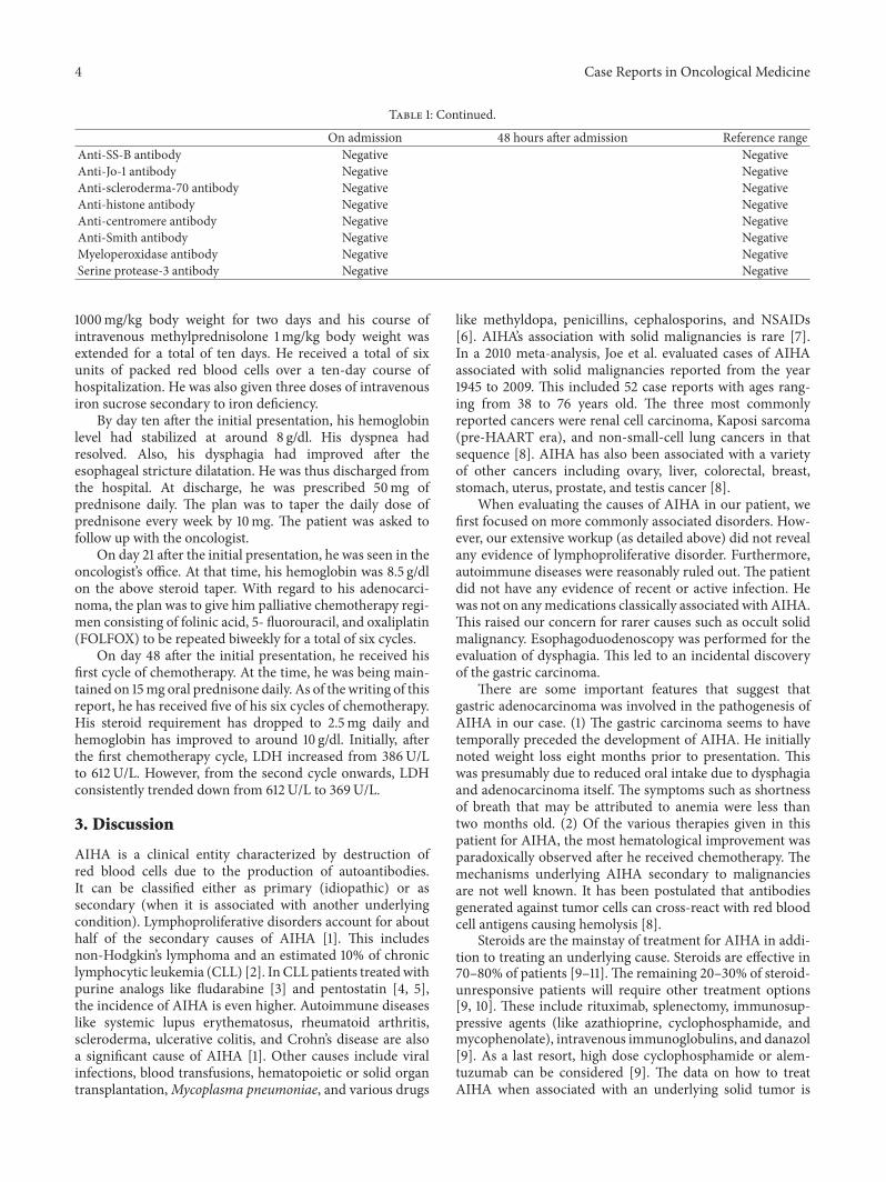
Table 1: Laboratory data.
On admission 48 hours after admission Reference rangeWhite cell count (per 𝜇l) 6160 8810 4500–11000Hemoglobin (g/dl) 6.1 8.6 14.0–17.0 (men)Hematocrit (%) 18.2 24.4 39.0–50.0Red cell count (per pl) 1.60 2.46 4.20–5.70Mean corpuscular volume (fl) 113.8 99.2 80.2–99.4Platelet count (per 𝜇l) 363,000 380,000 150,000–450,000Differential count (%)Neutrophils 66.7 40.0–80.0Lymphocytes 27.9 15.0–40.0Monocytes 4.9 4.0–12.0Basophils 0.2 0.0–2.0Eosinophils 0.0 0.10–0.20Sodium (mmol/l) 137 135 135–145Potassium (mmol/l) 3.5 3.8 3.2–4.9Chloride (mmol/l) 103 99 95–110Carbon dioxide (mmol/l) 24 29 21–32Glucose (mg/dl) 100 113 70–100Blood urea nitrogen (mg/dl) 14 18 7–18Creatinine (mg/dl) 1.3 1.1 0.6–1.3Calcium (mg/dl) 7.8 8.0 8.5–10.2Total protein (g/dl) 6.9 6.6 6.0–8.2Albumin (g/dl) 2.7 2.6 3.4–5.0Aspartate aminotransferase (U/liter) 25 21 15–37Alanine aminotransferase (U/liter) 10 10 12–78Alkaline phosphatase (U/liter) 80 68 45–117Total bilirubin (U/liter) 3.4 2.7 0.0–1.1Conjugated bilirubin (U/liter) 1.2 0.0–0.2Lactate dehydrogenase (U/liter) 614 454 85–240Haptoglobin (mg/dl) <8 30–200Reticulocyte count (%) 19.7 0.4–2.7Prothrombin time (seconds) 15.5 12.0–15.0International normalized ratio (seconds) 1.23Activated prothrombin thromboplastin time (seconds) 29.8 23.0–37.0Folic acid (ng/ml) 4.8 4.0–18.0Vitamin B12 (ng/l) 669 193–986Iron level (𝜇g/dl) 56 65–175Transferrin (mg/dl) 191 200–360Saturation (%) 20 20–44Ferritin (𝜇g/l) 93 5–244IgG,Mycoplasma pneumoniae 0.70 ≤0.90IgM,Mycoplasma pneumoniae 0.36 ≤0.90Carcinoembryonic antigen (𝜇g/l) 3.3 0.0–3.0CA 19-9 (U/ml) 4.6 0.0–35.0Anti-nuclear antibody Positive NegativeAnti-double-stranded antibody Negative NegativeRibonucleoprotein antibody Negative NegativeAnti-SS-A antibody Negative Negative
4 Case Reports in Oncological Medicine
Table 1: Continued.
On admission 48 hours after admission Reference rangeAnti-SS-B antibody Negative NegativeAnti-Jo-1 antibody Negative NegativeAnti-scleroderma-70 antibody Negative NegativeAnti-histone antibody Negative NegativeAnti-centromere antibody Negative NegativeAnti-Smith antibody Negative NegativeMyeloperoxidase antibody Negative NegativeSerine protease-3 antibody Negative Negative
1000mg/kg body weight for two days and his course ofintravenous methylprednisolone 1mg/kg body weight wasextended for a total of ten days. He received a total of sixunits of packed red blood cells over a ten-day course ofhospitalization. He was also given three doses of intravenousiron sucrose secondary to iron deficiency.
By day ten after the initial presentation, his hemoglobinlevel had stabilized at around 8 g/dl. His dyspnea hadresolved. Also, his dysphagia had improved after theesophageal stricture dilatation. He was thus discharged fromthe hospital. At discharge, he was prescribed 50mg ofprednisone daily. The plan was to taper the daily dose ofprednisone every week by 10mg. The patient was asked tofollow up with the oncologist.
On day 21 after the initial presentation, he was seen in theoncologist’s office. At that time, his hemoglobin was 8.5 g/dlon the above steroid taper. With regard to his adenocarci-noma, the plan was to give him palliative chemotherapy regi-men consisting of folinic acid, 5- fluorouracil, and oxaliplatin(FOLFOX) to be repeated biweekly for a total of six cycles.
On day 48 after the initial presentation, he received hisfirst cycle of chemotherapy. At the time, he was being main-tained on 15mg oral prednisone daily. As of the writing of thisreport, he has received five of his six cycles of chemotherapy.His steroid requirement has dropped to 2.5mg daily andhemoglobin has improved to around 10 g/dl. Initially, afterthe first chemotherapy cycle, LDH increased from 386U/Lto 612U/L. However, from the second cycle onwards, LDHconsistently trended down from 612U/L to 369U/L.
3. Discussion
AIHA is a clinical entity characterized by destruction ofred blood cells due to the production of autoantibodies.It can be classified either as primary (idiopathic) or assecondary (when it is associated with another underlyingcondition). Lymphoproliferative disorders account for abouthalf of the secondary causes of AIHA [1]. This includesnon-Hodgkin’s lymphoma and an estimated 10% of chroniclymphocytic leukemia (CLL) [2]. In CLL patients treatedwithpurine analogs like fludarabine [3] and pentostatin [4, 5],the incidence of AIHA is even higher. Autoimmune diseaseslike systemic lupus erythematosus, rheumatoid arthritis,scleroderma, ulcerative colitis, and Crohn’s disease are alsoa significant cause of AIHA [1]. Other causes include viralinfections, blood transfusions, hematopoietic or solid organtransplantation,Mycoplasma pneumoniae, and various drugs
like methyldopa, penicillins, cephalosporins, and NSAIDs[6]. AIHA’s association with solid malignancies is rare [7].In a 2010 meta-analysis, Joe et al. evaluated cases of AIHAassociated with solid malignancies reported from the year1945 to 2009. This included 52 case reports with ages rang-ing from 38 to 76 years old. The three most commonlyreported cancers were renal cell carcinoma, Kaposi sarcoma(pre-HAART era), and non-small-cell lung cancers in thatsequence [8]. AIHA has also been associated with a varietyof other cancers including ovary, liver, colorectal, breast,stomach, uterus, prostate, and testis cancer [8].
When evaluating the causes of AIHA in our patient, wefirst focused on more commonly associated disorders. How-ever, our extensive workup (as detailed above) did not revealany evidence of lymphoproliferative disorder. Furthermore,autoimmune diseases were reasonably ruled out. The patientdid not have any evidence of recent or active infection. Hewas not on anymedications classically associated with AIHA.This raised our concern for rarer causes such as occult solidmalignancy. Esophagoduodenoscopy was performed for theevaluation of dysphagia. This led to an incidental discoveryof the gastric carcinoma.
There are some important features that suggest thatgastric adenocarcinoma was involved in the pathogenesis ofAIHA in our case. (1) The gastric carcinoma seems to havetemporally preceded the development of AIHA. He initiallynoted weight loss eight months prior to presentation. Thiswas presumably due to reduced oral intake due to dysphagiaand adenocarcinoma itself. The symptoms such as shortnessof breath that may be attributed to anemia were less thantwo months old. (2) Of the various therapies given in thispatient for AIHA, the most hematological improvement wasparadoxically observed after he received chemotherapy. Themechanisms underlying AIHA secondary to malignanciesare not well known. It has been postulated that antibodiesgenerated against tumor cells can cross-react with red bloodcell antigens causing hemolysis [8].
Steroids are the mainstay of treatment for AIHA in addi-tion to treating an underlying cause. Steroids are effective in70–80% of patients [9–11]. The remaining 20–30% of steroid-unresponsive patients will require other treatment options[9, 10]. These include rituximab, splenectomy, immunosup-pressive agents (like azathioprine, cyclophosphamide, andmycophenolate), intravenous immunoglobulins, and danazol[9]. As a last resort, high dose cyclophosphamide or alem-tuzumab can be considered [9]. The data on how to treatAIHA when associated with an underlying solid tumor is
Case Reports in Oncological Medicine 5
sparse. In our literature search, we came across four casereports of AIHA secondary to gastric carcinoma [12–15]. Inone case, steroids were intolerable due to underlying diabetesand the patient had refused surgical resection of cancer [12].A rapid decrease in reticulocyte count and LDH was seenafter the initiation of second-line therapy with rituximab[12]. In two other cases, patients were initially treated withsteroids and hematological improvement was observed [13,14]. However, either corticosteroid dependence or relapseof hemolysis was observed with time. Patients underwentsurgical resection of cancer with or without splenectomyand thereafter remained in remission [13, 14]. In the lastcase, hemoglobin improved with steroids and adjunctivetreatment with high dose intravenous immunoglobulins [15].In our patient, initially, a similar observation was noted withsteroids and intravenous immunoglobulins. However, afterfew weeks, hemoglobin started trending down. Hemoglobinimproved and steroids were gradually weaned with the initia-tion of FOLFOX chemotherapy.This could possibly be due toreduction in the cancer antigenic load with the introductionof chemotherapy. AIHA secondary to solid tumors is thoughtto be less responsive to steroids and the treatment of theunderlying condition is very important [7]. Based on thepatient’s response in our case, we believe that FOLFOXchemotherapy can be utilized as a potential treatment optionto control autohemolysis associated with underlying gastriccancer.
4. Conclusion
Our report highlights a case of an 80-year-old male diag-nosedwith IgG-mediated autohemolysis in associationwith ametastatic gastric adenocarcinoma.The association of AIHAwith solid tumors is rare. Also, this case adds to our limitedliterature on the treatment of AIHA secondary to gastriccancer. In our patient, there was a relapse of autohemolysisafter an initial treatment with steroids and immunoglobulins.Hemoglobin was stabilized and steroids were successfullyweaned with the initiation of FOLFOX chemotherapy. Webelieve that FOLFOX can be utilized as an alternativetreatment for steroid-resistant AIHA with underlying gastriccancer.
Conflicts of Interest
The authors declare that there are no conflicts of interestregarding the publication of this paper.
References
[1] C.H. Packman, “The clinical pictures of autoimmune hemolyticanemia,” Transfusion Medicine and Hemotherapy, vol. 42, no. 5,pp. 317–324, 2015.
[2] F. R. Mauro, R. Foa, R. Cerretti et al., “Autoimmune hemolyticanemia in chronic lymphocytic leukemia: clinical, therapeutic,and prognostic features,” Blood, vol. 95, no. 9, pp. 2786–2792,2000.
[3] H. Myint, J. A. Copplestone, J. Orchard et al., “Fludar-abine-related autoimmune haemolytic anaemia in patients with
chronic lymphocytic leukaemia,” British Journal of Haematol-ogy, vol. 91, no. 2, pp. 341–344, 1995.
[4] B. C. Gehrs and R. C. Friedberg, “Autoimmune hemolyticanemia,” American Journal of Hematology, vol. 69, no. 4, pp.258–271, 2002.
[5] Z. M. Sthoeger, D. Sthoeger, M. Shtalrid, E. Sigler, D. Geltner,and A. Berrebi, “Mechanism of autoimmune hemolytic anemiain chronic lymphocytic leukemia,” American Journal of Hema-tology, vol. 43, no. 4, pp. 259–264, 1993.
[6] J. C. Byrd, A. A. Hertler, R. B. Weiss, J. Freiman, S. L. Kweder,and L. F. Diehl, “Fatal recurrence of autoimmune hemolyticanemia following pentostatin therapy in a patient with ahistory of fludarabine-associated hemolytic anemia,” Annals ofOncology, vol. 6, no. 3, pp. 300-301, 1995.
[7] M. A. Spira and E. C. Lynch, “Autoimmune hemolytic anemiaand carcinoma: An unusual association,”The American Journalof Medicine, vol. 67, no. 5, pp. 753–758, 1979.
[8] J. Puthenparambil, K. Lechner, and G. Kornek, “Autoimmunehemolytic anemia as a paraneoplastic phenomenon in solidtumors: A critical analysis of 52 cases reported in the literature,”Wiener Klinische Wochenschrift, vol. 122, no. 7-8, pp. 229–236,2010.
[9] A. Zanella and W. Barcellini, “Treatment of autoimmunehemolytic anemias,” Haematologica, vol. 99, no. 10, pp. 1547–1554, 2014.
[10] K. Lechner and U. Jager, “How I treat autoimmune hemolyticanemias in adults,” Blood, vol. 116, no. 11, pp. 1831–1838, 2010.
[11] F. Yilmaz and F. Vural, “Autoimmune Hemolytic Anemia:Focusing on Therapy According to Classification,” SOJImmunol, vol. 4, no. 1, pp. 1–6, 2017.
[12] H. Hanamoto, K. Sano, R. Fujiwara et al., “Successful treatmentof autoimmune hemolytic anemia concurrent with gastriccancer by rituximab: A case report of Evans syndrome,” InternMed Open J, vol. 1, no. 1, pp. 1–5, 2016.
[13] N. Guillaume, G. Alimardani, D. Chatelain, X. Henry, and J.-F.Claisse, “Disappearance of auto-antibody-induced haemolysisafter resection of a gastric stromal tumor. Case report andreview of the literature,” Revue de Medecine Interne, vol. 24, no.2, pp. 131–135, 2003.
[14] Y. Inoue, K. Kaku, T. Kaneko, andN.Matsumoto, “Autoimmunehemolytic anemia and gastric cancer: case report and review ofthe literature,”Nippon Ketsueki Gakkai Zasshi, vol. 46, no. 4, pp.836–841, 1983.
[15] N. Wakata, T. Kiyozuka, S. Konno et al., “Autoimmune throm-bocytopenic purpura, autoimmune hemolytic anemia and gas-tric cancer appeared in a patient with myasthenia gravis,”Internal Medicine, vol. 45, no. 7, pp. 479–481, 2006.
Submit your manuscripts athttps://www.hindawi.com
Stem CellsInternational
Hindawi Publishing Corporationhttp://www.hindawi.com Volume 2014
Hindawi Publishing Corporationhttp://www.hindawi.com Volume 2014
MEDIATORSINFLAMMATION
of
Hindawi Publishing Corporationhttp://www.hindawi.com Volume 2014
Behavioural Neurology
EndocrinologyInternational Journal of
Hindawi Publishing Corporationhttp://www.hindawi.com Volume 2014
Hindawi Publishing Corporationhttp://www.hindawi.com Volume 2014
Disease Markers
Hindawi Publishing Corporationhttp://www.hindawi.com Volume 2014
BioMed Research International
OncologyJournal of
Hindawi Publishing Corporationhttp://www.hindawi.com Volume 2014
Hindawi Publishing Corporationhttp://www.hindawi.com Volume 2014
Oxidative Medicine and Cellular Longevity
Hindawi Publishing Corporationhttp://www.hindawi.com Volume 2014
PPAR Research
The Scientific World JournalHindawi Publishing Corporation http://www.hindawi.com Volume 2014
Immunology ResearchHindawi Publishing Corporationhttp://www.hindawi.com Volume 2014
Journal of
ObesityJournal of
Hindawi Publishing Corporationhttp://www.hindawi.com Volume 2014
Hindawi Publishing Corporationhttp://www.hindawi.com Volume 2014
Computational and Mathematical Methods in Medicine
OphthalmologyJournal of
Hindawi Publishing Corporationhttp://www.hindawi.com Volume 2014
Diabetes ResearchJournal of
Hindawi Publishing Corporationhttp://www.hindawi.com Volume 2014
Hindawi Publishing Corporationhttp://www.hindawi.com Volume 2014
Research and TreatmentAIDS
Hindawi Publishing Corporationhttp://www.hindawi.com Volume 2014
Gastroenterology Research and Practice
Hindawi Publishing Corporationhttp://www.hindawi.com Volume 2014
Parkinson’s Disease
Evidence-Based Complementary and Alternative Medicine
Volume 2014Hindawi Publishing Corporationhttp://www.hindawi.com