a rare case of upper ureter rupture: ureteral perforation ... · impairment. there are causativ e...
TRANSCRIPT
Korean Journal of UrologyⒸ The Korean Urological Association, 2012 131 Korean J Urol 2012;53:131-133
www.kjurology.orghttp://dx.doi.org/10.4111/kju.2012.53.2.131
Case Report
A Rare Case of Upper Ureter Rupture: Ureteral Perforation Caused by Urinary RetentionSeung-Kwon Choi, Solmin Lee, Sunchan Kim, Tae Gu Kim, Koo Han Yoo, Gyeong Eun Min, Hyung-Lae LeeDepartment of Urology, School of Medicine, Kyung Hee University, Seoul, Korea
Perforation of the ureter is a rare condition that causes a series of problems including retroperitoneal urinoma, urosepsis, abscess formation, infection, and subsequent renal impairment. There are causative factors that induce ureteric rupture, including malig-nancy, urinary calculi, idiopathic retroperitoneal fibrosis, recent iatrogenic manipu-lation, external trauma, degenerative kidney conditions, urography with external com-pression, and spontaneous causes. We report a rare case of ureteric rupture caused by urinary retention. The patient was treated with temporary percutaneous drainage and antibiotics. The present case illustrates that urinary retention can induce not only blad-der rupture, but also ureteric rupture. It is thus of paramount importance to effectively manage patients with voiding problems.
Key Words: Neurogenic bladder, neurogenic; Rupture; Ureter; Urinary retention
This is an Open Access article distributed under the terms of the Creative Commons Attribution Non-Commercial License (http://creativecommons.org/licenses/by-nc/3.0) which permits unrestricted non-commercial use, distribution, and reproduction in any medium, provided the original work is properly cited.
Article History:received 16 June, 2011accepted 26 July, 2011
Corresponding Author:Gyeong Eun Min Department of Urology, Kyung Hee University, Hospital at Gangdong, 892, Dongnam-ro, Gangdong-gu, Seoul 134-727, KoreaTEL: +82-2-440-7735FAX: +82-2-440-7744E-mail: [email protected]
Ureteric rupture is a potentially dangerous event, the diag-nosis of which is often delayed because of its rarity. We re-port a patient with neurogenic bladder who experienced urinary retention and subsequent ureteric rupture.
CASE REPORT
A 75-year-old woman was admitted to the emergency de-partment with acute right abdominal pain of 2-hour duration. Her medical history included a hysterectomy 30 years ago. From several decades ago, she had almost al-ways voided with abdominal strain. Also, she suffered from tenesmus and weak stream. On examination, she was ori-ented and cooperative. Her vital signs were as follows: blood pressure, 150/75 mmHg; pulse, 84 beats per minute and regular; respirations, 20 per minute; and body temper-ature, 36.0°C. Clinical examination revealed diffuse pain in the right abdomen with tenderness. However, costo-vertebral angle (CVA) tenderness was not prominent. Bowel sounds were increased.
Urinalysis showed 2 to 4 red cells and 0 to 1 white cells per field under high-power magnification. Complete blood cell count results were as follows: 8,500/mm3, with 44.5%
neutrophils. Serum chemical analysis showed a urea level of 26 mg/dl, a creatinine level of 1.5 mg/dl, and a C-reactive protein (CRP) level of 0.99 mg/dl. Other values were within the normal limits.
After analgesic injection, her pain subsided. With a pre-sumptive diagnosis of acute gastroenteritis, she was dis-charged with medication.
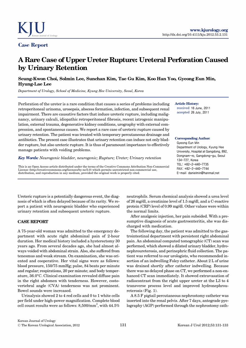
The following day, the patient was admitted to the gas-trointestinal department with persistent right abdominal pain. An abdominal computed tomographic (CT) scan was performed, which showed a dilated urinary bladder, hydro-nephroureterosis, and perinephric fluid collection. The pa-tient was referred to our urologists, who recommended in-sertion of an indwelling Foley catheter. About 2 L of urine was drained shortly after catheter indwelling. Because there was no delayed phase on CT, we performed a non-en-hanced CT scan immediately. It showed extravasation of radiocontrast from the right upper ureter at the L3 to 4 transverse process level and improved hydronephrou-reterosis (Fig. 1).
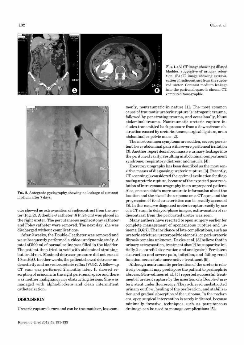
A 8.5 F pigtail percutaneous nephrostomy catheter was inserted into the renal pelvis. After 7 days, antegrade pye-lography (AGP) performed through the nephrostomy cath-
Korean J Urol 2012;53:131-133
132 Choi et al
FIG. 1. (A) CT image showing a dilated bladder, suggestive of urinary reten-tion. (B) CT image showing extrava-sation of radiocontrast from the ruptu-red ureter. Contrast medium leakage into the perirenal space is shown. CT, computed tomographic.
FIG. 2. Antegrade pyelography showing no leakage of contrast medium after 7 days.
eter showed no extravasation of radiocontrast from the ure-ter (Fig. 2). A double-J catheter (6 F, 24 cm) was placed in the right ureter. The percutaneous nephrostomy catheter and Foley catheter were removed. The next day, she was discharged without complications.
After 2 weeks, the Double-J catheter was removed and we subsequently performed a video-urodynamic study. A total of 500 ml of normal saline was filled in the bladder. The patient then tried to void with abdominal straining, but could not. Maximal detrusor pressure did not exceed 10 cmH2O. In other words, the patient showed detrusor un-deractivity and no vesicoureteric reflux (VUR). A follow-up CT scan was performed 2 months later. It showed re-sorption of urinoma in the right peri-renal space and there was neither malignancy nor obstructing lesions. She was managed with alpha-blockers and clean intermittent catheterization.
DISCUSSION
Ureteric rupture is rare and can be traumatic or, less com-
monly, nontraumatic in nature [1]. The most common cause of traumatic ureteric rupture is iatrogenic trauma, followed by penetrating trauma, and occasionally, blunt abdominal trauma. Nontraumatic ureteric rupture in-cludes transmitted back pressure from a downstream ob-struction caused by ureteric stones, surgical ligature, or an abdominal or pelvic mass [2].
The most common symptoms are sudden, severe, persis-tent lower abdominal pain with severe peritoneal irritation [3]. Another report described massive urinary leakage into the peritoneal cavity, resulting in abdominal compartment syndrome, respiratory distress, and anuria [4].
Excretory urography has been described as the most sen-sitive means of diagnosing ureteric rupture [3]. Recently, CT scanning is considered the optimal evaluation for diag-nosing ureteric rupture, because of the expected poor reso-lution of intravenous urography in an unprepared patient. Also, one can obtain more accurate information about the location and the size of the urinoma on a CT scan, and the progression of its characteristics can be readily assessed [5]. In this case, we diagnosed ureteric rupture easily by use of a CT scan. In delayed-phase images, extravasation of ra-diocontrast from the perforated ureter was seen.
Many authors have resorted to open surgery earlier for complete management of spontaneous rupture and ur-inoma [3,6,7]. The incidence of late complications, such as ureteric stricture, ureteropelvic stenosis, or peri-ureteric fibrosis remains unknown. Davies et al. [8] believe that in urinary extravasation, treatment should be supportive ini-tially (i.e., careful observation and analgesics). Persistent obstruction and severe pain, infection, and failing renal function necessitate more active treatment [9].
Although nontraumatic perforation of the ureter is rela-tively benign, it may predispose the patient to perinephric abscess. Stravodimos et al. [5] reported successful treat-ment of ureteric rupture by the insertion of a Double-J ure-teric stent under fluoroscopy. They achieved unobstructed urinary outflow, healing of the perforation, and stabiliza-tion and gradual absorption of the urinoma. In the modern era, open surgical intervention is rarely indicated, because minimally invasive techniques such as percutaneous drainage can be used to manage complications [5].
Korean J Urol 2012;53:131-133
Ureteral Perforation Caused by Urinary Retention 133
In our case, there was no evidence of factors that cause ureteric rupture other than urinary retention. Urine cytol-ogy was negative for malignancy. Video-urodynamic study showed detrusor underactivity without VUR. The fol-low-up CT scan showed neither malignancy nor an ob-structing lesion. Urinary retention may inhibit urinary flow to the bladder and result in high intraureteric pre-ssure. As intraureteric pressure rises, the ureteric wall fails to withstand the pressure and becomes ruptured.
Severe urinary retention may cause ureteric rupture in rare cases. Urologists should be aware of the possibility of ureteric rupture in the differential diagnosis of an acute ab-domen, particularly in patients with urinary retention. Management of these patients should include minimally invasive techniques accompanied by active management of urinary retention.
CONFLICTS OF INTERESTThe authors have nothing to disclose.
REFERENCES
1. Kaplan LM, Farrer JH, Lupu AN. Spontaneous rupture of ureter.
Urology 1987;29:313-6.2. Titton RL, Gervais DA, Hahn PF, Harisinghani MG, Arellano RS,
Mueller PR. Urine leaks and urinomas: diagnosis and imaging- guided intervention. Radiographics 2003;23:1133-47.
3. Diamond DA, Marshall FF. The diagnosis and management of spontaneous rupture of the ureter. J Urol 1982;128:808-10.
4. Katz R, Meretyk S, Gimmon Z. Abdominal compartment syn-drome due to delayed identification of a ureteric perforation fol-lowing abdomino-perineal resection for rectal carcinoma. Int J Urol 1997;4:615-7.
5. Stravodimos K, Adamakis I, Koutalellis G, Koritsiadis G, Grigoriou I, Screpetis K, et al. Spontaneous perforation of the ure-ter: clinical presentation and endourologic management. J Endourol 2008;22:479-84.
6. Chapman JP, Gonzales J, Diokno AC. Significance of urinary ex-travasation during renal colic. Urology 1987;30:541-5.
7. El-Boghdadly SA. Spontaneous rupture of the ureter proximal to ureteric stone. J R Soc Med 1985;78:255-7.
8. Davies P, Bates CP, Price HM. Chronic peripelvic extravasation treated conservatively. Br J Urol 1981;53:412-5.
9. Kettlewell M, Walker M, Dudley N, De Souza B. Spontaneous ex-travasation of urine secondary to ureteric obstruction. Br J Urol 1973;45:8-14.