a rare case report - diagnostic pathology
TRANSCRIPT
Okubo et al. Diagnostic Pathology 2013, 8:82http://www.diagnosticpathology.org/content/8/1/82
CASE REPORT Open Access
Pathophysiological implication of reversed CThalo sign in invasive pulmonary mucormycosis:a rare case reportYoichiro Okubo1, Takao Ishiwatari1, Haruka Izumi2, Fumitomo Sato3, Kyoko Aki1, Daisuke Sasai1, Tsunehiro Ando4,Minoru Shinozaki1, Kazuhiko Natori2, Naobumi Tochigi1, Megumi Wakayama1, Yoshinobu Hata3, Haruo Nakayama5,Tetsuo Nemoto1 and Kazutoshi Shibuya1,6*
Abstract
Background: It has been accepted that reversed halo sign (RHS) appeared on a computed tomography (CT) imagein immunocompromised patients indicates an invasive fungal infection, but its pathophysiology remains obscure asto what this image implies. Therefore, the present report describes detailed radiological and histopathologicalfindings of a case of invasive pulmonary mucormycosis (IPM) presenting RHS with comparison to those from alesion of discrete nodule caused by invasive pulmonary aspergillosis (IPA), and discusses the pathophysiologicalimplications of this characteristic image.
Case presentation: RHS had been clinically noted at the time of recovering of bone marrow function of a 64-year-oldJapanese man who had chemotherapy for his acute lymphoblastic leukemia. Histological examination of the surgicallyremoved lung revealed a lesion of IPM. This was composed of coagulation necrosis of septa at the center of lesionwith preservation of air content which was encompassed outer rim comprising triplet structure; liquefaction,consolidation, and organization from the inner to the outer layer. In addition, Micro-CT examination confirmed reticularstructure and monotonous high density at the central coagulation necrosis preserving air content and surroundingconsolidation, and organization lesion of the IPM lesion.
Conclusion: Our investigations suggest that RHS might be understood as a kind of immune reconstitution syndromeand be the initial and prior status of air crescent sign.
Virtual Slides: The virtual slide(s) for this article can be found here: http://www.diagnosticpathology.diagnomx.eu/vs/3480054198968132
Keywords: Reversed halo sign, Mucormycosis, Aspergillosis, Discrete nodule
BackgroundReversed halo sign (RHS) has been regarded as a sign oncomputed tomography (CT) image characterized bycentral ground-glass opacity (GGO) which is surroundedby a partial or complete rim of consolidation. Althoughthis sign was initially described specifically for cryptogenicorganizing pneumonia [1], it has also been associated withother diseases, including fungal infections [2,3]. However,
* Correspondence: [email protected] of Surgical Pathology, Toho University School of Medicine,6-11-1 Omori-Nishi, Ota-Ku, Tokyo 143-8541, Japan6Department of Dermatology, Peking University First Hospital, Beijing, ChinaFull list of author information is available at the end of the article
© 2013 Okubo et al.; licensee BioMed CentralCommons Attribution License (http://creativecreproduction in any medium, provided the or
only a few studies have conducted a detailed pathophysio-logical investigation of this phenomenon. Therefore,the present report describes detailed radiological andhistopathological findings of a case of invasive pulmonarymucormycosis (IPM) presenting RHS, with comparison toa discrete nodule (DN) [4,5] caused by invasive pulmonaryaspergillosis (IPA), and discusses the pathophysiologicalimplications of this characteristic image.
Case presentationA 64-year-old Japanese man reported dyspnea and generalfatigue two months before admission. He was referredto our hospital due to acute lymphoblastic leukemia,
Ltd. This is an Open Access article distributed under the terms of the Creativeommons.org/licenses/by/2.0), which permits unrestricted use, distribution, andiginal work is properly cited.
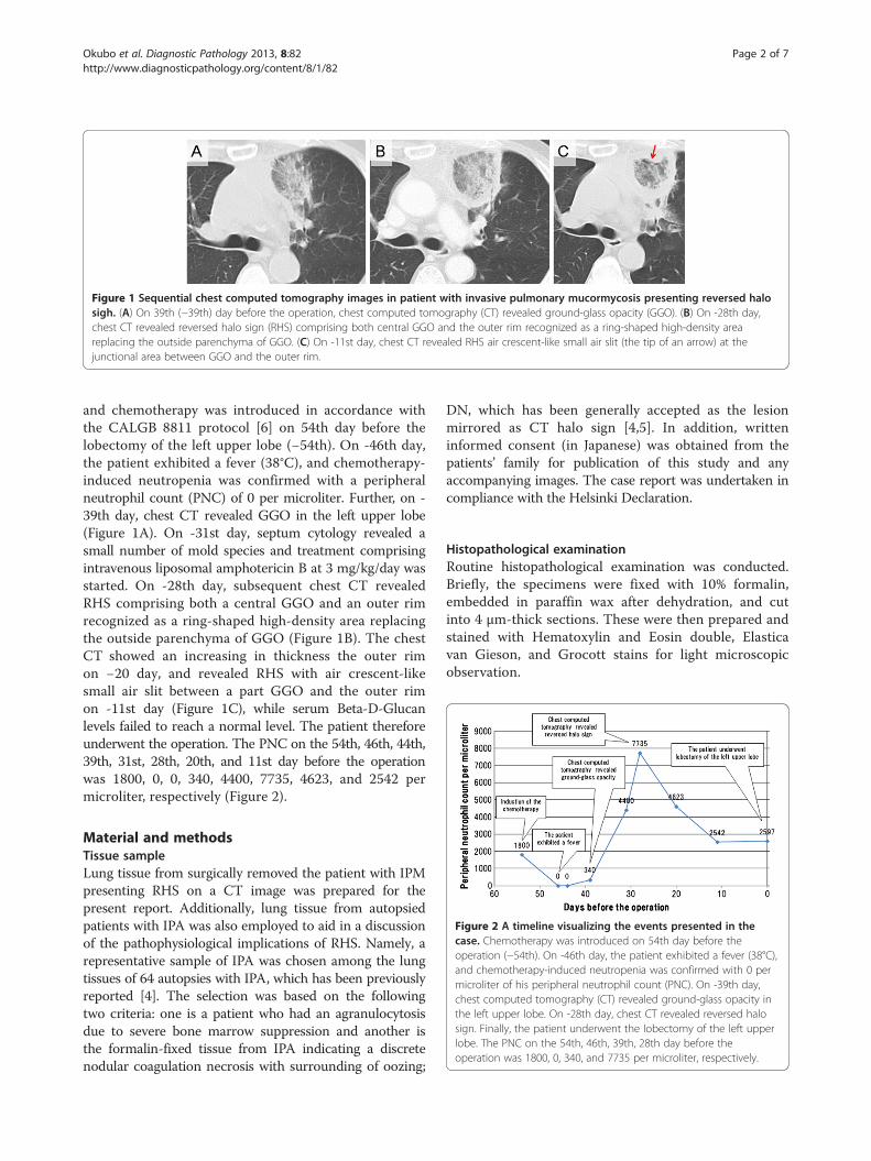
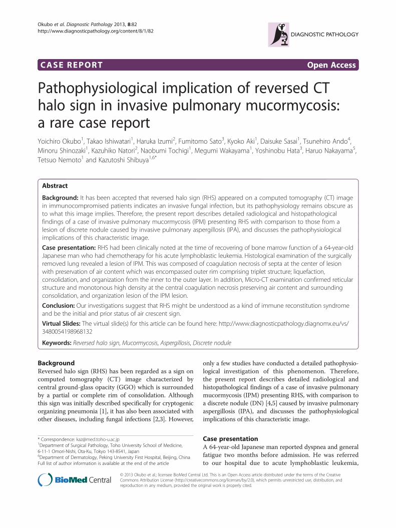
Figure 1 Sequential chest computed tomography images in patient with invasive pulmonary mucormycosis presenting reversed halosigh. (A) On 39th (−39th) day before the operation, chest computed tomography (CT) revealed ground-glass opacity (GGO). (B) On -28th day,chest CT revealed reversed halo sign (RHS) comprising both central GGO and the outer rim recognized as a ring-shaped high-density areareplacing the outside parenchyma of GGO. (C) On -11st day, chest CT revealed RHS air crescent-like small air slit (the tip of an arrow) at thejunctional area between GGO and the outer rim.
Figure 2 A timeline visualizing the events presented in thecase. Chemotherapy was introduced on 54th day before theoperation (−54th). On -46th day, the patient exhibited a fever (38°C),and chemotherapy-induced neutropenia was confirmed with 0 permicroliter of his peripheral neutrophil count (PNC). On -39th day,chest computed tomography (CT) revealed ground-glass opacity inthe left upper lobe. On -28th day, chest CT revealed reversed halosign. Finally, the patient underwent the lobectomy of the left upperlobe. The PNC on the 54th, 46th, 39th, 28th day before theoperation was 1800, 0, 340, and 7735 per microliter, respectively.
Okubo et al. Diagnostic Pathology 2013, 8:82 Page 2 of 7http://www.diagnosticpathology.org/content/8/1/82
and chemotherapy was introduced in accordance withthe CALGB 8811 protocol [6] on 54th day before thelobectomy of the left upper lobe (−54th). On -46th day,the patient exhibited a fever (38°C), and chemotherapy-induced neutropenia was confirmed with a peripheralneutrophil count (PNC) of 0 per microliter. Further, on -39th day, chest CT revealed GGO in the left upper lobe(Figure 1A). On -31st day, septum cytology revealed asmall number of mold species and treatment comprisingintravenous liposomal amphotericin B at 3 mg/kg/day wasstarted. On -28th day, subsequent chest CT revealedRHS comprising both a central GGO and an outer rimrecognized as a ring-shaped high-density area replacingthe outside parenchyma of GGO (Figure 1B). The chestCT showed an increasing in thickness the outer rimon −20 day, and revealed RHS with air crescent-likesmall air slit between a part GGO and the outer rimon -11st day (Figure 1C), while serum Beta-D-Glucanlevels failed to reach a normal level. The patient thereforeunderwent the operation. The PNC on the 54th, 46th, 44th,39th, 31st, 28th, 20th, and 11st day before the operationwas 1800, 0, 0, 340, 4400, 7735, 4623, and 2542 permicroliter, respectively (Figure 2).
Material and methodsTissue sampleLung tissue from surgically removed the patient with IPMpresenting RHS on a CT image was prepared for thepresent report. Additionally, lung tissue from autopsiedpatients with IPA was also employed to aid in a discussionof the pathophysiological implications of RHS. Namely, arepresentative sample of IPA was chosen among the lungtissues of 64 autopsies with IPA, which has been previouslyreported [4]. The selection was based on the followingtwo criteria: one is a patient who had an agranulocytosisdue to severe bone marrow suppression and another isthe formalin-fixed tissue from IPA indicating a discretenodular coagulation necrosis with surrounding of oozing;
DN, which has been generally accepted as the lesionmirrored as CT halo sign [4,5]. In addition, writteninformed consent (in Japanese) was obtained from thepatients’ family for publication of this study and anyaccompanying images. The case report was undertaken incompliance with the Helsinki Declaration.
Histopathological examinationRoutine histopathological examination was conducted.Briefly, the specimens were fixed with 10% formalin,embedded in paraffin wax after dehydration, and cutinto 4 μm-thick sections. These were then prepared andstained with Hematoxylin and Eosin double, Elasticavan Gieson, and Grocott stains for light microscopicobservation.
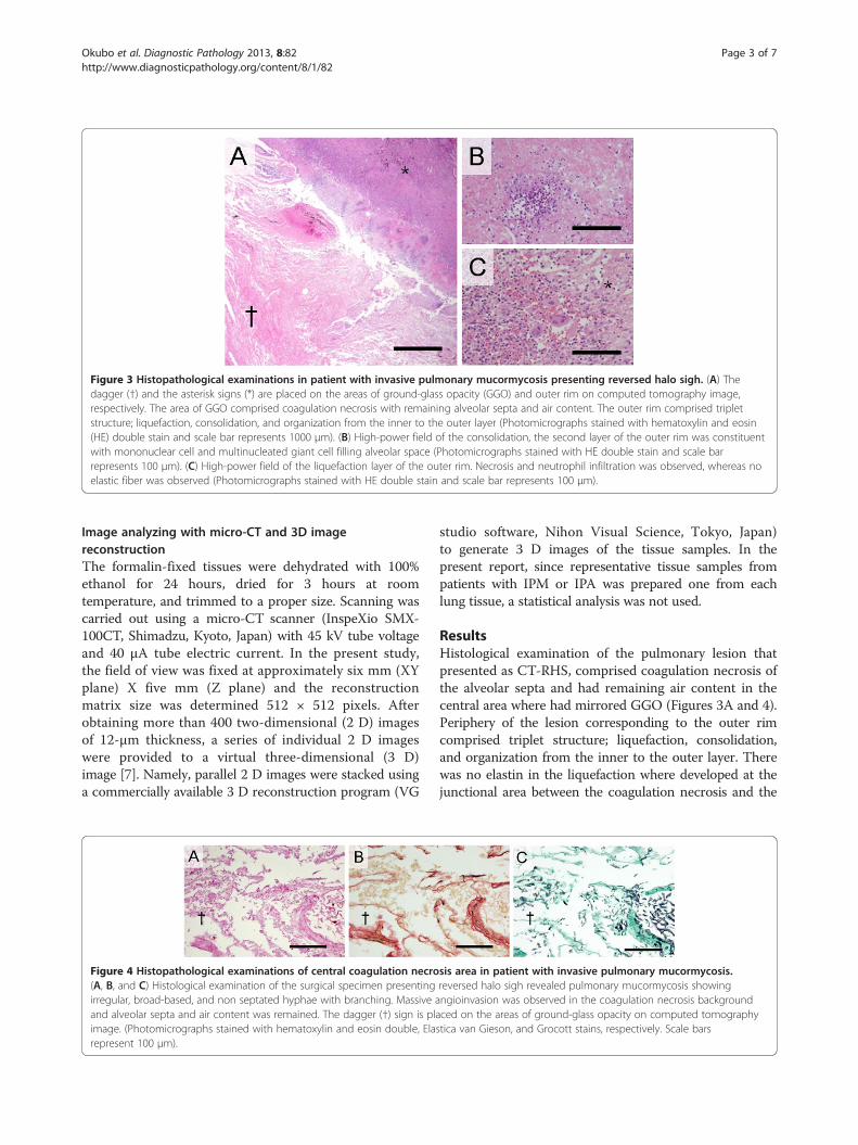
Figure 3 Histopathological examinations in patient with invasive pulmonary mucormycosis presenting reversed halo sigh. (A) Thedagger (†) and the asterisk signs (*) are placed on the areas of ground-glass opacity (GGO) and outer rim on computed tomography image,respectively. The area of GGO comprised coagulation necrosis with remaining alveolar septa and air content. The outer rim comprised tripletstructure; liquefaction, consolidation, and organization from the inner to the outer layer (Photomicrographs stained with hematoxylin and eosin(HE) double stain and scale bar represents 1000 μm). (B) High-power field of the consolidation, the second layer of the outer rim was constituentwith mononuclear cell and multinucleated giant cell filling alveolar space (Photomicrographs stained with HE double stain and scale barrepresents 100 μm). (C) High-power field of the liquefaction layer of the outer rim. Necrosis and neutrophil infiltration was observed, whereas noelastic fiber was observed (Photomicrographs stained with HE double stain and scale bar represents 100 μm).
Okubo et al. Diagnostic Pathology 2013, 8:82 Page 3 of 7http://www.diagnosticpathology.org/content/8/1/82
Image analyzing with micro-CT and 3D imagereconstructionThe formalin-fixed tissues were dehydrated with 100%ethanol for 24 hours, dried for 3 hours at roomtemperature, and trimmed to a proper size. Scanning wascarried out using a micro-CT scanner (InspeXio SMX-100CT, Shimadzu, Kyoto, Japan) with 45 kV tube voltageand 40 μA tube electric current. In the present study,the field of view was fixed at approximately six mm (XYplane) X five mm (Z plane) and the reconstructionmatrix size was determined 512 × 512 pixels. Afterobtaining more than 400 two-dimensional (2 D) imagesof 12-μm thickness, a series of individual 2 D imageswere provided to a virtual three-dimensional (3 D)image [7]. Namely, parallel 2 D images were stacked usinga commercially available 3 D reconstruction program (VG
Figure 4 Histopathological examinations of central coagulation necro(A, B, and C) Histological examination of the surgical specimen presentingirregular, broad-based, and non septated hyphae with branching. Massive aand alveolar septa and air content was remained. The dagger (†) sign is plaimage. (Photomicrographs stained with hematoxylin and eosin double, Elarepresent 100 μm).
studio software, Nihon Visual Science, Tokyo, Japan)to generate 3 D images of the tissue samples. In thepresent report, since representative tissue samples frompatients with IPM or IPA was prepared one from eachlung tissue, a statistical analysis was not used.
ResultsHistological examination of the pulmonary lesion thatpresented as CT-RHS, comprised coagulation necrosis ofthe alveolar septa and had remaining air content in thecentral area where had mirrored GGO (Figures 3A and 4).Periphery of the lesion corresponding to the outer rimcomprised triplet structure; liquefaction, consolidation,and organization from the inner to the outer layer. Therewas no elastin in the liquefaction where developed at thejunctional area between the coagulation necrosis and the
sis area in patient with invasive pulmonary mucormycosis.reversed halo sigh revealed pulmonary mucormycosis showingngioinvasion was observed in the coagulation necrosis backgroundced on the areas of ground-glass opacity on computed tomographystica van Gieson, and Grocott stains, respectively. Scale bars
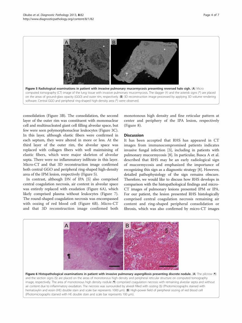
Figure 5 Radiological examinations in patient with invasive pulmonary mucormycosis presenting reversed halo sigh. (A) Microcomputed tomography (CT) image of the lung tissue with invasive pulmonary mucormycosis. The dagger (†) and the asterisk signs (*) are placedon the areas of ground-glass opacity (GGO) and outer rim, respectively. (B) 3D reconstruction image processed by applying 3D volume renderingsoftware. Central GGO and peripheral ring-shaped high-density area (*) were observed.
Okubo et al. Diagnostic Pathology 2013, 8:82 Page 4 of 7http://www.diagnosticpathology.org/content/8/1/82
consolidation (Figure 3B). The consolidation, the secondlayer of the outer rim was constituent with mononuclearcell and multinucleated giant cell filling alveolar space, butfew were seen polymorphonuclear leukocytes (Figure 3C).In this layer, although elastic fibers were confirmed ineach septum, they were altered in more or less. At thethird layer of the outer rim, the alveolar space wasreplaced with collagen fibers with well maintaining ofelastic fibers, which were major skeleton of alveolarsepta. There were no inflammatory infiltrate in this layer.Micro-CT and that 3D reconstruction image confirmedboth central GGO and peripheral ring-shaped high-densityarea of the IPM lesion, respectively (Figure 5).In contrast, although DN of IPA [5] also comprised
central coagulation necrosis, air content in alveolar spacewas entirely replaced with exudation (Figure 6A), whichlikely comprised plasma without leukocytes (Figure 7).The round-shaped coagulation necrosis was encompassedwith oozing of red blood cell (Figure 6B). Micro-CTand that 3D reconstruction image confirmed both
Figure 6 Histopathological examinations in patient with invasive pulmand the section signs (§) are placed on the areas of monotonous high denimage, respectively. The area of monotonous high density nodule (¶) compair content due to inflammatory exudation. The necrosis was surrounded bhematoxylin and eosin (HE) double stain and scale bar represents 1000 μm(Photomicrographs stained with HE double stain and scale bar represents 1
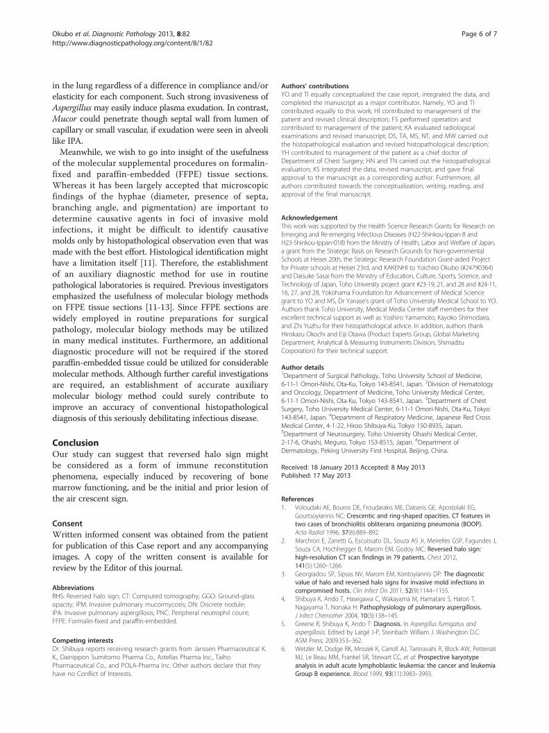
monotonous high density and fine reticular pattern atcenter and periphery of the IPA lesion, respectively(Figure 8).
DiscussionIt has been accepted that RHS has appeared in CTimages from immunocompromised patients indicatesinvasive fungal infection [3], including in patients withpulmonary mucormycosis [8]. In particular, Busca A et al.described that RHS may be an early radiological signof mucormycosis and emphasized the importance ofrecognizing this sign as a diagnostic strategy [8]. However,detailed pathophysiology of the sign remains obscure.Therefore, we would like to discuss how RHS develops incomparison with the histopathological findings and micro-CT images of pulmonary lesions presented IPM or IPA.For our patient, the lesion presented RHS histologicallycomprised central coagulation necrosis remaining aircontent and ring-shaped peripheral consolidation orfibrosis, which was also confirmed by micro-CT images
onary aspergillosis presenting discrete nodule. (A) The pilcrow (¶)sity and peripheral reticular structure on computed tomographyrised coagulation necrosis with remaining alveolar septa and withouty alveoli filled with oozing (§) (Photomicrographs stained with). (B) High-power field of peripheral oozing of red blood cell00 μm).
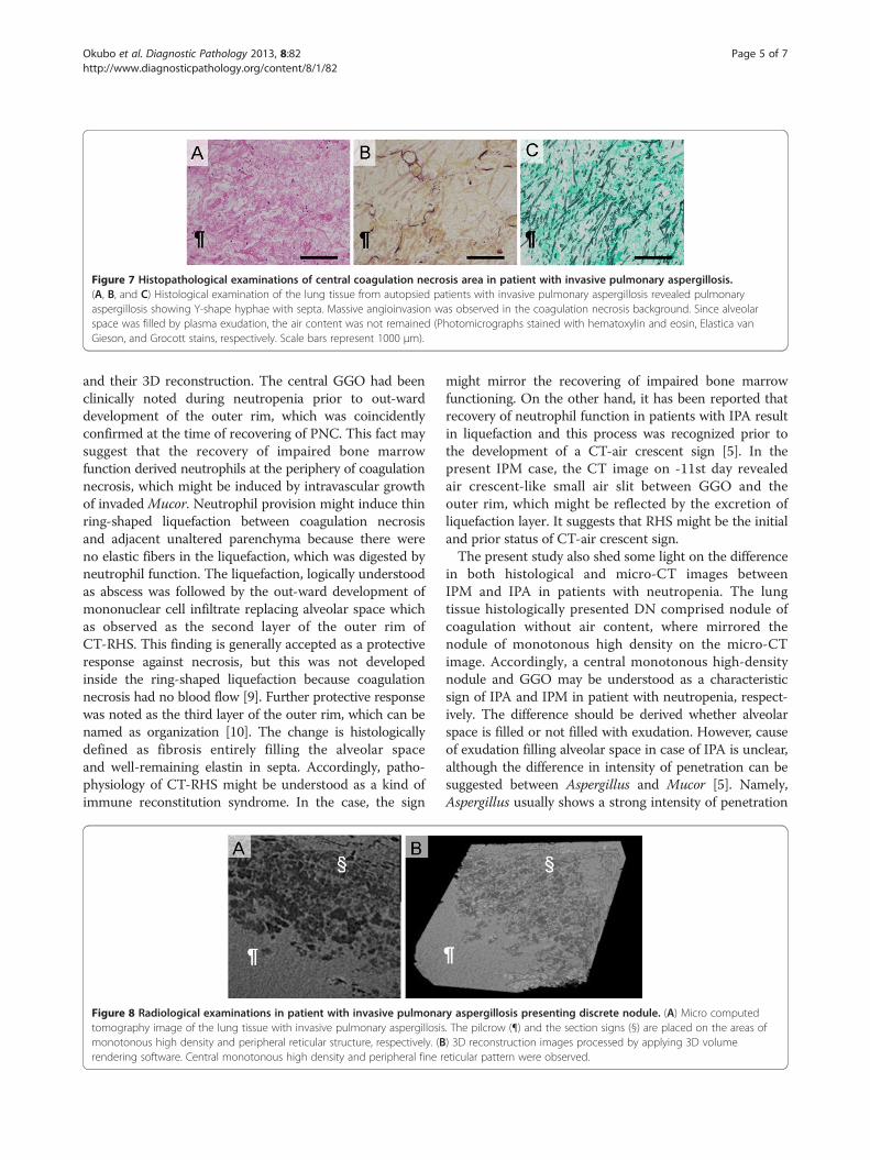
Figure 7 Histopathological examinations of central coagulation necrosis area in patient with invasive pulmonary aspergillosis.(A, B, and C) Histological examination of the lung tissue from autopsied patients with invasive pulmonary aspergillosis revealed pulmonaryaspergillosis showing Y-shape hyphae with septa. Massive angioinvasion was observed in the coagulation necrosis background. Since alveolarspace was filled by plasma exudation, the air content was not remained (Photomicrographs stained with hematoxylin and eosin, Elastica vanGieson, and Grocott stains, respectively. Scale bars represent 1000 μm).
Okubo et al. Diagnostic Pathology 2013, 8:82 Page 5 of 7http://www.diagnosticpathology.org/content/8/1/82
and their 3D reconstruction. The central GGO had beenclinically noted during neutropenia prior to out-warddevelopment of the outer rim, which was coincidentlyconfirmed at the time of recovering of PNC. This fact maysuggest that the recovery of impaired bone marrowfunction derived neutrophils at the periphery of coagulationnecrosis, which might be induced by intravascular growthof invaded Mucor. Neutrophil provision might induce thinring-shaped liquefaction between coagulation necrosisand adjacent unaltered parenchyma because there wereno elastic fibers in the liquefaction, which was digested byneutrophil function. The liquefaction, logically understoodas abscess was followed by the out-ward development ofmononuclear cell infiltrate replacing alveolar space whichas observed as the second layer of the outer rim ofCT-RHS. This finding is generally accepted as a protectiveresponse against necrosis, but this was not developedinside the ring-shaped liquefaction because coagulationnecrosis had no blood flow [9]. Further protective responsewas noted as the third layer of the outer rim, which can benamed as organization [10]. The change is histologicallydefined as fibrosis entirely filling the alveolar spaceand well-remaining elastin in septa. Accordingly, patho-physiology of CT-RHS might be understood as a kind ofimmune reconstitution syndrome. In the case, the sign
Figure 8 Radiological examinations in patient with invasive pulmonatomography image of the lung tissue with invasive pulmonary aspergillosismonotonous high density and peripheral reticular structure, respectively. (Brendering software. Central monotonous high density and peripheral fine r
might mirror the recovering of impaired bone marrowfunctioning. On the other hand, it has been reported thatrecovery of neutrophil function in patients with IPA resultin liquefaction and this process was recognized prior tothe development of a CT-air crescent sign [5]. In thepresent IPM case, the CT image on -11st day revealedair crescent-like small air slit between GGO and theouter rim, which might be reflected by the excretion ofliquefaction layer. It suggests that RHS might be the initialand prior status of CT-air crescent sign.The present study also shed some light on the difference
in both histological and micro-CT images betweenIPM and IPA in patients with neutropenia. The lungtissue histologically presented DN comprised nodule ofcoagulation without air content, where mirrored thenodule of monotonous high density on the micro-CTimage. Accordingly, a central monotonous high-densitynodule and GGO may be understood as a characteristicsign of IPA and IPM in patient with neutropenia, respect-ively. The difference should be derived whether alveolarspace is filled or not filled with exudation. However, causeof exudation filling alveolar space in case of IPA is unclear,although the difference in intensity of penetration can besuggested between Aspergillus and Mucor [5]. Namely,Aspergillus usually shows a strong intensity of penetration
ry aspergillosis presenting discrete nodule. (A) Micro computed. The pilcrow (¶) and the section signs (§) are placed on the areas of) 3D reconstruction images processed by applying 3D volumeeticular pattern were observed.
Okubo et al. Diagnostic Pathology 2013, 8:82 Page 6 of 7http://www.diagnosticpathology.org/content/8/1/82
in the lung regardless of a difference in compliance and/orelasticity for each component. Such strong invasiveness ofAspergillusmay easily induce plasma exudation. In contrast,Mucor could penetrate though septal wall from lumen ofcapillary or small vascular, if exudation were seen in alveolilike IPA.Meanwhile, we wish to go into insight of the usefulness
of the molecular supplemental procedures on formalin-fixed and paraffin-embedded (FFPE) tissue sections.Whereas it has been largely accepted that microscopicfindings of the hyphae (diameter, presence of septa,branching angle, and pigmentation) are important todetermine causative agents in foci of invasive moldinfections, it might be difficult to identify causativemolds only by histopathological observation even that wasmade with the best effort. Histological identification mighthave a limitation itself [11]. Therefore, the establishmentof an auxiliary diagnostic method for use in routinepathological laboratories is required. Previous investigatorsemphasized the usefulness of molecular biology methodson FFPE tissue sections [11-13]. Since FFPE sections arewidely employed in routine preparations for surgicalpathology, molecular biology methods may be utilizedin many medical institutes. Furthermore, an additionaldiagnostic procedure will not be required if the storedparaffin-embedded tissue could be utilized for considerablemolecular methods. Although further careful investigationsare required, an establishment of accurate auxiliarymolecular biology method could surely contribute toimprove an accuracy of conventional histopathologicaldiagnosis of this seriously debilitating infectious disease.
ConclusionOur study can suggest that reversed halo sign mightbe considered as a form of immune reconstitutionphenomena, especially induced by recovering of bonemarrow functioning, and be the initial and prior lesion ofthe air crescent sign.
ConsentWritten informed consent was obtained from the patientfor publication of this Case report and any accompanyingimages. A copy of the written consent is available forreview by the Editor of this journal.
AbbreviationsRHS: Reversed halo sign; CT: Computed tomography; GGO: Ground-glassopacity; IPM: Invasive pulmonary mucormycosis; DN: Discrete nodule;IPA: Invasive pulmonary aspergillosis; PNC: Peripheral neutrophil count;FFPE: Formalin-fixed and paraffin-embedded.
Competing interestsDr. Shibuya reports receiving research grants from Janssen Pharmaceutical K.K., Dainippon Sumitomo Pharma Co., Astellas Pharma Inc., TaihoPharmaceutical Co., and POLA-Pharma Inc. Other authors declare that theyhave no Conflict of Interests.
Authors’ contributionsYO and TI equally conceptualized the case report, integrated the data, andcompleted the manuscript as a major contributor. Namely, YO and TIcontributed equally to this work; HI contributed to management of thepatient and revised clinical description; FS performed operation andcontributed to management of the patient; KA evaluated radiologicalexaminations and revised manuscript; DS, TA, MS, NT, and MW carried outthe histopathological evaluation and revised histopathological description;YH contributed to management of the patient as a chief doctor ofDepartment of Chest Surgery; HN and TN carried out the histopathologicalevaluation; KS integrated the data, revised manuscript, and gave finalapproval to the manuscript as a corresponding author. Furthermore, allauthors contributed towards the conceptualization, writing, reading, andapproval of the final manuscript.
AcknowledgementThis work was supported by the Health Science Research Grants for Research onEmerging and Re-emerging Infectious Diseases (H22-Shinkou-Ippan-8 andH23-Shinkou-Ippan-018) from the Ministry of Health, Labor and Welfare of Japan,a grant from the Strategic Basis on Research Grounds for Non-governmentalSchools at Heisei 20th, the Strategic Research Foundation Grant-aided Projectfor Private schools at Heisei 23rd, and KAKENHI to Yoichiro Okubo (#24790364)and Daisuke Sasai from the Ministry of Education, Culture, Sports, Science, andTechnology of Japan, Toho University project grant #23-19, 21, and 28 and #24-11,16, 27, and 28, Yokohama Foundation for Advancement of Medical Sciencegrant to YO and MS, Dr Yanase’s grant of Toho University Medical School to YO.Authors thank Toho University, Medical Media Center staff members for theirexcellent technical support as well as Yoshiro Yamamoto, Kayoko Shimodaira,and Zhi Yuzhu for their histopathological advice. In addition, authors thankHirokazu Okochi and Eiji Otawa (Product Experts Group, Global MarketingDepartment, Analytical & Measuring Instruments Division, ShimadzuCorporation) for their technical support.
Author details1Department of Surgical Pathology, Toho University School of Medicine,6-11-1 Omori-Nishi, Ota-Ku, Tokyo 143-8541, Japan. 2Division of Hematologyand Oncology, Department of Medicine, Toho University Medical Center,6-11-1 Omori-Nishi, Ota-Ku, Tokyo 143-8541, Japan. 3Department of ChestSurgery, Toho University Medical Center, 6-11-1 Omori-Nishi, Ota-Ku, Tokyo143-8541, Japan. 4Department of Respiratory Medicine, Japanese Red CrossMedical Center, 4-1-22, Hiroo Shibuya-Ku, Tokyo 150-8935, Japan.5Department of Neurosurgery, Toho University Ohashi Medical Center,2-17-6, Ohashi, Meguro, Tokyo 153-8515, Japan. 6Department ofDermatology, Peking University First Hospital, Beijing, China.
Received: 18 January 2013 Accepted: 8 May 2013Published: 17 May 2013
References1. Voloudaki AE, Bouros DE, Froudarakis ME, Datseris GE, Apostolaki EG,
Gourtsoyiannis NC: Crescentic and ring-shaped opacities. CT features intwo cases of bronchiolitis obliterans organizing pneumonia (BOOP).Acta Radiol 1996, 37(6):889–892.
2. Marchiori E, Zanetti G, Escuissato DL, Souza AS Jr, Meirelles GSP, Fagundes J,Souza CA, Hochhegger B, Marom EM, Godoy MC: Reversed halo sign:high-resolution CT scan findings in 79 patients. Chest 2012,141(5):1260–1266.
3. Georgiadou SP, Sipsas NV, Marom EM, Kontoyiannis DP: The diagnosticvalue of halo and reversed halo signs for invasive mold infections incompromised hosts. Clin Infect Dis 2011, 52(9):1144–1155.
4. Shibuya K, Ando T, Hasegawa C, Wakayama M, Hamatani S, Hatori T,Nagayama T, Nonaka H: Pathophysiology of pulmonary aspergillosis.J Infect Chemother 2004, 10(3):138–145.
5. Greene R, Shibuya K, Ando T: Diagnosis. In Aspergillus fumigatus andaspergillosis. Edited by Latgé J-P, Steinbach William J. Washington D.C:ASM Press; 2009:353–362.
6. Wetzler M, Dodge RK, Mrozek K, Carroll AJ, Tantravahi R, Block AW, PettenatiMJ, Le Beau MM, Frankel SR, Stewart CC, et al: Prospective karyotypeanalysis in adult acute lymphoblastic leukemia: the cancer and leukemiaGroup B experience. Blood 1999, 93(11):3983–3993.
Okubo et al. Diagnostic Pathology 2013, 8:82 Page 7 of 7http://www.diagnosticpathology.org/content/8/1/82
7. Kato A, Ohno N: Construction of three-dimensional tooth model bymicro-computed tomography and application for data sharing.Clin Oral Investig 2009, 13(1):43–46.
8. Busca A, Limerutti G, Locatelli F, Barbui A, De Rosa FG, Falda M: Thereversed halo sign as the initial radiographic sign of pulmonaryzygomycosis. Infection 2012, 40(1):77–80.
9. Kumar V, Abbas AK, Aster JC, Robbins SL: Inflammation and repair. InRobbins basic pathology. 9th edition. Edited by Kumar V, Abbas AK, Aster JC,Robbins SL. Philadelphia: Elsevier/Saunders; 2013:29–74.
10. Travis WD, Colby TV, Koss MN, Rosado-de-Christenson ML, Müller NL,Talmadge E, King J: In Atlas of nontumor pathology Non-Neoplastic Disordersof the Lower Respiratory Tract. Edited by West King D. Washington DC:American Registry of Pathology; 2001:592–636.
11. Hofman V, Dhouibi A, Butori C, Padovani B, Gari-Toussaint M, Garcia-Hermoso D, Baumann M, Venissac N, Cathomas G, Hofman P: Usefulnessof molecular biology performed with formaldehyde-fixed paraffinembedded tissue for the diagnosis of combined pulmonary invasivemucormycosis and aspergillosis in an immunocompromised patient.Diagn Pathol 2010, 5:1.
12. Shinozaki M, Okubo Y, Sasai D, Nakayama H, Murayama SY, Ide T, WakayamaM, Ishiwatari T, Tochigi N, Nemoto T, et al: Development of a peptidenucleic acid probe to Trichosporon species and identification oftrichosporonosis by use of in situ hybridization in formalin-fixed andparaffin-embedded (FFPE) sections. J Clin Microbiol 2013, 51(1):295–298.
13. Okubo Y, Shinozaki M, Wakayama M, Nakayama H, Sasai D, Ishiwatari T,Nemoto T, Naobumi T, Shibuya K: Applied gene histopathology:identification of Fusarium species in FFPE tissue sections by in situhybridization. Methods Mol Biol 2013, 968:141–147.
doi:10.1186/1746-1596-8-82Cite this article as: Okubo et al.: Pathophysiological implication of reversedCT halo sign in invasive pulmonary mucormycosis: a rare case report.Diagnostic Pathology 2013 8:82.
Submit your next manuscript to BioMed Centraland take full advantage of:
• Convenient online submission
• Thorough peer review
• No space constraints or color figure charges
• Immediate publication on acceptance
• Inclusion in PubMed, CAS, Scopus and Google Scholar
• Research which is freely available for redistribution
Submit your manuscript at www.biomedcentral.com/submit