a reverse genetics platform that spans the zika virus ... · a reverse genetics platform that spans...
TRANSCRIPT
A Reverse Genetics Platform That Spansthe Zika Virus Family Tree
Douglas G. Widman,a Ellen Young,a Boyd L. Yount,a Kenneth S. Plante,c
Emily N. Gallichotte,b Derek L. Carbaugh,b Kayla M. Peck,d Jessica Plante,a
Jesica Swanstrom,a Mark T. Heise,b,c Helen M. Lazear,b Ralph S. Barica,b
Department of Epidemiology, Gillings School of Global Public Health, University of North Carolina, Chapel Hill,North Carolina, USAa; Department of Microbiology and Immunology, School of Medicine, University of NorthCarolina, Chapel Hill, North Carolina, USAb; Department of Genetics, School of Medicine, University of NorthCarolina, Chapel Hill, North Carolina, USAc; Department of Biology, University of North Carolina, Chapel Hill,North Carolina, USAd
ABSTRACT Zika virus (ZIKV), a mosquito-borne flavivirus discovered in 1947, hasonly recently caused large outbreaks and emerged as a significant human pathogen.In 2015, ZIKV was detected in Brazil, and the resulting epidemic has spread through-out the Western Hemisphere. Severe complications from ZIKV infection include neu-rological disorders such as Guillain-Barré syndrome in adults and a variety of fetalabnormalities, including microcephaly, blindness, placental insufficiency, and fetaldemise. There is an urgent need for tools and reagents to study the pathogenesis ofepidemic ZIKV and for testing vaccines and antivirals. Using a reverse genetics plat-form, we generated six ZIKV infectious clones and derivative viruses representing di-verse temporal and geographic origins. These include three versions of MR766, theprototype 1947 strain (with and without a glycosylation site in the envelope pro-tein), and H/PF/2013, a 2013 human isolate from French Polynesia representative ofthe virus introduced to Brazil. In the course of synthesizing a clone of a circulatingBrazilian strain, phylogenetic studies identified two distinct ZIKV clades in Brazil. Wereconstructed viable clones of strains SPH2015 and BeH819015, representing ances-tral members of each clade. We assessed recombinant virus replication, binding tomonoclonal antibodies, and virulence in mice. This panel of molecular clones and re-combinant virus isolates will enable targeted studies of viral determinants of patho-genesis, adaptation, and evolution, as well as the rational attenuation of contempo-rary outbreak strains to facilitate the design of vaccines and therapeutics.
IMPORTANCE Viral emergence is a poorly understood process as evidenced by thesudden emergence of Zika virus in Latin America and the Caribbean. Malleable re-agents that both predate and span an expanding epidemic are key to understand-ing the virologic determinants that regulate pathogenesis and transmission. We havegenerated representative cDNA molecular clones and recombinant viruses that spanthe known ZIKV family tree, including early Brazilian isolates. Recombinant virusesreplicated efficiently in cell culture and were pathogenic in immunodeficient mice,providing a genetic platform for rational vaccine and therapeutic design.
Zika virus (ZIKV), a member of the Flavivirus genus and closely related to denguevirus (DENV), was first discovered in 1947 in Uganda (1) and until recently had been
responsible for only sporadic human infections in Africa and Asia (2). In the past decade,however, ZIKV has emerged into new areas, including a large outbreak in Micronesia in2007, where it is estimated that 73% of the population was infected within a 4-monthperiod (3), followed by a 2013-2014 outbreak in French Polynesia and subsequentspread throughout Oceania (4). The ZIKV outbreak in the South Pacific is thought to bethe source of virus introduced to Brazil, supported by the close genetic relationship of
Received 1 November 2016 Accepted 14February 2017 Published 7 March 2017
Citation Widman DG, Young E, Yount BL,Plante KS, Gallichotte EN, Carbaugh DL, PeckKM, Plante J, Swanstrom J, Heise MT, LazearHM, Baric RS. 2017. A reverse genetics platformthat spans the Zika virus family tree. mBio 8:e02014-16. https://doi.org/10.1128/mBio.02014-16.
Editor Vincent R. Racaniello, ColumbiaUniversity College of Physicians & Surgeons
Copyright © 2017 Widman et al. This is anopen-access article distributed under the termsof the Creative Commons Attribution 4.0International license.
Address correspondence to Ralph S. Baric,[email protected].
RESEARCH ARTICLE
crossm
March/April 2017 Volume 8 Issue 2 e02014-16 ® mbio.asm.org 1
m
bio.asm.org
on April 9, 2017 - P
ublished by m
bio.asm.org
Dow
nloaded from

South Pacific and epidemic Latin American strains (5). The first ZIKV cases in Brazil werereported in early 2015, with the outbreak initially concentrated in the northeasternregion of the country (6, 7). Although early case reports were consistent with theself-limited febrile illness observed in previous outbreaks, a surge in cases of micro-cephaly was reported in the northeastern region of Brazil in the fall of 2015 (8).Thereafter, a growing body of molecular, immunologic, and epidemiological evidencehas demonstrated a causal role for ZIKV infection in microcephaly as well as a spectrumof other neurodevelopmental defects, now collectively referred to as “congenital Zikasyndrome” (9). To date, it is unknown why transplacental transmission and teratoge-nicity have been observed during the current ZIKV epidemic in Latin America but notin previous outbreaks. Furthermore, this epidemic has revealed a role for sexualtransmission in the spread of ZIKV, a transmission mode not reported for otherflaviviruses (10). While some have speculated that genetic changes in the virus could beresponsible for new pathogenic phenotypes, testing this hypothesis would be aided bya tractable reverse genetics system to generate panels of isogenic mutants of proposedviral determinants of pathogenesis.
Although cDNA-based infectious clones (ICs) have been generated for other flavi-viruses, including West Nile virus, yellow fever virus, DENV, and ZIKV (11–17), flavivirusreverse genetics systems can be more challenging than those for many other viruses,because of sequence instability in bacterial vectors (18). Recent efforts to generate ZIKVrecombinant viruses have resulted in different cloning strategies, all of different ZIKVstrains, that have used DNA plasmids with introns or as multipiece systems designed toovercome these fundamental stability issues (15–17, 19).
Here, we have developed a panel of ZIKV infectious clones, patterned after DENVand coronavirus strategies (20–22). The ZIKV panel includes three allelic variants of ahistorical African strain (MR766) as well as contemporary outbreak strains from FrenchPolynesia (H/PF/2013) and early epidemic Brazilian strains (SPH2015 and BeH819015),enabling experimental testing of viral determinants that distinguish the current ZIKVepidemic from earlier outbreaks. In the process of generating clones of the twoBrazilian strains, we identified sequence abnormalities that impacted virus viability.Phylogenetic analysis of currently available full-length genomes suggests that twoclades of ZIKV are circulating in Brazil (5). We generated molecular clones and recov-ered recombinant viruses representing early isolates from both Brazilian ZIKV clades.
Based on our previous experience with DENV infectious clones (20–22), we wereable to partition toxic regions of the ZIKV genome into stable plasmid subclones.Furthermore, we used nonpalindromic restriction endonuclease sites naturally occur-ring in the ZIKV genome, allowing directional ligation of digested subgenomic frag-ments into full-length cDNAs from which full-length infectious transcripts can besynthesized in vitro. The resulting ZIKV recombinant viruses grow to similar peak titersas their parental isolate viruses and are recognized by cross-reactive DENV monoclonalantibodies (MAbs) but not DENV serotype-specific MAbs. Recombinant viruses werevirulent in Ifnar1�/� � Ifngr1�/� C57BL/6J mice, though slightly attenuated comparedto natural isolate viruses. Interestingly, there was a large difference in murine patho-genesis between the two Brazilian recombinant viruses, driven by one or more of6 amino acid differences that distinguish the two strains. This highly tractable geneticplatform will be useful for evaluating viral determinants of ZIKV pathogenesis and forthe development and testing of interventions to combat ZIKV disease.
RESULTSComplete genomic sequence determination of multiple ZIKV strains. Construct-
ing an infectious clone requires knowledge of the complete viral genome sequence.However, many early ZIKV sequences deposited in GenBank annotated as being a “com-plete genome” were actually incomplete (accession numbers KU729218, KU707826,KF383116, etc.), consisting of a complete open reading frame sequence with incom-plete sequences of the 5= and/or 3= untranslated regions (UTRs). We therefore soughtto determine whether our laboratory stocks of MR766 and H/PF/2013 matched the
Widman et al. ®
March/April 2017 Volume 8 Issue 2 e02014-16 mbio.asm.org 2
m
bio.asm.org
on April 9, 2017 - P
ublished by m
bio.asm.org
Dow
nloaded from
published sequences. MR766 is the prototype ZIKV strain, isolated in Uganda in 1947(1), and has undergone extensive in vivo and in vitro passaging resulting in substrains.Conversely, H/PF/2013 is a human clinical isolate from French Polynesia, 2013, withlimited passage in Vero cells (23). Our laboratory stocks of both of these viruses weregrown in C6/36 cells, and cDNA was prepared from infected cell supernatant. Sangersequencing was performed on PCR products to obtain a consensus sequence of theopen reading frame, and these exactly matched deposited sequences for each virus(MR766, accession no. KU955594, and H/PF/2013, accession no. KJ776791). Of interest,we identified our MR766 strain as being among those substrains that carry a four-codondeletion that ablates a canonical N-linked glycosylation site in the envelope (E) glyco-protein (Fig. 1B), rather than an alternative MR766 substrain (and other ZIKV strains)that encodes a glycosylation site at this position (N154).
We next obtained complete sequences of the 5= and 3= genomic termini for bothstrains. We used a modified 5=-3= rapid amplification of cDNA ends (RACE) methodbecause conventional RACE protocols are dependent upon a 3= poly(A) tail which isnot present on flavivirus genomes (see Fig. S1A in the supplemental material). Thedeposited genomic sequence of MR766 included complete 5= and 3= termini, and oursequences matched these exactly (Fig. S1B and C). As the deposited sequence ofH/PF/2013 consisted of a complete open reading frame with only partial UTR se-quences, our analysis added 60 nucleotides (nt) to the 5= end and 130 nucleotides tothe 3= end to generate a new complete genomic sequence for this virus (Fig. S1B andC). An additional contemporary outbreak strain, PRVABC59, a human clinical isolatefrom Puerto Rico, 2015, had the same 5= and 3= UTR sequences as H/PF/2013 except fora single difference in the 3= UTR at nucleotide 10391 (Fig. S1B and C). Interestingly, thenucleotide change at position 10391 also occurs in both of the early Brazilian ZIKVisolates (SPH2015 and BeH819015) and matches the PRVABC59 sequence as well. Thisrepresents a diversity of only 0.23% between the 3= UTRs of H/PF/2013 and PRVABC59,whereas the H/PF/2013 and MR766 3= UTRs differ by 17 nucleotides (3.90%). Theseresults indicate a high degree of conservation of the 5= and 3= UTR genomic regions,especially in natural clinical isolates that have not undergone extensive in vitro and invivo passages.
Development of a ZIKV reverse genetics system using unidirectional assemblyof a quadripartite genome. We previously developed reverse genetics platforms thatare comprised of multipartite genomes with unidirectional assembly to generate stableinfectious clones for coronaviruses (24, 25) and DENV (20–22). Given the high geneticrelatedness between DENV and ZIKV, we adapted this strategy to design a clone of ourMR766 ZIKV substrain, which lacks E glycosylation due to a 4-amino-acid (aa) deletion(�Gly MR766), using a quadripartite subgenomic system to disrupt toxic genomicregions by partitioning them onto four high-copy-number plasmids for bacterial am-plification (Fig. 1A and B). The partition sites were chosen to mimic those used toconstruct DENV infectious clones by this method (20) and by the location of naturallyoccurring class IIG restriction sites which cleave outside their recognition site, gener-ating nonpalindromic ends that allow for unidirectional ligation of the purified fourgenomic fragments into full-length genomes. A T7 RNA polymerase promoter site wasoriented directly upstream of the first ZIKV nucleotide and used for in vitro transcriptionof full-length ligated genomes, while a hepatitis delta virus ribozyme immediatelyfollowing the last ZIKV nucleotide generates an authentic 3= terminus on the full-lengthinfectious genomic RNA (Fig. 1A). These transcripts were electroporated into C6/36Aedes albopictus cells, which released high-titer infectious virus (approximately 106 to107 focus-forming units [FFU]/ml) in the culture supernatants (Fig. 1D). These werecollected and passaged once or twice on C6/36 cells to generate virus stocks used forexperiments. To verify the presence of molecularly cloned viruses, we sequenced thefull viral genome, confirming the genetic origins of each recombinant virus. Since theMR766 infectious clone included engineered ablation of duplicate restriction sites, wealso digested cDNA from the clone and isolate viruses, confirming the expectedrestriction fragment sizes from two of the clones (Fig. 1E).
Zika Virus Reverse Genetics ®
March/April 2017 Volume 8 Issue 2 e02014-16 mbio.asm.org 3
m
bio.asm.org
on April 9, 2017 - P
ublished by m
bio.asm.org
Dow
nloaded from
ZIKV MR766
C prM E E NS1 NS2A NS2B NS2B NS3 NS4A NS4B NS5 NS55’ UTR 3’ UTR
EcoRV PfIMI SmaIPfIMI BglI BglI BstAPI BstAPI
CCANNNNNTGG2132 GCCNNNNNGC3987 GCANNNNNTGC7324Vector Vector
A B C D+Gly / +del 2156 NT 1844 NT 3368 NT 3593 NT
-Gly 2144 NT
*PflMI *PflMI *SmaI *BstAPI
GCGGCCGGCGATATCTAATACGACTCACTATAGAGTTNotI EcoRV T7 Promoter ZIKV 5’ UTR
GGTTTCT GGCCGGCATGGTCCCAGCCTCCTCGCTGGCGCCGGCTGGGCAACATTCCGAGGGGACCGTCCCCTCGGTAATGGCGAATGGGACCCGGGZIKV 3’ UTR SmaIHDV Ribozyme
B
C
D
100012001517
500
700
MR766Isolate
MR766Clone (+del)
MR766Clone (-gly)
E
MR766isolate
MR766 IC - gly P1
MR766 IC +del P1
MR766 IC +gly P1
A
-gly MR766 IC AMR39835
+gly MR766 IC AMK02027+del MR766 IC BAP47441
75K
50KMR766 IC +gly
MR766 isolate
MR766 IC -gly
MR766 IC +del
F MR766 IsolateSmaI
903 532
MR766 Clones1435 NT
*
0 50 100100
101
102
103
104
105
106
107
108
109
Time post infection (hours)
FFU/
ml
MR766 IsolateMR766 IC Del+ p1MR766 IC Gly- p1MR766 IC Gly+ p1
FIG 1 (A) Schematic diagram of ZIKV MR766 infectious clone. The genome of the virus is divided into 4 fragments using the diagrammedrestriction endonucleases and cloned into high-copy-number vectors. A T7 promoter is placed before the first nucleotide of the ZIKV genome,and a hepatitis delta virus ribozyme is placed after the final genomic nucleotide for RNA stability. Four restriction endonuclease sites shownbeneath the genome (two PflMI, one SmaI, and one BstAPI) were removed using synonymous changes. Sizes of each fragment are shown, andthe larger size is listed along with the location of the N154 allelic mutations (green star). (B) Amino acid sequences of the 3 allelic MR766 variantsin the region of the N154 glycosylation site on E. Numbers (440 and 460) correspond to amino acid positions in the complete ZIKV genome. (C)Virus focus images on Vero-81 cells. (D) Growth curves of MR766 isolate and 3 infectious clones done at 37°C. MR766 �gly IC had significantlyhigher early growth than the other 3 strains by 2-way analysis of variance with Tukey’s test. (E) After reverse transcriptase-PCR, viral ampliconswere subjected to SmaI digestion; only the DNA from the natural isolate was cut. (F) Western blot of E protein from MR766 isolate and 3 infectiousclones performed using the pan-flavivirus MAb 4G2. Size differences of the E protein are shown based on their sequence and glycosylation status.
Widman et al. ®
March/April 2017 Volume 8 Issue 2 e02014-16 mbio.asm.org 4
m
bio.asm.org
on April 9, 2017 - P
ublished by m
bio.asm.org
Dow
nloaded from
Although our substrain of MR766 exactly matched deposited sequences of this virus(accession number KU955594), other deposited MR766 substrain sequences differ andencode an intact N-linked glycosylation site at N154 of E. Since this N154 glycosylationsite is a known determinant of virulence, neuroinvasion, and vector competence forother flaviviruses (26, 27), we constructed an additional MR766 variant with an intactglycosylation site based on this published sequence (accession number KU720415). Athird clone restoring the 4-aa deletions but not encoding the N-linked glycosylation sitewas also generated (accession number LC002520). An amino acid alignment of theregion surrounding the N154 glycosylation site highlights the differences in the cloneviruses (Fig. 1B). Restoration of the 4-aa deletion and functional glycosylation wasconfirmed by Western blot analysis of E protein. As expected, glycosylated E migratesat the highest molecular weight followed by the �Del and �Gly clones and naturalisolate (Fig. 1F). While peak infectious titers of all three clone viruses reached similarlevels as our isolate virus with the 4-aa deletion (Fig. 1D), the viral clone with restoredglycosylation appears to have an early growth advantage and to make larger infectiousfoci than the other allelic mutants (Fig. 1C; Fig. S2).
Utilizing similar strategies, we constructed an infectious clone system for ZIKV strainH/PF/2013, considered to be a near-predecessor of the current outbreak in the Amer-icas. Because of the genetic heterogeneity between MR766 and H/PF/2013, differentrestriction endonuclease sites were used in clone design (Fig. 2A). After electroporationof C6/36 cells, titers reached 106 to 107 FFU/ml, equivalent to that of the natural isolate(Fig. 2B), but the cells demonstrated slightly smaller infectious focus size and morphol-ogy (Fig. 2C; Fig. S2). We performed multistep growth curves on Vero-81 cells at two
A ZIKV H/PF/2013
C prM E E NS1 NS2A NS2B NS2B NS3 NS4A NS4B NS5 NS55’ UTR 3’ UTR
EcoRV Bsu36I SmaIBsu36I BstXI BstXI SfiI SfiI
CCTNAGG2390 CCANNNNNNTGG4124 GGCCNNNNNGGCC8361
GCGGCCGGCGATATCTAATACGACTCACTATAGAGTTNotI EcoRV T7 Promoter ZIKV 5’ UTR
Vector Vector
TGGGTCTGGCCGGCATGGTCCCAGCCTCCTCGCTGGCGCCGGCTGGGCAACATTCCGAGGGGACCGTCCCCTCGGTAATGGCGAATGGGACCCGGGZIKV 3’ UTR SmaIHDV Ribozyme
A B C D2477 NT 1731 NT 4143 NT 2456 NT
H/PF/2013isolate
H/PF/2013IC p0
B DC
0 50 100100
101
102
103
104
105
106
107
growth curve 320C and 37 0C
Time post infection (hours)
FFU/
ml
PF 2013 isolate 320C
PF 2013 IC p1 32 0C
PF 2013 isolate 370C
PF 2013 IC p1 37 0C
H/PF/2013 isolate
H/PF/2013 IC p0
105
106
107
108
Titre FFU/ml
FFU/
ml
FIG 2 (A) Schematic diagram of ZIKV H/PF/2013 infectious clone. The genome of the virus is divided into 4 fragments using diagrammed restrictionendonucleases and cloned into high-copy-number vectors. A T7 promoter and a hepatitis delta virus ribozyme flank the genome. Sizes of each fragment areshown. (B) H/PF/2013 virus isolate was harvested 4 days after infection of C6/36 cells at an MOI of 0.1. Recombinant virus was harvested 4 to 7 days afterelectroporation of RNA into C6/36 cells. Titrations were performed in triplicate. (C) Virus focus images on Vero-81 cells. (D) Growth curves of H/PF/2013 naturalisolate and infectious clone at 32°C and 37°C. At both temperatures, the isolate grew significantly better than the infectious clone by 2-way analysis of variance.
Zika Virus Reverse Genetics ®
March/April 2017 Volume 8 Issue 2 e02014-16 mbio.asm.org 5
m
bio.asm.org
on April 9, 2017 - P
ublished by m
bio.asm.org
Dow
nloaded from
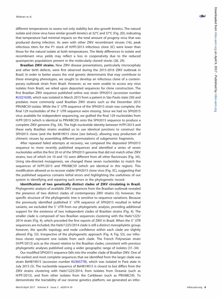
different temperatures to assess not only stability but also growth kinetics. The naturalisolate and clone virus have similar growth kinetics at 32°C and 37°C (Fig. 2D), indicatingthat temperature had minimal impacts on the total amount of progeny virus that wasproduced during infection. As seen with other ZIKV recombinant viruses (16), peakinfectious titers for the P1 stock of H/PF/2013 infectious clone (IC) were lower thanthose for the natural isolate at both temperatures. The likely differences in isolate andrecombinant virus yields may reflect a loss in cooperativity due to the reducedquasispecies populations present in the molecularly cloned stocks (28, 29).
Brazilian ZIKV strains. New ZIKV disease presentations, particularly microcephalyand other birth defects, were first observed during the 2015-2016 ZIKV outbreak inBrazil. In order to better assess the viral genetic determinants that may contribute tothese emerging phenotypes, we sought to develop an infectious clone of a contem-porary outbreak strain from Brazil. However, as we were unable to access any virusisolates from Brazil, we relied upon deposited sequences for clone construction. Thefirst Brazilian ZIKV sequence published online was strain SPH2015 (accession numberKU321639), which was isolated in March 2015 from a patient in São Paulo state (30) andpredates more commonly used Brazilian ZIKV strains such as the December 2015PRVABC59 isolate. While the 5= UTR sequence of the SPH2015 strain was complete, thefinal 129 nucleotides of the 3= UTR sequence were missing. Since we had no SPH2015virus available for independent sequencing, we grafted the final 129 nucleotides fromH/PF/2013 (which is identical to PRVABC59) onto the SPH2015 sequence to produce acomplete ZIKV genome (Fig. 3A). The high nucleotide identity between H/PF/2013 andthese early Brazilian strains enabled us to use identical junctions to construct theSPH2015 clone (and the BeH819015 clone [see below]), allowing easy production ofchimeric viruses by assembling different permutations of subgenomic fragments.
After repeated failed attempts at recovery, we compared the deposited SPH2015sequence to more recently published sequences and identified a series of sevennucleotides within the first 20 nt of the SPH2015 genome that did not match other ZIKVstrains, two of which (nt 10 and 15) were different from all other flaviviruses (Fig. 3A).Using site-directed mutagenesis, we changed these seven nucleotides to match thesequences of H/PF/2013 and PRVABC59 (which are identical in this region). Thismodification allowed us to recover viable SPH2015 clone virus (Fig. 3C), suggesting thatthe published sequence contains lethal errors and highlighting the usefulness of oursystem in identifying and repairing such errors in the phylogenetic record.
Identification of two genetically distinct clades of ZIKV circulating in Brazil.Phylogenetic analysis of available ZIKV sequences from the Brazilian outbreak revealedthe presence of two distinct clades of contemporary ZIKV strains (5); however, thespecific structure of the phylogenetic tree is sensitive to sequence variations. Becausethe previously identified published 5= UTR sequence of SPH2015 resulted in lethalvariants, we excluded the 5= UTR from our phylogenetic analysis, providing additionalsupport for the existence of two independent clades of Brazilian strains (Fig. 4). Thesmaller clade is composed of two Brazilian sequences clustering with the Haiti/1225/2014 strain (Fig. 4), which preceded the first reports of ZIKV in Brazil. When the 5= UTRsequences are included, the Haiti/1225/2014 clade is still a distinct monophyletic group;however, the specific topology and node confidence within each clade are slightlyaltered (Fig. S3). Irrespective of the phylogenetic approach (Fig. 4; Fig. S3), our infec-tious clones represent one isolate from each clade. The French Polynesian strain(H/PF/2013) acts as the closest relative to the Brazilian clades, consistent with previousphylogenetic analyses published using a wider geographic range of isolates (31–34).
Our modified SPH2015 sequence falls into the smaller clade of Brazilian ZIKV. One ofthe earliest and most complete sequences that we identified from the larger clade wasstrain BeH819015 (accession number KU365778), which was isolated in Pará state inJuly 2015 (5). The nucleotide sequence of BeH819015 is closest to but differs from theZIKV strains clustering with Haiti/1225/2014, from isolates from Oceania (such asH/PF/2013), and from other isolates from the Caribbean (such as PRVABC59). Todemonstrate the tractability of our reverse genetics platform, we generated an infec-
Widman et al. ®
March/April 2017 Volume 8 Issue 2 e02014-16 mbio.asm.org 6
m
bio.asm.org
on April 9, 2017 - P
ublished by m
bio.asm.org
Dow
nloaded from
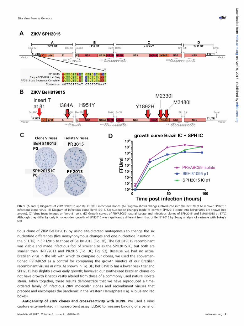
tious clone of ZIKV BeH819015 by using site-directed mutagenesis to change the sixnucleotide differences (five nonsynonymous changes and one nucleotide insertion inthe 5= UTR) in SPH2015 to those of BeH819015 (Fig. 3B). The BeH819015 recombinantwas viable and made infectious foci of similar size as the SPH2015 IC, but both aresmaller than H/PF/2013 and PR2015 (Fig. 3C; Fig. S2). Because we had no actualBrazilian virus in the lab with which to compare our clones, we used the abovemen-tioned PVRABC59 as a control for comparing the growth kinetics of our Brazilianrecombinant viruses in vitro. As shown in Fig. 3D, BeH819015 has a lower peak titer andSPH2015 has slightly slower early growth; however, our synthesized Brazilian clones donot have growth kinetics vastly altered from those of a commonly used natural isolatestrain. Taken together, these results demonstrate that we have reproduced a time-ordered family of infectious ZIKV molecular clones and recombinant viruses thatprecede and encompass the pandemic in the Western Hemisphere (Fig. 4, blue and redboxes).
Antigenicity of ZIKV clones and cross-reactivity with DENV. We used a viruscapture enzyme-linked immunosorbent assay (ELISA) to measure binding of a panel of
B ZIKV BeH819015
C prM E E NS1 NS2A NS2B NS2B NS3 NS4A NS4B NS5 NS55’ UTR 3’ UTR
EcoRV SmaIBsu36I BstXI BstXI SfiI SfiI
CCTNAGG2390 CCANNNNNNTGG4124 GGCCNNNNNGGCC8361Vector Vector
A ZIKV SPH2015
C prM E E NS1 NS2A NS2B NS2B NS3 NS4A NS4B NS5 NS55’ UTR 3’ UTR
EcoRV Bsu36I SmaIBsu36I BstXI BstXI SfiI SfiI
CCTNAGG2390 CCANNNNNNTGG4124 GGCCNNNNNGGCC8361Vector Vector
A B C D2477 NT 1731 NT 4143 NT 2456 NT
I384Ainsert Tat 81 H951Y Y1892H
M2330IM3480I
Isolate VirusesClone VirusesBeH 819015
P0SPH2015 IC
BeH 819015
0 50 100100
101
102
103
104
105
106
107
108
growth curve Brazil IC + SPH IC
Time post infection (hours)
FFU/
ml
PRVABC59 isolateBEH 81095 p1SPH2015 IC p1
DC
Bsu36I
FIG 3 (A and B) Diagrams of ZIKV SPH2015 and BeH819015 infectious clones. (A) Diagram shows changes introduced into the first 20 nt to recover SPH2015infectious clone virus. (B) Diagram of infectious clone BeH819015. Six nucleotide changes made to convert SPH2015 clone into BeH819015 are shown (redarrows). (C) Virus focus images on Vero-81 cells. (D) Growth curves of PRVABC59 natural isolate and infectious clones of SPH2015 and BeH819015 at 37°C.Although they differ by only 6 nucleotides, growth of SPH2015 was significantly different from that of BeH819015 by 2-way analysis of variance with Tukey’stest.
Zika Virus Reverse Genetics ®
March/April 2017 Volume 8 Issue 2 e02014-16 mbio.asm.org 7
m
bio.asm.org
on April 9, 2017 - P
ublished by m
bio.asm.org
Dow
nloaded from
DENV monoclonal antibodies (MAbs) to the panel of isolate and recombinant ZIKVs,compared to the four DENV serotypes. The MAb panel included four DENV serotype-specific neutralizing MAbs (1F4, 2D22, 5J7, and 5H2) (Table S1). As expected, none ofthese MAbs bound to any of the ZIKV strains tested (Fig. 5A). In contrast, MAbs 1C19and 1M7, which bind the highly conserved fusion-loop region of all DENV serotypes(Table S1), bound to all tested ZIKV strains (Fig. 5B). Dissimilarly, MAb 1B22, which bindsto prM on immature DENV virions, bound to all four DENV serotypes but none of theZIKV strains tested (Fig. 5B). To determine whether the lack of 1B22 binding to ZIKV wasdue to the absence of prM present on ZIKV virions (35), or antigenic divergencebetween DENV and ZIKV prM, we infected Vero-81 cells with DENV4 or ZIKV and stainedpermeabilized cells. While anti-fusion loop MAb 4G2 stained all infected cells, anti-prMDENV MAb 2H2 stained only DENV4-infected cells (Fig. S3), likely reflecting antigenicdivergence in prM between DENV and ZIKV. As there is extensive cross-reactivitybetween antiflavivirus immune sera and ZIKV (36, 37), defining cross-reactive andvirus-specific epitopes between ZIKV and DENV is a critical component of developingZIKV diagnostics and understanding the mechanisms of ZIKV pathogenesis.
ZIKV infectious clones are virulent in a mouse model of lethal ZIKV disease.Previous work has demonstrated that wild-type immunocompetent mice are resistantto ZIKV pathogenesis but that mice lacking alpha/beta interferon (IFN-�/�) signaling(e.g., Ifnar1�/�, A129, AG129, Irf3�/� � Irf5�/� � Irf7�/�) succumb to ZIKV infection(38, 39). We infected 6- to 8-week-old Ifnar1�/� � Ifngr1�/� (C57BL/6) mice with 102
to 103 FFU of ZIKV by a subcutaneous route and monitored weight loss and lethalitydaily for 14 days (Fig. 6A to D). Mice were infected with the ZIKV H/PF/2013 isolate (n �
9) or the H/PF/2013 infectious clone (n � 9) or were mock infected with phosphate-buffered saline (PBS) (n � 7). Mice that received H/PF/2013 virus began to lose weightat 5 days postinfection (Fig. 6A), while H/PF/2013 clone-infected mice exhibited moregradual weight loss than those infected with the isolate virus (P � 0.0001). AllZIKV-infected mice succumbed to infection (Fig. 6B), but there was a statistical differ-ence by log rank for recombinant viruses. Additional mice were infected with either of
KU509998 - Hait i/1225/2014KX197192 - ZIKV/H.sapiens/Brazil/PE243/2015KU321639 - ZIKV SPH2015KX520666 - HS-2015-BA-01KU729218 - BeH828305 Brazil 2015KU991811 - Brazil/2016/INMI1KU497555 - Brazil-ZKV2015KU365780 - BeH815744 Brazil 2015KU365777 - BeH818995 Brazil 2015KU365779 - BeH819966 Brazil 2015KU707826 - SSABR1 Brazil 2015KU365778 - BeH819015 Brazil 2015KU926309 - Rio-U1 Brazil 2016KU926310 - Rio-S1 Brazil 2016KU527068 - Natal RGN Brazil 2015KU729217 - BeH823339 Brazil 2015KX280026 - Paraiba 01 Brazil 2015KJ776791 - H/PF/2013 French Polynesia Com pleteKU955593 - H.sapiens-tc/KHM/2010/FSS13025 Cam bodia 2010
99
100100
62
59
97
100
79
60
85
70
SSABR1 Brazil 2015-
Ri U1 B il 2016
ZIKVK /H.sapiens/B-
HS 2015 BA 01
70
FIG 4 Phylogenetic tree of Brazilian ZIKV full-length sequences. ZIKV sequences were acquired fromGenBank and this study (see Materials and Methods), and the 5= UTR was removed from each lineage.Multiple sequence alignment was performed using MAFFT, and the phylogenetic tree was generatedusing maximum likelihood (RAxML software) with 100 bootstrap replicates. Only bootstrap supportvalues of �50 are displayed at each node. GenBank accession number and strain name are indicated oneach branch. Two distinct clades of Brazilian ZIKV sequences are highly supported (green box). SPH2015and BeH819015 are boxed in blue and red, respectively.
Widman et al. ®
March/April 2017 Volume 8 Issue 2 e02014-16 mbio.asm.org 8
m
bio.asm.org
on April 9, 2017 - P
ublished by m
bio.asm.org
Dow
nloaded from

the Brazilian clones (SPH2015 IC or BeH819015 IC) with a higher dose of 103 FFU dueto unpublished data suggesting that these infectious clones are attenuated in com-parison to the H/PF/2013 viruses in mice. SPH2015-infected mice started losing weightby day 10 (Fig. 6C), signs of illness appeared by day 12, and all mice succumbed toillness by day 18 (Fig. 6D). A statistical difference between the infected groups was seenat days 17 and 18 by Bonferroni test (P � 0.0184 and 0.009, respectively). Sixty-sixpercent of BeH819015-infected mice showed no overt disease signs, leading to astatistical difference in lethality when measured by a log-ranked Mantel test betweenstrains (Fig. 6D). In contrast, when we infected 6- to 8-week-old Ifnar1�/� mice with 105
FFU of ZIKV (clones SPH2015, H/PF/2013, and BeH819015), we observed no signs ofillness or weight loss over the course of 25 days (Fig. 6E). The increased morbidity andmortality in Ifnar1�/� � Ifngr1�/� mice compared to Ifnar1�/� mice for all virusestested reveal a specific role for gamma interferon (IFN-�) in controlling ZIKVpathogenesis.
DISCUSSION
The emergence of ZIKV is the latest example of an Old World arbovirus spreadingrapidly upon arrival in the Americas, reminiscent of Chikungunya virus in 2013, WestNile virus in 1999, multiple DENV serotypes in the 1980s, and, originally, the introduc-tion of yellow fever virus in the 17th century. The emergence of ZIKV in the Americas
1F4 2D22 5J7 5H20.0
0.5
1.0
1.5
DENV Type-Specific Antibodies Bi
ndin
g (O
D 40
5)
1C19 1M7 1B22 DT0000.0
0.5
1.0
1.5
Bind
ing
(OD
405)
DENV Cross-Reactive Antibodies
MR766 virusMR766 + del cloneMR766 - gly clone
BeH 81095DENV1DENV2DENV3DENV4
H/PF/2013 virusH/PF/2013 clone
A
B
FIG 5 Antibody binding to DENV and ZIKV clone viruses. (A) The highly DENV serotype-specific MAbs1F4, 2D22, 5J7, and 5H2 clearly bind only the appropriate DENV serotype but not ZIKV. (B) The DENVserotype-cross-reactive MAbs 1C19 and 1M7 bound all DENV and ZIKV strains tested, while 1B22, aprM-specific MAb, bound only the 4 DENV serotypes. DT000 is serum isolated from a traveler with repeatflavivirus vaccinations and infections and is used to control protein loading.
Zika Virus Reverse Genetics ®
March/April 2017 Volume 8 Issue 2 e02014-16 mbio.asm.org 9
m
bio.asm.org
on April 9, 2017 - P
ublished by m
bio.asm.org
Dow
nloaded from
and the concomitant association with congenital infection highlight the importance ofunderstanding the fundamental molecular mechanisms regulating flavivirus transmis-sion, spread, and emergence. Here, we report the generation of a ZIKV reverse geneticssystem, representing strains from both the African and Asian ZIKV lineages. We usedsynthetic approaches to recover two recombinant viruses (SPH2015 and BeH819015)representing early epidemic strains from two contemporary clades of ZIKV that arecirculating in Brazil. This panel of infectious clones also includes three allelic variants ofthe prototype ZIKV strain (MR766). Additionally, we generated a clone of an isolate fromFrench Polynesia (H/PF/2013) that immediately preceded the current epidemic in theAmericas and is likely representative of the ZIKV strain that was introduced to Brazil.
Several groups have reported ZIKV clones using low-copy-number plasmids, plas-mids with introns, or multipiece systems (15–17, 19) to overcome the bacterial toxicityof the plasmids. Shan et al. derived a full-length infectious cDNA clone of a 2010 ZIKVstrain from Cambodia (FSS13025) using a sequentially assembled multipiece system togenerate a single-piece infectious clone (40). FSS13025 falls within the Asian lineage ofZIKV strains but differs by approximately 19 amino acids from Latin American strainssuch as SPH2015 and BeH819015. Other infectious clones of ZIKV strain Paraiba_01/2015 (16) and MR766 (15) were propagated in a bacterial artificial chromosome ascomplete genomes containing engineered introns and launched from DNA, not RNA.Using our system, we generated a clone of the BeH819015 virus by introducing sixmutations to the SPH2015 background, which was straightforward because thechanges were distributed across the subgenomic fragments on four plasmids. Incontrast to other systems (16, 17), all of our subgenomic regions are maintained inhigh-copy-number plasmids. Our quadripartite assembly scheme allows for flexibleconstruction of chimeric viruses, as contemporary strains share sufficient sequencehomology to use identical restriction sites, and so subgenomic fragments from differentstrains can be interchanged. This allows rapid mapping of genetic determinantsbetween two virus strains. An overlapping quadripartite system was recently used to
FIG 6 In vivo pathogenesis studies. ZIKV H/PF/2013 isolate (n � 9), infectious clone virus (n � 9), or PBS (n � 7) was inoculated intoIFNGR-knockout mice by footpad inoculation with 102 FFU (A and B). In parallel, Ifnar1�/� and Ifngr1�/� mice were inoculated by footpad injectionwith 103 FFU of BeH819015 (n � 6) or SPH2015 (n � 6) infectious clone or PBS (n � 3) (C and D) or 105 FFU of recombinant SPH2015 (n � 5),BeH819015 (n � 5), or H/PF/2013 (n � 5) infectious clone (E), respectively. Weight loss and mortality were recorded through 25 days postinfectionor until all animals had succumbed to infection.
Widman et al. ®
March/April 2017 Volume 8 Issue 2 e02014-16 mbio.asm.org 10
m
bio.asm.org
on April 9, 2017 - P
ublished by m
bio.asm.org
Dow
nloaded from
construct a different MR766 strain incorporating green fluorescent protein (GFP) (19);however, the stability of these reporter viruses was unclear given that GFP flavivirusesare generally unstable (41).
Previous studies have analyzed ZIKV sequences from the current epidemic (5, 34, 42,43), and two clades can be seen in some analyses; however, these reports have notdiscussed the significance of the two distinct clades of Brazilian ZIKV isolates, as wasalso found in our analysis (5, 34, 42, 43). The presence of two distinct clades couldindicate independent introductions of ZIKV into Brazil, an earlier introduction thanpreviously appreciated, novel evolutionary patterns, or errors in the published se-quences. Additional factors to consider include geographic location, tissue source,and/or disease presentation of the isolate viruses, although no relationship betweenthese factors and the clade separation was evident in the sequences considered in ouranalysis. Better resolution of the tree topology and potential characteristics definingeach clade will be achieved as more isolates are sequenced and incorporated intophylogenetic analyses. We generated infectious clones of two Brazilian ZIKV strainsrepresenting each of the two identified clades, which will facilitate further analysis ofthe properties that distinguish these two groups of epidemic ZIKV strains.
The ZIKV virus clones from both Brazilian clades (SPH2015 and BeH819015) areantigenically similar to the Asian strain (H/PF/2013) and the African strain (MR766),consistent with the idea that ZIKV strains circulate as a single serotype. DENV serotype-specific antibodies do not bind any of the ZIKV isolates or clones, confirming the uniqueantigenic areas of E protein between DENV and ZIKV. Conversely, all ZIKV strains arerecognized by cross-reactive DENV fusion-loop antibodies, affirming the homology ofZIKV and DENV in this region. In contrast to the substantial antigenic cross-reactivitybetween ZIKV and DENV E protein, a MAb that recognizes prM of all four DENVserotypes did not bind to cells infected with any ZIKV strains, suggesting that prM is notantigenically conserved. Currently, available ZIKV diagnostics are confounded by cross-reactive binding to DENV in standard ELISAs (36, 37), so identifying antigenicallydistinct epitopes is important for developing diagnostics that can identify ZIKV infec-tion in DENV-seropositive individuals.
In Ifnar1�/� � Ifngr1�/� mice, the H/PF/2013 isolate was significantly more virulentthan recombinant H/PF/2013, likely reflecting the reduced quasispecies complexity ofP0 recombinant virus stocks (16), a known virulence determinant (28, 29). Although theBeH819015 and SPH2015 recombinant viruses differ by only six nucleotide changesthroughout their genomes, SPH2015 infection resulted in 100% mortality and earliermorbidity than the less pathogenic BeH819015 strain. Importantly, all three recombi-nant viruses were attenuated in Ifngr1�/� mice, demonstrating the critical importanceof IFN-� responses in controlling lethal ZIKV infections, as also reported by other groups(38). It is interesting that the northern Brazil BeH819015 isolate was more stronglyassociated with severe congenital disease than SPH2015 (5) but more attenuated in themouse model. Additional studies will be needed to identify the key residues respon-sible for reduced BeH819015 pathogenesis in vivo.
After ZIKV infection, an unexpected development is the severe disease presenta-tions, most significantly congenital infection and birth defects, associated with thecurrent ZIKV outbreak in Latin America and the Caribbean (44) and replicated in mousemodels of human disease (45, 46). An association between ZIKV infection and Guillain-Barré syndrome was first noted during the 2013-2014 outbreak in French Polynesia (47),and retrospective analyses of this outbreak identified previously unappreciated asso-ciations with fetal neurodevelopmental abnormalities (48). More recently, ZIKV-associated Guillain-Barré syndrome has been reported in the Americas as well (49, 50).It is unclear why severe manifestations of ZIKV infection have become evident only inthe most recent outbreaks, and possible explanations include host genetic background,host immune status, and the large size and intense surveillance of the current epidemic.However, it is plausible that viral genetic changes could result in new disease pheno-types. Previous analyses have speculated that this may be the case, based on sequencecomparisons of historic and contemporary ZIKV strains (42, 43). However, testing this
Zika Virus Reverse Genetics ®
March/April 2017 Volume 8 Issue 2 e02014-16 mbio.asm.org 11
m
bio.asm.org
on April 9, 2017 - P
ublished by m
bio.asm.org
Dow
nloaded from
hypothesis requires the ability to generate isogenic mutants to evaluate viral determi-nants of pathogenesis. Our panel of ZIKV infectious clones representing historical andcontemporary virus strains, as well as the relative ease of generating allelic variants andchimeric viruses using the quadripartite system, provides an essential toolkit fordetermining the mechanisms of ZIKV pathogenesis and furthering the development ofnew vaccines and antivirals.
MATERIALS AND METHODSCells and viruses. The MR766 strain of ZIKV was obtained from the World Reference Center for
Emerging Viruses and Arboviruses (R. Tesh, University of Texas Medical Branch). ZIKV strains H/PF/2013and PRVABC59 were provided by the U.S. Centers for Disease Control and Prevention. Virus stocks wereprepared in C6/36 Aedes albopictus cells (ATCC catalog no. CRL-1660) or, where indicated, Vero-81Cercopithecus aethiops cells (ATCC catalog no. CCL-81) and titrated in Vero-81 cells (51). C6/36 cells weregrown at 32°C with 5% CO2 in Eagle’s minimum essential medium (MEM) supplemented with 5%heat-inactivated (HI) fetal bovine serum, nonessential amino acids, and antibiotics/antimycotics. Vero-81cells were grown at 37°C with 5% CO2 in Dulbecco’s modified Eagle’s medium (DMEM)–F-12 50/50medium (Gibco) supplemented with 5% HI fetal bovine serum and antibiotics/antimycotics (52). Forgrowth curve analyses, cells were infected at a multiplicity of infection (MOI) of 0.1 in triplicate, andsupernatants were collected at 4, 24, 48, 72, and 96 h postinfection and frozen at �80°C until titrated onVero-81 cells. Viral foci were detected at 44 to 48 h after infection, following fixation/permeabilizationwith 4% paraformaldehyde-0.01% saponin using primary MAb E60 (38) or 4G2 (51) and secondaryhorseradish peroxidase (HRP)-conjugated goat anti-mouse IgG (Sigma), followed by TrueBlue substrate(KPL). Number and size of foci were analyzed with a CTL Immunospot instrument.
Viral genome sequencing and modified 5=-3= RACE. Viral RNA was isolated using a QIAamp viralRNA minikit (Qiagen), and cDNA libraries were prepared using Superscript III (Invitrogen). Sangersequencing was performed on PCR templates generated using Phusion High-Fidelity DNA polymerase(New England BioLabs) and analyzed using Sequencher (Gene Codes) and Lasergene (DNAStar) software.
The 5= and 3= UTR sequences were determined as previously described (53). Briefly, viral RNA wasisolated and treated with calf intestinal phosphatase (Ambion) for 1 h at 37°C to remove terminalphosphates of uncapped RNAs. Following a phenol-chloroform extraction and isopropanol precipitation,the RNA was treated with tobacco acid pyrophosphatase (Ambion) for 1 h at 37°C. After anotherphenol-chloroform extraction and isopropanol precipitation, the RNA was incubated with T4 RNA ligaseI (Ambion) for 1 h at 37°C and then overnight at 4°C, to ligate the 5= and 3= ends. cDNA (Superscript III;Invitrogen) was made and used to generate an amplicon containing both the 5= and 3= UTR sequences,which was Sanger sequenced.
ZIKV infectious clone design. We designed a quadripartite unidirectional molecular clone strategysimilar to that previously described for DENV and emerging coronaviruses (20–22, 24, 25). First, weidentified naturally occurring class IIG nonpalindromic restriction endonuclease sites within the six ZIKVfull-length genomes. For the clone of strain MR766, synonymous nucleotide changes were introduced atpositions 895, 901, 2983, 7834, 7837, 8467, and 8473 to eliminate four internal restriction enzyme sites(two PflMI sites, an SmaI site, and a BstAPI site, respectively), simultaneously leaving marker mutationsfor identifying recombinant viruses. A T7 promoter sequence was added to the immediate 5= end of thegenome, and a hepatitis delta virus ribozyme was added directly after the last nucleotide of each ZIKVgenome to generate an authentic 3= end. The four subgenomic fragments were synthesized into thepUC57 vector (BioBasic) and amplified in Escherichia coli strain MC1061. The resulting purified plasmidswere digested, ligated, in vitro transcribed, and electroporated into C6/36 cells as previously described(20–22). Supernatants from electroporated C6/36 cells were harvested after 6 to 7 days and passagedonce on C6/36 cells to generate virus stocks. Clone MR766 �Del and MR766 �Gly viruses weregenerated by site-directed mutagenesis of the MR766 �Gly clone. Except for the junction sites (Bsu36I,BstXI, and SfiI), we used an identical strategy as for the H/PF/2013 clone and for SPH2015. StrainBeH819015 was generated by site-directed mutagenesis of the SPH2015 clone in the six positions wherethe two strains differed (5= UTR, T81ins; E, I384V; NS1, H951Y; NS3, Y1892H; NS4B, M2330I; and NS4B,M2480I). We confirmed the sequence of all recombinant viruses.
Phylogenetic analysis. Full-length sequences (�10,000 nt) were obtained from GenBank and fromsequencing of laboratory clones/isolates. Sequences were either manually edited to remove the 5= UTR,left as downloaded, or manually modified to provide the viable 5= UTR sequence variants for Haiti/1225/2014 and SPH2015. Multiple sequence alignments were performed using MAFFT (54). The best substi-tution model for each alignment was evaluated using jModelTest (55) and identified to be a generaltime-reversible (GTR) model with an estimated proportion of invariable sites and estimated gammashape parameter (GTR � I � G). Maximum likelihood phylogenetic trees were generated using RAxML(56) using 100 bootstrap replicates. Trees were visualized using EvolView (57).
Virus capture ELISA. Virus particles were captured using mouse anti-DENV MAbs 4G2 and 2H2 incarbonate buffer. MAbs (see Table S1 in the supplemental material) were diluted to a concentration of20 ng/�l and added to captured virus for 1 h at 37°C. After incubation with alkaline phosphatase-conjugated secondary antibodies (Sigma), p-nitrophenylphosphate substrate (Sigma) was added andabsorbance at 405 nm was measured (Bio-Rad). Background signal (optical density at 405 nm [OD405]with no primary antibody) was subtracted from each virus sample, and absorbance was normalized tobinding of an antiflavivirus human polyclonal serum sample.
Widman et al. ®
March/April 2017 Volume 8 Issue 2 e02014-16 mbio.asm.org 12
m
bio.asm.org
on April 9, 2017 - P
ublished by m
bio.asm.org
Dow
nloaded from
Animal studies. Animal husbandry and experiments were performed under the approval of theUniversity of North Carolina at Chapel Hill Institutional Animal Care and Use Committee. Six- to8-week-old male and female Ifnar1�/� � Ifngr1�/� mice on a C57BL/6 background were infectedsubcutaneously via a footpad injection with 102 FFU of ZIKV H/PF/2013 isolate, infectious clone virus, orPBS. In a separate study, mice were infected with 103 FFU of BeH819015 or SPH2015 infectious clones orPBS. Mice were monitored daily for signs of morbidity or mortality and twice daily after losing 20% oftheir starting weight. Animals that exhibited dual hind limb paralysis or loss of 30% of their startingweight or that became moribund were humanely euthanized. Experiments were terminated 5 daysfollowing the last signs of illness resolving. Six- to 8-week-old male and female Ifnar1�/� mice (C57BL/6)were infected with 105 FFU of ZIKV clones of SPH2015, BeH819015, or H/PF/2013. Mice were monitoredfor 25 days with no adverse events noted.
SUPPLEMENTAL MATERIALSupplemental material for this article may be found at https://doi.org/10.1128/
mBio.02014-16.FIG S1, TIF file, 0.7 MB.FIG S2, EPS file, 0.9 MB.FIG S3, TIF file, 1.8 MB.FIG S4, EPS file, 2.7 MB.TABLE S1, DOCX file, 0.01 MB.
ACKNOWLEDGMENTSWe thank Aravinda de Silva (UNC) for providing 1C19, 1M7, and 1B22 monoclonal
antibodies.This work was funded by NIH grants AI 107810 (R.S.B.), AI 109761 (R.S.B.), R01 AI
107731 (de Silva), and U19 AI 100625 (R.S.B./M.T.H.), as well as startup funds from theLineberger Comprehensive Cancer Center (D.L.C. and H.M.L.). K.S.P. was funded by AI100625 and T32 AI 007151-36A1.
REFERENCES1. Dick GW, Kitchen SF, Haddow AJ. 1952. Zika virus. I. Isolations and
serological specificity. Trans R Soc Trop Med Hyg 46:509 –520. https://doi.org/10.1016/0035-9203(52)90042-4.
2. Macnamara FN. 1954. Zika virus: a report on three cases of humaninfection during an epidemic of jaundice in Nigeria. Trans R Soc TropMed Hyg 48:139 –145. https://doi.org/10.1016/0035-9203(54)90006-1.
3. Duffy MR, Chen TH, Hancock WT, Powers AM, Kool JL, Lanciotti RS,Pretrick M, Marfel M, Holzbauer S, Dubray C, Guillaumot L, Griggs A, BelM, Lambert AJ, Laven J, Kosoy O, Panella A, Biggerstaff BJ, Fischer M,Hayes EB. 2009. Zika virus outbreak on Yap Island, Federated States ofMicronesia. N Engl J Med 360:2536 –2543. https://doi.org/10.1056/NEJMoa0805715.
4. Tognarelli J, Ulloa S, Villagra E, Lagos J, Aguayo C, Fasce R, Parra B, MoraJ, Becerra N, Lagos N, Vera L, Olivares B, Vilches M, Fernández J. 2016. Areport on the outbreak of Zika virus on Easter Island, South Pacific, 2014.Arch Virol 161:665– 668. https://doi.org/10.1007/s00705-015-2695-5.
5. Faria NR, Azevedo RDS, Kraemer MU, Souza R, Cunha MS, Hill SC, ThezeJ, Bonsall MB, Bowden TA, Rissanen I, Rocco IM, Nogueira JS, Maeda AY,Vasami FG, Macedo FL, Suzuki A, Rodrigues SG, Cruz AC, Nunes BT,Medeiros DB, Rodrigues DS, Nunes Queiroz AL, da Silva EV, HenriquesDF, Travassos da Rosa ES, de Oliveira CS, Martins LC, Vasconcelos HB,Casseb LM, Simith DDB, Messina JP, Abade L, Lourenco J, Alcantara LCJ,de Lima MM, Giovanetti M, Hay SI, de Oliveira RS, Lemos PDS, de OliveiraLF, de Lima CP, da Silva SP, de Vasconcelos JM, Franco L, Cardoso JF,Vianez-Junior JL, Mir D, Bello G, Delatorre E, Khan K, Creatore M, CoelhoGE, de Oliveira WK, Tesh R, Pybus OG, Nunes MR, Vasconcelos PF. 2016.Zika virus in the Americas: early epidemiological and genetic findings.Science 352:345–349. https://doi.org/10.1126/science.aaf5036.
6. Campos GS, Bandeira AC, Sardi SI. 2015. Zika virus outbreak, Bahia, Brazil.Emerg Infect Dis 21:1885–1886. https://doi.org/10.3201/eid2110.150847.
7. Musso D. 2015. Zika virus transmission from French Polynesia to Brazil.Emerg Infect Dis 21:1887. https://doi.org/10.3201/eid2110.151125.
8. Oliveira Melo AS, Malinger G, Ximenes R, Szejnfeld PO, Alves Sampaio S,Bispo de Filippis AM. 2016. Zika virus intrauterine infection causes fetalbrain abnormality and microcephaly: tip of the iceberg? UltrasoundObstet Gynecol 47:6 –7. https://doi.org/10.1002/uog.15831.
9. Rasmussen SA, Jamieson DJ, Honein MA, Petersen LR. 2016. Zika virus
and birth defects—reviewing the evidence for causality. N Engl J Med374:1981–1987. https://doi.org/10.1056/NEJMsr1604338.
10. D’Ortenzio E, Matheron S, Yazdanpanah Y, de Lamballerie X, Hubert B,Piorkowski G, Maquart M, Descamps D, Damond F, Leparc-Goffart I.2016. Evidence of sexual transmission of Zika virus. N Engl J Med374:2195–2198. https://doi.org/10.1056/NEJMc1604449.
11. Butrapet S, Huang CY, Pierro DJ, Bhamarapravati N, Gubler DJ, KinneyRM. 2000. Attenuation markers of a candidate dengue type 2 vaccinevirus, strain 16681 (PDK-53), are defined by mutations in the 5= noncod-ing region and nonstructural proteins 1 and 3. J Virol 74:3011–3019.https://doi.org/10.1128/JVI.74.7.3011-3019.2000.
12. Khromykh AA, Westaway EG. 1994. Completion of Kunjin virus RNAsequence and recovery of an infectious RNA transcribed from stablycloned full-length cDNA. J Virol 68:4580 – 4588.
13. Rice CM, Grakoui A, Galler R, Chambers TJ. 1989. Transcription of infec-tious yellow fever RNA from full-length cDNA templates produced by invitro ligation. New Biol 1:285–296.
14. Weger-Lucarelli J, Duggal NK, Bullard-Feibelman K, Veselinovic M, RomoH, Nguyen C, Rückert C, Brault AC, Bowen RA, Stenglein M, Geiss BJ, EbelGD. 2017. Development and characterization of recombinant virus gen-erated from a New World Zika virus infectious clone. J Virol 91:e01765-16. https://doi.org/10.1128/JVI.01765-16.
15. Schwarz MC, Sourisseau M, Espino MM, Gray ES, Chambers MT, TortorellaD, Evans MJ. 2016. Rescue of the 1947 Zika virus prototype strain with acytomegalovirus promoter-driven cDNA clone. mSphere 1:e00246-16.https://doi.org/10.1128/mSphere.00246-16.
16. Tsetsarkin KA, Kenney H, Chen R, Liu G, Manukyan H, Whitehead SS,Laassri M, Chumakov K, Pletnev AG. 2016. A full-length infectious cDNAclone of Zika virus from the 2015 epidemic in Brazil as a genetic platformfor studies of virus-host interactions and vaccine development. mBio7:e01114-16. https://doi.org/10.1128/mBio.01114-16.
17. Shan C, Xie X, Muruato AE, Rossi SL, Roundy CM, Azar SR, Yang Y, TeshRB, Bourne N, Barrett AD, Vasilakis N, Weaver SC, Shi PY. 2016. Aninfectious cDNA clone of Zika virus to study viral virulence, mosquitotransmission, and antiviral inhibitors. Cell Host Microbe 19:891–900.https://doi.org/10.1016/j.chom.2016.05.004.
18. Pu SY, Wu RH, Yang CC, Jao TM, Tsai MH, Wang JC, Lin HM, Chao YS,
Zika Virus Reverse Genetics ®
March/April 2017 Volume 8 Issue 2 e02014-16 mbio.asm.org 13
m
bio.asm.org
on April 9, 2017 - P
ublished by m
bio.asm.org
Dow
nloaded from
Yueh A. 2011. Successful propagation of flavivirus infectious cDNAs by anovel method to reduce the cryptic bacterial promoter activity of virusgenomes. J Virol 85:2927–2941. https://doi.org/10.1128/JVI.01986-10.
19. Gadea G, Bos S, Krejbich-Trotot P, Clain E, Viranaicken W, El-Kalamouni C,Mavingui P, Desprès P. 2016. A robust method for the rapid generationof recombinant Zika virus expressing the GFP reporter gene. Virology497:157–162. https://doi.org/10.1016/j.virol.2016.07.015.
20. Messer WB, Yount B, Hacker KE, Donaldson EF, Huynh JP, de Silva AM,Baric RS. 2012. Development and characterization of a reverse geneticsystem for studying dengue virus serotype 3 strain variation and neu-tralization. PLoS Negl Trop Dis 6:e1486. https://doi.org/10.1371/journal.pntd.0001486.
21. Messer WB, Yount BL, Royal SR, de Alwis R, Widman DG, Smith SA, CroweJE, Jr, Pfaff JM, Kahle KM, Doranz BJ, Ibarra KD, Harris E, de Silva AM, BaricRS. 2016. Functional transplant of a dengue virus serotype 3 (DENV3)-specific human monoclonal antibody epitope into DENV1. J Virol 90:5090 –5097. https://doi.org/10.1128/JVI.00155-16.
22. Gallichotte EN, Widman DG, Yount BL, Wahala WM, Durbin A, WhiteheadS, Sariol CA, Crowe JE, Jr, de Silva AM, Baric RS. 2015. A new quaternarystructure epitope on dengue virus serotype 2 is the target of durabletype-specific neutralizing antibodies. mBio 6:e01461-15. https://doi.org/10.1128/mBio.01461-15.
23. Baronti C, Piorkowski G, Charrel RN, Boubis L, Leparc-Goffart I, deLamballerie X. 2014. Complete coding sequence of Zika virus from aFrench Polynesia outbreak in 2013. Genome Announc 2:e00500-14.https://doi.org/10.1128/genomeA.00500-14.
24. Yount B, Curtis KM, Fritz EA, Hensley LE, Jahrling PB, Prentice E, DenisonMR, Geisbert TW, Baric RS. 2003. Reverse genetics with a full-lengthinfectious cDNA of severe acute respiratory syndrome coronavirus. ProcNatl Acad Sci U S A 100:12995–13000. https://doi.org/10.1073/pnas.1735582100.
25. Scobey T, Yount BL, Sims AC, Donaldson EF, Agnihothram SS, MenacheryVD, Graham RL, Swanstrom J, Bove PF, Kim JD, Grego S, Randell SH, BaricRS. 2013. Reverse genetics with a full-length infectious cDNA of theMiddle East respiratory syndrome coronavirus. Proc Natl Acad Sci U S A110:16157–16162. https://doi.org/10.1073/pnas.1311542110.
26. Beasley DW, Whiteman MC, Zhang S, Huang CY, Schneider BS, Smith DR,Gromowski GD, Higgs S, Kinney RM, Barrett AD. 2005. Envelope proteinglycosylation status influences mouse neuroinvasion phenotype of ge-netic lineage 1 West Nile virus strains. J Virol 79:8339 – 8347. https://doi.org/10.1128/JVI.79.13.8339-8347.2005.
27. Shirato K, Miyoshi H, Goto A, Ako Y, Ueki T, Kariwa H, Takashima I. 2004.Viral envelope protein glycosylation is a molecular determinant of theneuroinvasiveness of the New York strain of West Nile virus. J Gen Virol85:3637–3645. https://doi.org/10.1099/vir.0.80247-0.
28. Vignuzzi M, Stone JK, Arnold JJ, Cameron CE, Andino R. 2006. Quasispe-cies diversity determines pathogenesis through cooperative interactionsin a viral population. Nature 439:344 –348. https://doi.org/10.1038/nature04388.
29. Xue KS, Hooper KA, Ollodart AR, Dingens AS, Bloom JD. 2016. Cooper-ation between distinct viral variants promotes growth of H3N2 influenzain cell culture. Elife 5:e13974. https://doi.org/10.7554/eLife.13974.
30. Cunha MS, Esposito DL, Rocco IM, Maeda AY, Vasami FG, Nogueira JS, deSouza RP, Suzuki A, Addas-Carvalho M, Barjas-Castro MDL, Resende MR,Stucchi RS, Boin IDF, Katz G, Angerami RN, da Fonseca BA. 2016. Firstcomplete genome sequence of Zika virus (Flaviviridae, Flavivirus) froman autochthonous transmission in Brazil. Genome Announc 4:e00032-16.https://doi.org/10.1128/genomeA.00032-16.
31. Brasil P, Calvet GA, Siqueira AM, Wakimoto M, de Sequeira PC, Nobre A,Quintana MDS, Mendonça MC, Lupi O, de Souza RV, Romero C, Zogbi H,Bressan CDS, Alves SS, Lourenço-de-Oliveira R, Nogueira RM, CarvalhoMS, de Filippis AM, Jaenisch T. 2016. Zika virus outbreak in Rio deJaneiro, Brazil: clinical characterization, epidemiological and virologicalaspects. PLoS Negl Trop Dis 10:e0004636. https://doi.org/10.1371/journal.pntd.0004636.
32. Calvet G, Aguiar RS, Melo AS, Sampaio SA, de Filippis I, Fabri A, AraujoES, de Sequeira PC, de Mendonça MC, de Oliveira L, Tschoeke DA,Schrago CG, Thompson FL, Brasil P, Dos Santos FB, Nogueira RM, TanuriA, de Filippis AM. 2016. Detection and sequencing of Zika virus fromamniotic fluid of fetuses with microcephaly in Brazil: a case study. LancetInfect Dis 16:653– 660. https://doi.org/10.1016/S1473-3099(16)00095-5.
33. Calvet GA, Filippis AM, Mendonça MC, Sequeira PC, Siqueira AM, VelosoVG, Nogueira RM, Brasil P. 2016. First detection of autochthonous Zika
virus transmission in a HIV-infected patient in Rio de Janeiro, Brazil. J ClinVirol 74:1–3. https://doi.org/10.1016/j.jcv.2015.11.014.
34. Lanciotti RS, Lambert AJ, Holodniy M, Saavedra S, Signor LDC. 2016.Phylogeny of Zika virus in Western Hemisphere, 2015. Emerg Infect Dis22:933–935. https://doi.org/10.3201/eid2205.160065.
35. Kostyuchenko VA, Lim EX, Zhang S, Fibriansah G, Ng TS, Ooi JS, Shi J, LokSM. 2016. Structure of the thermally stable Zika virus. Nature 533:425– 428. https://doi.org/10.1038/nature17994.
36. Rabe IB, Staples JE, Villanueva J, Hummel KB, Johnson JA, Rose L, Hills S,Wasley A, Fischer M, Powers AM. 2016. Interim guidance for interpreta-tion of Zika virus antibody test results. MMWR Morb Mortal Wkly Rep65:543–546. https://doi.org/10.15585/mmwr.mm6521e1.
37. Lanciotti RS, Kosoy OL, Laven JJ, Velez JO, Lambert AJ, Johnson AJ,Stanfield SM, Duffy MR. 2008. Genetic and serologic properties of Zikavirus associated with an epidemic, Yap State, Micronesia, 2007. EmergInfect Dis 14:1232–1239. https://doi.org/10.3201/eid1408.080287.
38. Lazear HM, Govero J, Smith AM, Platt DJ, Fernandez E, Miner JJ, DiamondMS. 2016. A mouse model of Zika virus pathogenesis. Cell Host Microbe19:720 –730. https://doi.org/10.1016/j.chom.2016.03.010.
39. Rossi SL, Tesh RB, Azar SR, Muruato AE, Hanley KA, Auguste AJ, Langs-joen RM, Paessler S, Vasilakis N, Weaver SC. 2016. Characterization ofa novel murine model to study Zika virus. Am J Trop Med Hyg 94:1362–1369. https://doi.org/10.4269/ajtmh.16-0111.
40. Shan C, Xie X, Muruato AE, Rossi SL, Roundy CM, Azar SR, Yang Y, TeshRB, Bourne N, Barrett AD, Vasilakis N, Weaver SC, Shi PY. 2016. Aninfectious cDNA clone of Zika virus to study viral virulence, mosquitotransmission, and antiviral inhibitors. Cell Host Microbe 19:891–900.https://doi.org/10.1016/j.chom.2016.05.004.
41. Julander JG, Winger QA, Rickords LF, Shi PY, Tilgner M, Binduga-Gajewska I,Sidwell RW, Morrey JD. 2006. West Nile virus infection of the placenta.Virology 347:175–182. https://doi.org/10.1016/j.virol.2005.11.040.
42. Wang L, Valderramos SG, Wu A, Ouyang S, Li C, Brasil P, Bonaldo M,Coates T, Nielsen-Saines K, Jiang T, Aliyari R, Cheng G. 2016. Frommosquitos to humans: genetic evolution of Zika virus. Cell Host Microbe19:561–565. https://doi.org/10.1016/j.chom.2016.04.006.
43. Zhu Z, Chan JF, Tee KM, Choi GK, Lau SK, Woo PC, Tse H, Yuen KY. 2016.Comparative genomic analysis of pre-epidemic and epidemic Zika virusstrains for virological factors potentially associated with the rapidlyexpanding epidemic. Emerg Microbes Infect 5:e22. https://doi.org/10.1038/emi.2016.48.
44. Mlakar J, Korva M, Tul N, Popovic M, Poljšak-Prijatelj M, Mraz J, Kolenc M,Resman Rus K, Vesnaver Vipotnik T, Fabjan Vodušek V, Vizjak A, Pižem J,Petrovec M, Avšic Županc T. 2016. Zika virus associated with microceph-aly. N Engl J Med 374:951–958. https://doi.org/10.1056/NEJMoa1600651.
45. Cugola FR, Fernandes IR, Russo FB, Freitas BC, Dias JL, Guimarães KP,Benazzato C, Almeida N, Pignatari GC, Romero S, Polonio CM, Cunha I,Freitas CL, Brandão WN, Rossato C, Andrade DG, Faria DDP, Garcez AT,Buchpigel CA, Braconi CT, Mendes E, Sall AA, Zanotto PM, Peron JP, MuotriAR, Beltrão-Braga PC. 2016. The Brazilian Zika virus strain causes birthdefects in experimental models. Nature 534:267–271. https://doi.org/10.1038/nature18296.
46. Yockey LJ, Varela L, Rakib T, Khoury-Hanold W, Fink SL, Stutz B, Szigeti-Buck K, Van den Pol A, Lindenbach BD, Horvath TL, Iwasaki A. 2016.Vaginal exposure to Zika virus during pregnancy leads to fetal braininfection. Cell 166:1247–1256. https://doi.org/10.1016/j.cell.2016.08.004.
47. Cao-Lormeau VM, Blake A, Mons S, Lastère S, Roche C, Vanhomwegen J,Dub T, Baudouin L, Teissier A, Larre P, Vial AL, Decam C, Choumet V,Halstead SK, Willison HJ, Musset L, Manuguerra JC, Despres P, FournierE, Mallet HP, Musso D, Fontanet A, Neil J, Ghawché F. 2016. Guillain-Barresyndrome outbreak associated with Zika virus infection in FrenchPolynesia: a case-control study. Lancet 387:1531–1539. https://doi.org/10.1016/S0140-6736(16)00562-6.
48. Jouannic JM, Friszer S, Leparc-Goffart I, Garel C, Eyrolle-Guignot D. 2016.Zika virus infection in French Polynesia. Lancet 387:1051–1052. https://doi.org/10.1016/S0140-6736(16)00625-5.
49. Brasil P, Sequeira PC, Freitas AD, Zogbi HE, Calvet GA, de Souza RV,Siqueira AM, de Mendonca MC, Nogueira RM, de Filippis AM, Solomon T.2016. Guillain-Barre syndrome associated with Zika virus infection. Lan-cet 387:1482. https://doi.org/10.1016/S0140-6736(16)30058-7.
50. Roze B, Najioullah F, Ferge JL, Apetse K, Brouste Y, Cesaire R, Fagour C,Fagour L, Hochedez P, Jeannin S, Joux J, Mehdaoui H, Valentino R, SignateA, Cabie A, GBS Zika Working Group. 2016. Zika virus detection in urine frompatients with Guillain Barre syndrome on Martinique, January 2016. EuroSurveill 21(9). https://doi.org/10.2807/1560-7917.ES.2016.21.9.30154.
Widman et al. ®
March/April 2017 Volume 8 Issue 2 e02014-16 mbio.asm.org 14
m
bio.asm.org
on April 9, 2017 - P
ublished by m
bio.asm.org
Dow
nloaded from
51. Swanstrom JA, Plante JA, Plante KS, Young EF, McGowan E, GallichotteEN, Widman DG, Heise MT, de Silva AM, Baric RS. 2016. Dengue virusenvelope dimer epitope monoclonal antibodies isolated from denguepatients are protective against Zika virus. mBio 7:e01123-16. https://doi.org/10.1128/mBio.01123-16.
52. Brien JD, Lazear HM, Diamond MS. 2013. Propagation, quantification,detection, and storage of West Nile virus. Curr Protoc Microbiol 31:15D.3.1–15D.3.18. https://doi.org/10.1002/9780471729259.mc15d03s31.
53. Mandl CW, Heinz FX, Puchhammer-Stöckl E, Kunz C. 1991. Sequencingthe termini of capped viral RNA by 5’-3’ ligation and PCR. Biotechniques10:484 – 486.
54. Katoh K, Misawa K, Kuma K, Miyata T. 2002. MAFFT: a novel method
for rapid multiple sequence alignment based on fast Fourier trans-form. Nucleic Acids Res 30:3059 –3066. https://doi.org/10.1093/nar/gkf436.
55. Darriba D, Taboada GL, Doallo R, Posada D. 2012. JModelTest 2: moremodels, new heuristics and parallel computing. Nat Methods 9:772.https://doi.org/10.1038/nmeth.2109.
56. Stamatakis A. 2014. RAxML version 8: a tool for phylogenetic analysisand post-analysis of large phylogenies. Bioinformatics 30:1312–1313.https://doi.org/10.1093/bioinformatics/btu033.
57. Zhang H, Gao S, Lercher MJ, Hu S, Chen WH. 2012. EvolView, an onlinetool for visualizing, annotating and managing phylogenetic trees. Nu-cleic Acids Res 40:W569 –W572. https://doi.org/10.1093/nar/gks576.
Zika Virus Reverse Genetics ®
March/April 2017 Volume 8 Issue 2 e02014-16 mbio.asm.org 15
m
bio.asm.org
on April 9, 2017 - P
ublished by m
bio.asm.org
Dow
nloaded from