a review of synthesis of nickel oxide by different routes ... · dept of chemistry, dhempe...
TRANSCRIPT

Research Journal of Recent Sciences
Vol. 6(11), 16-22, November (201
International Science Community Association
Review Paper A review of synthesis of Nickel Oxide by different routes and its
Photocatalytic and Microbial study
Dept of Chemistry, Dhempe
Available online at: Received 2nd August
Abstract
NiO nanoparticles are synthesised by co-
have resulted in maximum yield. The synthesised nanoparticles were
crystallite size obtained from XRD using Debye Scherrer formula is found to be in nano range from all 3 methods. The
precursor method has resulted in smallest nanoparticles of 12nm.The SEM image shows fo
co-precipitation method, nanocomposites have formed spherical clusters and agglomerates by precursor method. The
synthesised NiO nanoparticles have found to be an efficient photocatalyst for degradation of methylene blue and
to have antimicrobial properties.
Keywords: Co-precipitation, Nanocomposites, Co
Introduction
Nickel Oxide NiO, a well characterised
compared to its other oxides. It exists in two forms
rhombohedral, black in colour and is antiferromagnetic in
nature. The other Cubic form, green in colour and is
paramagnetic in nature. It is used as catalyst in hydrogenation,
antiferromagnetic material, and a metal deficient p
semiconductor with 3.6 ev band gap1.
Figure-1: Cubic form of NiO
Nickel is one of the transition metal that has magnetic property
in relation with bulk state and has interesting applications such
as hydrogen storage and catalytic property. Among magnetic
nanoparticles it is difficult to synthesize NiO nanoparticles as
they easily get oxidise3.
Sciences ___________________________________________
(2017)
International Science Community Association
A review of synthesis of Nickel Oxide by different routes and its
Photocatalytic and Microbial study Deepa M. Audi
Dept of Chemistry, Dhempe College of Arts and Science, Miramar, Goa, India
Available online at: www.isca.in, www.isca.me August 2017, revised 6th October 2017, accepted 25th October 2017
-precipitation, nanocomposite and precursor routes. The co
have resulted in maximum yield. The synthesised nanoparticles were charecterised by x-ray diffraction, SEM methods. The
crystallite size obtained from XRD using Debye Scherrer formula is found to be in nano range from all 3 methods. The
precursor method has resulted in smallest nanoparticles of 12nm.The SEM image shows formation of tetragonal grains by
precipitation method, nanocomposites have formed spherical clusters and agglomerates by precursor method. The
synthesised NiO nanoparticles have found to be an efficient photocatalyst for degradation of methylene blue and
precipitation, Nanocomposites, Co-precipitation, Precursor, Crystallite, Clusters, Photocatalyst.
oxide of Nickel
compared to its other oxides. It exists in two forms
rhombohedral, black in colour and is antiferromagnetic in
nature. The other Cubic form, green in colour and is
It is used as catalyst in hydrogenation,
agnetic material, and a metal deficient p-type
Cubic form of NiO
2.
Nickel is one of the transition metal that has magnetic property
in relation with bulk state and has interesting applications such
as hydrogen storage and catalytic property. Among magnetic
nanoparticles it is difficult to synthesize NiO nanoparticles as
Green NiO has a cubic rock salt structure and is stoichiometric
unlike the black form which is non
Temperature plays a very significant role in determining the
magnetic behaviour of NiO .Bulk NiO is rhom
antiferromagnetic below 329 K and above this temperature it is
found to exhibit Cubic structure and is paramagnetic. Nickel is
one of the transitional metals that has a magnetic property in
relation with its bulk state and thus has interest
and properties such as hydrogen storage and catalytic
properties4.
NiO nanoparticles differ from the bulk in terms of crystal
structure and physical properties due to energy state and
symmetry changes taking place at the surface. The crys
structure of the surface is affected by the development of lattice
strain5.
Synthesis
I have prepared NiO by chemical precipitation method, Nano
composite method, and by precursor method of [Ni(NH
followed by decomposition of the products and the final oxide
product.
Method-1: By Chemical Precipitation method
0.1M Ni(NO3)2.6H2O solution was prepared by dissolving 0.4g
of Ni(NO3)2.6H2O in distilled water and dilute the solution in a
100ml standard measuring flask. Pour this solution of Nickel
nitrate in a 250 ml beaker.
100ml of 0.1M NaOH Solution was prepared by dissolving
2.908g of NaOH in distilled water and dilute the solution in a
_____________ISSN 2277-2502
Res. J. Recent Sci.
16
A review of synthesis of Nickel Oxide by different routes and its
precipitation, nanocomposite and precursor routes. The co-precipitation method
ray diffraction, SEM methods. The
crystallite size obtained from XRD using Debye Scherrer formula is found to be in nano range from all 3 methods. The
rmation of tetragonal grains by
precipitation method, nanocomposites have formed spherical clusters and agglomerates by precursor method. The
synthesised NiO nanoparticles have found to be an efficient photocatalyst for degradation of methylene blue and have found
precipitation, Precursor, Crystallite, Clusters, Photocatalyst.
Green NiO has a cubic rock salt structure and is stoichiometric
unlike the black form which is non- stoichiometric in nature3.
Temperature plays a very significant role in determining the
.Bulk NiO is rhombohedral and is
antiferromagnetic below 329 K and above this temperature it is
found to exhibit Cubic structure and is paramagnetic. Nickel is
one of the transitional metals that has a magnetic property in
relation with its bulk state and thus has interesting applications
and properties such as hydrogen storage and catalytic
NiO nanoparticles differ from the bulk in terms of crystal
structure and physical properties due to energy state and
symmetry changes taking place at the surface. The crystal
structure of the surface is affected by the development of lattice
I have prepared NiO by chemical precipitation method, Nano
composite method, and by precursor method of [Ni(NH3)6] NO3
followed by decomposition of the products and the final oxide
By Chemical Precipitation method: 100ml of
O solution was prepared by dissolving 0.4g
O in distilled water and dilute the solution in a
ml standard measuring flask. Pour this solution of Nickel
100ml of 0.1M NaOH Solution was prepared by dissolving
2.908g of NaOH in distilled water and dilute the solution in a

Research Journal of Recent Sciences ______________________________________________________________ ISSN 2277-2502
Vol. 6(11), 16-22, November (2017) Res. J. Recent Sci.
International Science Community Association 17
100ml standard measuring flask. Pour this solution in a 250ml
separating funnel.
NaOH solution from separating funnel was slowly added drop
wise to Ni(NO3)2.6H2O by vigorously stirring, followed by
filtration and washing with water. Precipitate obtained was oven
dried for 70°C overnight.
The product obtained was decomposed at 300°C in the furnace,
later cooled and weighed.
Figure-2: Precipitation of Nickel Nano particles.
Figure-3: Nickel oxide by co-precipitation.
Experimental yield = 0.544g, % Yield = 72.8%.
Method-2: Preparation of nickel oxide by nancomposite:
500ml of 0.1M NiCl2 was prepared by dissolving 11.885g of
NiCl2 powder in distilled water. Similarly 500ml 0.1M KMnO4
was prepared by dissolving 7.901g in distilled water. Also
500ml 0.05M Urea was prepared by dissolving 1.5015g in
distilled water. KMnO4 was added to NiCl2, to this urea was
added very slowly which acts as the fuel and the mixture was
stirred at room temperature. The solution was kept for boiling
for about 4-5 hour. The product obtained is centrifuged and
placed in an electric oven for 12hours. The Color of NiO
composite obtained is black in color. The product obtained was
decomposed at 800°C in the furnace.
Figure-4: Nickel oxide prepared by Nano composite method.
Figure-5: Nanocomposite NiOMnO.
Experimental yield = 1.763g, % Yield = 50.54%.
Method-3: By Precurssor method: 100 ml of 2 M NH4OH
solution was added drop wise into 50 ml of 0.5 M
Ni(NO3)2.6H2O solution which was continuously stirred at 70°C
temperature.
The resultant light-green suspension was filtered, and then
washed with deionized water and ethanol in molar ratio of 1:1
for 5 to 10 times.
It was then dried at 70°C for 24 h, and finally calcined at 400°C
for 2-3 hrs.
The product was weighed to obtain the yield.
Research Journal of Recent Sciences ______________________________________________________________ ISSN 2277-2502
Vol. 6(11), 16-22, November (2017) Res. J. Recent Sci.
International Science Community Association 18
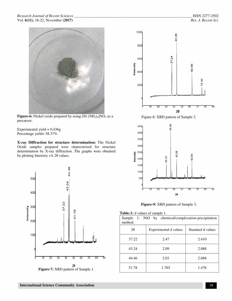
Figure-6: Nickel oxide prepared by using [Ni (NH3)6]NO3 as a
precursor.
Experimental yield = 0.436g
Percentage yield= 58.37%.
X-ray Diffraction for structure determination: The Nickel
Oxide samples prepared were charecterised for structure
determination by X-ray diffraction. The graphs were obtained
by plotting Intensity v/s 2θ values.
Figure-7: XRD pattern of Sample 1.
Figure-8: XRD pattern of Sample 2.
Figure-9: XRD pattern of Sample 3.
Table-1: d values of sample 1.
Sample 1: NiO by chemical/complexation-precipitation
method.
2θ Experimental d values Standard d values
37.22 2.47 2.410
43.24 2.09 2.088
44.46 2.03 2.088
51.78 1.765 1.476

Research Journal of Recent Sciences ______________________________________________________________ ISSN 2277-2502
Vol. 6(11), 16-22, November (2017) Res. J. Recent Sci.
International Science Community Association 19
Table-2: d values of sample 2.
Sample 2: NiO by nano-composite method.
2θ Experimental d values Standard d values
30.22 2.957 2.410
35.58 2.523 2.410
43.26 2.091 2.088
62.84 1.478 1.476
Table-3: d values of sample 3
Sample 3: NiO by using [Ni(NH3)6]NO3 as a precursor.
2θ Experimental d values Standard d values
37.24 2.414 2.410
43.26 2.09 2.088
62.86 1.478 1.476
75.4 1.260 1.259
The interplanar spacing values so calculated using Braggs law
nλ = 2dsinθ, agrees well with standard card data (JCPDS card
no- 47-1049). The peaks are sharp and intense which proves that
samples are crystalline in nature. There is broadening of peaks
in all three samples which indictes that the crystallites are of
nano size6. The XRD graph of NiO prepared by Nano-
composite method differs from others as it has different peaks
wherein we can conclude that it is not present as a single entity
of nickel oxide but we can assign a probable formula for this
composite as [NiO.MnO]. The particle size was calculated using
Debye Scherrer formula has given the following results D= 0.9
λ / β cosθ1.
Co-precipitation method— 23nm
Nanocomposite— 14nm
Precurssor method--- 12 nm
All the three methods have resulted in nanoparticles. The crystal
structure is size dependent which is clearly revealed from their
XRD which shows some additional peaks in XRD graphs7.
Scanning Electron Microscopy
The surface morphology was determined by Scanning Electron
Microscopy. The SEM analysis was carried out at
Instrumentation centre Goa University.
Figure-10: SEM image of Sample 1.
Figure-11: SEM image of Sample 2.
Figure-12: SEM image Sample 3 shows agglomeration.

Research Journal of Recent Sciences ____________________________________
Vol. 6(11), 16-22, November (2017)
International Science Community Association
Since SEM images do not focus on grain boundaries it cannot
be used to determine the exact particle size.
Microbial studies: Method 1 Using agar gel.
Preparation of nutrient plates: 1.2grams of nutrient agar powder
was weighed and heated in a heat proof dish with the mixing of
60ml hot water.The solution was boiled till all the powder
dissolves and clear liquid is obtained. When the solution is
warm it is poured into petri dish and the top half of the petri
dishis closed immediately to avoid contamination with air
bacteria. The petri dishis kept in refrigerator for 24 hours till
agar hardens.The Nickel oxide sample is stored in the sterile
tubes ,a pinch of sample to another sterile tube and to this about
1-2ml of NaCl saline is added . The solution is mixed by
shaking for about 5 minutes.The petri dish containing agar is
removed and the solution of NiO sample is poured with the help
of micropipette on the surface of agar drop wise and spread well
with the help of a glass rod.The petri dish is labelled and kept
in the incubator for 24 hours at a room temperature.
The petri dish was observed for the presence/absence of
bacteria.
Figure-13: Petridish with NiO and agar gel
Method 2 Using Saline: A pinch of NiO sample was transfered
to a sterile tube and add to it 1-2ml of saline of NaCl was added.
The solution was shaken well for about 5 minutes and keep it in
the incubator at a room temperature for 24 hours.The tubes for
the presence/absence of bacteria.
Both the methods did not show growth of bacteria which proved
that NiO has antimicrobial properties.
Photocatalytic study: Dye degradation is a process in which
large dye molecule is broken down chemically into smaller
molecules by certain chemical compounds
Photodegradation this is carried out in presence of light. The
rate of this photocatalytic degradation depends on the basic
________________________________________________________
International Science Community Association
Since SEM images do not focus on grain boundaries it cannot
Preparation of nutrient plates: 1.2grams of nutrient agar powder
was weighed and heated in a heat proof dish with the mixing of
60ml hot water.The solution was boiled till all the powder
s and clear liquid is obtained. When the solution is
warm it is poured into petri dish and the top half of the petri
dishis closed immediately to avoid contamination with air-borne
bacteria. The petri dishis kept in refrigerator for 24 hours till
dens.The Nickel oxide sample is stored in the sterile
tubes ,a pinch of sample to another sterile tube and to this about
2ml of NaCl saline is added . The solution is mixed by
shaking for about 5 minutes.The petri dish containing agar is
solution of NiO sample is poured with the help
of micropipette on the surface of agar drop wise and spread well
with the help of a glass rod.The petri dish is labelled and kept
in the incubator for 24 hours at a room temperature.
ved for the presence/absence of
Petridish with NiO and agar gel.
A pinch of NiO sample was transfered
2ml of saline of NaCl was added.
5 minutes and keep it in
the incubator at a room temperature for 24 hours.The tubes for
Both the methods did not show growth of bacteria which proved
adation is a process in which
large dye molecule is broken down chemically into smaller
molecules by certain chemical compounds8. In
Photodegradation this is carried out in presence of light. The
rate of this photocatalytic degradation depends on the basic
structure of catalyst and the nature of auxiliary group attached to
aromatic dye. The catalytic action of NiO on dye degradation
was experimented and to understand this Methylene blue was
selected as dye as it has well resolved spectrum in visible
region9. i. To prepare 16ppm of methylene blue, 0.016g of the
same was weighed and diluted to 1000ml using a standard
measuring flask. ii. 50ml of this solution was taken using a
graduated pipette and to this 100ml of distilled water was added.
iii. 0.5g of the prepared NiO sample was then added to this
diluted solution and kept under sunlight.
solution was found out using a UV
after intervals of 30, 60, 90, 120minutes.
Figure-14: Tubes containing N
Figure-15: Structure of Methylene Blue
Table-4: Absorbance values of NiO nanoparticles in
degaradation of Methylene Blue.
Time Blank Sample
0 0.85
30 0.85
60 0.85
90 0.85
120 0.85
_______________ ISSN 2277-2502
Res. J. Recent Sci.
20
structure of catalyst and the nature of auxiliary group attached to
aromatic dye. The catalytic action of NiO on dye degradation
was experimented and to understand this Methylene blue was
selected as dye as it has well resolved spectrum in visible
To prepare 16ppm of methylene blue, 0.016g of the
same was weighed and diluted to 1000ml using a standard
50ml of this solution was taken using a
graduated pipette and to this 100ml of distilled water was added.
0.5g of the prepared NiO sample was then added to this
diluted solution and kept under sunlight. iv. Absorbance of this
solution was found out using a UV-Visible spectrophotometer
after intervals of 30, 60, 90, 120minutes.
Tubes containing NiO in saline medium.
Structure of Methylene Blue
10.
Absorbance values of NiO nanoparticles in
Sample-1 Sample-2
- -
0.83 0.82
0.74 0.81
0.68 0.78
0.66 0.75

Research Journal of Recent Sciences ______________________________________________________________ ISSN 2277-2502
Vol. 6(11), 16-22, November (2017) Res. J. Recent Sci.
International Science Community Association 21
The NiO nanoparticles show a decrease in the absorbance with
an interval of 30min, we can say that NiO helps in the
photodegradation of methylene blue.
Figure-16: Graph showing photodegradation of Methylene blue
with NiO.
Therefore NiO can be used for treatment of waste waters likely
to be contaminated with organic based pollutants.
Results and Discussion
In the present work, NiO has been synthesized by co-
precipitation, Nanocomposite and precursor methods. The co-
precipitation method has given maximum yield. All methods
have resulted in non-stoichiometric oxide.
The XRD spectra shows resemblance of peaks and d values with
standard values which confirms the formation of NiO. The XRD
of Nanocomposite is slightly different than others, which
reveals that product exists as composite NiOMnO The particle
size of the three samples were calculated using Debye Schherer
formula have confirmed the formation of nano particles. The
precursor method has resulted in smallest nanoparticles of NiO
with dimensions of 12 nm. The SEM images have shown
formation of tetragonal grains and spherical clusters for the first
two methods and agglomerates by last one. The NiO
nanoparticles were shown to exhibit antimicrobial properties.
The synthesized NiO nanoparticles were found to be an
excellent photocatalyst for treatement of waste waters likely to
be contaminated with organic based pollutants. These could be
preferred over TiO2 which has low photonic efficiency and
requires u.v light for band gap excitation10
. In contrast to this
NiO is a p-type semiconductor with a band gap of Eg= 3.5 ev11
.
Conclusion
NiO prepared by all 3 methods have yielded black colour
nanoparticles, with rhombohedral structure. Size dependence of
crystal structure is verified from XRD graphs. SEM images
show formation of tetragonal grains and spherical clusters for
first two samples and agglomerates in case of Precurssor. NiO
nanoparicles have also shown to exhibit anti microbial
properties. NiO is found to be an excellent photocatalyst and
can be used in treatment of waste waters.
Acknowledgements
I acknowledge Dhempe College of Arts and Science Miramar
Goa for rendering the Chemistry lab facilities for carrying out
research work. I sincerely thank XRD department, Microbial
department at National Institute of Oceanography Donapaula
Goa for carrying out XRD analysis and permitting facilities of
Microbiology lab.
References
1. Fasaki I., Koutoulaki A., Kompitsas M. and Charitidis C.
(2010). Structural ,electrical and Mechanical properties of
NiO thin films grown by pulsed laser deposition. Applied
Surface Science, 257, 429-433.
2. Markwinter
https://www.webelements.com/compounds/Nickel/Nickel_
oxide.html 03/10/17.
3. Taghizadeh Fardin (2016). The study of Structural and
Magnetic properties of NiO nanoparticles. Optics and
Photonics Journal, 6, 164-169.
4. Taghizadeh Fardin (2016). The study of Structural and
magnetic properties of NiO. Scientific research, 6, No8B
164-169.
5. Tajiri Takayuki, Saisho Seiya, Mito Masaki, Deguchi
Hiroyuki, Kohno Atushi (2014). Particle size and distortion
effects on NiO nanoparticles embedded in one-dimensional
pores of mesoporous silica. Photon activity report 32,
(2015)B.
6. El-kemar M., Naggy N. and El-Mehasseb I. (2013).
Nickeloxide nanoparticles: Synthesis and spectral studies of
interactions with glucose. Materials Sience in
semiconductor processing, 16(6), 1747-1752.
7. Tajiri Takayuki, Saisho Seiya, Mito Masaki, Deguchi
Hiroyuki and Kohno Atushi (2015). Size dependence of
Crystal Structure and Magnetic properties of NiO
Nanoparticles in Mesoporous Silica. The Journal of
Physical Chemistry, 119(2), 1194-1200.
8. Mehra Meethi and Sharma T.R. (2012). Photocatalytic
degradation of two commercial dyes in aqeous phase using
photocatalyst TiO2. Advances in Applied Research, 3(2),
849-853.
9. Devi L.G., Raju K.A. and Kumar S.G. (2009).
Photodegradation of methyl red by advanced and
homogeneous Photo Fenton,s process. A comparative study
and kinetic approach. Journal of Environmental
Monitoring, 11(7), 1397-1404.

Research Journal of Recent Sciences ______________________________________________________________ ISSN 2277-2502
Vol. 6(11), 16-22, November (2017) Res. J. Recent Sci.
International Science Community Association 22
10. Hameed Abdul, Gombac Valentina, Tiziano Montini,
Graziani Mauro and siero PauloForna (2009). Synthesis
Charecterisation and Photocatalytic activity of NiO-Bi2O3
nanocomposites. Chemical Physics Letters, 472, 212-216.
11. Hameed A. and Gondal M.A. (2004). Laser Induced
Photocatalytic Generation of Hydrogen & Oxygen over
NiO & TiO2. J Mol. Catal. A-Chem, 219, 109-119.