a selective fluorescent sensor for detecting lead in living cells
TRANSCRIPT
A Selective Fluorescent Sensor for Detecting Lead in Living Cells
Qiwen He, Evan W. Miller, Audrey P. Wong, and Christopher J. Chang*
Department of Chemistry, UniVersity of California, Berkeley, California 94720
Received May 1, 2006; E-mail: [email protected]
Lead pollution is an ongoing danger to human health and theenvironment, as most of the 300 million tons of this heavy metalmined to date are still circulating in soil and groundwater.1 Despiteefforts to reduce global emissions, lead poisoning remains theworld’s most common environmentally caused disease.2 Onceintroduced into the body, lead is a potent neurotoxin that caninterfere with brain development,3 slow nerve conduction velocity,4
and trigger behavioral problems.5 Plausible molecular targets oflead include calcium- and zinc-binding proteins that control cellsignaling and gene expression, respectively,2,6-10 but mechanismsof lead toxicity remain an open question for study.
Interest in elucidating these pathways, as well as public concernsover toxic lead exposure, provides a need for devising new waysto track Pb2+ in natural samples. Whereas standard techniques, suchas atomic absorption or anodic stripping voltammetry, can onlymeasure total lead content, fluorescence imaging with Pb2+-sensitivechemosensors can, in principle, provide information on bioavailablelead pools with spatial and temporal resolution. The majorchallenges to achieving this goal are creating systems that areselective for Pb2+ and can function in water. Pb2+-responsivefluorescent probes based on peptide,11 protein,12 DNAzyme,13-16
polymer,17 and small-molecule18-27 scaffolds have been reported.However, none of these systems have been utilized successfullyfor tracking Pb2+ in living cells, as current probes can be limitedby interfering background fluorescence or nonspecific quenchingfrom competing metal ions, incompatibility with water, and/orultraviolet excitation that can damage or trigger autofluorescencefrom biological samples. We now present the synthesis andproperties of Leadfluor-1 (LF1,6), a new turn-on fluorescent sensorfor selective detection of Pb2+ in water and in living cells. LF1features visible wavelength excitation and emission profiles and aca. 18-fold fluorescence enhancement upon Pb2+ binding. Moreover,confocal microscopy experiments establish that LF1 can monitorchanges in Pb2+ levels within living mammalian cells.
LF1 combines a fluorescein-type scaffold with attractive opticalproperties and biological compatibility with a dicarboxylatepseudocrown receptor designed to satisfy the size and chargerequirements of the Pb2+ cation.28 The synthetic route to LF1 isshown in Scheme 1. Reaction of 2-anilinoethanol with 2-(2-chloroethoxy)ethanol affords diol1 in 66% yield. Alkylation of1with bromoacetic acid followed by acid-catalyzed esterificationprovides diester2 in 61% yield. Vilsmeier formylation of2 usingPOCl3/DMF followed by basic workup generates aldehyde3 in 50%yield, which is then coupled with 2 equiv of resorcinol to producethe tetrahydroxy intermediate4 in 14% yield. DDQ oxidation of4furnishes xanthenone5 in 41% yield. Ester deprotection of5 underbasic conditions proceeds quantitatively to give LF1.
Spectroscopic measurements with LF1 were performed undersimulated physiological conditions (20 mM HEPES, buffer pH 7).LF1 displays a characteristic fluorescein-like absorption band inthe visible region centered at 490 nm (ε ) 2.5 × 10⁴ M-1 cm-1)and weak fluorescence (Φ < 0.001,λem ) 514 nm). Upon addition
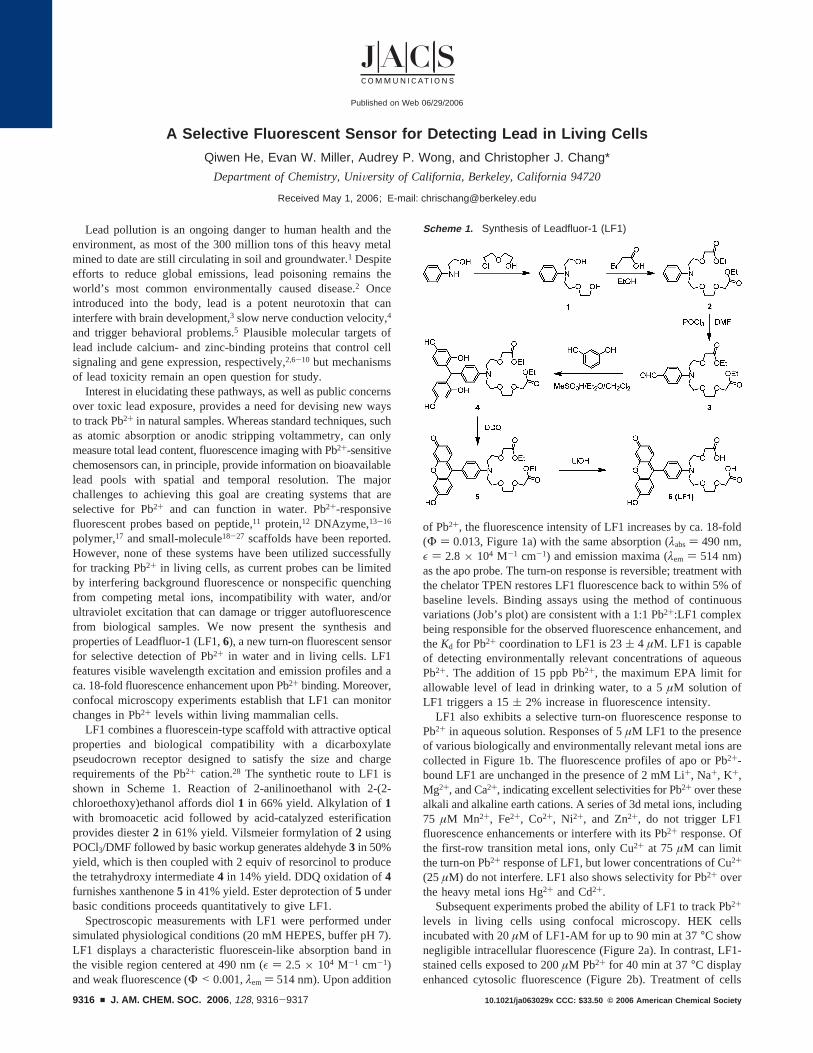
of Pb2+, the fluorescence intensity of LF1 increases by ca. 18-fold(Φ ) 0.013, Figure 1a) with the same absorption (λabs) 490 nm,ε ) 2.8 × 10⁴ M-1 cm-1) and emission maxima (λem ) 514 nm)as the apo probe. The turn-on response is reversible; treatment withthe chelator TPEN restores LF1 fluorescence back to within 5% ofbaseline levels. Binding assays using the method of continuousvariations (Job’s plot) are consistent with a 1:1 Pb2+:LF1 complexbeing responsible for the observed fluorescence enhancement, andtheKd for Pb2+ coordination to LF1 is 23( 4 µM. LF1 is capableof detecting environmentally relevant concentrations of aqueousPb2+. The addition of 15 ppb Pb2+, the maximum EPA limit forallowable level of lead in drinking water, to a 5µM solution ofLF1 triggers a 15( 2% increase in fluorescence intensity.
LF1 also exhibits a selective turn-on fluorescence response toPb2+ in aqueous solution. Responses of 5µM LF1 to the presenceof various biologically and environmentally relevant metal ions arecollected in Figure 1b. The fluorescence profiles of apo or Pb2+-bound LF1 are unchanged in the presence of 2 mM Li+, Na+, K+,Mg2+, and Ca2+, indicating excellent selectivities for Pb2+ over thesealkali and alkaline earth cations. A series of 3d metal ions, including75 µM Mn2+, Fe2+, Co2+, Ni2+, and Zn2+, do not trigger LF1fluorescence enhancements or interfere with its Pb2+ response. Ofthe first-row transition metal ions, only Cu2+ at 75µM can limitthe turn-on Pb2+ response of LF1, but lower concentrations of Cu2+
(25 µM) do not interfere. LF1 also shows selectivity for Pb2+ overthe heavy metal ions Hg2+ and Cd2+.
Subsequent experiments probed the ability of LF1 to track Pb2+
levels in living cells using confocal microscopy. HEK cellsincubated with 20µM of LF1-AM for up to 90 min at 37°C shownegligible intracellular fluorescence (Figure 2a). In contrast, LF1-stained cells exposed to 200µM Pb2+ for 40 min at 37°C displayenhanced cytosolic fluorescence (Figure 2b). Treatment of cells
Scheme 1. Synthesis of Leadfluor-1 (LF1)
Published on Web 06/29/2006
9316 9 J. AM. CHEM. SOC. 2006 , 128, 9316-9317 10.1021/ja063029x CCC: $33.50 © 2006 American Chemical Society
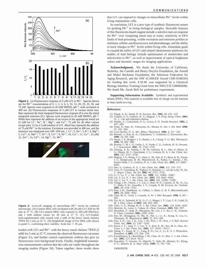
loaded with LF1 and Pb2+ with the heavy metal chelator TPEN (2mM) for 5 min at 25°C reverses the observed fluorescence increases(Figure 2c), and further control experiments without dye give nofluorescence over background levels. Finally, brightfield transmis-sion measurements confirm that the cells are viable throughout theimaging studies (Figure 2d). Taken together, these results show
that LF1 can respond to changes in intracellular Pb2+ levels withinliving mammalian cells.
In conclusion, LF1 is a new type of synthetic fluorescent sensorfor probing Pb2+ in living biological samples. Desirable featuresof this fluorescein-based reagent include a selective turn-on responsefor Pb2+ over competing metal ions in water, sensitivity to EPAlimits of lead poisoning, visible excitation and emission profiles tominimize cellular autofluorescence and photodamage, and the abilityto track changes in Pb2+ levels within living cells. Immediate goalsto expand the utility of LF1 and related chemosensor platforms forstudies of lead biology include optimization of sensitivities andselectivities to Pb2+, as well as improvement of optical brightnessvalues and dynamic ranges for imaging applications.
Acknowledgment. We thank the University of California,Berkeley, the Camille and Henry Dreyfus Foundation, the Arnoldand Mabel Beckman Foundation, the American Federation forAging Research, and the NSF (CAREER Award CHE-0548245)for funding this work. E.W.M. was supported by a ChemicalBiology Interface Training Grant from the NIH (T32 GM066698).We thank Ms. Sarah Bell for preliminary experiments.
Supporting Information Available: Synthetic and experimentaldetails (PDF). This material is available free of charge via the Internetat http://pubs.acs.org.
References
(1) Flegal, A. R.; Smith, D. R.EnViron. Res.1992, 58, 125-133.(2) Claudio, E. S.; Godwin, H. A.; Magyar, J. S.Prog. Inorg. Chem.2003,
51, 1-144 and references therein.(3) Winder, C.; Carmichael, N. G.; Lewis, P. D.Trends Neurosci.1982, 5,
207-209.(4) Araki, S.; Sato, H.; Yokoyama, K.; Murata, K.Am. J. Ind. Med.2000,
37, 193-204.(5) Cory-Slechta, D. A.AdV. BehaV. Pharmacol.1984, 4, 211-255.(6) Bressler, J.; Kim, K.-A.; Chakraborti, T.; Goldstein, G.Neurochem. Res.
1999, 24, 595-600.(7) Hanas, J. S.; Rodgers, J. S.; Bantle, J. A.; Cheng, Y.-G.Mol. Pharmacol.
1999, 56, 982-988.(8) Bouton, C. M. L. S.; Frelin, L. P.; Forde, C. E.; Godwin, H. A.; Pevsner,
J. J. Neurochem.2001, 76, 1724-1735.(9) Ghering, A. B.; Jenkins, L. M. M.; Schenck, B. L.; Deo, S.; Mayer, R.
A.; Pikaart, M. J.; Omichinski, J. G.; Godwin, H. A.J. Am. Chem. Soc.2005, 127, 3751-3759.
(10) Magyar, J. S.; Weng, T.-C.; Stern, C. M.; Dye, D. F.; Rous, B. W.; Payne,J. C.; Bridgewater, B. M.; Mijovilovich, A.; Parkin, G.; Zaleski, J. M.;Penner-Hahn, J. E.; Godwin, H. A.J. Am. Chem. Soc.2005, 127, 9495-9505.
(11) Deo, S.; Godwin, H. A.J. Am. Chem. Soc.2000, 122, 174-175.(12) Chen, P.; Greenberg, B.; Taghavi, S.; Romano, C.; van der Lelie, D.; He,
C. Angew. Chem., Int. Ed.2005, 44, 2715-2719.(13) Li, J.; Lu, Y. J. Am. Chem. Soc.2000, 122, 10466-10467.(14) Liu, J.; Lu, Y.J. Am. Chem. Soc.2003, 125, 6642-6643.(15) Liu, J.; Lu, Y.J. Am. Chem. Soc.2004, 126, 12298-12305.(16) Chang, I.-H.; Tulock, J. J.; Liu, J.; Kim, W.-S.; Cannon, D. M., Jr.; Lu,
Y.; Bohn, P. W.; Sweedler, J. V.; Cropek, D. M.EnViron. Sci. Technol.2005, 39, 3756-3761.
(17) Kim, I.-K.; Dunkhorst, A.; Gilbert, J.; Bunz, U. H. F.Macromolecules2005, 38, 4560-4562.
(18) Chae, M.-Y.; Yoon, J.; Czarnik, A. W.J. Mol. Recognit.1996, 9, 297-303.
(19) Xia, W.-S.; Schmehl, R. H.; Li, C.-J.; Mague, J. T.; Luo, C.-P.; Guldi, D.M. J. Phys. Chem. B2002, 106, 833-843.
(20) Chen, C.-T.; Huang, W.-P.J. Am. Chem. Soc.2002, 124, 6246-6247.(21) Metivier, R.; Leray, I.; Valeur, B.Chem. Commun.2003, 996-997.(22) Hayashita, T.; Qing, D.; Minagawa, M.; Lee, J. C.; Ku, C. H.; Teramae,
N. Chem. Commun.2003, 2160-2161.(23) Sun, M.; Shangguan, D.; Ma, H.; Nie, L.; Li, X.; Xiong, S.; Liu, G.;
Thiemann, W.Biopolymers2003, 72, 413-420.(24) Kim, S. K.; Lee, J. K.; Lim, J. M.; Kim, J. W.; Kim, J. S.Bull. Korean
Chem. Soc.2004, 25, 1247-1250.(25) Kwon, J. Y.; Jang, Y. J.; Lee, Y. J.; Kim, K. M.; Seo, M. S.; Nam, W.;
Yoon, J.J. Am. Chem. Soc.2005, 127, 10107-10111.(26) Zhang, Y.; Xiang, W. C.; Yang, R. H.; Liu, F.; Li, K. A.J. Photochem.
Photobiol. A2005, 173, 264-270.(27) Kavallieratos, K.; Rosenberg, J. M.; Chen, W.-Z.; Ren, T.J. Am. Chem.
Soc.2005, 127, 6514-6515.(28) Hayashita, T.; Sawano, H.; Higuchi, T.; Indo, M.; Hiratani, K.; Zhang,
Z.-Y.; Bartsch, R. A.Anal. Chem.1999, 71, 791-795.
JA063029X
Figure 1. (a) Fluorescence response of 5µM LF1 to Pb2+. Spectra shownare for Pb2+ concentrations of 0, 1, 2, 3, 4, 5, 10, 15, 20, 25, 35, 50, and75 µM. Spectra were acquired in 20 mM HEPES, pH 7, with excitation at490 nm. (b) Fluorescence responses of 5µM LF1 to various metal ions.Bars represent the final integrated fluorescence response (Ff) over the initialintegrated emission (Fi). Spectra were acquired in 20 mM HEPES, pH 7.White bars represent the addition of an excess of the appropriate metal ion(2 mM for Li+, Na+, K+, Mg2+, and Ca2+, 75 µM for all other cationsexcept entry 11) to a 5µM solution of LF1. Black bars represent the additionof 75 µM Pb2+ to the solution. Excitation was provided at 490 nm, and theemission was integrated over 500-650 nm. 1, Li+; 2, Na+; 3, K+; 4, Mg2+;5, Ca2+; 6, Mn2+; 7, Fe2+; 8, Co2+; 9, Ni2+; 10, Cu2+; 11, Cu2+, 25 µM;12, Zn2+; 13, Cd2+; 14, Hg2+; 15, Pb2+.
Figure 2. Live-cell imaging of intracellular Pb2+ levels by confocalmicroscopy. (A) Control HEK cells incubated with 20µM LF1-AM for 90min at 37°C. (B) LF1-stained HEK cells exposed to 200µM Pb(OAc)2and 1 mM sodium citrate for 40 min at 37°C. (C) LF1-loaded,lead-supplemented cells treated with 2 mM of the heavy metal chelatorTPEN for 5 min at 25°C. (D) Brightfield image of live HEK cells shownin panel C, confirming their viability. Scale bar) 10 µm.
C O M M U N I C A T I O N S
J. AM. CHEM. SOC. 9 VOL. 128, NO. 29, 2006 9317