a shift in perspective decentering through mindful …a shift in perspective: decentering through...
TRANSCRIPT

Neuropsychologia 75 (2015) 505–524
Contents lists available at ScienceDirect
Neuropsychologia
http://d0028-39
n CorrMA 024
E-mURL
journal homepage: www.elsevier.com/locate/neuropsychologia
A shift in perspective: Decentering through mindful attention to ima-gined stressful events
Lauren A.M. Lebois a,b,n, Esther K. Papies c, Kaundinya Gopinath d,e, Romeo Cabanban e,Karen S. Quigley f,g, Venkatagiri Krishnamurthy d,e, Lisa Feldman Barrett f,h,Lawrence W. Barsalou a
a Department of Psychology, Emory University, United Statesb McLean Hospital/Harvard Medical School, MA, United Statesc Department of Social and Organizational Psychology, Utrecht University, The Netherlandsd Department of Radiology and Imaging Sciences, United Statese Center for Systems Imaging, Emory School of Medicine, United Statesf Department of Psychology, Northeastern University, United Statesg Edith Nourse Rogers Memorial (Bedford) VA Hospital, United Statesh Massachusetts General Hospital/Harvard Medical School, United States
a r t i c l e i n f o
Article history:Received 19 November 2014Received in revised form19 May 2015Accepted 28 May 2015Available online 22 June 2015
Keywords:DecenteringMental simulationMindfulnessNeuroimagingSelfStress
x.doi.org/10.1016/j.neuropsychologia.2015.05.032/& 2015 Elsevier Ltd. All rights reserved.
esponding author at: McLean Hospital/Harva78, United States.ail address: [email protected] (L.A.M: http://laurenamcdonough.weebly.com (L.A.M
a b s t r a c t
Ruminative thoughts about a stressful event can seem subjectively real, as if the imagined event werehappening in the moment. One possibility is that this subjective realism results from simulating the selfas engaged in the stressful event (immersion). If so, then the process of decentering—disengaging the selffrom the event—should reduce the subjective realism associated with immersion, and therefore per-ceived stressfulness. To assess this account of decentering, we taught non-meditators a strategy fordisengaging from imagined events, simply viewing these events as transient mental states (mindfulattention). In a subsequent neuroimaging session, participants imagined stressful and non-stressfulevents, while either immersing themselves or adopting mindful attention. In conjunction analyses,mindful attention down-regulated the processing of stressful events relative to baseline, whereas im-mersion up-regulated their processing. In direct contrasts between mindful attention and immersion,mindful attention showed greater activity in brain areas associated with perspective shifting and effortfulattention, whereas immersion showed greater activity in areas associated with self-processing andvisceral states. These results suggest that mindful attention produces decentering by disengaging em-bodied senses of self from imagined situations so that affect does not develop.
& 2015 Elsevier Ltd. All rights reserved.
1. Introduction
1.1. Stress and its consequences
Perseverating about difficult events through rumination andworry elicits bodily stress responses that can affect one's healthadversely (Brosschot et al., 2006; Keller et al., 2012). As much re-search demonstrates, chronic stress responses translate into wearand tear on the body and brain, together with reductions in psy-chological well-being (Black and Garbutt, 2002; Ganzel et al.,2010; Hänsel et al., 2010; McEwen, 1998; Juster et al., 2010; Ro-drigues et al., 2009; Rozanski et al., 1999, 2005; Schiffrin and
30
rd Medical School, Belmont,
. Lebois).. Lebois).
Nelson, 2010; Zautra, 2003). Because of the many negative con-sequences associated with chronic stress, it is important to un-derstand the mechanisms that, first, produce stressful thoughtsand, second, reduce their negative impact. In a neuroimaging ex-periment, we examined the neural mechanisms that underliestressful thoughts and a brief decentering intervention for reg-ulating them (mindful attention).
1.2. Why some thoughts are stressful
Much of the stress literature is devoted to establishing whysome thoughts are stressful and others are not (e.g., Almeida,2005; Lazarus, 1993, 1999; Scherer, 2001). One definition suggeststhat stress occurs when a mismatch takes place between an eventone anticipates in the world and what actually happens (Ursin andEriksen, 2004). Together with this expectation violation, a com-bination of additional factors contributes to making an event

L.A.M. Lebois et al. / Neuropsychologia 75 (2015) 505–524506
stressful, in particular: perceived self-threat, perceived inability tocope effectively (inefficacy), the objective severity of the stressor,the individual's resilience and vulnerability, negative emotion, andthe associated neuroendocrine response (Almeida, 2005; Almeidaet al., 2002; Lazarus, 1993, 1999; Scherer, 2001). In recent work,we have similarly found that an imagined event appears stressfulwhen inability to cope effectively with a threatening situation isexperienced such that negative emotion and perseveration result(Lebois et al., 2015 also see Brosschot (2010); Brosschot et al.(2005); Dickerson et al. (2004); Higgins (1989)).
Here we further propose that immersion plays a central role instressful thoughts. By immersion we mean that people experiencea strong sense of self-engagement with an imagined situation. As aconsequence of self-engagement, people often experience vividsensory details, emotions, feelings, and physical sensations, as ifthey were entering into a vivid daydream that they experiencefully. As a further consequence, the imagined event seems sub-jectively real, as if it were happening in the present moment viamental time travel (Papies et al., 2012, 2015; also see cognitivefusion in Acceptance and Commitment Therapy or ACT, Hayes(2004)). Once immersion in a stressful situation produces sub-jective realism, negative emotion, bodily stress responses, andrumination are likely to result.
1.2.1. Neurobiology of stressful thinkingResearch on the neural bases of stress and other emotional
states finds that a consistent set of neural regions tends to becomeactive during stressful thoughts. The anterior insula, amygdala,orbitofrontal cortex (OFC), and their reciprocal connections tosensory areas help determine the relevance of a stimulus for anindividual (Barrett et al., 2007; Ganzel et al., 2010). Specifically, theOFC may initially categorize an event as stressful and can facilitateits perseveration in working memory (Dedovic et al., 2009a,b). Inturn, the anterior cingulate cortex (ACC), amygdala, and dor-somedial and ventromedial prefrontal cortex (dmPFC and vmPFC,respectively) contribute to appraisals related to personal sig-nificance, emotional intensity, and valence (Barrett et al., 2007;Dedovic et al., 2009b,c; Ganzel et al., 2010).
Through connections with the hypothalamus and brainstem,the aforementioned brain regions initiate physiological, hormonal,and behavioral responses to stress (Barrett et al., 2007; Chida andHamer, 2008; Dedovic et al., 2009a; Greenberg et al., 2002). Sev-eral additional areas regulate the neuroendocrine stress responsevia the hypothalamic–pituitary–adrenal axis (HPA axis). The hip-pocampus, for example, helps evaluate the extent to which thestressor affects one's goals and self (Dedovic et al., 2009a). Ad-ditionally, activation of the hippocampus, together with the mPFC,can inhibit the HPA axis (Dedovic et al., 2009a). Conversely, whenthe hippocampus and mPFC deactivate, the HPA axis is disin-hibited, thereby initiating the cascade of stress hormone release. Incontrast to the inhibitory role of the hippocampus and mPFC, theamygdala potentiates HPA axis activation (Dedovic et al., 2009a;Rodrigues et al., 2009). The amygdala, however, is not alwaysconsistently active during stressful cognition, with some contextsbeing more likely to activate it than others (Ganzel et al., 2010).
1.3. Mindfulness
Mindfulness offers one method for intervening on the neuro-biological and cognitive mechanisms that produce stress, wheremindfulness is often characterized as present-centered non-eva-luative awareness of one's thoughts, emotions, and other experi-ences in the moment (Bishop et al., 2004; Kabat-Zinn, 1990). Re-search increasingly documents the benefits of mindfulness acrossdiverse domains of well-being, including reductions in perceivedstress, stress symptoms, rumination, negative thought avoidance,
and emotional reactivity, coupled with enhanced attention andemotion regulation (for reviews see Bishop et al. (2004), Brownet al. (2007), Chiesa and Serretti (2010), Gard et al. (2014), Kenget al. (2011), Lutz et al. (2008), Tang et al. (2012)). Clinical inter-ventions have incorporated aspects of mindfulness to improvefunctioning in mood, attention, and eating disorders (includingACT, Mindfulness Based Stress Reduction or MBSR, Dialectical Be-havioral Therapy or DBT, and Mindfulness Based Cognitive Therapyor MBCT; for reviews see, Grossman et al. (2004), Rubia (2009),Hofmann et al. (2010)).
Neural mechanisms associated with mindfulness can varywidely across expertise and training regimen. Novices and inter-mediate practitioners of mindfulness, for example, typically acti-vate brain areas associated with voluntary effortful attention (e.g.,lateral prefrontal cortex lPFC, parietal cortex PC), whereas expertstypically exhibit reduced activity in these areas and in the defaultmode network (e.g., medial prefrontal cortex mPFC, posteriorcingulate cortex PCC), while at the same time exhibiting greateractivity in dorsal anterior cingulate cortex (dACC), left insula, andstriatum (Brefczynski-Lewis et al., 2007; for reviews see, Chiesaand Serretti (2010), Fox et al. (2014), Tang et al. (2012), Vago(2014)). Regarding training regimens, meditators whose practicesfocus on body awareness (e.g., Vipassana) often show increasedfunctional activity and structural differences in the insula, whereaspractices that lack this focus do not (see Fox et al. (2014) for arecent review).
1.3.1. Exploring brief mindfulness interventionsMost research has focused on experts and experienced practi-
tioners who have at least completed an extended mindfulnesscourse (e.g., 8 weeks of MBSR). Of primary interest has been howthese interventions change psychological states and the under-lying neural activity. Participants in an MBSR course, for example,exhibited reductions in perceived stress, together with less graymatter density in the amygdala (Hölzel et al., 2009). MBSR parti-cipants have also demonstrated reduced neural reactivity to sad-ness, especially in cortical midline areas associated with self-re-ferential processing, relative to a wait list control group (Farb et al.,2010).
Much less research addresses relevant cognitive abilities thatalready exist in individuals before mindfulness training that con-tribute to acquiring mindfulness skills during an intervention.Does mindfulness draw on preexisting cognitive abilities, or is itcompletely acquired in meditation training? Various con-templative approaches assume that individuals have natural con-templative abilities waiting to be uncovered through relevanttraining and experience (e.g., Dzogchen and Mahamudra in Tibe-tan Buddhism; Thrangu, 1996; Nyima, 2004). Several researchershave also made this claim (Brown and Ryan, 2003; Brown et al.,2007; Kabat-Zinn, 2003; Taylor et al., 2011).
In particular, Bishop et al. (2004) proposed that pre-existingcognitive abilities underlie two basic components of mindfulness:attentional awareness and perspective shifting. The attentioncomponent makes it possible to maintain focus on present ex-perience by regulating attention and inhibiting elaborative pro-cessing. The perspective shifting component makes it possible toapproach thoughts and reactions with curiosity, openness, andacceptance – observing all reactions without efforts to changetheir content.
An important outcome of shifting perspective is an insightknown as decentering: The realization that thoughts, feelings, andreactions are transitory patterns of mental activity, that they arenot necessarily true representations of the self and events, andthat they are not actually happening (Bishop et al., 2004; Brownet al., 2007; Teasdale et al., 1995; also see “reperceiving,” Shapiroet al. (2006); “cognitive defusion,” Hayes (2004)). Adopting this

L.A.M. Lebois et al. / Neuropsychologia 75 (2015) 505–524 507
perspective makes it possible for individuals to view theirthoughts and reactions to events as arising and dissipating in themoment, without becoming engaged in sustained affective re-sponses to them (Kross and Ayduk, 2008). From our perspective,decentering prevents subjective realism by disengaging a person'ssense of self from an imagined situation, thereby decreasing im-mersion and mental time traveling.
Consistent with the pre-existence of basic mindfulness abilities,increasing research demonstrates that brief mindfulness inter-ventions can produce immediate benefits via the attention com-ponent and/or the decentering/perspective shifting component.First, consider studies that have examined brief interventions fortraining the attention component (Arch and Craske, 2006; Deli-zonna et al., 2009; Dickenson et al., 2013; Ditto et al., 2006; Farbet al., 2007). In Dickenson et al. (2013), for example, a brief breath-focused meditation recruited more areas involved in internal stateawareness (insula) and in attentional control and shifting (dlPFC,angular gyrus (AG)) compared to a mind wandering condition,especially in participants high in trait mindfulness. In Farb et al.(2007), a simple attentional shift to more present-centeredawareness decreased activation in areas associated with self-re-ferential (posterior cingulate cortex, mPFC) and visceral stateprocessing (subgenual ACC).
Other brief intervention research has examined both attentionand decentering together (Alberts and Thewissen, 2011; Broderick,2005; Lutz et al., 2014; Singer and Dobson, 2007; Zeidan et al.,2010a,b,c). In several related studies, 20 min of mindfulnesspractice for 3–4 days improved sustained attention, visuospatialprocessing, working memory, and executive functioning, whilereducing fatigue, anxiety, heart rate, and subjective experiences ofpain compared to controls and sham meditation groups (Zeidanet al., 2010a,b,c). After training a mindfulness group with briefwritten instructions before a functional magnetic resonance ima-ging (fMRI) scan session, Lutz et al. (2014) found that mindfulnesswas associated with greater emotion regulation (increased super-ior mPFC) in anticipation of negative pictures, and decreasedemotional responding during perception of emotional pictures(decreased amygdala and parahippocampal gyrus activity) com-pared to a control group.
Only a handful of brief intervention studies have emphasizeddecentering explicitly (Erisman and Roemer, 2010; Kross et al.,2009; Papies et al., 2012, 2015; Tincher et al., in press). In Papieset al. (2012, 2015), a 15 min mindful attention induction modu-lated implicit approach responses toward desirable, unhealthyfoods, and also choices to consume them. In Tincher et al. (2015), a20 min mindful attention induction modulated stereotype biasesto in-group and out-group members. In Kross et al. (2009), lessself-referential, emotional, and visceral state integration occurredin mPFC and sgACC for negative autobiographical memories in amindful condition compared to a ruminative condition.
The majority of the work just described, however, is behavioral,with relatively little emphasis on the underlying neural mechan-isms. In the experiment reported here, we assessed the neuralmechanisms underlying immersion in stressful thoughts, togetherwith the neural mechanisms underlying disengagement from suchthoughts. We adapted a brief mindfulness intervention—mindfulattention—from Papies et al. (2012) that utilizes the perspectiveshifting mechanism of mindfulness, specifically, decentering. Pre-vious mindfulness interventions, reviewed above, often lack thisspecific focus on decentering, and none has emphasized de-centering in the context of stressful cognition. By contrasting thecognition associated with immersion vs. mindful attention in aneuroimaging paradigm, we hoped to establish the neural me-chanisms that make imagined events seem subjectively real andstressful, and conversely, the mechanisms that make it possible todisengage from these immersion experiences.
1.4. Experiment overview
During a brief initial instruction, participants learned a mindfulattention strategy for disengaging from imagined events (de-centering), and also practiced an immersion strategy for engagingwith imagined events (mentally time travelling). During a sub-sequent fMRI session, in a completely repeated measures design,participants performed blocks using the mindful attention strategyand blocks using the immersion strategy. Within each mindfulattention and immersion block, participants imagined experien-cing stressful events in one sub-block (e.g., Your professor just ac-cused you of cheating on an exam) and non-stressful events in asecond sub-block (e.g., Your professor just passed out lecture notesin preparation for the next class). Within the initial reading periodof each trial, participants read and comprehended a stressful ornon-stressful event, and then, during the subsequent strategyperiod, performed either the mindful attention or immersionstrategy for that event. Finally, participants rated their ability toperform mindful attention or immersion during the trial.
Of interest was the neural activity that each strategy (mindfulattention vs. immersion) exhibited over the course of each period(reading vs. strategy) in each event condition (stressful vs. non-stressful). Most generally, we predicted that immersion wouldutilize brain areas that produce sensorimotor simulation of anevent, together with areas that contribute to the experience thatthe event is subjectively real, including areas associated with self,emotion, and visceral states. In contrast, we predicted that mindfulattention would utilize brain areas that contribute to disengagingthe self from the simulated event, including areas associated withperspective shifting, effortful attention, and regulatory processing.
Because trials for immersion and mindful attention wereblocked, it is possible that mindful attention and immersion pro-cessing operated across entire blocks, thereby entering into boththe reading and strategy period on every trial. Rather than onlybeing restricted to the strategy period, the strategy could be dis-tributed across both periods. If, for example, mindful attentiondown-regulates affect associated with stressful events, then muchinitial regulatory activity during the reading period could be fol-lowed by less affective activity during the strategy period. Con-versely, if immersion in an event increases across the reading andstrategy periods, then neural activity might also increase.
Finally, we expected that mindful attention and immersionwould differ more in neural activity for stressful events than fornon-stressful events. Because stressful events generate strong af-fective responses, they are likely to produce strong experiences ofimmersion, and thus afford salient responses that can be regulatedvia mindful attention (see Papies et al., 2015, for related proposalsand findings). In contrast, non-stressful events might not offer thesame opportunities for differentially applying the two strategies,such that neural activity would be more similar.
2. Method
2.1. Design and participants
2.1.1. DesignThe scanning session contained three independent variables—
strategy (mindful attention vs. immersion) X event (stressful vs.non-stressful) X period (reading vs. strategy)—in a completelycrossed repeated-measures design. As Fig. 1 illustrates, four criticalconditions occurred in the strategy X event sub-design (associatedonce with the reading period and once with the strategy period):(1) mindful attention stressful, (2) mindful attention non-stressful,(3) immersion stressful, (4) immersion non-stressful. For reasonsexplained shortly, each of these four conditions contained 30

Fig. 1. The experimental design. The top panel summarizes the training procedure. The middle panel presents the four event types in the design, which occured in both thereading and strategy periods. The bottom panel describes the trial sequence using the immersion stress condition as an example. Mindful attention trials and non-stresstrials followed the same procedure. The first sequence depicts a complete trial. The second sequence depicts a catch trial.
L.A.M. Lebois et al. / Neuropsychologia 75 (2015) 505–524508
reading period trials and 24 strategy period trials. Although manymindfulness experiments include a mindfulness group and sepa-rate control group in a between-groups manipulation, we chose arepeated-measures design after piloting both designs. Specifically,pilot participants reported finding it easier to learn the immersionand mindful attention strategies together (contrasting them witheach other) rather than alone (without the other).
A mix of complete trials and catch trials allowed us to separateBOLD activations during the reading period from those during the
strategy period (details provided later). Catch trials constituted20% of the total trials, enough to successfully isolate activationsduring the two adjacent periods (Ollinger et al., 2001a, 2001b).Each of the 4 critical conditions defined above contained 24complete trials and 6 catch trials.
An active baseline task (visual detection) was used instead of aresting state baseline (details provided later). Because participantshad to press a button to respond on the baseline task, it wasanalogous to the critical task that also required a button press. By

L.A.M. Lebois et al. / Neuropsychologia 75 (2015) 505–524 509
subtracting the active baseline from the critical conditions, weremoved uninteresting activations associated with visual andmotor processing that were not central to the event and strategyactivations of interest. If we had used a resting baseline instead, itwould likely have produced mind wandering associated with self-related processing (e.g., Mason et al., 2007). Because self-relatedprocessing is central to stress (e.g., Dedovic et al., 2009a; Dick-erson and Kemeny, 2004), a resting baseline would have removedpotentially germane activations from later analyses (e.g., corticalmidline activity; Mason et al., 2007). An active baseline, therefore,was deemed more appropriate.
2.1.2. ParticipantsThirty participants (15 female) were drawn from the student
populations of Emory University and Georgia Institute of Tech-nology, with this sample size providing sufficient power to test ourhypotheses (Mumford and Nichols, 2008). Participants were 18–23years old, including 50% Caucasian, 20% Asian, 17% other, 10%African American, and 3% Native American (1 individual alsoidentified as Hispanic or Latino). Three participants were droppeddue to excessive head movement in the scanner, and one partici-pant was dropped after disclosing failure to follow instructions(during the exit interview). These 4 participants were replaced tomaintain a 30-participant sample that exhibited the aforemen-tioned demographics. Typical imaging exclusion criteria were en-forced. Any individuals who were left-handed, had metal implants,or claustrophobia were excluded, as were individuals who werecurrently taking psychotropic medication, or who had experiencedsignificant head injury associated with loss of consciousness. Par-ticipants also had to be native English speakers with normal orcorrected vision. Additionally, we excluded individuals withmeditation experience, as we wanted to examine the mechanismsunderlying mindful attention in non-meditators. The Emory In-stitutional Review Board approved the protocol, and informedconsent was obtained from all participants. Participants received$80 compensation for their time.
2.2. Materials
2.2.1. ScenariosCritical events were 120 one-sentence scenarios (60 stressful,
60 non-stressful) that averaged 15 words in length. Each scenariocontained second person (“you”) references to promote self-en-gagement. Stressful scenarios all included interpersonal tensionsrelevant to college life, for example, “You have to tell your parentsyou failed a class and need to take summer school,” “Your room-mates had a party while you were gone, and now your landlord isthreatening to evict you,” and “Your professor asks for take-homemidterms, and you realize you left yours at home.” Non-stressfulscenarios were written to match scene and character details fromthe stressful scenarios, but with non-threatening social interac-tions, for example, “You tell your parents you're considering takinga class over the summer to free up your fall schedule,” “Yourroommate says they decided to stay home while you went awaylast weekend,” and “Your professor asks everyone to talk amongstthemselves while taking a quick phone call outside.”
To make the scenarios more ecologically valid, we drew ideasfor events from a nation-wide database of stressful events (Al-meida et al., 2002), and from undergraduate research assistants. Intotal, 572 stressful and non-stressful scenarios (286 each) wereconstructed and normed by 12 participants in a separate beha-vioral study for stressfulness, self-threat, perseverative thought,expectation violation, efficacy, experience, familiarity, plausibility,valence, arousal, and certainty (Lebois et al., 2015). The 60 moststressful scenarios with the least amount of variance in stressful-ness were selected for use in the imaging experiment, along with
their 60 matched non-stressful scenarios. Stressful and non-stressful scenarios did not differ in sentence length (stressfulM¼15.33, SD¼3.07; non-stressful M¼15.52, SD¼2.46; t(59)¼� .39, SE¼ .47, p4 .250). On a Likert scale of 1 (low) to 7(high), stressful scenarios were higher in perceived stressfulness(M¼5.86, SD¼ .37) compared to non-stressful scenarios (M¼1.34,SD¼ .29; t(59)¼75.01, SE¼ .06, po .001). Stressful scenarios werealso significantly different on core features that predict stress,including, threat, arousal, perseveration, negative valence, bodilyimagery, violation of expectations, efficacy, and positive valence(Lebois et al., 2015). The Supplementary Materials (SM) report thedetails of these additional norming results.
2.3. Procedure
As Fig. 1 illustrates, each participant performed two trainingsessions, one scanning session, and a post-scan question period.Each is addressed in turn.
2.3.1. Training session 1Training session 1 was approximately 1.5 h in duration from
initial self-report questionnaires to final task practice. Participantsfirst completed self-report measures of absorption, rumination,and mindfulness. The results for these measures do not bear onthe current analyses and are not discussed further.
To ensure that participants fully understood and were com-fortable performing mindful attention and immersion, a detailedinstruction protocol was followed (see the SM for more completedetails). We adapted key concepts for this instruction from pre-vious research (Lebois et al., 2015; Papies et al., 2012; Wilson-Mendenhall et al., 2011). Papies et al. (2012) elicited reliable be-havioral differences between mindful attention and immersion onan implicit approach–avoidance task using a similar but moreconcise instruction.
First, we introduced the concept of immersion, provided a de-finition, and presented examples. As described earlier, participantswere asked to become completely absorbed in the experience ofthe scenarios, as if they were happening in the moment. Theywere to mentally time travel and experience the sensory details,physical sensations, feelings, emotions, and bodily states asso-ciated with engaging in the scenario vividly. Participants practicedimmersing themselves in presented scenarios through a series oftasks that built up to the timing and procedure of the critical task.
Second, participants learned the distinction between completeand catch trials. As Fig. 1 illustrates, complete trials contained areading period, a strategy period, and a rating period (detailsprovided later). During the reading period, participants were in-structed to comprehend a presented event; during the strategyperiod, participants were instructed to perform either immersionor mindful attention (as described shortly) on the event; duringthe rating period, participants rated how well they were able toperform the strategy. As Fig. 1 further illustrates, catch trials wereexactly the same as the complete trials except that they onlyconsisted of the reading period, with the strategy and rating per-iods excluded. Following instruction, participants practiced per-forming both complete and catch trials to become comfortablewith each.
Third, participants received instructions on the left-right visualdetection task that served as the active baseline (details providedlater). Participants then practiced the baseline task so that theywould be comfortable performing it later in the context of com-plete and catch trials.
Fourth, we introduced the concept of mindful attention, pro-vided a definition, and presented examples, following the samestructure as the immersion instruction. Participants were asked toremain aware of their current physical location while thinking

L.A.M. Lebois et al. / Neuropsychologia 75 (2015) 505–524510
about the scenarios. They were further asked to notice the kinds ofreactions that they normally have during immersion, but ratherthan ‘living’ the event, they were instructed to simply observetheir thoughts and reactions to it in the present moment. Parti-cipants were asked to perceive their thoughts about the stimuli astransitory mental states, not as parts of the scenarios, but as theirpsychological responses to them. Essentially, we briefly taughtparticipants the decentering component of mindfulness, allowingthem to disengage from the events being imagined.
Lastly, participants practiced one run of the experimental task,including complete trials, catch trials, and the active baseline task.All these elements had been practiced previously in training, buthad not yet been implemented together. The practice run con-tained 1 block of 10 immersion trials mixed with baseline trials,and 1 block of 10 mindful attention trials also mixed with baselinetrials (16 complete trials, and 4 catch trials). Each block containeda mini-block of 5 stressful scenarios and a mini-block of 5 non-stressful scenarios, counterbalanced for order.
At the onset of a 10-event block, participants received a cue,“IMMERSION” or “MINDFUL,” presented in white font on a blackbackground that lasted for 2.3 s followed by 2.3 s of a black screen.Cues only occurred at the beginning of a strategy block, not beforeeach trial, nor when participants switched between stressful andnon-stressful scenario mini-blocks within a strategy block.
As Fig. 1 illustrates, a complete trial consisted of the followingevents. (1) During the reading period, a one-sentence scenario waspresented visually in white font on a black background for 6.9 s.During this period, the task was simply to read and understand thesentence. (2) During the strategy period, the sentence changed to adark gray font, cuing participants to adopt either the mindful at-tention or immersion strategy for 6.9 s, depending on the type ofblock. (3) During the rating period, the screen switched to “Im-mersion rating?” or “Mindful rating?” for 2.3 s in a lime green fonton a black background. Participants' task was to rate their ability toimmerse (or mindfully attend) on a scale of 1 (not at all) to 5(high). On catch trials, only the reading period occurred, not fol-lowed by the strategy and rating periods. One trial in each mini-block of five trials was randomly chosen to be a catch trial.
After every complete trial or catch trial, a left–right visual de-tection trial occurred (i.e., the active baseline task). For a randomlyjittered interval of 4.6–9.2 s, the following sentence appeared on theblack screen in a dark gray font, “Find the cue and then get ready topress the direction indicated by it.” This sentence was of compar-able length to the critical scenarios. At a random point during thevariable interval, the word “left” or “right” appeared somewherewithin the sentence, occluding letters within the sentence (e.g.,“Finlefte cue and then get ready to press the direction indicated byit.” where “left” occludes part of “Find the”). A rating screen thenappeared for 2.3 s with the word “Direction?” in lime green font.Participants pressed the left-most button on the response box ifthey saw the word “left,” the right-most button if they saw theword “right,” and the middle button if they missed the directionword. The rating screen was followed by 2.3 s of a blank blackscreen before proceeding to the next trial. A 6.9 s black screen ap-peared between the immersion and mindful attention blocks, and a16 s black screen occurred at the end of each run. All cues, stimuli,and rating requests appeared in the center of the screen.
2.3.2. Training session 2Participants were not asked to practice any of the strategies
outside of the lab training sessions. Training session 2 occurred 1–2 days after the first session. The total duration of day two fromtraining to post-scan rating was approximately 2.5 h. Duringtraining, participants reviewed the immersion and mindful at-tention strategies, completed one more practice run, and thenproceeded immediately to the MRI scanner.
2.3.3. Scanning sessionIn the scanner, participants completed six runs that followed
the same procedure as the aforementioned practice run. To avoidrepetition effects, participants viewed novel scenarios during theexperimental task in the scanner not seen during practice. Thescan session lasted approximately 1 h, including one T1 anatomi-cal scan and 54 min of critical functional scans on the experi-mental task. While in the scanner, measures of heart rate, re-spiration, and electrodermal activity were also collected. Thesephysiological data will be reported in a later manuscript.
Each of six runs, lasting about 9 min each, contained twostrategy blocks, one for mindful attention and one for immersion.Each strategy block contained one mini-block of 5 stressful eventsand one mini-block of 5 non-stressful events, with each mini-block containing 4 complete trials and 1 catch trial, randomly or-dered. Within each strategy block, the assigned strategy alwaysremained constant across the two mini-blocks (e.g., mindful at-tention was performed first for stressful or non-stressful eventsand then for the other type of event). Eight different versions ofthe experiment were constructed, counterbalancing run order,block order, mini-block order, and the assignment of each event tomindful attention or immersion (each participant only saw a givenevent once, performing either mindful attention or immersion onit).
2.3.4. Post scan sessionAs a manipulation check, participants rated the critical sce-
narios for overall stressfulness on a 1 (not at all stressful) to 7(highly stressful) scale. Finally, participants completed an exit in-terview in which they described what they were doing duringeach strategy, and how difficult it was for them.
2.4. Scan sequence
All scans were completed on a Siemens 3T Trio scanner with a32-channel head coil. The functional scans were acquired with awhole-brain multiband slice-accelerated gradient-echo echo planarimaging (EPI) sequence (Feinberg et al., 2010; Moeller et al., 2010):TR/TE/FA¼1150 ms/24 ms/45°, FOV¼220 mm�220 mm; imageacquisition matrix¼74�74; 64 slices with thickness of 2 mm for a3 mm�3 mm�2 mm voxel resolution. The high-resolution anato-mical scans were acquired with a sagittal 3D T1-weighted MPRAGEsequence (FOV¼240 mm�240 mm; TR/TI/TE/FA¼2250 ms/900 ms/3 ms/9° image acquisition matrix¼256�256�160;0.9 mm�0.9 mm�1 mm resolution). Sequences were chosen toreduce susceptibility artifacts.
2.5. Image preprocessing and statistical analyses
AFNI was used to perform standard preprocessing includingskull stripping and slice time correction (Cox, 1996). FSL (Smithet al., 2004) was used to correct spatial intensity variations (Zhanget al., 2001) and to perform spatial normalization and co-regis-tration (Andersson et al., 2010; Jenkinson and Smith, 2001; Jen-kinson et al., 2002). See the SM for further details on various as-pects of the preprocessing and analyses described below.
During preprocessing, data for individual participants weretemporally shifted, registered to a base volume, co-registered to ananatomical volume, spatially normalized to MNI152 templatespace, resampled to 3x3x3 voxels, and smoothed with an isotropicGaussian (FWHM¼6 mm) kernel. Multiple linear regression ana-lysis was then performed on individual participant's data. Re-gressors were constructed using a vector of onset time points foreach respective condition. These blocks of stimulus times wereconvolved with a gamma-variate function. Each block was thenrescaled to have an amplitude of 1 multiplied by the estimated

L.A.M. Lebois et al. / Neuropsychologia 75 (2015) 505–524 511
stimulus type beta-weight. For each voxel, the signal was modeledas the weighted sum of the aforementioned convolutions, the 3rdorder polynomial drifts, and the constant baseline.
The 29 regressors (beta weights) included 4 during the readingperiod for the 4 critical conditions (mindful attention stress,mindful attention non-stress, immersion stress, immersion non-stress conditions), 4 during the strategy period for the same4 critical conditions, 1 for the fixation cue, 6 for motion para-meters, 6 for the 6 rest periods, and 8 for the 8 strategy and
Fig. 2. The conjunction analysis procedure. Step 1 across Panels A and B illustrates the tstressful (S) and non-stressful events (N) during the reading period. The arrows point tocommon across both stressful and non-stressful events during reading (A). In Step 2, we reach of the four conditions: mindful attention stressful (U-MS), immersion stressful (UPanels C and D specify the same procedures for the strategy periods.
baseline rating periods (removing the regressors for the rest andrating periods had no effect on the results).
Each individual's beta coefficients for the 8 critical conditionswere entered into a random effects whole brain ANOVA to obtaingroup level maps. All group-level maps mentioned in the con-junction and contrast analyses to follow were thresholded at avoxel-wise level of po .005 and a corrected extent threshold ofpo .05 (26 3 mmx3 mm�3 mm voxels), estimated using AFNI'sMonte Carlo 3dClustsim program. All x y z coordinates reported
wo initial conjunction analyses that each identified neural activity common acrossa third conjunction analysis of the voxels in S and N to establish the neural activityemoved the common activity to extract the unique neural activity during reading in-IS), mindful attention non-stressful (U-MN), and immersion non-stressful (U-IN).

L.A.M. Lebois et al. / Neuropsychologia 75 (2015) 505–524512
are in Talairach space.
2.5.1. Conjunction analysesAs Fig. 2 illustrates, the eight condition maps in the period X
strategy X event design (relative to the active baseline) were ex-amined in a series of conjunction analyses. Significance in allconjunction analyses was established using the po .005 level forindividual voxels and the po .05 level for spatial extent just de-scribed. A primary interest was to examine the neural activity ineach condition relative to baseline across the reading and strategyperiods. How much did mindful attention and immersion alterneural activity relative to baseline during the reading and strategyperiods? During each period, what brain areas became activeabove baseline for each strategy? What activations did the stra-tegies have in common? What activations were unique?.
To assess these issues, we first performed two conjunctionanalyses for the reading period, one for the two stressful condi-tions (mindful attention stressful, immersion stressful), and onefor the two non-stressful conditions (mindful attention non-stressful, immersion non-stressful). As Step 1 across Panels A and Bof Fig. 2 illustrates, these two initial conjunction analyses identi-fied neural activity common across stressful events (S in Fig. 2) andacross non-stressful events (N in Fig. 2) during the reading period.We then performed a third conjunction analysis of the voxelscommon across stressful events (S) and across non-stressful events(N) to establish the neural activity common across all eventsduring reading (A in Fig. 2). Finally, in Step 2, we removed allcommon activity to extract the unique neural activity duringreading in each of the four critical conditions: mindful attentionstressful, immersion stressful, mindful attention non-stressful, andimmersion non-stressful (in Fig. 2, U-MS, U-IS, U-IN, U-MN,respectively).
As Fig. 2 further illustrates in Panels C and D, three analogousconjunction analyses were performed for the strategy period.Again, shared voxels for stressful events (S), non-stressful events(N), and all events (A) were established, as were unique voxels forthe four critical conditions (U-MS, U-IS, U-IN, U-MN).
As described in the Results section, assessing these 6 sets ofshared voxels and 8 sets of unique voxels allowed us to char-acterize changes in brain activity relative to baseline for eachcondition. As we will see in the unique activation results, mindfulattention and immersion differed significantly in how neural ac-tivity changed relative to baseline across the reading and strategyperiods for stressful and non-stressful events.
We also examined the shared voxel sets and each of the eightunique voxel sets for the extent to which they contained voxelsfrom important neural networks. Using masks that Yeo et al.(2011) established from a large-scale resting state study, wecounted the number of voxels in each voxel set that resided in Yeoet al.'s visual, somatosensorimotor, limbic, default mode, fronto-parietal control, ventral attention, and dorsal attention networks.Of interest was the extent to which these seven networks werepresent in neural activity across the eight critical conditions.
2.5.2. Linear contrast analysesFinally, we performed linear contrasts within the reading per-
iod and the strategy period. Of interest during each period waswhether neural activity differed significantly between the mindfulattention vs. immersion strategies.
3. Results
3.1. Behavioral results
Participants' task ratings during the scan session on the 1 to
5 scale indicated that they were able to perform the mindful at-tention and immersion strategies effectively (mindful attentionstressful: M¼3.75, SD¼ .70, bootstrapped 95% CI [3.50, 4.01];mindful attention non-stressful: M¼3.89, SD¼ .63, bootstrapped95% CI [3.66, 4.12]; immersion stressful: M¼3.85, SD¼ .73, boot-strapped 95% CI [3.59, 4.09]; immersion non-stressful: M¼3.77,SD¼ .71, bootstrapped 95% CI [3.51, 4.02]). The lack of main effectsfor strategy type and event type indicate that both strategies wereperformed equally easily for both event types (strategy type: F(1,29)¼ .06, p4 .250, ηp²¼ .002; event type: F(1, 29)¼ .13, p4 .250,ηp²¼ .132; interaction, F(1, 29)¼2.28, p¼ .142, ηp²¼ .073).
Participants' event ratings during the post-scan session ratingon the 1 to 7 scale indicated that when the scenarios occurred onmindful attention trials, they were rated as significantly lessstressful (M¼3.57, SD¼2.13, bootstrapped 95% CI [3.47, 3.66]) thanwhen they occurred on immersion trials (M¼3.66, SD¼2.11,bootstrapped 95% CI [3.57, 3.74]), F(1, 118)¼4.77, p¼ .031,ηp²¼ .039, indicating that the instruction had a small sustainedeffect on self-report ratings of stress. Not surprisingly, the stressfulscenarios were rated higher in stressfulness (M¼5.66, SD¼ .50,bootstrapped 95% CI [5.55, 5.78]) than the non-stressful scenarios(M¼1.56, SD¼ .38. bootstrapped 95% CI [1.45, 1.68]), F(1, 118)¼2561.82, po .001, ηp²¼ .96.
During the exit interview, participants provided accurate verbaldescriptions of the immersion and mindful attention strategies,and generally reported being able to perform both. A majority ofparticipants experienced immersion as producing more vivid ex-periences of the imagined scenarios, especially with bodily sen-sations of tension, compared to mindful attention. Most foundmindful attention more effortful than immersion, and in contrast,those who reported more difficulty with immersion often ex-pressed finding it unpleasant to project themselves into stressfulscenarios.
3.2. Unique activations in the conjunction analyses
As Step 1 of Fig. 2 illustrates, conjunction analyses establishedshared activations across all four conditions during the readingand strategy period (labeled A in Fig. 2). Additional conjunctionanalyses established shared activations for just the stressful events(S) and non-stressful events (N). The SM describe these sharedactivations in detail (SM Tables 1 and 2, SM Figs. 1, 2, and 3).
As Step 2 of Fig. 2 further illustrates, all shared clusters wereremoved to establish the unique clusters that became active abovebaseline in each condition (voxels labeled U-MS, U-IS, U-MN, U-INin both panels of Fig. 2). Of interest was how much mindful at-tention and immersion significantly increased neural activityabove baseline during the reading and strategy periods, and whatbrain areas became active for each strategy.
As Fig. 3 illustrates, the four conditions differed in their re-spective distributions of unique neural activity across the readingand strategy periods (where “unique neural activity” is the totalnumber of voxels across significantly active clusters). As Fig. 4further illustrates, the four conditions also exhibited large differ-ences in the neural networks active across these periods.Tables 1 and 2 provide the complete lists of unique clusters thatbecame active above baseline in each condition, for the readingand strategy periods, respectively. Fig. 5 illustrates examples ofthese unique activations (panels A–D).
3.2.1. Stressful eventsDuring mindful attention to stressful events, participants ex-
hibited much more unique neural activity above baseline duringthe reading period than during the strategy period (Fig. 3, top left).The immersion condition exhibited the opposite pattern, showingmuch more unique neural activity during the strategy period for

Fig. 3. Total unique neural activity for each of the four strategy-event type conditions from conjunction analyses illustrated in Fig. 2 (as measured in total voxels acrosssignificantly active clusters relative to the active baseline). All shared activations across mindful attention and immersion have been removed.
L.A.M. Lebois et al. / Neuropsychologia 75 (2015) 505–524 513
the stressful events (Fig. 3, top right). Thus, the distributions ofunique activity above baseline across the reading and strategyperiods differed significantly for mindful attention vs. immersion,χ2(1)¼ 2247, po .001.
3.2.2. Non-stressful eventsFor the non-stressful events, more processing generally oc-
curred during the strategy period for both mindful attention andimmersion (Fig. 3, bottom left and right). Thus, the distributions ofunique neural activity for mindful attention across the reading andstrategy periods differed for stressful vs. non-stressful events,χ2(1)¼ 1568, po .001.
3.3. Network analysis of the unique activations
Using the seven resting state networks established in Yeo et al.(2011), we examined the unique clusters above baseline in thevisual, somatosensorimotor, limbic, default mode, frontoparietalcontrol, ventral, and dorsal attention networks. As Fig. 4 illustrates,unique activations in these networks varied considerably acrossthe four conditions.
3.3.1. Stressful eventsFor mindful attention to stressful events, the distribution of
unique clusters across the seven networks differed significantlybetween the reading and strategy periods, χ2(6)¼455, po .001.Initially during the reading period, large amounts of unique ac-tivity occurred in the somatosensorimotor, visual, and limbicnetworks, with some dorsal attention network activity. During thestrategy period, these activations decreased. Mindful attention tostressful events also produced large amounts of default modenetwork (DMN) activity during both the reading and strategyperiods.
For immersion in stressful events, the reading and strategyperiods also exhibited large differences in the distributions ofunique neural activity across networks, χ2(6)¼476, po .001. Dur-ing the strategy period, large increases in neural activity relative tothe reading period occurred in the somatosensorimotor, limbic,default mode, and ventral attention networks.
3.3.2. Non-stressful eventsFor mindful attention to non-stressful events, the distribution
of unique neural activity differed across the reading and strategyperiods, χ2(6)¼715, po .001. During the strategy period, visualactivity decreased while somatosensorimotor activity increased.

Fig. 4. Total unique neural activity for each of the four strategy-event type conditions lying within the Yeo et al. (2011) network masks from conjunction analyses illustratedin Fig. 2 (as measured in total voxels across significantly active clusters relative to the active baseline). All shared activations across mindful attention and immersion havebeen removed. Abbreviations for the Yeo et al. networks are: Visual¼visual network, Somatomotor¼somatosensorimotor network, limbic¼ limbic network, DMN¼defaultmode network, FPCN¼frontoparietal control network, VAN¼ventral attention network, DAN¼dorsal attention network.
L.A.M. Lebois et al. / Neuropsychologia 75 (2015) 505–524514
Activity in the DMN, the frontoparietal control network, and bothattention networks also increased during the strategy period.
For immersion in non-stressful events, the distribution of un-ique neural activity again differed across the reading and strategyperiods, χ2(6)¼248, po .001. Similar to mindful attention, soma-tosensorimotor activity increased, but unlike mindful attention,DMN activity decreased. Similar to immersion in stressful events,activity in the ventral attention network increased.
3.3.3. Critical comparisons between conditionsA first pair of critical comparisons demonstrates how differ-
ently mindful attention and immersion operated for stressfulevents across the reading and strategy periods (Fig. 4). During thereading period, the distributions of network activity differedsubstantially between mindful attention and immersion for thestressful events, χ2(6)¼369, po .001. Specifically, mindful atten-tion exhibited much more activity in the visual, somatosensor-imotor, and limbic networks than did immersion. Mindful atten-tion also exhibited greater activity in the DMN, frontoparietal

Table 1Uniquely active clusters for mindful attention and immersion for stressful eventsduring the reading and strategy periods (from two conjunction analyses, one foreach period).
Cluster Brain region Brodmann area Spatialextent
Peak Center
t x y z
Reading Period: Mindful Attention (Stressful Events)1 R MTG 21 633 7.07 48 3 �21
R STSR STG 22, 39R ITG 20R TemporalPole
38
R FusiformGyrus
20
R PHG 36, 35R lOFC 47R AmygdalaR CulmenR TuberR CerebellarTonsilR Inf Semi-Lunar LobuleR PyramisR Uvula
2 L ITG 20 570 7.43 �42 �9 �30L TemporalPole
38
L STSL MTG 21L FusiformGyrus
36, 37
L IOG 18L LingualGyrus
19
L Uncus 20, 36L PHG 35, 28L Hippo-campusL ThalamusB PCC 31B Precuneus 7L CulmenL Declive
3 R SFG 9 134 6.57 �3 54 �18R dmPFC 9B vmPFC 10B mOFC 11B vACC 32
4 B CerebellarTonsil
92 5.05 12 �42 �42
5 R PrecentralGyrus
4 67 4.51 36 �24 45
6 R IOG 18 65 4.39 30 �93 0R LingualGyrus
18, 17
7 R SFG/FEF 8 65 4.77 12 36 54R SMA 6
8 R MFG 46 57 4.64 �51 24 24R dlPFC/MFG 9
9 L Pyramis 50 4.68 �24 �75 �33L Inf Semi-Lu-nar Lobule
10 L LingualGyrus
18, 17 49 4.67 �12 �96 �12
11 B Brainstem 48 4.61 �9 �21 �3012 L MFG 6 47 4.73 �36 15 4513 L STG 22 42 4.76 �54 �45 1514 B SMA 6 42 4.29 �12 0 60
L dACC 3215 R STG 41 34 5.68 42 �21 12
R PosteriorInsula
13
16 B SMA 6 27 3.59 �6 �21 5717 L Frontopolar
Cortex10 26 3.97 �18 45 39
Table 1 (continued )
Cluster Brain region Brodmann area Spatialextent
Peak Center
t x y z
Reading Period: Immersion (Stressful Events)1 B Culmen 98 4.79 �15 �33 �9
B Brainstem2 B vACC 32 39 5.56 �3 24 �6
B sgACC 25B mOFC 11
3 B dmPFC 9 28 4.43 0 45 30
Strategy Period: Mindful Attention (Stressful Events)1 L vlPFC 10 281 5.59 �3 54 �15
L mOFC 112 L STG 39 188 6.17 �51 �60 39
L AG 39L IPL 40L Precuneus 19
3 L PrecentralGyrus
4 164 5.32 �15 �18 63
L SMA 6B ParacentralLobule
4 L ITG 20 148 5.12 �63 �42 �9L MTG 21
5 L vlPFC 44, 45 88 4.46 �36 18 6L AnteriorInsula
13
6 R lOFC 47 77 4.77 42 24 �12R TemporalPole
38
7 B Brainstem 62 4.16 15 �36 �36R CerebellarTonsil
8 R Pyramis 60 5.82 33 �78 �339 B dmPFC 9 59 5.15 �3 48 42
B MFG/FEF 810 L PHG 54 5.65 �15 �36 6
L Thalamus11 R Precentral
Gyrus4 42 3.76 39 �24 48
R PostcentralGyrus
3, 40
12 L Brainstem 41 4.43 �18 �36 �33L CerebellarTonsil
13 L LentiformNucleus
40 4.20 �21 3 12
L Lateral Glo-bus PallidusL Thalamus
14 R Inf Semi-Lunar Lobule
32 4.15 24 �69 �42
Strategy Period: Immersion (Stressful Events)1 L mOFC 11 796 6.55 �12 �24 39
B vmPFC 10B dACC 32B MCC 24B SMA 6B ParacentralLobuleR dmPFC 9
2 L FusiformGyrus
20, 37 376 8.01 �39 �30 �12
L PHG 36, 34L UncusL AmygdalaL Culmen
3 R STG 22 278 6.11 57 �66 9R MTG 21
4 L PostcentralGyrus
2 221 5.34 �36 �18 45
L Precentral 4
L.A.M. Lebois et al. / Neuropsychologia 75 (2015) 505–524 515

Table 1 (continued )
Cluster Brain region Brodmann area Spatialextent
Peak Center
t x y z
GyrusL SMA 6
5 L Putamen 178 7.32 �12 15 9L CaudateL Lateral Glo-bus PallidusL vACC/vmPFC 32, 10L mOFC 11
6 B RSC 29, 30 166 5.78 �12 �51 12B Precuneus 31, 7B dPCC 31R PCC 23
7 R Putamen 140 5.69 18 12 9R Caudate
8 R PHG 111 4.85 21 �12 �18R Mid Insula 13R Claustrum
9 R MTG 21 80 5.02 42 21 �24R TemporalPole
38
10 L Uvula 59 5.18 �24 �75 �30L PyramisL Inf Semi-Lu-nar Lobule
11 L MFG/vlPFC 10 57 5.16 �30 42 27L SFG/dlPFC 9
12 L MTG 37, 19 56 4.32 �42 �78 27L SOG 19
13 R CerebellarTonsil
46 4.90 45 �45 �42
14 L CerebellarTonsil
34 5.04 �42 �60 �33
15 R v AnteriorPremotorCortex
44 31 5.33 60 9 12
16 R FusiformGyrus
20 30 4.77 42 �24 �3
R STG 22R PHG 36
17 L TemporalPole
38 29 4.83 �48 9 �6
L AnteriorInsula
13
18 L TemporalPole
38 26 3.89 �36 18 �27
L lOFC 47
Note. Clusters were thresholded at a voxel-wise level of po .005 and a correctedextent threshold of po .05 (26 3 mmx3 mmx3 mm voxels). L¼ left, R¼right,B¼bilateral, ACC¼anterior cingulate cortex, AG¼angular gyrus, d¼dorsal, FEF¼frontal eye fields, Inf¼ inferior, IOG¼ inferior occipital gyrus, IPL¼ inferior parietallobule, ITG¼ inferior temporal gyrus, l¼ lateral, m¼medial, MCC¼middle cingulategyrus, MFG¼middle frontal gyrus, Mid¼middle, MTG¼middle temporal gyrus,OFC¼orbitofrontal, PCC¼posterior cingulate cortex, PFC¼prefrontal cortex,PHG¼parahippocampal gyrus, RSC¼retrosplenial cortex, SFG¼superior frontalgyrus, SFG¼superior frontal gyrus, sg¼subgenual, SMA¼supplemental motor area,SOG¼superior occipital gyrus, STG¼superior temporal gyrus, STS¼superior tem-poral sulcus, v¼ventral.
L.A.M. Lebois et al. / Neuropsychologia 75 (2015) 505–524516
control networks, and both attention networks.The distributions of network activity for mindful attention and
immersion also differed substantially during the strategy periodfor stressful events, χ2(6)¼562, po .001. Whereas somatosensor-imotor activity was higher for immersion, DMN activity was higherfor mindful attention. Activity in both attention networks was alsohigher during immersion.
A second critical pair of comparisons demonstrates how dif-ferently mindful attention operated for stressful vs. non-stressful
events (Fig. 4). During the reading period, mindful attention wasassociated with higher activity across all seven networks for thestressful events than for the non-stressful events, χ2(6)¼831,po .001. In particular, mindful attention especially engaged areasassociated with processing stressful situations both physically(visual, somatosensorimotor) and internally (limbic, DMN). Con-versely, during the strategy period, greater network activity gen-erally occurred for the non-stressful events, χ2(6)¼338, po .001.
3.4. Linear contrast analyses
In the conjunction analyses just presented, we focused on howneural activity increased significantly above baseline differentlyacross conditions. As we saw, mindful attention and immersiondiffered considerably in their distributions of neural activity acrossthe reading and strategy periods for the stressful and non-stressfulevents. Next we address direct differences between mindful at-tention and immersion in neural activity, rather than contrastingthe two strategies with respect to differences in significant neuralactivity above baseline. Specifically, we report the results of linearcontrasts between mindful attention and immersion, first in thereading period, and then in the strategy period. In the results re-ported here, we collapsed across event type, given that the in-dividual contrasts for stressful and non-stressful events werecomparable but weaker (SM Table 3 presents the individualcontrasts).
3.4.1. Reading periodThe contrast between mindful attention and immersion during
the reading period exhibited one small cluster with greater activityfor mindful attention in the right inferior occipital gyrus (BA 18,spatial extent¼27, peak t¼3.75, center¼27, �81, �9). No othersignificant clusters emerged.
Notably, the relative lack of direct significant differences be-tween mindful attention and immersion during the reading periodcontrasts with the large differences in significant neural activityabove baseline reported earlier in Figs. 3 and 4 (also inTables 1 and 2). Although mindful attention and immersion dif-fered considerably in how neural activity increased significantlyabove baseline in the conjunction analyses, they did not differ asmuch in their overall levels of neural activity when contrastedagainst each other.
Examination of activation levels across conditions suggests thatthe following explanation underlies this pattern of results. In theconjunction analyses, activation typically increased above baselinefor both mindful attention and immersion in similar brain areas.Interestingly, however, these activations above baseline were oftenlarge enough to achieve significance for either mindful attention orimmersion, but not for both (i.e., the significantly active clustersTables 1 and 2; as SM Tables 1 and 2 illustrate, however, manyadditional clusters reached significance for both strategies). Mostimportantly, mindful attention sometimes activated brain areassignificantly above baseline, with activity in the same areas alsoabove baseline for immersion, but not significantly so (and viceversa). As a consequence, direct contrasts between activation le-vels for mindful attention and immersion often did not reachsignificance, because both had increased above baseline. Con-sistent with this conclusion, additional clusters became significantin the linear contrasts when voxel and/or spatial extent thresholdswere lowered.
Thus, our results offer two perspectives on the neural activityassociated with mindful attention and immersion. On the onehand, the two strategies differed considerably in the neural clus-ters that they activated significantly above baseline. On the otherhand, they engaged similar brain areas, such that direct contrastsbetween them were often not significant at standard thresholds.

Table 2Uniquely active clusters for mindful attention and immersion for non-stressfulevents during the reading and strategy periods (from two conjunction analyses,one for each period).
Cluster Brain region Brodmann area Spatialextent
Peak Center
t x y z
Reading Period: Mindful Attention (Non-stressful Events)1 R Temporal
Pole38 237 6.40 33 12 �30
R MTG 21R ITG 20R PHG 35, 28R UncusR AmygdalaR Culmen
2 L MTG 21 138 6.17 �45 6 �27L ITG 20L Uncus 20L PHGL Hippo-campusL AmygdalaL Culmen
3 L IOG 18 90 4.42 �36 �75 �18L FusiformGyrus
18
L LingualGyrus
18, 17
L Declive4 R IOG 18 73 4.64 27 �87 0
R LingualGyrus
18, 17
R Declive5 B Cerebellar
Tonsil54 4.17 3 �51 �42
6 R Pyramis 50 5.65 24 �72 �39R Inf Semi-Lunar Lobule
7 L MFG/dlPFC 46, 8 38 4.39 �42 15 248 L PHG 36 31 4.10 �9 �30 �6
L Culmen
Reading Period: Immersion (Non-stressful Events)1 B vmPFC 10 150 4.68 �9 66 9
L dmPFC 92 L SFG/FEF 8 99 5.68 �36 15 51
L PremotorCortex
6
3 R MTG 39 92 4.85 51 �66 21R STG 22, 39
4 R FusiformGyrus
20, 37 67 6.63 36 �30 �18
R PHG 36R Uncus 20
5 L PHG 36, 28 59 5.86 �12 �33 �18L Culmen
6 L lOFC 47 51 5.47 �27 18 �27L TemporalPole
38
7 R Tuber 49 4.95 24 �63 �30R CerebellarTonsil
8 R PostcentralGyrus
3 44 4.98 39 �21 45
R PrecentralGyrus
4
9 R ITG 21 42 5.05 66 �6 �12R STS
10 B mOFC 11 42 5.30 0 30 �2111 L Culmen 33 4.30 �15 �42 �612 L AG 39 32 5.02 �30 �78 39
L Precuneus 19L SOG 19
13 R TemporalPole
38 31 5.74 48 3 �39
14 L Culmen 30 5.20 �42 �36 �27L Tuber
Table 2 (continued )
Cluster Brain region Brodmann area Spatialextent
Peak Center
t x y z
Strategy Period: Mindful Attention (Non-stressful Events)1 L ITG 20 1140 7.57 �48 6 �24
L MTG 21L STSL STG 22, 39L FusiformGyrus
20
L TemporalPole
38
B PHG 35L UncusL Hippo-campusL AmygdalaL Supramar-ginal Gyrus
40
L AG 39L IPL 39, 40L Precuneus 19L MFG/vlPFC 46L AnteriorInsula
13
L lOFC 11, 47B BrainstemB CulmenL CerebellarTonsilL Fastigium
2 L PremotorCortex
6 750 5.67 �18 15 48
L PrecentralGyrus
4
B SFG/MFG 6L PostcentralGyrus
3
L dmPFC 9B dACC 32L MCC 24B ParacentralLobuleB SMA 6
3 R CerebellarTonsil
301 8.89 18 �78 �33
R Inf Semi-Lunar LobuleR PyramisR Uvula
4 L MFG/vlPFC 46, 10 175 5.93 �18 57 3L vmPFC 10
5 L Inf Semi-Lu-nar Lobule
80 4.93 �18 �78 �36
6 R STS 78 5.32 57 �21 �3R MTG 21R FusiformGyrus
20
R PHG 367 L MFG/FEF 8 55 5.01 �36 18 398 L Mid Insula 13 32 3.74 �33 �6 9
L ClaustrumL Putamen
9 R PostcentralGyrus
2, 3, 40 30 3.48 33 �24 45
10 R CerebellarTonsil
29 4.87 9 �45 �39
11 L mOFC 11 27 4.19 �3 45 �1812 L Putamen 27 4.13 �15 9 �6
L Caudate13 R IFG/vlPFC 45 27 4.00 57 21 6
Strategy Period: Immersion (Non-stressful Events)1 L Paracentral
Lobule175 5.19 �6 �18 39
L MCC 24
L.A.M. Lebois et al. / Neuropsychologia 75 (2015) 505–524 517
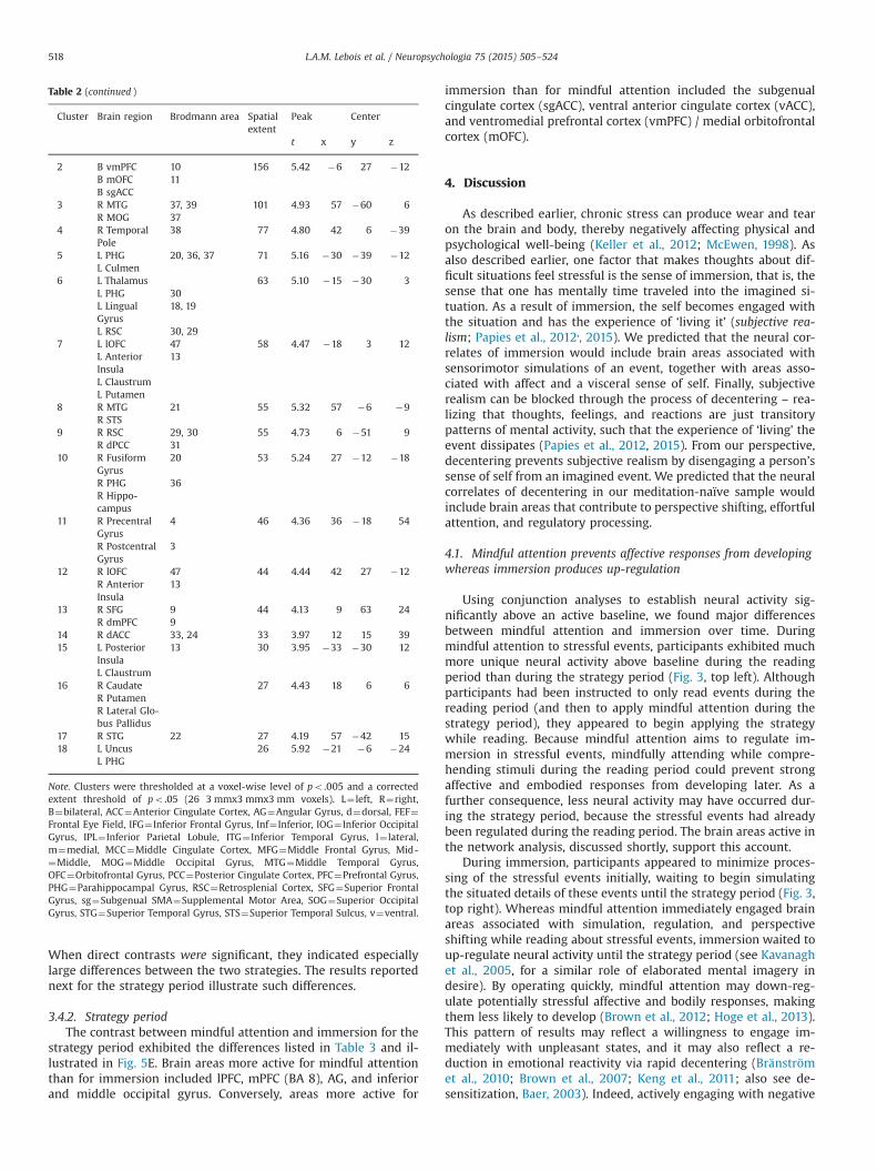
Table 2 (continued )
Cluster Brain region Brodmann area Spatialextent
Peak Center
t x y z
2 B vmPFC 10 156 5.42 �6 27 �12B mOFC 11B sgACC
3 R MTG 37, 39 101 4.93 57 �60 6R MOG 37
4 R TemporalPole
38 77 4.80 42 6 �39
5 L PHG 20, 36, 37 71 5.16 �30 �39 �12L Culmen
6 L Thalamus 63 5.10 �15 �30 3L PHG 30L LingualGyrus
18, 19
L RSC 30, 297 L lOFC 47 58 4.47 �18 3 12
L AnteriorInsula
13
L ClaustrumL Putamen
8 R MTG 21 55 5.32 57 �6 �9R STS
9 R RSC 29, 30 55 4.73 6 �51 9R dPCC 31
10 R FusiformGyrus
20 53 5.24 27 �12 �18
R PHG 36R Hippo-campus
11 R PrecentralGyrus
4 46 4.36 36 �18 54
R PostcentralGyrus
3
12 R lOFC 47 44 4.44 42 27 �12R AnteriorInsula
13
13 R SFG 9 44 4.13 9 63 24R dmPFC 9
14 R dACC 33, 24 33 3.97 12 15 3915 L Posterior
Insula13 30 3.95 �33 �30 12
L Claustrum16 R Caudate 27 4.43 18 6 6
R PutamenR Lateral Glo-bus Pallidus
17 R STG 22 27 4.19 57 �42 1518 L Uncus 26 5.92 �21 �6 �24
L PHG
Note. Clusters were thresholded at a voxel-wise level of po .005 and a correctedextent threshold of po .05 (26 3 mmx3 mmx3 mm voxels). L¼ left, R¼right,B¼bilateral, ACC¼Anterior Cingulate Cortex, AG¼Angular Gyrus, d¼dorsal, FEF¼Frontal Eye Field, IFG¼ Inferior Frontal Gyrus, Inf¼ Inferior, IOG¼ Inferior OccipitalGyrus, IPL¼ Inferior Parietal Lobule, ITG¼ Inferior Temporal Gyrus, l¼ lateral,m¼medial, MCC¼Middle Cingulate Cortex, MFG¼Middle Frontal Gyrus, Mid-¼Middle, MOG¼Middle Occipital Gyrus, MTG¼Middle Temporal Gyrus,OFC¼Orbitofrontal Gyrus, PCC¼Posterior Cingulate Cortex, PFC¼Prefrontal Gyrus,PHG¼Parahippocampal Gyrus, RSC¼Retrosplenial Cortex, SFG¼Superior FrontalGyrus, sg¼Subgenual SMA¼Supplemental Motor Area, SOG¼Superior OccipitalGyrus, STG¼Superior Temporal Gyrus, STS¼Superior Temporal Sulcus, v¼ventral.
L.A.M. Lebois et al. / Neuropsychologia 75 (2015) 505–524518
When direct contrasts were significant, they indicated especiallylarge differences between the two strategies. The results reportednext for the strategy period illustrate such differences.
3.4.2. Strategy periodThe contrast between mindful attention and immersion for the
strategy period exhibited the differences listed in Table 3 and il-lustrated in Fig. 5E. Brain areas more active for mindful attentionthan for immersion included lPFC, mPFC (BA 8), AG, and inferiorand middle occipital gyrus. Conversely, areas more active for
immersion than for mindful attention included the subgenualcingulate cortex (sgACC), ventral anterior cingulate cortex (vACC),and ventromedial prefrontal cortex (vmPFC) / medial orbitofrontalcortex (mOFC).
4. Discussion
As described earlier, chronic stress can produce wear and tearon the brain and body, thereby negatively affecting physical andpsychological well-being (Keller et al., 2012; McEwen, 1998). Asalso described earlier, one factor that makes thoughts about dif-ficult situations feel stressful is the sense of immersion, that is, thesense that one has mentally time traveled into the imagined si-tuation. As a result of immersion, the self becomes engaged withthe situation and has the experience of ‘living it’ (subjective rea-lism; Papies et al., 2012,, 2015). We predicted that the neural cor-relates of immersion would include brain areas associated withsensorimotor simulations of an event, together with areas asso-ciated with affect and a visceral sense of self. Finally, subjectiverealism can be blocked through the process of decentering – rea-lizing that thoughts, feelings, and reactions are just transitorypatterns of mental activity, such that the experience of ‘living’ theevent dissipates (Papies et al., 2012, 2015). From our perspective,decentering prevents subjective realism by disengaging a person’ssense of self from an imagined event. We predicted that the neuralcorrelates of decentering in our meditation-naïve sample wouldinclude brain areas that contribute to perspective shifting, effortfulattention, and regulatory processing.
4.1. Mindful attention prevents affective responses from developingwhereas immersion produces up-regulation
Using conjunction analyses to establish neural activity sig-nificantly above an active baseline, we found major differencesbetween mindful attention and immersion over time. Duringmindful attention to stressful events, participants exhibited muchmore unique neural activity above baseline during the readingperiod than during the strategy period (Fig. 3, top left). Althoughparticipants had been instructed to only read events during thereading period (and then to apply mindful attention during thestrategy period), they appeared to begin applying the strategywhile reading. Because mindful attention aims to regulate im-mersion in stressful events, mindfully attending while compre-hending stimuli during the reading period could prevent strongaffective and embodied responses from developing later. As afurther consequence, less neural activity may have occurred dur-ing the strategy period, because the stressful events had alreadybeen regulated during the reading period. The brain areas active inthe network analysis, discussed shortly, support this account.
During immersion, participants appeared to minimize proces-sing of the stressful events initially, waiting to begin simulatingthe situated details of these events until the strategy period (Fig. 3,top right). Whereas mindful attention immediately engaged brainareas associated with simulation, regulation, and perspectiveshifting while reading about stressful events, immersion waited toup-regulate neural activity until the strategy period (see Kavanaghet al., 2005, for a similar role of elaborated mental imagery indesire). By operating quickly, mindful attention may down-reg-ulate potentially stressful affective and bodily responses, makingthem less likely to develop (Brown et al., 2012; Hoge et al., 2013).This pattern of results may reflect a willingness to engage im-mediately with unpleasant states, and it may also reflect a re-duction in emotional reactivity via rapid decentering (Bränströmet al., 2010; Brown et al., 2007; Keng et al., 2011; also see de-sensitization, Baer, 2003). Indeed, actively engaging with negative

L.A.M. Lebois et al. / Neuropsychologia 75 (2015) 505–524 519
experience has the potential to reduce experiential avoidance, akey goal in DBT and ACT (Hayes et al., 2006; Keng et al., 2011).
Interestingly, mindful attention exhibited different distribu-tions of neural activity for stressful vs. non-stressful events (Fig. 3,left). During the reading period, more neural activity occurredabove baseline for stressful events, suggesting that they affordsalient affective and bodily responses that mindful attention canregulate (Papies et al., 2015). Conversely, non-stressful eventsproduced greater neural activity during the strategy period, sug-gesting that greater effort was required to generate appropriatethoughts relevant for applying mindful attention. Similarly, forimmersion, participants again appeared to delay immersingthemselves in the non-stressful events until the reading period, asthey had done for the stressful events.
Analyses using Yeo et al.'s (2011) seven resting state networkscorroborated the results for the conjunction analyses just de-scribed. For mindful attention to stressful events, the distributionof unique clusters across Yeo et al.'s networks changed sig-nificantly from the reading period to the strategy period (Fig. 4,top left). Initially during the reading period, large amounts ofunique activity occurred in somatosensorimotor, visual, and limbicnetworks, with some activity in the dorsal attention network,suggesting that participants were simulating the scenarios (Da-masio, 1999; Ganis et al., 2004), and attempting to regulate re-sponses to them by shifting attention (Corbetta and Shulman,2002; Froeliger et al., 2012). During the strategy period, theseactivations decreased, suggesting that participants were no longersimulating the scenarios and emotional reactions to them as vi-vidly, given that they had been down-regulated during the readingperiod (consistent with the results in Fig. 3, top left).
Mindful attention to stressful events also produced largeamounts of default mode network (DMN) activity during both thereading and strategy periods. Other mindfulness research onlydemonstrates decreased activation in DMN hubs (e.g., mPFC, PCC)for expert meditators, or sometimes for novices at lower thresh-olds (Farb et al., 2007). Because the DMN is implicated in internalgoal-directed activity (e.g., Spreng et al., 2010), it may be highlyengaged when first learning mindfulness practices.
For immersion in stressful events, the reading and strategyperiods also exhibited large differences in the distributions ofunique neural activity across networks (Fig. 4, top right). Relativeto the reading period, large increases occurred during the strategyperiod in the somatosensorimotor, limbic, default mode, andventral attention networks. As suggested earlier, participants mayhave waited until the strategy period to immerse themselves inthe stressful events, simulating both the external situations andtheir internal reactions to them, especially personal salience.
For mindful attention to non-stressful events, the distribution ofunique neural activity differed across the reading and strategyperiods (Fig. 4, bottom left). During the strategy period, visual ac-tivity decreased while somatosensorimotor activity increased,suggesting that participants increasingly imagined acting in thenon-stressful events. Increased activity in the DMN, the frontopar-ietal control network, and both attention networks during thestrategy period further suggests that self-referential (Buckner et al.,2008) and effortful goal oriented processing (Spreng et al., 2013)increased as well. Because the non-stressful events did not readilyafford emotional and bodily reactions, participants may haveworked harder to produce thoughts relevant for mindful attention.
For immersion in non-stressful events, the distribution of un-ique neural activity again differed across the reading and strategyperiods (Fig. 4, bottom right). Similar to mindful attention, so-matosensorimotor activity increased, suggesting increased actionengagement in the non-stressful situations. Similar to immersionin stressful events, activity in the ventral attention network in-creased, suggesting that effortful processing related to personal
salience increased, perhaps working to generate affective andbodily responses. Unlike mindful attention, DMN activity de-creased, perhaps reflecting a greater focus on the physical situa-tion for immersion than on mental states for mindful attention(Buckner et al., 2008).
4.2. Mindful attention promotes perspective shifting and regulatoryactivity whereas immersion engages a visceral sense of self
Direct contrasts between mindful attention and immersionfound, first, that both strategies activated many similar areas, andsecond, that a small subset of areas were significantly more activefor one strategy vs. the other (Table 3 and Fig. 5E). Specifically, thebrain areas more active for mindful attention than for immersionare associated with perspective shifting (AG; Seghier, 2013), ex-ecutive and attentional control (lPFC; Spreng et al., 2013), aug-mented inhibitory control (mPFC, BA 8; Tang et al., 2012), andvisual processing (inferior and middle occipital gyrus). Lack ofexpertise with mindful attention may have required greater shiftsin perspective than did the more natural and familiar process ofimmersion. To implement this newly-learned mode of perspectiveshifting, participants may have needed to exert greater effortduring mindful attention than during immersion, thereby enga-ging executive and regulatory areas. Higher visual activity mayhave reflected increased attention on imagined situations.
Conversely, areas more active for immersion than for mindfulattention included the subgenual cingulate cortex (sgACC), ventralanterior cingulate cortex (vACC), and ventromedial prefrontalcortex (vmPFC)/medial orbitofrontal cortex (mOFC). As establishedelsewhere, these areas are often involved when integrating visc-eral states (Vogt, 2005), monitoring and processing reward (Elliotet al., 2000), attending to feelings (Kross et al., 2009), and labelingstimuli as self-relevant (Northoff and Bermpohl, 2004). Thus, im-mersion appeared to engage stronger self, bodily, and affectiveresponses than did mindful attention, consistent with engagingoneself in events physically, becoming immersed in them, andexperiencing them as subjectively real.
Overall, the results for these contrasts are consistent with thefollowing conclusion: During the strategy period, mindful atten-tion caused a shift in perspective that disengaged an embodiedsense of self from simulated events (decentering). On the onehand, activations in AG, lPFC, and mPFC suggest that mindful at-tention shifted perspective through the use of regulatoryprocesses. On the other hand, decreased activations in sgACC,vACC, vmPFC, and mOFC suggest that an embodied sense of selfwas less active for imagined events during mindful attention thanduring immersion. As a consequence, imagined events were ex-perienced as transitory mental states in the current moment.An important goal for future research is to examine this patternof neural activity further, establishing whether a causal relationexists between perspective shifting and reductions in self-engagement.
4.3. Relations to previous neuroimaging findings
As mentioned above, relative to immersion, mindful attentionexhibited significantly less neural activity in sgACC, vmPFC, andmOFC. As previous research shows, these areas are associated withintegrating visceral, autonomic, and affective states, representingthe reward value of stimuli, and establishing self-relevance (e.g.,Ressler and Mayberg, 2007; Kross et al., 2009; Greicius et al., 2007;Northoff and Bermpohl, 2004). Together, these areas may con-tribute to the experience of subjective realism. Several recentstudies have similarly found that mindfulness is associated withlow activity in these areas (e.g., Farb et al., 2007; Kross et al., 2009;Westbrook et al., 2013). Thus, our findings suggest that mindful

Fig. 5. Panels A–D illustrate unique activations from the conjunction analyses across mindful attention and immersion reported in Tables 1 and 2 and Figs. 4 and 5 (A.Mindful Attention Stressful; B. Immersion Stressful; C. Mindful Attention Non-stressful; D. Immersion Non-stressful). Panel E illustrates activations from the linear contrastbetween mindful attention and immersion during the strategy period, collapsed across stressful and non-stressful events.
L.A.M. Lebois et al. / Neuropsychologia 75 (2015) 505–524520
attention, too, is associated with lower activity in these areas,consistent with our conclusion that decentering results from dis-engaging the self from imagined situations.
Conversely, mindful attention produced higher activations thanimmersion in brain areas associated with executive processing(lPFC) and inhibitory control (mPFC, BA 8). Activity in these re-gions is increasingly recognized as a hallmark of mindfulness inless experienced meditators, requiring more effort than for experts(e.g., Creswell et al., 2007; Farb et al., 2007, 2010; Tang et al., 2012).Likewise, several activations for mindful attention overlapped withthe frontoparietal control network (e.g., lPFC, dmPFC, AG) and thedorsal attention network (e.g., superior frontal, frontal eye fields,middle temporal, occipital cortex). Whereas the frontoparietal
control network facilitates goal-directed cognition through ex-ecutive control, the dorsal attention network controls externallydirected attention (Spreng et al., 2013).
Greater activity for mindful attention also occurred in parietalcortex (AG, inferior parietal lobule IPL). Similar areas are asso-ciated with high trait mindfulness (Dickenson et al., 2013), andwith increased gray matter following MBSR (Hölzel et al., 2011).AG activity is also associated with transferring attention to re-levant targets (Gottlieb, 2007; Seghier, 2013), and with shiftingbetween first and third person bodily perspectives (Blanke et al.,2005). Interestingly, our AG coordinates are closest to those foractivity associated with external agency attribution (Seghier, 2013;Sperduti et al., 2011), again suggesting that decentering may
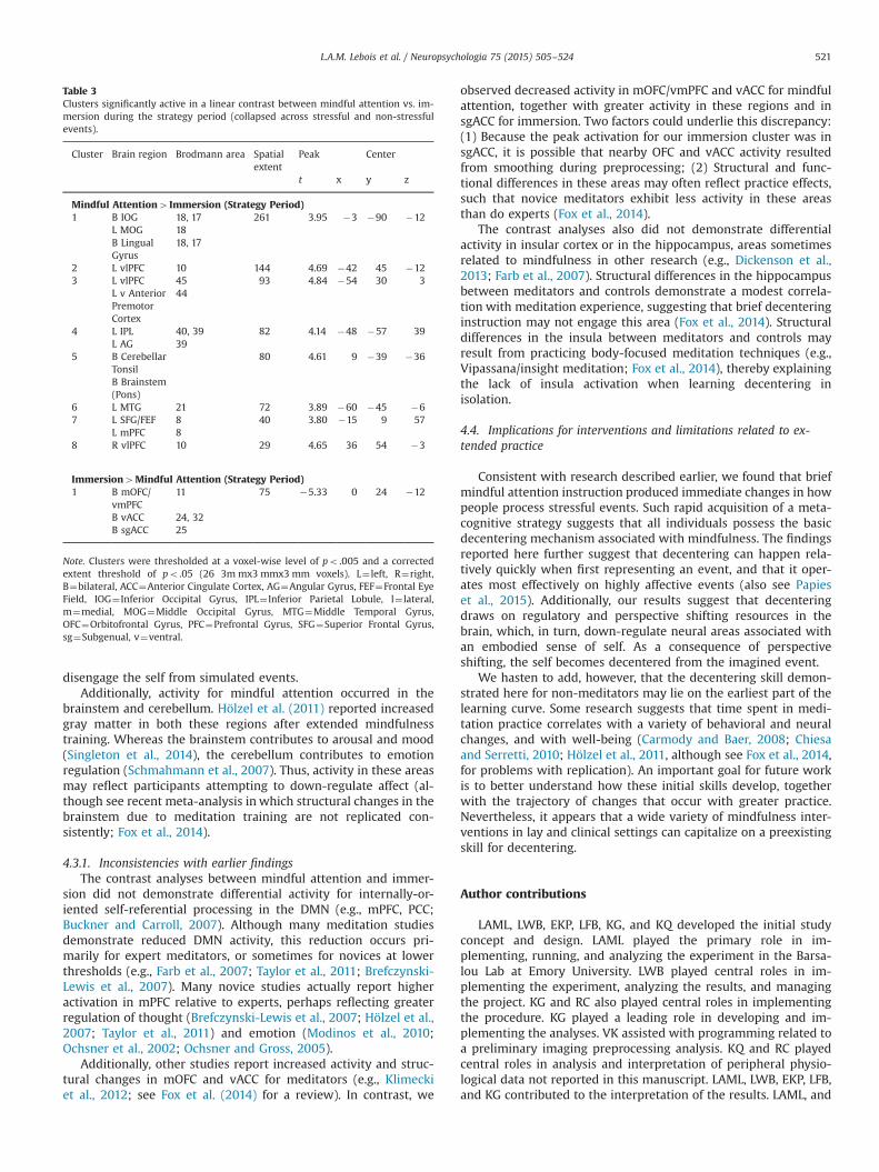
Table 3Clusters significantly active in a linear contrast between mindful attention vs. im-mersion during the strategy period (collapsed across stressful and non-stressfulevents).
Cluster Brain region Brodmann area Spatialextent
Peak Center
t x y z
Mindful Attention4Immersion (Strategy Period)1 B IOG 18, 17 261 3.95 �3 �90 �12
L MOG 18B LingualGyrus
18, 17
2 L vlPFC 10 144 4.69 �42 45 �123 L vlPFC 45 93 4.84 �54 30 3
L v AnteriorPremotorCortex
44
4 L IPL 40, 39 82 4.14 �48 �57 39L AG 39
5 B CerebellarTonsil
80 4.61 9 �39 �36
B Brainstem(Pons)
6 L MTG 21 72 3.89 �60 �45 �67 L SFG/FEF 8 40 3.80 �15 9 57
L mPFC 88 R vlPFC 10 29 4.65 36 54 �3
Immersion4Mindful Attention (Strategy Period)1 B mOFC/
vmPFC11 75 �5.33 0 24 �12
B vACC 24, 32B sgACC 25
Note. Clusters were thresholded at a voxel-wise level of po .005 and a correctedextent threshold of po .05 (26 3m mx3 mmx3 mm voxels). L¼ left, R¼right,B¼bilateral, ACC¼Anterior Cingulate Cortex, AG¼Angular Gyrus, FEF¼Frontal EyeField, IOG¼ Inferior Occipital Gyrus, IPL¼ Inferior Parietal Lobule, l¼ lateral,m¼medial, MOG¼Middle Occipital Gyrus, MTG¼Middle Temporal Gyrus,OFC¼Orbitofrontal Gyrus, PFC¼Prefrontal Gyrus, SFG¼Superior Frontal Gyrus,sg¼Subgenual, v¼ventral.
L.A.M. Lebois et al. / Neuropsychologia 75 (2015) 505–524 521
disengage the self from simulated events.Additionally, activity for mindful attention occurred in the
brainstem and cerebellum. Hölzel et al. (2011) reported increasedgray matter in both these regions after extended mindfulnesstraining. Whereas the brainstem contributes to arousal and mood(Singleton et al., 2014), the cerebellum contributes to emotionregulation (Schmahmann et al., 2007). Thus, activity in these areasmay reflect participants attempting to down-regulate affect (al-though see recent meta-analysis in which structural changes in thebrainstem due to meditation training are not replicated con-sistently; Fox et al., 2014).
4.3.1. Inconsistencies with earlier findingsThe contrast analyses between mindful attention and immer-
sion did not demonstrate differential activity for internally-or-iented self-referential processing in the DMN (e.g., mPFC, PCC;Buckner and Carroll, 2007). Although many meditation studiesdemonstrate reduced DMN activity, this reduction occurs pri-marily for expert meditators, or sometimes for novices at lowerthresholds (e.g., Farb et al., 2007; Taylor et al., 2011; Brefczynski-Lewis et al., 2007). Many novice studies actually report higheractivation in mPFC relative to experts, perhaps reflecting greaterregulation of thought (Brefczynski-Lewis et al., 2007; Hölzel et al.,2007; Taylor et al., 2011) and emotion (Modinos et al., 2010;Ochsner et al., 2002; Ochsner and Gross, 2005).
Additionally, other studies report increased activity and struc-tural changes in mOFC and vACC for meditators (e.g., Klimeckiet al., 2012; see Fox et al. (2014) for a review). In contrast, we
observed decreased activity in mOFC/vmPFC and vACC for mindfulattention, together with greater activity in these regions and insgACC for immersion. Two factors could underlie this discrepancy:(1) Because the peak activation for our immersion cluster was insgACC, it is possible that nearby OFC and vACC activity resultedfrom smoothing during preprocessing; (2) Structural and func-tional differences in these areas may often reflect practice effects,such that novice meditators exhibit less activity in these areasthan do experts (Fox et al., 2014).
The contrast analyses also did not demonstrate differentialactivity in insular cortex or in the hippocampus, areas sometimesrelated to mindfulness in other research (e.g., Dickenson et al.,2013; Farb et al., 2007). Structural differences in the hippocampusbetween meditators and controls demonstrate a modest correla-tion with meditation experience, suggesting that brief decenteringinstruction may not engage this area (Fox et al., 2014). Structuraldifferences in the insula between meditators and controls mayresult from practicing body-focused meditation techniques (e.g.,Vipassana/insight meditation; Fox et al., 2014), thereby explainingthe lack of insula activation when learning decentering inisolation.
4.4. Implications for interventions and limitations related to ex-tended practice
Consistent with research described earlier, we found that briefmindful attention instruction produced immediate changes in howpeople process stressful events. Such rapid acquisition of a meta-cognitive strategy suggests that all individuals possess the basicdecentering mechanism associated with mindfulness. The findingsreported here further suggest that decentering can happen rela-tively quickly when first representing an event, and that it oper-ates most effectively on highly affective events (also see Papieset al., 2015). Additionally, our results suggest that decenteringdraws on regulatory and perspective shifting resources in thebrain, which, in turn, down-regulate neural areas associated withan embodied sense of self. As a consequence of perspectiveshifting, the self becomes decentered from the imagined event.
We hasten to add, however, that the decentering skill demon-strated here for non-meditators may lie on the earliest part of thelearning curve. Some research suggests that time spent in medi-tation practice correlates with a variety of behavioral and neuralchanges, and with well-being (Carmody and Baer, 2008; Chiesaand Serretti, 2010; Hölzel et al., 2011, although see Fox et al., 2014,for problems with replication). An important goal for future workis to better understand how these initial skills develop, togetherwith the trajectory of changes that occur with greater practice.Nevertheless, it appears that a wide variety of mindfulness inter-ventions in lay and clinical settings can capitalize on a preexistingskill for decentering.
Author contributions
LAML, LWB, EKP, LFB, KG, and KQ developed the initial studyconcept and design. LAML played the primary role in im-plementing, running, and analyzing the experiment in the Barsa-lou Lab at Emory University. LWB played central roles in im-plementing the experiment, analyzing the results, and managingthe project. KG and RC also played central roles in implementingthe procedure. KG played a leading role in developing and im-plementing the analyses. VK assisted with programming related toa preliminary imaging preprocessing analysis. KQ and RC playedcentral roles in analysis and interpretation of peripheral physio-logical data not reported in this manuscript. LAML, LWB, EKP, LFB,and KG contributed to the interpretation of the results. LAML, and

L.A.M. Lebois et al. / Neuropsychologia 75 (2015) 505–524522
LWB drafted the manuscript, and all authors contributed to re-vising it. LWB and LFB are joint senior authors. All authors ap-proved the final version for submission.
Declaration of conflicting interests
The authors declared that they had no conflicts of interest withrespect to their authorship or the publication of this article.
Funding
This work was supported by the National Center for Com-plementary and Integrative Health, United States at the NationalInstitute of Health with a predoctoral Ruth L. Kirschstein NationalResearch Service Award to LAML at Emory University (Grantnumbers 1 F31 AT007130-01, 5 F31 AT007130-02), and by a Na-tional Institute of Health Director's Pioneer Award to LFB atNortheastern University (Grant number DPI OD003312), with asub-contract to LWB at Emory University.
Acknowledgments
We are grateful to David Almeida for sharing his stressor da-tabase, Taryn Colton, Hailey Friedman, and Bridget Warren forassistance with sampling the stressful life events, Michael Larchefor assistance with MRI set up, Wendy Hasenkamp for assistancewith an earlier design of this experiment, and Robyn Fivush, JimRilling, Kim Wallen, and Christine Wilson-Mendenhall for com-ments on an earlier version of this manuscript.
Appendix A. Supplementary Materials
Supplementary materials associated with this article can befound in the online version at http://dx.doi.org/10.1016/j.neuropsychologia.2015.05.030.
References
Alberts, H.J., Thewissen, R., 2011. The effect of a brief mindfulness intervention onmemory for positively and negatively valenced stimuli. Mindfulness 2, 73–77.
Almeida, D., 2005. Resilience and vulnerability to daily stressors assessed via diarymethods. Curr. Dir. Psychol. Sci. 14 (2), 64.
Almeida, D., Wethington, E., Kessler, R., 2002. The daily inventory of stressfulevents: an interview-based approach for measuring daily stressors. Assessment9 (1), 41.
Andersson, J., Jenkinson, M., Smith, S., 2010. Non-linear Optimisation. Non-linearRegistration, Aka Spatial Normalisation. FMRIB Technical Report TR07JA2.Available at: ⟨http://www.fmrib.ox.ac.uk/analysis/techrep⟩.
Arch, J.J., Craske, M.G., 2006. Mechanisms of mindfulness: emotion regulation fol-lowing a focused breathing induction. Behav. Res. Ther. 44, 1849–1858.
Baer, R.A., 2003. Mindfulness training as a clinical intervention: a conceptual andempirical review. Clin. Psychol.: Sci. Pract. 10, 125–143.
Barrett, L.F., Mesquita, B., Ochsner, K.N., Gross, J.J., 2007. The experience of emotion.Annu. Rev. Psychol. 58, 373–403.
Bishop, S.R., Lau, M., Shapiro, S., Carlson, L., Anderson, N.D., Carmody, J., Devins, G.,et al., 2004. Mindfulness: a proposed operational definition. Clin. Psychol.: Sci.Pract. 11, 230–241.
Black, P.H., Garbutt, L.D., 2002. Stress, inflammation and cardiovascular disease. J.Psychosom. Res. 52 (1), 1–23.
Blanke, O., Mohr, C., Michel, C.M., Pascual-Leone, A., Brugger, P., Seeck, M., Thut, G.,et al., 2005. Linking out-of-body experience and self processing to mental own-body imagery at the temporoparietal junction. J. Neurosci. 25, 550–557.
Bränström, R., Kvillemo, P., Brandberg, Y., Moskowitz, J.T., 2010. Self-report mind-fulness as a mediator of psychological well-being in a stress reduction inter-vention for cancer patients—a randomized study. Ann. Behav. Med. 39,151–161.
Brefczynski-Lewis, J.A., Lutz, A., Schaefer, H.S., Levinson, D.B., Davidson, R.J., 2007.
Neural correlates of attentional expertise in long-term meditation practitioners.Proc. Natl. Acad. Sci. 104, 11483–11488.
Broderick, P.C., 2005. Mindfulness and coping with dysphoric mood: contrasts withrumination and distraction. Cogn. Ther. Res. 29, 501–510.
Brosschot, J.F., 2010. Markers of chronic stress: prolonged physiological activationand (un) conscious perseverative cognition. Neurosci. Biobehav. Rev. 35 (1),46–50.
Brosschot, J.F., Gerin, W., Thayer, J.F., 2006. The perseverative cognition hypothesis:a review of worry, prolonged stress-related physiological activation, and health.J. Psychosom. Res. 60, 113–124.
Brosschot, J.F., Pieper, S., Thayer, J.F., 2005. Expanding stress theory: prolongedactivation and perseverative cognition. Psychoneuroendocrinology 30 (10),1043–1049.
Brown, K.W., Ryan, R.M., 2003. The benefits of being present: mindfulness and itsrole in psychological well-being. J. Personal. Soc. Psychol. 84, 822.
Brown, K.W., Ryan, R.M., Creswell, J.D., 2007. Mindfulness: Theoretical foundationsand evidence for its salutary effects. Psychol. Inquiry 18, 211–237.
Brown, K.W., Weinstein, N., Creswell, J.D., 2012. Trait mindfulness modulates neu-roendocrine and affective responses to social evaluative threat. Psychoneur-oendocrinology. 37, 2037–2041.
Buckner, R.L., Andrews‐Hanna, J.R., Schacter, D.L., 2008. The brain's default net-work. Ann. New York Acad. Sci. 1124, 1–38.
Buckner, R.L., Carroll, D.C., 2007. Self-projection and the brain. Trends Cogn. Sci. 11,49–57.
Carmody, J., Baer, R.A., 2008. Relationships between mindfulness practice and le-vels of mindfulness, medical and psychological symptoms and well-being in amindfulness-based stress reduction program. J. Behav. Med. 31, 23–33.
Chida, Y., Hamer, M., 2008. Chronic psychosocial factors and acute physiologicalresponses to laboratory-induced stress in healthy populations: a quantitativereview of 30 years of investigations. Psychol. Bull. 134 (6), 829–885.
Chiesa, A., Serretti, A., 2010. A systematic review of neurobiological and clinicalfeatures of mindfulness meditations. Psychol. Med. 40, 1239.
Corbetta, M., Shulman, G.L., 2002. Control of goal-directed and stimulus-drivenattention in the brain. Nat. Rev. Neurosci. 3, 201–215.
Cox, R.W., 1996. AFNI: software for analysis and visualization of functional mag-netic resonance neuroimages. Comput. Biomed. Res. 29, 162–173.
Creswell, J.D., Way, B.M., Eisenberger, N.I., Lieberman, M.D., 2007. Neural correlatesof dispositional mindfulness during affect labeling. Psychosomatic Medicine 69,560–565.
Damasio, A.R., 1999. The Feeling of What Happens. Harcourt, New York.Dedovic, K., D’Aguiar, C., Pruessner, J., 2009. What stress does to your brain: a re-
view of neuroimaging studies. Can. J. Psychiatry (Revue canadienne de psy-chiatrie) 54 (1), 6.
Dedovic, K., Duchesne, A., Andrews, J., Engert, V., Pruessner, J.C., 2009. The brainand the stress axis: the neural correlates of cortisol regulation in response tostress. Neuroimage 47 (3), 864–871.
Dedovic, K., Rexroth, M., Wolff, E., Duchesne, A., Scherling, C., Beaudry, T., Pruessner,J.C., et al., 2009. Neural correlates of processing stressful information: an event-related fMRI study. Brain Res. 1293, 49–60.
Delizonna, L.L., Williams, R.P., Langer, E.J., 2009. The effect of mindfulness on heartrate control. J. Adult Dev. 16, 61–65.
Dickenson, J., Berkman, E.T., Arch, J., Lieberman, M.D., 2013. Neural correlates offocused attention during a brief mindfulness induction. Soc. Cogn. Affect.Neurosci. 8, 40–47.
Dickerson, S.S., Gruenewald, T.L., Kemeny, M.E., 2004. When the social self isthreatened: shame, physiology, and health. J. Personal. 72 (6), 1191–1216.
Dickerson, S.S., Kemeny, M.E., 2004. Acute stressors and cortisol responses: a the-oretical integration and synthesis of laboratory research. Psychol. Bull. 130,355–391.
Ditto, B., Eclache, M., Goldman, N., 2006. Short-term autonomic and cardiovasculareffects of mindfulness body scan meditation. Ann. Behav. Med. 32, 227–234.
Elliott, R., Dolan, R.J., Frith, C.D., 2000. Dissociable functions in the medial andlateral orbitofrontal cortex: evidence from human neuroimaging studies. Cer-ebral cortex 10 (3), 308–317.
Erisman, S.M., Roemer, L., 2010. A preliminary investigation of the effects of ex-perimentally induced mindfulness on emotional responding to film clips.Emotion 10, 72–82.
Farb, N.A., Segal, Z.V., Mayberg, H., Bean, J., McKeon, D., Fatima, Z., Anderson, A.K.,2007. Attending to the present: mindfulness meditation reveals distinct neuralmodes of self-reference. Soc. Cogn. Affect. Neurosci. 2, 313–322.
Farb, N.A., Anderson, A.K., Mayberg, H., Bean, J., McKeon, D., Segal, Z.V., 2010.Minding one's emotions: mindfulness training alters the neural expression ofsadness. Emotion 10, 25–33.
Feinberg, D.A., Moeller, S., Smith, S.M., Auerbach, E., Ramanna, S., Glasser, M.F.,Yacoub, E., et al., 2010. Multiplexed echo planar imaging for sub-second wholebrain FMRI and fast diffusion imaging. PLoS One 5, e15710.
Fox, K.C., Nijeboer, S., Dixon, M.L., Floman, J.L., Ellamil, M., Rumak, S.P., Christoff, K.,2014. Is meditation associated with altered brain structure? A systematic re-view and meta-analysis of morphometric neuroimaging in meditation practi-tioners. Neurosci. Biobehav. Rev. 43, 48–73.
Froeliger, B., Garland, E.L., Kozink, R.V., Modlin, L.A., Chen, N.K., McClernon, F.J.,Sobin, P., 2012. Meditation-state functional connectivity (msFC): strengtheningof the dorsal attention network and beyond. Evid. Based Complement. Alternat.Med. 2012, 1–9.
Ganis, G., Thompson, W.L., Kosslyn, S.M., 2004. Brain areas underlying visual mentalimagery and visual perception: an fMRI study. Cogn. Brain Res. 20, 226–241.

L.A.M. Lebois et al. / Neuropsychologia 75 (2015) 505–524 523
Ganzel, B.L., Morris, P.A., Wethington, E., 2010. Allostasis and the human brain:Integrating models of stress from the social and life sciences. Psychol. Rev. 117(1), 134–174.
Gard, T., Hölzel, B.K., Lazar, S.W., 2014. The potential effects of meditation on age‐related cognitive decline: a systematic review. Ann. New York Acad. Sci. 1307,89–103.
Gottlieb, J., 2007. From thought to action: the parietal cortex as a bridge betweenperception, action, and cognition. Neuron 53, 9–16.
Greenberg, N., Carr, J., Summers, C., 2002. Causes and consequences of stress. Integr.Comp. Biol. 42 (3), 508.
Greicius, M.D., Flores, B.H., Menon, V., Glover, G.H., Solvason, H.B., Kenna, H.,Schatzberg, A.F., et al., 2007. Resting-state functional connectivity in majordepression: abnormally increased contributions from subgenual cingulatecortex and thalamus. Biol. Psychiatry 62, 429–437.
Grossman, P., Niemann, L., Schmidt, S., Walach, H., 2004. Mindfulness-based stressreduction and health benefits: a meta-analysis. J. Psychosom. Res. 57, 35–43.
Hänsel, A., Hong, S., Cámara, R.J., Von Kaenel, R., 2010. Inflammation as a psycho-physiological biomarker in chronic psychosocial stress. Neurosci. Biobehav. Rev.35 (1), 115–121.
Hayes, S.C., 2004. Acceptance and commitment therapy, relational frame theory,and the third wave of behavioral and cognitive therapies. Behav. Ther. 35,639–665.
Hayes, S.C., Luoma, J.B., Bond, F.W., Masuda, A., Lillis, J., 2006. Acceptance andcommitment therapy: Model, processes and outcomes. Behav. Res. Ther. 44,1–25.
Higgins, E.T., 1989. Self-discrepancy theory: what patterns of self-beliefs causepeople to suffer. Adv. Exp. Soc. Psychol. 22, 93–136.
Hofmann, S.G., Sawyer, A.T., Witt, A.A., Oh, D., 2010. The effect of mindfulness-basedtherapy on anxiety and depression: a meta-analytic review. J. Consult. Clin.Psychol. 78, 169–183.
Hoge, E.A., Bui, E., Marques, L., Metcalf, C.A., Morris, L.K., Robinaugh, D.J., Simon, N.M., 2013. Randomized controlled trial of mindfulness meditation for general-ized anxiety disorder: effects on anxiety and stress reactivity. J. Clin. Psychiatry74, 786–792.
Hölzel, B.K., Carmody, J., Evans, K.C., Hoge, E.A., Dusek, J.A., Morgan, L., Lazar, S.W.,2009. Stress reduction correlates with structural changes in the amygdala. Soc.Cogn. Affect. Neurosci. 5, 11–17. http://dx.doi.org/10.1093/scan/nsp034.
Hölzel, B.K., Ott, U., Hempel, H., Hackl, A., Wolf, K., Stark, R., Vaitl, D., 2007. Dif-ferential engagement of anterior cingulate and adjacent medial frontal cortexin adept meditators and non-meditators. Neurosci. Lett. 421, 16–21.
Hölzel, B.K., Carmody, J., Vangel, M., Congleton, C., Yerramsetti, S.M., Gard, T., Lazar,S.W., 2011. Mindfulness practice leads to increases in regional brain gray matterdensity. Psychiatry Res.: Neuroimaging 191, 36–43.
Hölzel, B.K., Lazar, S.W., Gard, T., Schuman-Olivier, Z., Vago, D.R., Ott, U., 2011. Howdoes mindfulness meditation work? Proposing mechanisms of action from aconceptual and neural perspective. Perspect. Psychol. Sci. 6, 537–559.
Jenkinson, M., Smith, S., 2001. A global optimisation method for robust affine re-gistration of brain images. Med. Image Anal. 5, 143–156.
Jenkinson, M., Bannister, P., Brady, M., Smith, S., 2002. Improved optimization forthe robust and accurate linear registration and motion correction of brainimages. Neuroimage 17, 825–841.
Juster, R.P., McEwen, B.S., Lupien, S.J., 2010. Allostatic load biomarkers of chronicstress and impact on health and cognition. Neurosci. Biobehav. Rev. 35 (1),2–16.
Kabat-Zinn, J., 1990. Full Catastrophe Living: Using the Wisdom of Your Mind toFace Stress, Pain and Illness. Dell, New York.
Kabat‐Zinn, J., 2003. Mindfulness‐based interventions in context past, present, andfuture. Clin. Psychol.: Sci. Pract. 10, 144–156.
Kavanagh, D.J., Andrade, J., May, J., 2005. Imaginary relish and exquisite torture: theelaborated intrusion theory of desire. Psychol. Rev. 112, 446–467.
Keller, A., Litzelman, K., Wisk, L.E., Maddox, T., Cheng, E.R., Creswell, P.D., Witt, W.P.,2012. Does the perception that stress affects health matter? The associationwith health and mortality. Health Psychol. 31, 677–684.
Keng, S.L., Smoski, M.J., Robins, C.J., 2011. Effects of mindfulness on psychologicalhealth: a review of empirical studies. Clin. Psychol. Rev. 31, 1041–1056.
Klimecki, O.M., Leiberg, S., Lamm, C., Singer, T., 2012. Functional neural plasticityand associated changes in positive affect after compassion training. Cereb.Cortex 23, 1552–1561. http://dx.doi.org/10.1093/cercor/bhs142.
Kross, E., Ayduk, O., 2008. Facilitating adaptive emotional analysis: Distinguishingdistanced-analysis of depressive experiences from immersed-analysis anddistraction. Personal. Soc. Psychol. Bull. 34 (7), 924.
Kross, E., Davidson, M., Weber, J., Ochsner, K., 2009. Coping with emotions past: theneural bases of regulating affect associated with negative autobiographicalmemories. Biol. Psychiatry 65, 361–366.
Lazarus, R., 1993. From psychological stress to the emotions: a history of changingoutlooks. Annu. Rev. Psychol. 44 (1), 1–22.
Lazarus, R., 1999. Stress and Emotion: A New Synthesis. Springer, New York.Lebois, L.A.M., Hertzog, C., Slavich, G.M., Barrett, L.F., Barsalou L.W., 2015. Estab-
lishing the Situated Features Associated with Perceived Stressfulness (Sub-mitted Manuscript Under Review).
Lutz, J., Herwig, U., Opialla, S., Hittmeyer, A., Jäncke, L., Rufer, M., Brühl, A.B., et al.,2014. Mindfulness and emotion regulation—an fMRI study. Soc. Cogn. Affect.Neurosci. 9, 776–785.
Lutz, A., Slagter, H.A., Dunne, J.D., Davidson, R.J., 2008. Attention regulation andmonitoring in meditation. Trends Cogn. Sci. 12, 163–169.
Mason, M.F., Norton, M.I., Van Horn, J.D., Wegner, D.M., Grafton, S.T., Macrae, C.N.,
2007. Wandering minds: the default network and stimulus-independentthought. Science 315, 393–395.
McEwen, B.S., 1998. Stress, adaptation, and disease: allostasis and allostatic load.Ann. New York Acad. Sci. 840, 33–44.
Modinos, G., Ormel, J., Aleman, A., 2010. Individual differences in dispositionalmindfulness and brain activity involved in reappraisal of emotion. Soc. Cogn.Affect. Neurosci.. 5, 369–377.
Moeller, S., Yacoub, E., Olman, C.A., Auerbach, E., Strupp, J., Harel, N., Uğurbil, K.,2010. Multiband multislice GE‐EPI at 7 T, with 16‐fold acceleration using partialparallel imaging with application to high spatial and temporal whole‐brainfMRI. Magn. Reson. Med. 63, 1144–1153.
Mumford, J.A., Nichols, T.E., 2008. Power calculation for group fMRI studies ac-counting for arbitrary design and temporal autocorrelation. NeuroImage 39,261–268.
Nyima, C., 2004. Present Fresh Wakefulness: a Meditation Manual on Non-conceptual Wisdom. Rangjung Yeshe Publications.
Northoff, G., Bermpohl, F., 2004. Cortical midline structures and the self. TrendsCogn. Sci. 8, 102–107.
Ochsner, K.N., Gross, J.J., 2005. The cognitive control of emotion. Trends Cogn. Sci. 9,242–249.
Ochsner, K.N., Bunge, S.A., Gross, J.J., Gabrieli, J.D., 2002. Rethinking feelings: anfMRI study of the cognitive regulation of emotion. J. Cogn. Neurosci. 14,1215–1229.
Ollinger, J.M., Corbetta, M., Shulman, G.L., 2001. Separating processes within a trialin event-related functional MRI: II. Analysis. Neuroimage 13, 218–229.
Ollinger, J.M., Shulman, G.L., Corbetta, M., 2001. Separating processes within a trialin event-related functional MRI: I. The method. Neuroimage 13, 210–217.
Papies, E.K., Barsalou, L.W., Custers, R., 2012. Mindful attention prevents mindlessimpulses. Soc. Psychol. Personal. Sci. 3, 291–299.
Papies, E.K., Pronk, T.M., Keesman, M., Barsalou, L.W., 2015. The benefits of simplyobserving: mindful attention modulates the link between motivation and be-havior. J. Personal. Soc. Psychol. 108, 148–170.
Ressler, K.J., Mayberg, H.S., 2007. Targeting abnormal neural circuits in mood andanxiety disorders: from the laboratory to the clinic. Nat. Neurosci. 10,1116–1124.
Rodrigues, S.M., LeDoux, J.E., Sapolsky, R.M., 2009. The influence of stress hormoneson fear circuitry. Annu. Rev. Neurosci. 32, 289–313.
Rozanski, A., Blumenthal, J.A., Davidson, K.W., Saab, P.G., Kubzansky, L., 2005. Theepidemiology, pathophysiology, and management of psychosocial risk factorsin cardiac practice: the emerging field of behavioral cardiology. J. Am. Coll.Cardiol. 45 (5), 637–651.
Rozanski, A., Blumenthal, J.A., Kaplan, J., 1999. Impact of psychological factors onthe pathogenesis of cardiovascular disease and implications for therapy. Cir-culation 99 (16), 2192–2217.
Rubia, K., 2009. The neurobiology of meditation and its clinical effectiveness inpsychiatric disorders. Biol. Psychol. 82, 1–11.
Scherer, K.R., 2001. Appraisal considered as a process of multilevel sequentialchecking. In: Scherer, K.R., Schorr, A., Johnstone, T. (Eds.), Appraisal Processes inEmotion: Theory, Methods, Research. Oxford University Press, Oxford,pp. 92–120.
Schiffrin, H.H., Nelson, S.K., 2010. Stressed and happy? Investigating the relation-ship between happiness and perceived stress. J. Happiness Stud. 11 (1), 33–39.
Schmahmann, J.D., Weilburg, J.B., Sherman, J.C., 2007. The neuropsychiatry of thecerebellum—insights from the clinic. Cereb. 6, 254–267.
Seghier, M.L., 2013. The angular gyrus multiple functions and multiple subdivisions.Neurosci. 19, 43–61.
Shapiro, S.L., Carlson, L.E., Astin, J.A., Freedman, B., 2006. Mechanisms of mind-fulness. J. Clin. Psychol. 62, 373–386.
Singer, A.R., Dobson, K.S., 2007. An experimental investigation of the cognitivevulnerability to depression. Behav. Res. Ther. 45, 563–575.
Singleton, O., Hölzel, B.K., Vangel, M., Brach, N., Carmody, J., Lazar, S., 2014. Changein brainstem gray matter concentration following a mindfulness-based inter-vention is correlated with improvement in psychological well-being. Front.Hum. Neuroscience 8, 33.
Smith, S.M., Jenkinson, M., Woolrich, M.W., Beckmann, C.F., Behrens, T.E., Johansen-Berg, H., Matthews, P.M., 2004. Advances in functional and structural MR imageanalysis and implementation as FSL. Neuroimage 23, S208–S219.
Sperduti, M., Delaveau, P., Fossati, P., Nadel, J., 2011. Different brain structures re-lated to self-and external-agency attribution: a brief review and meta-analysis.Brain Struct. Funct. 216, 151–157.
Spreng, R.N., Stevens, W.D., Chamberlain, J.P., Gilmore, A.W., Schacter, D.L., 2010.Default network activity, coupled with the frontoparietal control network,supports goal-directed cognition. Neuroimage 53, 303–317.
Spreng, R.N., Sepulcre, J., Turner, G.R., Stevens, W.D., Schacter, D.L., 2013. Intrinsicarchitecture underlying the relations among the default, dorsal attention, andfrontoparietal control networks of the human brain. J. Cogn. Neurosci. 25,74–86.
Tang, Y.Y., Rothbart, M.K., Posner, M.I., 2012. Neural correlates of establishing,maintaining, and switching brain states. Trends Cogn. Sci. 16, 330–337.
Taylor, V.A., Grant, J., Daneault, V., Scavone, G., Breton, E., Roffe-Vidal, S., Beau-regard, M., 2011. Impact of mindfulness on the neural responses to emotionalpictures in experienced and beginner meditators. Neuroimage. 57, 1524–1533.
Teasdale, J.D., Segal, Z.V., Williams, J.M.G., Mark, G., 1995. How does cognitivetherapy prevent depressive relapse and why should attentional control(mindfulness training help)? Behav. Res. Ther. 33, 25–39.
Thrangu, K., 1996. Essentials of Mahamudra: Looking Directly at the Mind. Wisdom

L.A.M. Lebois et al. / Neuropsychologia 75 (2015) 505–524524
Publications, Massachusetts.Tincher, M., Lebois, L.A.M., Barsalou, L.W., Mindful Attention Reduces Linguistic
Expectancy Bias. Mindfulness. in press.Ursin, H., Eriksen, H.R., 2004. The cognitive activation theory of stress. Psycho-
neuroendocrinology 29 (5), 567–592.Vago, D.R., 2014. Mapping modalities of self‐awareness in mindfulness practice: a
potential mechanism for clarifying habits of mind. Ann. New York Acad. Sci.1307, 28–42.
Vogt, B.A., 2005. Pain and emotion interactions in subregions of the cingulate gyrus.Nat. Rev. Neurosci. 6, 533–544.
Westbrook, C., Creswell, J.D., Tabibnia, G., Julson, E., Kober, H., Tindle, H.A., 2013.Mindful attention reduces neural and self-reported cue-induced craving insmokers. Soc. Cogn. Affect. Neurosci. 8, 73–84.
Wilson-Mendenhall, C.D., Barrett, L.F., Simmons, W.K., Barsalou, L.W., 2011.Grounding emotion in situated conceptualization. Neuropsychologia 49,1105–1127.
Yeo, B.T., Krienen, F.M., Sepulcre, J., Sabuncu, M.R., Lashkari, D., Hollinshead, M.,
Buckner, R.L., et al., 2011. The organization of the human cerebral cortex esti-mated by intrinsic functional connectivity. J. Neurophysiol. 106, 1125–1165.
Zautra, A., 2003. Emotions, Stress, and Health. Oxford University Press, Oxford.Zeidan, F., Johnson, S.K., Gordon, N.S., Goolkasian, P., 2010. Effects of brief and sham
mindfulness meditation on mood and cardiovascular variables. J. Altern.Complement. Med. 16, 867–873.
Zeidan, F., Gordon, N.S., Merchant, J., Goolkasian, P., 2010. The effects of briefmindfulness meditation training on experimentally induced pain. J. Pain 11,199–209.
Zeidan, F., Johnson, S.K., Diamond, B.J., David, Z., Goolkasian, P., 2010. Mindfulnessmeditation improves cognition: evidence of brief mental training. Conscious.Cogn. 19, 597–605.
Zhang, Y., Brady, M., Smith, S., 2001. Segmentation of brain MR images through ahidden Markov random field model and the expectation-maximization algo-rithm. IEEE Trans. Med. Imaging 20, 45–57.