a ssessment of cutaneous drug delivery using microdialysis
TRANSCRIPT

Advanced Drug Delivery Reviews 54 Suppl. 1 (2002) S99–S121www.elsevier.com/ locate/drugdeliv
A ssessment of cutaneous drug delivery using microdialysis*Mads Kreilgaard
Department of Neurochemistry and Discovery ADME, H. Lundbeck A /S, Ottiliavej 9, DK-2500 Valby, Denmark
Abstract
During the last decade microdialysis has been successfully applied to assess cutaneous drug delivery of numeroussubstances, indicating the large potential for bioequivalence/bioavailability evaluation of topical formulations. The techniquehas been shown to be minimally invasive and supply pharmacokinetic information directly in the target organ for cutaneousdrug delivery with high temporal resolution without further intervention with the tissue after implantation. However, thereare a few challenges that need to be addressed before microdialysis can be regarded as a generally applicable routinetechnique for cutaneous drug delivery assessments. Firstly, the technique is currently not suitable for sampling of highlylipophilic compounds and, secondly, more studies are desirable for elucidation of the variables associated with the techniqueto increase reproducibility. The present literature indicates that the condition of the skin at the individual assessment sites isthe main variable, but also variables associated with relative recovery, differentiation between the pharmacokineticparameters (i.e., lag time, distribution, absorption and elimination rate) can influences the reproducibility of the technique.Furthermore, it has been indicated that cutaneous microdialysis in rats may be useful for prediction of dermalpharmacokinetic properties of novel drugs/ topical formulations in man. 2002 Elsevier Science B.V. All rights reserved.
Keywords: Microdialysis; Cutaneous drug delivery; Bioequivalence; Bioavailability; Skin; In vitro–vivo correlation; Relative recovery;Variability
Contents
1 . Introduction ............................................................................................................................................................................ S1002 . Theory and principles of microdialysis...................................................................................................................................... S101
2 .1. Principles of microdialysis ................................................................................................................................................ S1012 .2. Features of microdialysis .................................................................................................................................................. S102
2 .3. Recovery.................................................................................................................................................................. S1022 .3.1. Retrodialysis ................................................................................................................................................ S104
2 .4. Invasiveness ............................................................................................................................................................. S1052 .5. Current limitations and challenges.............................................................................................................................. S106
3 . Animal investigations .............................................................................................................................................................. S1063 .1. Single formulation studies ................................................................................................................................................ S1063 .2. Bioequivalence/bioavailability studies............................................................................................................................... S107
4 . Human investigations .............................................................................................................................................................. S1094 .1. Single formulation studies ................................................................................................................................................ S109
*Tel.: 145-3630-1311; fax:145-3644-0043.E-mail address: [email protected](M. Kreilgaard).
0169-409X/02/$ – see front matter 2002 Elsevier Science B.V. All rights reserved.PI I : S0169-409X( 02 )00117-5

S100 M. Kreilgaard / Advanced Drug Delivery Reviews 54 Suppl. 1 (2002)S99–S121
4 .2. Bioequivalence/bioavailability studies............................................................................................................................... S1105 . Reproducibility of the microdialysis technique .......................................................................................................................... S111
5 .1. Inter- and intra-individual variability of recovery................................................................................................................ S1135 .2. Pharmacokinetic compartmental models ............................................................................................................................ S115
6 . Correlation between in vivo microdialysis and alternative methods to assess cutaneous drug delivery............................................ S1166 .1. In vitro models................................................................................................................................................................. S1166 .2. In vivo models ................................................................................................................................................................. S116
7 . Prediction of cutaneous drug delivery in humans from rat microdialysis studies ........................................................................... S1178 . Conclusions ............................................................................................................................................................................ S118References .................................................................................................................................................................................. S119
1 . Introduction corneum structure due to water uptake, and that themethod actually determines percutaneous permeation
During the recent decades the advantages of instead of cutaneous penetration.cutaneous and percutaneous drug delivery has gained To obtain clinically relevant information aboutincreasing attention for novel drug formulations. The pharmacokinetic profiles in the skin, in vivo tech-percutaneous route may be an attractive solution for niques must be applied. Previously, tape stripping ofsystemic delivery of very potent drugs with low oral the skin has been a frequent technique to assessbioavailability, low systemic clearance and narrow cutaneous drug delivery. This implies removal of thetherapeutic window, due to the avoidance of hepatic stratum corneum cell layers by consecutive adhesionfirst-pass metabolism and potential of long-term of tape pieces to the skin surface and stripping of thecontrolled release. However, the greatest potential top cell layers. The technique is, therefore, ratherfor the topical administration route is targeted drug invasive, only assesses the penetration of drug intodelivery to the skin itself, where dramatically higher the stratum corneum (which is usually not theskin-to-plasma ratios can be obtained compared to therapeutic target of cutaneous drug delivery) andsystemic drug delivery, and thereby maintain thera- can only determine a single concentration–time pointpeutically effective drug concentrations in the target per administration site. Furthermore, indirect radio-organ without the risk of inducing side-effects due to chemical methods, skin biopsies, pharmacodynamichigh systemic exposure [1]. While the in vivo methods and more rarely suction blisters (which areefficiency of a topical formulation for percutaneous usually applied to assess skin drug levels, followingdrug delivery is trivial to quantify in terms of systemic administration) have also been applied tobioequivalence/bioavailability by measurement of estimate in vivo skin penetration.plasma concentrations, routine methods for assess- During the last decade microdialysis has beenment of cutaneous drug delivery are still not well shown, by an accelerating number of publicationsestablished. [1,3–12,12–23], to be a very promising technique
The most prevalent method for estimation of for assessment of cutaneous drug delivery. Thecutaneous drug delivery is still diffusion through technique is continuously evolving, but as it is still inexcised animal /human skin or artificial membranes its infancy in the dermatological research area, therein the classical two-compartment Franz-type diffu- are still a few issues that need to be addressed,sion cells [2]. Even though this in vitro system has before it can be regarded as a generally applicableproven to be a robust screening system for early routine technique for cutaneous drug delivery assess-qualitative prediction of bioequivalence/bioavail- ments.ability, the method obviously have several limita- The aim of this paper is to review the applicationtions. Among the most critical is the lack of elimina- of the microdialysis technique to investigate cuta-tion routes in terms of the vascular system and viable neous drug delivery in animals and humans withmetabolising enzymes, alterations in the stratum focus on the variables associated with the method

M. Kreilgaard / Advanced Drug Delivery Reviews 54 Suppl. 1 (2002)S99–S121 S101
and the prediction of human cutaneous bioequival-ence/bioavailability (in this paper used as localbioavailability in the skin) from animal studies.
2 . Theory and principles of microdialysis
2 .1. Principles of microdialysis
A microdialysis fibre consists of a semipermeablemembrane forming a thin hollow ‘tube’ (typically0.2–0.5 mm diameter), which functionally resemblesa blood vessel. The fibre only allows passage of
Fig. 2. Illustration of the implantation procedure of linear mi-molecules with a volume smaller than the openingscrodialysis probes via a guide cannula. Two probes have alreadyin the membrane (termed ‘cut-off’ value). Dependingbeen implanted and the guide cannula withdrawn in the upper part
on the probe design, the fibre has one end connectedof the illustration (but have not yet been connected with efferentto an afferent impermeable tube, which leads to a sampling tubes), and a third probe is being implanted via insertionmicropump, and the other end to an efferent sam- through a guide cannula (bottom part of the illustration).
pling tube. The sampling tube should possess assmall a dead volume as possible, to minimise probe is implanted in the dermis of the skin via aconcentration gradient diffusions of the sampled drug guide cannula (Fig. 2). The microdialysis fibre isafter dialysis. The most prevalent designs are the slowly perfused (typically 0.1–5ml /min) with alinear probe, which is presently not commercially physiological solution, which equilibrates with theavailable, but is simple and inexpensive to manufac- extracellular fluid (ECF) of the surrounding tissue,ture from artificial kidney fibres [16,24], and the exchanging substances smaller than the cut-off valuecommercially available concentric probe, which is of the membrane during the passage through the fibrepresently fairly costly relative to the homemade (Fig. 3). Entering the microdialysis fibre, the solutionversion (Fig. 1). For cutaneous microdialysis, the is termed perfusate, and following dialysis of sub-
Fig. 1. Illustrations of the linear (a) and concentric (b) microdialysis probe design. Arrows indicate direction of perfusate flow.

S102 M. Kreilgaard / Advanced Drug Delivery Reviews 54 Suppl. 1 (2002)S99–S121
Fig. 3. Sampling of substances from a topical application with the microdialysis technique in the dermis of the skin.
stances, the solution exiting the fibre is termed obtained from each sampling site without furtherdialysate. intervention [27]. The pharmacokinetic profile can
The exchange of substances occurs due to con- principally be of very high temporal resolutioncentration gradients according to Fick’s second law depending on the flow rate of the perfusate and theof diffusion [25], and the speed of equilibrium is analytical method (online analytical systems haveconsequently proportional to the size of the gradient been established which continuously monitor theand diffusion rate of the substance in the medium, in drug levels).addition to the surface area of the fibre membrane. Furthermore, the technique is minimally invasive,
As substances are able to diffuse in both directions and only implies a minor reversible trauma bythrough the membrane, microdialysis can principally insertion of the guide cannula used for the implanta-be used to both extract and deliver substances in the tion of the microdialysis probe [16,28–31].tissues.
2 .3. Recovery2 .2. Features of microdialysis The partition of a substance between the perfusate
and the ECF depends on the composition of theThe main feature of microdialysis is the possibility perfusate and the hydrophilic / lipophilic properties of
of assessing substance levels directly in the target the ECF surrounding the microdialysis fibre. Further-tissue, which is very useful for comparison of more, due to the short passage duration of thepharmacokinetic and pharmacodynamic responses. perfusate through the fibre, complete equilibration isThe technique enables estimation of both endogen- often not attained between the perfusate and theous and exogenous substances in most tissues and ECF. The fraction of drug, which is collected in theorgans, and may also be used for delivery of dialysate, relative to the actual ECF concentration ofsubstances to tissues. Due to the typically low cut-off unbound drug is termed relative recovery. The totalvalues of microdialysis membranes, samples are amount of drug collected in the dialysate is definedprotein free and readily analysable without the need as absolute recovery.for further analytical purification. The free protein- Relative recovery is theoretically independent ofunbound drug fraction is, therefore, determined the compound concentration since the concentrationdirectly. As the level of unbound drug generally gradient and partition coefficient is proportional tocorresponds with the pharmacodynamic response in the amount, which diffuses into the perfusate. This isthe tissues, this feature further adds to the pharmaco- a prerequisite for application of the microdialysislogical relevance. Fibres with higher cut-off values technique to estimate true unbound extracellular(100–3000 kDa) are also available if macromolecule levels of a compound. However, technical problems,or protein sampling is the target [26]. e.g., adhesion of the compound of interest to the
Once probe implantation has been done, full microdialysis probe, can render the concentrationpharmacokinetic profiles (up to several days) can be independency obsolete, and should hence be ex-

M. Kreilgaard / Advanced Drug Delivery Reviews 54 Suppl. 1 (2002)S99–S121 S103
amined in vitro prior to onset of a microdialysis 5. Higher affinity of the substance to the perfusate,study [32]. which will increase the partition coefficient of
The absolute recovery of a substance is a critical drug between the perfusate and ECF. Depend-parameter for the success of the microdialysis tech- ing on the physico-chemical properties of thenique. A low absolute recovery will proportionally drug, the composition/pH of the perfusate maydiminish the sample concentrations and/or lead to be altered accordingly to increase the solubilityunacceptable long sampling periods. In addition to of the drug, relative to that of the surroundingthe low sample volumes (typically 10–100ml) ECF.obtained in microdialysis due to the low flow rate ofthe perfusate, this will stress the analytical method Absolute recovery increases with:and require a very sensitive detector to enablequantification of the samples. 1. The factors which increases relative recovery
Drug recovery is affected by several parameters, (potentially apart from decreasing flow rate).which can be manipulated to increase drug content in 2. Absolute unbound tissue concentrations of thethe samples (Table 1). substance, i.e., lower tissue clearance of the
Relative recovery increases with: drug and protein-bound fraction of the sub-stance [32].
1. Larger surface area of the microdialysis mem- 3. Higher perfusate flow (?). If the correspondingbrane [32,33], which will increase the total area decrease in drug concentration (relative re-available for diffusion of the drug into the covery) does not exceed the proportional in-perfusate, and thereby accelerate the equilib- crease in total sampled drug amount by higherrium process. If the drug is completely equili- sample volume, higher absolute recovery willbrated between the perfusate and the ECF, an be obtained by increasing the perfusate flow.increase in surface area will not influence 4. More superficial probe implantation depth, rela-relative recovery. tive to the application site [23]. When the
2. Declining perfusate flow [32,34]. A lower stratum corneum constitutes the rate limitingperfusate flow will allow more time for the drug barrier for cutaneous drug delivery and sinkto enter the perfusate and, comparable to the conditions are maintained in the deeper subcuta-increase in membrane surface area, increase the neous layers, a concentration gradient is formedequilibrium process. after topical application of a compound (Fig. 4).
3. Higher temperature will also accelerate the However, the significance of this correlation onequilibrium process, according to the standard cutaneous microdialysis is questionable as thelaws of physics for diffusion [32,35]. implantation range in the dermis typically varies
4. Higher diffusivity of the drug in the surrounding between 0.5 and 1.0 mm, where the corre-tissues/ECF, i.e., local clearance in the vicinity sponding concentration gradient is not veryof the probe. Substances with lower MV will pronounced for the majority of studied com-diffuse faster in the tissue. The diffusion rate of pounds [3,4,8,9,32].a substance also depends on the structure,charge, and surface activity [36]. Most of these parameters are optimised prior to
Table 1Theoretical correlation between various factors influencing relative and absolute recovery of microdialysis probes relative to total freeunbound drug concentration in the skin
Recovery Membrane Perfusate Temp. Diffusivity in Perfusate Unbound free Implantationarea flow surrounding solubility fraction in depth
tissue ECF
Relative ↑ ↓ ↑ ↑ ↑ ↔ ↔Absolute ↑ ↓↑ ↑ ↑ ↑ ↑ ↓

S104 M. Kreilgaard / Advanced Drug Delivery Reviews 54 Suppl. 1 (2002)S99–S121
crodialysis samples in order to quantify the relativedifference between pharmacokinetic parameters. Inorder to enable comparisons between studies, and tocorrelate pharmacodynamic effects to the phar-macokinetic profile of a drug, emphasis should bemade to obtain relative recovery values as close totrue values as possible.
Several methods have been used to assess relativerecovery in microdialysis experiments. Results fromin vitro methods are often not reliable indicators forin vivo recovery [32]; however, these methods areoften applied initially to ensure a concentration-independent relative recovery of the applied mi-crodialysis equipment, and can provide a roughestimate of the magnitude of in vivo recovery.Among the in vivo methods, the most reliablemethods are the ‘point of no net-flux’, which re-quires steady state drug concentrations and utilisesthe principle of diffusion of the substance from theperfusate into the tissue will occur for perfusateconcentrations lower than the true tissue value andopposite for higher concentrations [24], and the
Fig. 4. Theoretical correlation between drug skin concentrationretrodialysis method [37]. For extensive reviews ofand depth following topical application of a drug (AP), where thethe various methods for recovery estimation, seestratum corneum (SC) is the main diffusion barrier and sink
˚Kehr [38], Parsons and Justice [33] and Stahle [25].conditions is maintained through drug elimination in the epidermal(ED) and dermal (D) layers. The most prevalent experimental method for re-
covery estimation in cutaneous microdialysis in vivoonset of a microdialysis experiment, and can be is the retrodialysis method.regarded as constants, which do not induce vari-ability in the study results. However, diverging inter- 2 .3.1. Retrodialysisand intra-individual tissue properties, causes the The retrodialysis method assesses the loss of therelative recovery to differ between experiments drug from the perfusate, and relies on the assumption[11,12,23] and may even be subject to changes that net drug transport, through the microdialysiswithin an experiment [11,12]. Relative recovery membrane, from the perfusate into the surroundingshould therefore preferably be determined individual- tissues, equals the net drug transport from the tissuesly for each probe and, if possible, monitored during into the perfusate. Relative recovery (RR) is calcu-the experiment. The two main variables in the skin lated as following:are clearance of the drug from the tissues surround-
C 2Cing the microdialysis membrane, which is affected perfusate dialysate]]]]]]RR5 (1)S Dby enzymatic activity and capillary blood supply C 2Cmedium perfusate
(depending on the major route of elimination) andthe partition coefficient between the tissues and the A prerequisite for correct estimation of relativeperfusate, which is affected by lipophilicity and pH recovery with the retrodialysis method is therefore aof the surrounding tissues/ECF. negligible concentration of the compound in the
While exact estimation of unbound tissue con- medium (ECF) surrounding the probe (attainment ofcentrations is not crucial to bioequivalence studies, ‘sink’ conditions), which is generally assured by thethe method of recovery estimation should reliably rapid vascular clearance of substances from thereflect changes in relative recovery between mi- dermis. The equation for calculation of relative

M. Kreilgaard / Advanced Drug Delivery Reviews 54 Suppl. 1 (2002)S99–S121 S105
recovery with the retrodialysis method is hence often probe design, species and anaesthesia are importantabbreviated to: factors, which determines the duration of the trauma.
Groth et al. studied skin traumas in both rats [29,46]C 2C and humans [30] following insertion of linear mi-perfusate dialysate]]]]]]RR5 (2)S D crodialysis probes with a 21-gauge (0.8 mm innerCperfusate
diameter (i.d.)) cannula. The studies showed aninitial increase in blood flow, skin thickness andTo date, (sub-)cutaneous microdialysis recoveryhistamine levels in rats, which were normalisedvalues have typically been estimated by the re-approximately 30 min after probe insertion. Skintrodialysis method with the substance of interest inwheal (30% of normal thickness) was, however, notseparate experiments [4,5,23], prior or subsequent tosignificantly reduced during the experiments. Similarthe experiment [8], which does not enable correctionobservations were made in humans; however, theof interindividual differences in relative recovery.vascular effects required a minimum of 90 min toAdditionally, when relative recovery varies duringnormalise. Concurrent injection of lidocaine reducedthe experiment, this approach may lead to skewedthe vascular effects of the trauma. Anderson et al.corrected estimates of the true unbound tissue con-found a normalisation of the increased blood flowcentrations [11,12].[28] in humans 60 min after insertion of a 0.5-mmTo monitor recovery during the experiment, re-outer diameter (o.d.) concentric probe, and a normali-trodialysis by calibrator (also referred to as ‘internalsation of the increased histamine levels [45] after 40standard’ or ‘reference’ method) has successfullymin. A decrease in vascular effect if the subjectsbeen introduced to microdialysis in lung/blood [39],received local anaesthesia (mepivacaine) prior to thebrain [40] and to monitor endogenous glucose in theinsertion was also demonstrated by these studies. Byskin [37,41–43]. Recently, this technique has alsoinitial application of EMLA cream (lidocaine,successfully been applied to cutaneous drug deliveryprilocaine), Petersen [31] observed a return to bloodstudies [11,12]. The calibrator should have physico-flow baseline levels after 40 min, following insertionchemical and local pharmacokinetic properties thatof a linear probe with a 23-gauge guide cannula (0.6are similar to the drug to be a reliable indicator ofmm i.d.) in humans. Generally it appears that smallerchanges in recovery level. If relative recovery of thetraumas develop from implantation of probes (orcalibrator differs significantly from the drug ofmore correctly, guide cannula used for the implanta-interest, the ratio between the two should be de-tion) with smaller diameter, and the increase in bloodtermined subsequent or prior to the experiment byflow subsides faster in rats compared to humans andaddition of both to the perfusate [40] and relativeby concurrent application of local anaesthetics.recovery of the drug during the experiment can be
A histological study of the cell layers of rat skincalculated according to:following implantation of a linear probe (using a25-gauge cannula) over 32 h, have indicated that no
C 2C RRperfusate dialysate drug significant oedema or blood accumulation occurs]]]]]] ]]]RR5 3 (3)S DS DC RRperfusate calibrator around the probe after implantation [16]. However,infiltration of lymphocytes after 6 h and developmentof scar tissue after 24 h was observed. In another2 .4. Invasivenessstudy, Ault et al. demonstrated an increase in trans-The skin traumas induced by the probe implanta-dermal flux in vitro of 5-fluorouracil when a conce-tion procedure, includes an increase in blood flowntric probe (via a 21-gauge cannula) was implantedand erythema, wheal of the skin, and histamine[47] compared to a linear probe (via a 25-gaugerelease [28–31,44–46]. As drug recovery is verycannula) [16], indicating that the tissue disruptionmuch affected by alterations in blood flow and otherwas greater by insertion of a concentric probe. Theparameters which influences elimination, the minorobservation was most likely due to the smallertrauma, which is inflicted by implantation of thediameter of the guide cannula used for insertion ofmicrodialysis probe, should be diminished beforethe linear probe.onset of the experiment. Studies have shown that
S106 M. Kreilgaard / Advanced Drug Delivery Reviews 54 Suppl. 1 (2002)S99–S121
2 .5. Current limitations and challenges sensitive detectors, e.g., mass spectrometers, biosen-The microdialysis technique has been demonstra- sors, etc., to analyse microdialysis samples are
ted to be applicable to multiple tissues and organs for methods which has extensively broaden the range ofsampling of numerous different substances. The most substances that can be sampled and analysed by thesubstantial challenges for the microdialysis technique microdialysis technique. The analytical aspects oftoday is sampling of lipophilic substances, due to the microdialysis have been described in detail bylow relative recovery [32,48]. The current limitations Davies et al. [54].for sampling of very lipophilic substances are relatedto the hydrophilic nature of the perfusate applied formost microdialysis experiments, and possibilities of 3 . Animal investigationsadherence of the drug to the microdialysis equip-ment. Presently, an isotonic aqueous buffer, e.g., To date, all cutaneous drug delivery studies usingRingers solution, is often used as perfusate, in which microdialysis in animals have been performed inlipophilic substances have a very low solubility and rats. An overview of the studies is presented in Tablehence low relative recovery. The low solubility of 2, and are described more detailed in the followinglipophilic substances can principally be solved by the section.addition of solvents (e.g., polyethylene glycol, cyclo-dextrins, lipids or proteins) to the perfusate [49–51], 3 .1. Single formulation studiesor by changing the pH of the perfusate if thesubstance is acidic or alkaline. However, considera- The feasibility of microdialysis to sample 5-fluoro-tions should be made to insure compatibility between uracil in the skin has been demonstrated by Ault etthe perfusate and the tissues surrounding the mi- al. [16]. Steady-state levels of the drug followingcrodialysis fibre. topical application were determined in six awake
Previously, sampling of substances with high MV fuzzy rats over a period of 12 h. A more than 10-foldhas also been considered a limitation for the mi- difference in the observedC (ranging fromss
crodialysis technique. However, with the recent 0.03360.008 to 0.3860.30 mg/ml), illustrated theintroduction of microdialysis membranes with cut-off relative large variability of dermal levels of thevalues around 3000 kDa [26,52], the challenge has penetrated drug assessed by the microdialysis tech-instead shifted to the analytical methods where nique.sample preparation may be required due to the Benfeldt and Serup [5] have studied the penetra-subsequent introduction of macromolecules and pro- tion of salicylic acid in hairless rats with theteins in the dialysate. This can be problematic with objective to investigate barrier function of the skin,the small sample volumes typically collected (10–50 following treatment by acetone, sodium lauryl sul-ml). On-line sample purification with, e.g., turbulent phate and tape stripping, respectively. The AUCs offlow chromatography [53] or similar analytical meth- salicylic acid determined by microdialysis samplingods, has diminished this challenge, though. in the dermis of the skin, correlated well with
The current limitations and challenges of mi- transepidermal water loss and erythema, indicatingcrodialysis sampling today with low relative re- that microdialysis is an effective assessment tech-covery is, therefore, also very much related to nique for skin barrier function. Average relativeanalytical limitations. Past studies have mainly used recovery, determined by the retrodialysis method inregular and narrowbore HPLC with UV-detection to separate experiments (n511), was 2964% and wasquantify substance levels in microdialysis samples, indicated to be independent of anatomic region of thewhich may be adequate if relative recovery of the rat.substance is high. However, these conventional Following 3 days occluded application of highanalytical methods are often the limitation to the drug amounts, Benfeldt and Groth [48] attempted toapplication of the microdialysis techniques for sub- sample fusidic acid (logP52.7, 97% protein bind-stances with low recovery [48]. Recent introduction ing) and betamethasone-17-valerate (logP53.5, lowof the microbore/capillary LC methods and more protein binding) in rats by the microdialysis tech-
M. Kreilgaard / Advanced Drug Delivery Reviews 54 Suppl. 1 (2002)S99–S121 S107
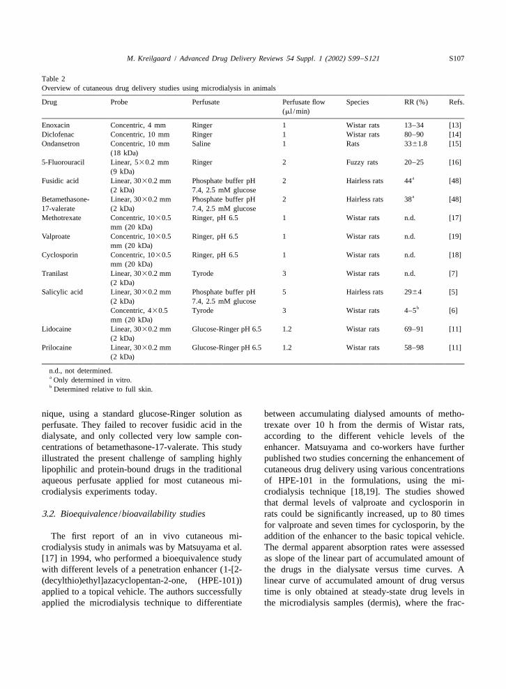
Table 2Overview of cutaneous drug delivery studies using microdialysis in animals
Drug Probe Perfusate Perfusate flow Species RR (%) Refs.(ml /min)
Enoxacin Concentric, 4 mm Ringer 1 Wistar rats 13–34 [13]Diclofenac Concentric, 10 mm Ringer 1 Wistar rats 80–90 [14]Ondansetron Concentric, 10 mm Saline 1 Rats 3361.8 [15]
(18 kDa)5-Fluorouracil Linear, 530.2 mm Ringer 2 Fuzzy rats 20–25 [16]
(9 kDa)aFusidic acid Linear, 3030.2 mm Phosphate buffer pH 2 Hairless rats 44 [48]
(2 kDa) 7.4, 2.5 mM glucoseaBetamethasone- Linear, 3030.2 mm Phosphate buffer pH 2 Hairless rats 38 [48]
17-valerate (2 kDa) 7.4, 2.5 mM glucoseMethotrexate Concentric, 1030.5 Ringer, pH 6.5 1 Wistar rats n.d. [17]
mm (20 kDa)Valproate Concentric, 1030.5 Ringer, pH 6.5 1 Wistar rats n.d. [19]
mm (20 kDa)Cyclosporin Concentric, 1030.5 Ringer, pH 6.5 1 Wistar rats n.d. [18]
mm (20 kDa)Tranilast Linear, 3030.2 mm Tyrode 3 Wistar rats n.d. [7]
(2 kDa)Salicylic acid Linear, 3030.2 mm Phosphate buffer pH 5 Hairless rats 2964 [5]
(2 kDa) 7.4, 2.5 mM glucosebConcentric, 430.5 Tyrode 3 Wistar rats 4–5 [6]
mm (20 kDa)Lidocaine Linear, 3030.2 mm Glucose-Ringer pH 6.5 1.2 Wistar rats 69–91 [11]
(2 kDa)Prilocaine Linear, 3030.2 mm Glucose-Ringer pH 6.5 1.2 Wistar rats 58–98 [11]
(2 kDa)
n.d., not determined.a Only determined in vitro.b Determined relative to full skin.
nique, using a standard glucose-Ringer solution as between accumulating dialysed amounts of metho-perfusate. They failed to recover fusidic acid in the trexate over 10 h from the dermis of Wistar rats,dialysate, and only collected very low sample con- according to the different vehicle levels of thecentrations of betamethasone-17-valerate. This study enhancer. Matsuyama and co-workers have furtherillustrated the present challenge of sampling highly published two studies concerning the enhancement oflipophilic and protein-bound drugs in the traditional cutaneous drug delivery using various concentrationsaqueous perfusate applied for most cutaneous mi- of HPE-101 in the formulations, using the mi-crodialysis experiments today. crodialysis technique [18,19]. The studies showed
that dermal levels of valproate and cyclosporin in3 .2. Bioequivalence /bioavailability studies rats could be significantly increased, up to 80 times
for valproate and seven times for cyclosporin, by theThe first report of an in vivo cutaneous mi- addition of the enhancer to the basic topical vehicle.
crodialysis study in animals was by Matsuyama et al. The dermal apparent absorption rates were assessed[17] in 1994, who performed a bioequivalence study as slope of the linear part of accumulated amount ofwith different levels of a penetration enhancer (1-[2- the drugs in the dialysate versus time curves. A(decylthio)ethyl]azacyclopentan-2-one, (HPE-101)) linear curve of accumulated amount of drug versusapplied to a topical vehicle. The authors successfully time is only obtained at steady-state drug levels inapplied the microdialysis technique to differentiate the microdialysis samples (dermis), where the frac-

S108 M. Kreilgaard / Advanced Drug Delivery Reviews 54 Suppl. 1 (2002)S99–S121
tional increase in accumulated amount is the same. between AUC and AUC diverged betweendermal plasma
The bioequivalence parameters is hence actually an the formulations, leading he authors to the assump-assessment ofC , which is proportional to the tion that several of the vehicles enabled retention ofss
absorption rate and inverse proportional to the the drug in the skin, increasing dermal drug deliveryelimination rate and volume of distribution, pro- relative to transdermal. These results emphasise theviding an average of these parameters. relevance of estimating cutaneous drug delivery in
A substantial increase in dermal tranilast levels, in the dermis, which can currently only be done con-terms ofC and AUC, by topical application in a tinuously in vivo by the microdialysis technique.max
vehicle containing up to 20% oleic acid and 0–10% Recovery of salicylic acid was determined as thepropylene glycol (PG) has also been demonstrated concentration ratio between the dialysate (Tyrodeby use of the microdialysis technique [7]. The study solution) and excised skin of the sacrificed rat, anddemonstrated a good quantitative agreement between was estimated to be 4–5%. This is 6–7-fold lower,relative increase inC and AUC between the six than that estimated by the retrodialysis method [5],max
formulations in the dermis and in plasma, respective- which is presumably attributable to the distributionly, indicating that the enhancers increased both between unbound drug in the ECF and total skindermal and transdermal delivery. The dermal levels concentrations.(determined by AUC) were indicated to be more Microdialysis has also recently been applied tothan 400 times higher following topical application, assess cutaneous drug delivery using iontophoresiscompared to intravenous injection of a similar dose [13,14], for which the technique appears to be atranilast. The study was an excellent demonstration promising tool, as iontophoresis is mainly used withof topical administration as the primary route of ionised and highly polar molecules. Fang et al. havechoice for future treatment of keloid and hyper- examined the influence of hydrogel formulations ontrophic scars with tranilast. dermal penetration of diclofenac [14] and enaxin
Relative recovery in vivo was not estimated in any [13], respectively. Two polymers (polyvinylpyrroli-of the above-mentioned studies and the reported done and hydroxypropyl methylcellulose) were useddermal concentrations of the drugs are, therefore, not for the diclofenac hydrogels, either alone or com-reliable indicators of true unbound tissue concen- bined, and the latter in addition to enhancer pre-trations and comparisons between different tissues, treatment with cardamom oil. Even though in vivoassessment techniques or studies should be done with relative recovery of was high (80–90%), diclofenaccaution. Nevertheless, these early studies indicated was barely detectable in the dialysate when nothe tremendous potential of the microdialysis tech- enhancers were applied. Twelve hours pre-treatmentnique for bioequivalence/bioavailability studies to with cardamom oil significantly increased dermaloptimise topical vehicles for cutaneous drug deliv- drug levels, and further elevation of dermal ECFery. levels was observed for all four formulations when
The enhancer effect of oleic acid on dermal iontophoresis was applied. In the latter study, thedelivery has also been demonstrated by Ding et al. effect of pH and addition of Azone to the hydrogels[15], using ondensetron hydrochloride as model on the iontophoretic delivery of enaxin to the skindrug. Both 2 and 5% oleic acid in PG linearly was examined. Significant higher dermal drug levelsincreased relative steady-state delivery rates com- could be demonstrated by use of unbuffered hydrogelpared to neat PG, and also lag time was substantially or addition of Azone compared with a hydrogelreduced. buffered at pH 5.
Murakami et al. [6] have published a study The influence of topical microemulsion composi-evaluating the effect of different topical vehicles on tion and internal structure on the cutaneous absorp-the dermal absorption of salicylic acid compared to tion of a lipophilic (lidocaine) and a hydrophilicsystemic levels (transdermal). A 100-fold difference model drug (prilocaine hydrochloride), respectively,was observed betweenC and AUC of dermal has been investigated in Wistar rats [11]. By meansmax
levels of salicylic acid, with a w/o emulsion pro- of the retrodialysis by calibrator method a highviding the most extensive cutaneous drug delivery relative recovery was found in vivo for both lido-and water-soluble solbase vehicle the least. The ratio caine (69–91%) and prilocaine (58–98%) using an

M. Kreilgaard / Advanced Drug Delivery Reviews 54 Suppl. 1 (2002)S99–S121 S109
isotonic aqueous perfusate buffered at pH 6.5. To Table 3. The studies are described more detailed inmonitor relative recovery during the experiments, the the following section.study introduced the retrodialysis by calibrator meth-od. Relative recovery was demonstrated to vary, not 4 .1. Single formulation studiesonly between probes, but also within the individualprobes during the experiment, generally decreasing The very first publication of a cutaneous mi-slightly, which emphasised the relevance of moni- crodialysis was a study of ethanol absorption into thetoring recovery during the experiments. Furthermore, skin by Anderson et al. [9]. The study demonstratedthe microdialysis technique enabled demonstration of that ethanol does penetrate the skin and that maxi-significant influence of microemulsion composition mum dermal levels varied from 15 to 800mg/mlon the dermal drug delivery of both drugs. between subjects during the sampling period.
Dialysate was, however, sampled for 50 min andonly collected two times in five of the subjects, andone time in four of the subjects. In a later study,
4 . Human investigations Anderson et al. [10] extended the study of percuta-neous absorption of organic solvents sampled by
An overview of the cutaneous drug delivery microdialysis, to include isopropanol and by meansstudies using microdialysis in humans is presented in of an increased temporal resolution (samples col-
Table 3Overview of cutaneous drug delivery studies using microdialysis in humans
Drug Probe Perfusate Perfusate flow RR (%) Refs.(ml /min)
a8-Methoxypsoralen Concentric, 3030.6 mm Water 2 25 [1](20 kDa)
Propranolol Linear, 1030.2 mm Ringer, lactate 3 53614 [20](20 kDa)
aMethyl nicotinate Concentric, 1030.5 mm Ringer 5/1 26/65 [21](20 kDa)
Lidocaine Linear, 3030.2 mm Glucose-Ringer 1.2 56–95 [12](2 kDa) pH 6.5Concentric, 430.5 mm Ringer 3 n.d. [22](20 kDa)
Prilocaine Concentric, 430.5 mm Ringer 3 n.d. [22](20 kDa)
Ethanol Concentric, 1030.5 mm Ringer 1 90 [9](20 kDa)Concentric, 1030.5 mm Ringer 3 n.d. [10](20 kDa)
Isopropanol Concentric, 1030.5 mm Ringer 3 n.d. [10](20 kDa)
Salicylic acid Linear, 3030.2 mm Phosphate buffer pH 5 2464 [4](2 kDa) 7.4, 2.5 mM glucose
aNicotine Concentric, 1030.5 mm Ringer 10 13 [3](20 kDa)Concentric, 1630.5 mm Ringer, 7% albumin 1.5 25–36 [23](20 kDa)
Estradiol Concentric, 1630.5 mm Ringer, 7% albumin 1.5 1–3 [23](20 kDa)
Diclofenac Concentric, 1630.5 mm Ringer 1.5 66612 [8](20 kDa)
n.d., not determined.a Determined in vitro.

S110 M. Kreilgaard / Advanced Drug Delivery Reviews 54 Suppl. 1 (2002)S99–S121
lected every 10 min for 140 min), detectable levels patch (Nicotinell TTS 30, Ciba-Geigy (21 mg/24¨of ethanol and isopropanol could be demonstrated in h)), has additionally been investigated by Muller et
the dermis after 20 min in all subjects and appeared al. [23]. Microdialysis probes where implanted 2–9to reach a plateau after 100 min. The research group mm beneath the skin surface, and nicotine washas also published a preliminary report, which detectable in all skin layers. A correlation was founddemonstrated that lidocaine and prilocaine can be between AUC of concentration–time curves,C andss
sampled from the dermis by the microdialysis tech- probe depth. This correlation was even higher bynique, following topical application of EMLA [22]. day-to-day assessments in a single individual, in-
Comparable to their studies in rats, Benfeldt et al. dicating interindividual variability to be largest. It[4] have investigated the cutaneous penetration of was also attempted to investigate the transdermalsalicylic acid through normal and perturbed skin in permeation of estradiol from a patch (Estraderm TTShumans. The average relative recovery of the drug 100mg/24 h); however, this failed to recover thewas determined in two individuals (two probes drug in detectable amounts in the dialysate witheach), independently of the experiments, and was probe depths ranging from 1.5 to 10 mm. In vivoassessed to be 2464%, which differed substantially relative recovery was estimated on separate studyfrom the determined in vitro recovery (8063%), days by the retrodialysis method and ranged from 25illustrating the unreliability of basing in vivo re- to 36% for nicotine and 1 to 3% for estradiol.covery on in vitro results. Salicylic acid could be Dermal methyl nicotinate could only be detecteddetected in the dialysate, already 10 min after in two out of three subjects when a high dose (100application in both normal and perturbed skin, mM) was applied topically for 10 min, even thoughindicating a very short lag time of the drug, and the in vitro studies indicated a high relative recoverycutaneous penetration of the drug was substantially (66% at 1ml /min flow) [21]. When the formulationincreased, in terms of AUC of the concentration– was applied for 1 min, nothing could be detected intime curves in the skin, with increasing barrier five subjects. Fast absorption and elimination wasperturbation. The microdialysis technique was dem- demonstrated in excised skin in vitro using mi-onstrated to be substantially more sensitive than crodialysis with the same application periods, but thetransepidermal water loss and erythema to assess in vivo study may have been hampered by the shortbarrier function of the skin by weak barrier perturba- application periods, rapid degradation of the drug intions. Penetration was assessed in four different the skin in conjugation with elevated capillary bloodlocations on the left volar forearm, equally distanced flow due to extensive development of erythema/from the elbow to the hand. No intraregional vari- edema at the application site. Also, in vivo recovery,ation in barrier function of the skin was observed. which was not determined, could have been much
The feasibility of microdialysis to study ion- lower than that indicated in vitro.tophoretic drug delivery in humans has been demon- Transdermal delivery of diclofenac in subcuta-strated with propranolol as model drug [20]. neous tissues has additionally been investigated by
¨The pharmacokinetic profile of nicotine in the Muller et al. [8]. Probes were placed in two defineddermis following application of a commercially tissue layers (3.960.3 mm and 9.360.5 mm). Di-available patch (Nicotinell TTS 20, Ciba-Geigy), has clofenac was only detected in the dialysate (Ringer’sbeen reported by Hegemann et al. [3]. Detectable solution) in 11 out of 20 subjects, independently oflevels of the drug in the microdialysis samples were the probe depth, and the average AUC of thedemonstrated approximately 120–150 min after ap- concentration–time curves from the two probe im-plication of the patch in nine subjects, and reached a plantation levels was not significantly different.plateau within 330–360 min. However, large inter-individual variability of C was observed, ranging 4 .2. Bioequivalence /bioavailability studiesmax
from 500 to 2140 ng/ml. In vivo recovery was notdetermined. In spite of the many successful reports of bio-
Nicotine levels in defined cutaneous/subcutaneous equivalence/bioavailability studies in rats, only twotissue layers following application of a transdermal human studies have yet been reported [1,12]. The
M. Kreilgaard / Advanced Drug Delivery Reviews 54 Suppl. 1 (2002)S99–S121 S111
first compared dermal absorption rate and lag time of study design also allowed a clear establishment oflidocaine from a microemulsion vehicle and a com- t with both administration route where add-onmax
mercially available o/w emulsion (Xylocain 5%) in therapy with ultraviolet A exposure should beeight subjects, with an experimental design similar to initiated for maximum therapeutic effect.an earlier rat study by the same group [11]. Assess- Cutaneous levels of a substantial amount of drugsment of pharmacokinetic parameters according to a have been determined in humans by the mi-compartmental model, demonstrated a significant crodialysis technique (in single formulation studies),increase in absorption rate and decrease in lag time indicating the tremendous potential for bioequival-when lidocaine was applied in a microemulsion ence studies, which yet has to be realised.compared with an o/w emulsion. Prilocaine wasused as relative recovery calibrator, and recoverywas monitored during the experiments for each probe 5 . Reproducibility of the microdialysis techniquevia the retrodialysis method. Relative recoveryranged from 56 to 95% during the study, and varied As indicated by many of the studies described inboth within and between probes, indicating a benefit the review section of animal and human investiga-of monitoring relative recovery during the experi- tions, a relative large variability is often found inments. cutaneous drug delivery determined by the mi-
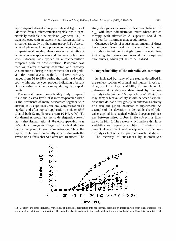
The second human bioavailability study compared crodialysis technique (CV typically 50–100%). Thistissue and plasma levels of 8-methoxypsoralen (used may hamper bioavailability studies between formula-in the treatments of many dermatoses together with tions that do not differ greatly in cutaneous deliveryultraviolet A exposure) after oral administration (1 of a drug and general precision of experiments. Anmg/kg) and after topical application in either 95% example of the deviation in dermal levels of lido-ethanol bath (3 mg/ l) or a cream (0.1%, w/w) [1]. caine applied in a topical vehicle between subjectsVia dermal microdialysis the study elegantly showed and between paired probes in the subjects is illus-that skin /plasma ratio of 8-methoxypsoralen was trated in Fig. 5. The factors which induce this large3–5 orders of magnitude larger with topical adminis- variability are frequently a subject of debate in thetration compared to oral administration. Thus, the current development and acceptance of the mi-topical route could potentially greatly diminish the crodialysis technique for pharmacokinetic studies.severe side-effects observed after oral treatment. The The recovery of substances by microdialysis
Fig. 5. Inter- and intra-individual variability of lidocaine penetration into the dermis, sampled by microdialysis from eight subjects (twoprobes under each topical application). The paired probes in each subject are indicated by the same symbols / lines. Raw data from Ref. [12].

S112 M. Kreilgaard / Advanced Drug Delivery Reviews 54 Suppl. 1 (2002)S99–S121
probes has, in controlled environments (glass beak- comparison to interindividually (46%). A study byers) in vitro, been shown to be highly reproducible Stagni et al. [20] indicated that day-to-day intrasub-(CV typically ,5%) independently of probe design ject variability (n53) of iontophoretically deliveredand perfusate [3,11,19,55], indicating that the vari- propranolol in terms of AUC was not significantlyability is not attributable to the general dialysis different from intersubject variability (n511). Thesampling technique. authors concluded that AUC variability was more
The significance of probe implantation depth on dependent on the condition of the skin at the time ofthe reproducibility of microdialysis experiments is the experiment, than genetic differences amongstill a subject of debate in the current literature subject. The correlation between propranolol AUC[3–5,8,9,20,23]. While most authors have not found and various subject demographics was also ex-a correlation between penetrated drug levels in the amined, but neither sex, race, age nor weight couldskin and probe depth [3,4,8,9,20,32], two studies account for the variability.
¨have indicated a correlation [5,23]. Muller et al. [23] In vitro / in vivo correlation of the variability offound a correlation between absolute recovery of dermal steady-state levels of topically applied 5-nicotine in humans from a transdermal patch and fluorouracil in rat skin, have demonstrated a 10-foldprobe positions ranging from 2 to 9 mm beneath the difference in in vivo levels determined by mi-
2skin surface (estimated by AUC,r 50.6; estimated crodialysis sampling, and an almost 5-fold difference2by C , r 50.7). In contrast, Hegemann et al. [3] did in excised rat skin levels in vitro similarly de-ss
not find a correlation between probe depths (ranging termined by microdialysis sampling in the dermisfrom 0.57 to 1.22 mm) andC of nicotine in the [16]. The in vitro experiments were carried out in amax
dermis, delivered from a transdermal patch. In a controlled environment by Franz-type diffusion cells,¨later, experimentally similar study, Muller et al. [8] with a fixed area and direction of the diffusion (0.6
2did not observe a statistical significant correlation cm ), and the eliminating environment was limitedbetween dermal AUCs of a topically applied ‘finite’ to passive diffusion into the aqueous receptor phasedose of diclofenac and probe depth. A weak correla- from the surface area of the dermis, i.e., no active
2tion (r 50.3) between dermal delivery of salicylic elimination by enzymes of vascular supply. Theseacid (estimated by AUC) and probe depths in rat skin results suggest that the main variability is thereforeranging from 0.5 to 1.1 mm has been indicated [5]. attributable to individual differences in skin barrierThe correlation could, however, not be confirmed in function. However, the diminished variability of thean experimentally similar study in humans [4]. in vitro experiment indicates that distribution and
The applied dosage amount, the partitioning and elimination of the drug are important factors, whichelimination pathway of the current drug are factors also contribute to the variability of penetrationwhich will affect the concentration gradient of the studies using the microdialysis technique.drug in the skin layers, and thereby influence the The good reproducibility of the microdialysisdegree of correlation found in the above-mentioned technique itself by sampling in the skin (and subcuta-studies. neous tissues [56]) is also well illustrated by the
Variability of pharmacokinetic parameters from generally lower variability of dermal drug levels,microdialysis experiments generally appears to be following systemic administration (CV typicallylarger between individuals compared to within in- ,30%), which eliminates the variability induced bydividuals, when multiple probes are used for the differences in skin barrier function [56–58]. In theseassessment of cutaneous penetration from a formula- studies variability of pharmacokinetic parameterstion [5,11,12,32]. In a human study [12], phar- determined with microdialysis in the skin was notmacokinetic profiles of cutaneous lidocaine concen- substantially different from variability of parameterstrations tended to be similar for paired microdialysis determined directly from plasma samples [56,58].probes under the same application site). This was The investigated pharmacokinetic parameter addi-reflected in substantially lower mean intra-individual tionally influences the size of the variability. Cuta-CV (4%) of lag time compared to the interindividual neous drug levels are dependent on both skin barrierCV (38%). Also mean CV of absorption coefficients properties (i.e., penetration rate and lag time), vol-was substantially lower intra-individually (30%), in ume of distribution, elimination rate and additionally

M. Kreilgaard / Advanced Drug Delivery Reviews 54 Suppl. 1 (2002)S99–S121 S113
for the microdialysis technique, factors which affect [37,40,42,43,59–61], only few microdialysis studiesrecovery (described in section). Accumulation of of cutaneous drug delivery have yet included thethese variables in the assessed pharmacokinetic method to assess variability between individualparameter, e.g., AUC andC , will inevitably in- assessment sites and/or fluctuations within the ex-ss
crease overall variability. Thus, to increase precision periments.¨in estimation of drug penetration rates, it would be Using the retrodialysis by drug method, Muller et
useful to differentiate between these factors and al. [23] have demonstrated in 15 subjects a substan-eliminate the additional deviations originating from tial variation in individual recovery in (sub-)cuta-other variables than the penetration rate. The benefit neously implanted probes (2–9 mm from the skinof this is illustrated by the low variability (CV surface) ranging from 25 to 36% for nicotine andtypically ,25%) of the classical Franz-type diffu- 1–3% for estradiol, and in a later study in 11sion cell in vitro studies using a confined receptor subjects, had a relative recovery of 63615%and donor chamber to estimate permeation of sub- (mean6standard error [S.E.]) for diclofenac, withstances though excised skin, where the major vari- probes implanted 3.960.3 mm from the skin surfaceable in steady-state permeation rate is the skin barrier [8].function (roughly assessed). A significant reduction A large deviation in relative recovery values ofin variability of assessed cutaneous lidocaine pene- lidocaine and prilocaine has also been demonstratedtration in humans from two different formulations in both rats [11] and humans [12]. Inter-5individual(microemulsion and Xylocain 5%), has been demon- relative calibrator recovery of prilocaine and lido-strated by individual assessment of apparent absorp- caine in the dermis of rats varied between 58–98%tion rate, lag time and elimination rate in comparison and 69–91%, respectively. Furthermore, recoveryto AUC [12]. CVs of AUCs (n58) from concen- fluctuations were also observed within the experi-tration–time curves of lidocaine penetration from the ments for each probe, and for some probes with amicroemulsion formulation was 91%, and was re- slightly decreasing recovery (most prevalent in theduced to, respectively, 62 and 37% by estimation of initial phase) during the experiment. The averageabsorption coefficient and lag time. Similarly for the recovery fluctuation within experiments forXylocain 5% formulation, CV was reduced from prilocaine was 11.364.2% (n529) with a maximum63% (AUC) to, respectively, 30% (absorption coeffi- fluctuation of 21.2% during a single experiment, andcient) and 39% (lag time). However, this clear trend for lidocaine the average was 6.163.1% (n520)could not be demonstrated in a similar microdialysis with a maximum of 11.4%. Similarly in the humanstudies in rats [11], possibly due to the low number microdialysis study, relative recovery varied betweenof replicates (n53). A significantly lower CV has 56 and 95% between the experiments, and a slightlyalso been reported for propranolol elimination rate decreasing recovery was also occasionally observed(40%) compared to AUC (130%) in human skin during the experiments. The average recovery fluc-[20]. tuation within experiments was 12.464.7% (n531)
The variability of cutaneous drug delivery assess- with a maximum fluctuation of 23.1% during aments by the microdialysis technique is therefore single experiment. The demonstrated inconsistentprobably mainly attributable to differences in barrier relative recovery within the experiments, correlatesfunction of the skin, but also to differences in lag well with previous microdialysis reports in alter-time, elimination rate and possibly volume of dis- native tissues and with other drugs, where largetribution. Furthermore, probe implantation depth and fluctuations and up to 50% decrease in relativerelative recovery (elaborated below) may also con- recovery has been observed during the experimenttribute to the variability. [16,39,41,43,60,62,63]. This suggests a substantial
increase in accuracy of assessing true extracellular5 .1. Inter- and intra-individual variability of tissue concentrations by estimating recovery duringrecovery the experiments, contrary to the current habit of only
assessing one mean recovery value in cutaneousWhile individual relative recovery assessment is microdialysis studies. However, not all drugs and
widely integrated into brain microdialysis tissues display time-dependent recovery

S114 M. Kreilgaard / Advanced Drug Delivery Reviews 54 Suppl. 1 (2002)S99–S121
[40,59,60,64]. Thus, the fluctuations in relative re- hampering sink conditions [63]. These observationscovery of a substance during the experiment appear were not evident in the human experiments of theto be dependent on the diffusion/distribution prop- study, however. The hypothesis may be justified forerties of the substance, the tissue and the time this specific study, considering that the probes wereinterval studied. implanted in the subcutaneous fatty tissues, which
While the mechanism behind the time-dependent may have impeded the outwards diffusion of thefluctuation in relative recovery observed in several extremely hydrophilic model drug. However, thisstudies has not yet been elucidated, several hypoth- hypothesis is not a likely explanation of the time-esis on the subject have been published. Larsson [39] dependent decrease in recovery observed, for exam-suggested that the observed large continuous de- ple, in microdialysis studies in the blood vessels,crease in recovery of caffeine in the jugular veins where the vicinity of the probe is continuouslyand lungs of rats over a 6-h period, was attributable perfused.to a deteriorating systemic circulation during nar- A decrease in lidocaine/prilocaine relative re-cosis, which decreased diffusion (clearance) of the covery was observed in both rats [11] and humansdrug in the tissues/blood in the vicinity of the probe. [12] during the first 60–90 min of the majority ofSauernheimer et al. [62], suggested that a time- experiments. Lidocaine is substantially influenced bydependent 50% decrease in relative recovery of capillary blood flow, and topical application ofacetaminophen in the jugular vein of freely moving lidocaine on rat skin, stripped for stratum corneum,rats over 15 h, may be due to an accumulation of is rapidly cleared from the dermis [65]. It is thereforefree cells and macromolecules in the blood on the likely that the observed slight decrease and fluctua-microdialysis membrane, hindering drug diffusion. tion of relative recovery over time in these studies,Also a rapid decline of recovery in the brain was may be attributed to variations in blood flow. It isobserved (up to 1 h after probe implantation), generally acknowledged that implantation of mi-followed by a steady-state level. The hypothesis that crodialysis probes are associated with a temporarythe actual probe recovery efficiency may change over increase in cutaneous blood perfusion, skin thicknesstime, e.g., due to clotting of the fibre membrane, has and histamine release [29,30,41], which slowly sub-been supported in a study by Wang et al. [43]. By sides during the following hours. Additionally, minorvalidating the relative recovery of microdialysis local bleeding from disruption of the capillaries byprobes (CMA/10) in vitro before and after an in implantation has been observed [41]. All thesevivo experiment in the ventricle and brain of rabbits, factors influence the environment of the probe,a decrease in relative recovery during a 6-h in vivo which can affect diffusion and clearance of theexperiment was demonstrated to be attributed to a substance i.e., relative recovery. Lidocaine is a weakdecrease in actual probe recovery efficiency. In vasoconstrictor, which could also have attributed tocontrast to the above-mentioned hypothesises, decreased clearance. Also a significant correlation
2¨Lonroth and collaborators [41,63] have suggested (r 50.51, P,0.002,n516) has been demonstratedthat the frequently observed declining recovery between mean relative recovery and elimination rateassessed by the retrodialysis method, does not reflect of lidocaine in humans [12], supporting the influenceactual changes in relative recovery, but is due to an of vascular clearance.error of the assessment method. The retrodialysis by A drawback concerning the reproducibility of thecalibrator method estimates loss of the calibrator retrodialysis method to estimate relative recoveryfrom the perfusate, which is assumed5 to reflect and the linear probe design used for the majority ofrecovery of a similar compound of interest. A cutaneous drug delivery studies today, is that varia-prerequisite for this method is sink conditions in the tions in active fibre length outside the actual applica-vicinity of the probe. A decrease in relative recovery tion area (when the membrane ‘window’ has notof subcutaneous glucose in rats over a 3-h period, been confined to the application area), will influenceindicated by simultaneous retrodialysis with radio- loss of the calibrator in a reverse proportionallabelled glucose, was presumed to be attributable to direction to the actual recovery of the drug, and leadan accumulation of the calibrator around the probe, to an overestimation of relative recovery. Further-

M. Kreilgaard / Advanced Drug Delivery Reviews 54 Suppl. 1 (2002)S99–S121 S115
more, analytical errors will also result in inverse (R ), one compartment, and first-order elimination0
proportional errors in estimation of relative recovery, (k) model, including a lag time (t ):lag
which will be particularly sensitive for the correctionKabs 2k t2ts lagdof dialysate sample concentrations if relative re- ]]C 5 12 e (4)s dkcovery is very low and lead to large fluctuation of
corrected concentration–time curves [40]. whereK is the absorption coefficient. The absorp-abs
tion coefficient K estimates the initial rate ofabs
concentration change (at time5t ). Mean correla-lag
5 .2. Pharmacokinetic compartmental models tion between observed and predicted drug concen-trations was 0.98160.029 (n549) for the rat study
The main parameters of interest in topical bio- and 0.99460.007 (n529) for the human studyequivalence/bioavailability studies are the duration (example of average fit: Fig. 6). The current studyof time before the substance enters the skin from design did not allow for individual estimation oftime of administration (i.e., lag time), and the speed absorption rate and volume of distribution (V ), andd
of which the substance is absorbed into the skin (i.e., apparent absorption rate was, therefore, estimated aspenetration rate). Application of an appropriate com- an absorption coefficient (K 5R /V ). It was as-abs 0 d
partmental pharmacokinetic model to fit concentra- sumed thatV was similar (within the limitations ofd
tion–time curves, will enable estimation of these general biological variability) for a drug applied inparameters. A further advantage of introducing a the same anatomical region and species and that thecompartmental pharmacokinetic model to analyse absorption coefficient, therefore, was a reliable in-microdialysis data, is the possibility of individual dicator of apparent absorption rate.estimation of the pharmacokinetic parameters, which In a microdialysis study of transdermal drugcan lead to a lower variability of the results as delivery, although not performed in the subcutaneous
¨described above. layer, Muller et al. [8] used two standard compart-A relatively simple compartmental model has been mental pharmacokinetic models to assessC andmax
introduced for analysis of cutaneous microdialysis AUC of topically applied diclofenac in two defineddata from topically applied drugs (lidocaine and tissue layers, 4 and 9 mm, respectively, below theprilocaine hydrochloride) in both rats [11] and skin surface. The concentration–time curves fromhumans [12] according to a zero-order absorption the upper and lower layer, were fitted to a two-
Fig. 6. Typical pharmacokinetic model fit to cutaneous microdialysis concentration–time curves. Open circles represent actual dialysateconcentrations (corrected for recovery) and solid line represents predicted concentrations based on the pharmacokinetic model [12].
S116 M. Kreilgaard / Advanced Drug Delivery Reviews 54 Suppl. 1 (2002)S99–S121
compartment model and a one-compartment model, A good qualitative and relative quantitative corre-respectively, with a first-order elimination and ab- lation has been demonstrated between in vitro per-sorption rate, without lag time. However, the com- meation using Franz-type diffusion cells [69] and inpartmental parameters were not reported. vivo dermal absorption coefficient determined by
microdialysis [11] of a lipophilic (lidocaine) andhydrophilic model drugs (prilocaine hydrochloride/
6 . Correlation between in vivo microdialysis and lidocaine hydrochloride), respectively, applied inalternative methods to assess cutaneous drug four different topical vehicles. A linear in vitro / indelivery vivo relationship of cutaneous absorption from the
vehicles was indicated for the lipophilic model drug2 2The microdialysis technique uniquely enables (r 50.97) and the hydrophilic model drugs (r 5
assessment of drug levels directly in the dermis and 0.86), respectively. However, the in vitro / in vivoappears to be a very sensitive method to investigate assessments did not correlate between the modelminor differences in cutaneous drug delivery, which drugs. In vitro flux of the formulations containing thehas not been possible with previous methods. How- hydrophilic model drugs, indicated that cutaneousever, as the technique has only recently been intro- delivery rate of the drugs would on average be 20%duced to cutaneous drug delivery studies, it is relative to the mean delivery rate of the lipophilicnaturally of great interest to evaluate the method and model drug from the four vehicles. With the mi-compare it to well-established in vitro / in vivo crodialysis technique though, it was demonstratedmodels and readdress the clinical relevance of the that the mean dermal delivery rate in vivo of the‘well established’ methods. hydrophilic model drugs was only approximately 2%
relative to that of the lipophilic drug. These studies6 .1. In vitro models suggest that in vitro Franz-type diffusion cells may
be a good qualitative, and relative quantitativeOne of the most acknowledged methods for indicator of bioequivalence between formulations
bioequivalence studies in vitro is the Franz-type with the same drug; however, this method is notdiffusion cell [2,66–68], which has been used for reliable for assessment of relative drug delivery ratedecades to assess qualitative permeation ranks of between different drugs, and does not enable estima-topical formulations. A very close relationship be- tion of actual dermal drug levels in vivo. Mi-tween permeated 5-flouruoracil levels in the receptor crodialysis has also been used to demonstrate signifi-compartment of a Franz-type diffusion cell, sampled cant increase of cutaneous diclofenac [14] andby regular sample withdrawal and the levels (cor- enaxin [13] delivery in rats by application of variousrected for relative recovery) assessed by mi- enhancers, which correlated qualitatively well withcrodialysis sampling in the same compartment has transdermal permeation in vitro in the former, butbeen demonstrated [47]. A good qualitative agree- not as clearly in the latter study.ment between flux through different rat skin samples(n53) determined from traditional receptor compart- 6 .2. In vivo modelsment sampling, and dermal levels assessed by themicrodialysis technique in an in vitro diffusion cell, To the author’s knowledge, no comparison studyhas furthermore been demonstrated for the drug [16]. of the microdialysis technique and the previouslyHowever, comparison of average dermal drug levels most prevalent assessment technique for direct esti-sampled by microdialysis from rat skin in vitro and mation of cutaneous penetration in vivo, tape strip-in vivo [16], has indicated that 5-fluorouracil levels ping, has yet been performed.assessed in vitro are approximately 40 times higher Two comparison studies by Murakami et al. [6,7]than the actual in vivo levels; hence suggesting that of cutaneous drug delivery in rats by the mi-quantitative assessments based on in vitro experi- crodialysis technique and by the indirect assessmentments are not reliable indicators of in vivo levels due of plasma concentrations have been published withto the intact clearance processes (vascular and en- diverging findings. One report [7] demonstrated anzymatic) in the latter. excellent quantitative correlation between dermal

M. Kreilgaard / Advanced Drug Delivery Reviews 54 Suppl. 1 (2002)S99–S121 S117
AUC/C of topically applied tranilast in six differ- cutaneous absorption coefficient of lidocaine inmax
ent vehicles, assessed by the microdialysis technique, Wistar rats (administered on the side of the thorax)and AUC/C from plasma concentration–time [11] and in humans (administered on the volarmax
curves. The other report [6], however, did not find surface of the forearm) [12] from a microemulsionany correlation between dermal AUC/C and vehicle and Xylocain 5%, respectively. As themax
plasma AUC/C of salicylic acid applied in five estimated absorption coefficients (K 5R /V ) didmax abs 0 d
different vehicles. The studies illustrate that plasma not account for differences inV between rat andd
levels do not always correlate with dermal drug human skin, the actual ratio between absorption ratesdelivery rate, and furthermore do not provide in- may differ slightly from the observed absorptionformation of actual drug levels in the skin. coefficients. Rat skin is generally thinner than human
Among the conventional in vivo methods to assess skin, both in regard to whole skin and stratumcutaneous drug delivery is, additionally, pharmaco- corneum thickness (10–15mm in rat skin compareddynamic models. A combination of the microdialysis to 15–20mm in human skin), and contains fewertechnique and a pharmacodynamic model is an cornified cell layers. It may therefore be assumedattractive solution to assess PK/PD relationships, as that the volume of distribution is lower in rat skin,the sampling can be performed in the target organ. compared to human skin, and that the assessedThe cutaneous absorption of lidocaine has been penetration rates in rats, are slightly overestimated,investigated in humans by the microdialysis tech- relative to those in humans. This would be innique and compared to the anaesthetic effect of a accordance with the general findings of other assess-microemulsion formulation and Xylocain 5% [12]. ment techniques, where rat skin is typically observedThe pharmacodynamic study showed a significant to be two to five times more permeable than humanreduction in pain perception of prods with von Frey skin [68]. Also, elimination rate of lidocaine inhairs during the application of the two lidocaine human and rat skin was not discernible (unpublishedformulations, discernible from placebo treatment. data from Refs. [11,12]). However, lag time forThe anaesthetic effect of the two formulations was lidocaine detection in the dermis was increasednot statistically discernible, although indicating simi- approximately 5.5-fold for both the microemulsionlar cutaneous absorption profile of lidocaine from the formulation and Xylocain 5%, which correspondstwo formulations. However, the cutaneous mi- well with the increased thickness and additional cellcrodialysis study demonstrated a 3-fold increase in layers of the human stratum corneum. This indicatesapparent absorption rate and a significant decrease in that the total time for lidocaine passage through thelag time of lidocaine applied in the microemulsion barrier layer of the skin increased more than the ratevehicle compared to Xylocain 5%, which resulted in of the passage.a more than four times increase in total amount of Comparative studies of cutaneous drug penetrationlidocaine absorbed into the skin from the former in hairless rats and the volar forearm of humans,vehicle. No correlation between the pharmacokinetic using the microdialysis technique, have also beenparameters and AUE could be demonstrated. It was done by Benfeldt et al. [4,5,70]. These studiessuggested that the efficacy of lidocaine in the indicated a 53-fold increase in dermal penetrationassessed concentration range was relatively low, (assessed by AUC ) of topically applied0–210 min
which hampered differentiation between the formula- salicylic acid for rats relative to humans (applied ontions in the pharmacodynamic study. Regardless, the the volar surface of the forearm). These findings arestudy illustrated the higher sensitivity and lower in contrast to an in vitro study, which demonstratedvariability of the microdialysis technique to assess similar permeation rates of salicylic acid throughbioequivalence of cutaneous drug delivery, compared excised skin from hairless rats and human breast andto the pharmacodynamic model. thigh [71]. While in vitro studies are not always
reliable indicators for the in vivo situation, the verylarge differences between the findings of these
7 . Prediction of cutaneous drug delivery instudies suggest that penetration rate alone cannot
humans from rat microdialysis studiesexplain the differences in AUC found by Benfeldt et
No significant difference was found between al., and that deviating elimination rate, lag time and

S118 M. Kreilgaard / Advanced Drug Delivery Reviews 54 Suppl. 1 (2002)S99–S121
possibly volume of distribution may have contributed of the skin. It has been demonstrated to enableto the observed differences. The different anatomical quantification of numerous hydrophilic and a fewregion of human skin of the two studies, may also moderately lipophilic drugs directly in the targethave contributed to the deviations. The in vivo organ—the viable layers of the skin. Studies in bothstudies did, however, indicate a good relative rat / humans and rats have shown a tremendous potentialhuman correlation between AUC ratios of penetra- of microdialysis to estimate bioequivalence/bioavail-tion of salicylic acid through normal skin and various ability of topical formulations, due to the highdegrees of perturbed skin, indicating that the rat sensitivity of the technique. However, a considerablemodel was a good indicator for relative decrease in variability of dermal drug levels by microdialysishuman skin barrier function to drug penetration in sampling of cutaneously absorbed drugs has beendifferent stages of perturbation. While both the observed in the early studies, which hampered theformulation and microdialysis probes were different precision of bioequivalence studies. The main vari-in the studies, hampering quantitative correlations, ability is indicated to be attributable to interindividu-diclofenac has been shown to penetrate the skin very al differences in barrier function of the skin; how-slowly when no enhancers are applied in both rats ever, differences in lag time, elimination rate and[14] and humans [8]. possibly distribution also contribute significantly to
Human/rat microdialysis correlation studies, have the variance between assessment sites. It has beenindicated that relative recovery does not differ shown that a substantial increase in precision ofsubstantially between the two species [4,5,11,12]. cutaneous drug delivery assessment can be obtainedBenfeldt et al. found an average relative recovery of by estimation of absorption rate, lag time andsalicylic acid in rats of 2964% and in humans of elimination rate through compartmental modelling of2464%. The average relative recovery of prilocaine microdialysis data. Furthermore, a few studies havehas been shown to be 8166% in rats [11] and indicated a lower variability of pharmacokinetic7767% in humans [12]. While in vitro recovery is parameters when assessment sites are correctedoften not a reliable indicator of relative recovery in individually for relative recovery. Probe depth ap-vivo [4,5,23], it appears that recovery in human skin pears mainly to influence reproducibility of themay be derived from rat experiments; however, more technique, when implantation ranges between largerstudies are desirable to establish the relationship distances than the cutaneous layers.more closely. Assessment of bioequivalence in terms of absorp-
The most important determinant for the rat–human tion rate by in vivo microdialysis appears to correlatecorrelation is most likely the properties of the model qualitatively well with established in vitro Franz-typedrug, which will be affected in various degrees by diffusion cell assessments of permeation rates forthe differences in stratum corneum structure and formulations with the same drug. However, therelipid composition between rat and human skin. does not appear to be a correlation between cuta-However, the multiple biological factors (i.e., ana- neous in vitro flux and in vivo absorption fortomical location of the administration), which in- formulations with different drugs, and in vitro meth-fluence the permeability of the skin, and the rat ods are not reliable indicators of actual tissue drugspecies/age [72], are most likely parameters that levels in vivo. Furthermore, assessment of systemicadditionally contribute to the observed differences in levels has been demonstrated to not always adequate-the various studies. ly estimate relative dermal absorption rates, and in
vivo microdialysis is currently the only technique todirectly assess unbound drug levels in the dermis of
8 . Conclusions the skin.Finally, a good correlation between in vivo cuta-
The microdialysis technique is substantially more neous absorption coefficient and elimination rate of asensitive and less invasive than previous in vitro / in lipophilic model drug in Wistar rats and in humanvivo methods in assessing differences in cutaneous forearm skin has been indicated by a single study.penetration rates of substances and barrier function However, lag time appears to be much longer in

M. Kreilgaard / Advanced Drug Delivery Reviews 54 Suppl. 1 (2002)S99–S121 S119
therapeutic drug, tranilast, by combined use of oleic acid andhuman skin than in rat skin, emphasising the impor-propylene glycol as a penetration enhancer: evaluation bytance of differentiating between these parameters, toskin microdialysis in rats, J. Pharm. Pharmacol. 50 (1998)
enable quantitative prediction of bioequivalence for 49–54.topical formulations in humans from rat mi- ¨[8] M. Muller, H. Mascher, C. Kikuta, S. Schafer, M. Brunner,crodialysis studies. More studies are needed to G. Dorner, H.G. Eichler, Diclofenac concentrations in de-
fined tissue layers after topical administration, Clin. Phar-confirm these relationships.macol. Ther. 62 (1997) 293–299.The future challenges for assessment of cutaneous
[9] C. Anderson, T. Andersson, M. Molander, Ethanol absorp-drug delivery using the microdialysis technique liestion across human skin measured by in vivo microdialysis
in the sampling of lipophilic compounds, which technique, Acta Dermatol. Venereol. 71 (1991) 389–393.constitutes the majority of novel drugs targeted to the [10] C. Anderson, T. Andersson, A. Boman, M. Molander,
Cutaneous microdialysis for the measurement in vivo of theskin. Thus, further work is needed in the optimi-percutaneous absorption of organic solvents, Curr. Probl.sation of perfusate, diminishing adhesion to mi-Dermatol. 25 (1996) 37–46.crodialysis materials and also to implement more
[11] M. Kreilgaard, Dermal pharmacokinetics of microemulsionsensitive analytical techniques. But as the many formulations determined by in vivo microdialysis, Pharm.authors cited in this review have clearly demon- Res. 18 (2001) 367–373.strated, the technique can already now be applied for [12] M. Kreilgaard, M.J. Kemme, J. Burggraaf, R.C. Schoemaker,
A.F. Cohen, Influence of a microemulsion vehicle on cuta-biopharmaceutical optimisation of topical formula-neous bioequivalence of a lipophilic model drug assessed bytions with the numerous successfully recoveredmicrodialysis and pharmacodynamics, Pharm. Res. 18
drugs. (2001) 593–599.[13] J.Y. Fang, L.R. Hsu, Y.B. Huang, Y.H. Tsai, Evaluation of
transdermal iontophoresis of enoxacin from polymer formu-lations: in vitro skin permeation and in vivo microdialysis
R eferences using Wistar rat as an animal model, Int. J. Pharm. 180(1999) 137–149.
[14] J.Y. Fang, K.C. Sung, H.H. Lin, C.L. Fang, Transdermal[1] I. Tegeder, L. Brautigam, M. Podda, S. Meier, R. Kaufmann,iontophoretic delivery of diclofenac sodium from variousG. Geisslinger, M. Grundmann-Kollmann, Time course ofpolymer formulations: in vitro and in vivo studies, Int. J.8-methoxypsoralen concentrations in skin and plasma afterPharm. 178 (1999) 83–92.topical (bath and cream) and oral administration of 8-
[15] P. Ding, H. Xu, G. Wei, J. Zheng, Microdialysis samplingmethoxypsoralen, Clin. Pharmacol. Ther. 71 (2002) 153–coupled to HPLC for transdermal delivery study of ondanset-161.ron hydrochloride in rats, Biomed. Chromatogr. 14 (2000)[2] T.J. Franz, Percutaneous absorption on the relevance of in141–143.vitro data, J. Invest. Dermatol. 64 (1975) 190–195.
[16] J.M. Ault, C.M. Riley, N.M. Meltzer, C.E. Lunte, Dermal[3] L. Hegemann, C. Forstinger, B. Partsch, I. Lagler, K. Wolff,microdialysis sampling in vivo, Pharm. Res. 11 (1994)Microdialysis in cutaneous pharmacology: kinetic analysis of1631–1639.transdermally delivered nicotine, J. Invest. Dermatol. 104
[17] K. Matsuyama, M. Nakashima, Y. Nakaboh, M. Ichikawa, S.(1995) 839–843.Satoh, Application of in vivo microdialysis to transdermal[4] E. Benfeldt, J. Serup, T. Menne, Effect of barrier perturba-absorption of methotrexate in rats, Pharm. Res. 11 (1994)tion on cutaneous salicylic acid penetration in human skin: in684–686.vivo pharmacokinetics using microdialysis and non-invasive
[18] M. Nakashima, M.F. Zhao, H. Ohya, M. Sakurai, H. Sasaki,quantification of barrier function, Br. J. Dermatol. 140K. Matsuyama, M. Ichikawa, Evaluation of in-vivo transder-(1999) 739–748.mal absorption of cyclosporin with absorption enhancer[5] E. Benfeldt, J. Serup, Effect of barrier perturbation onusing intradermal microdialysis in rats, J. Pharm. Pharmacol.cutaneous penetration of salicylic acid in hairless rats: in48 (1996) 1143–1146.vivo pharmacokinetics using microdialysis and non-invasive
[19] K. Matsuyama, M. Nakashima, M. Ichikawa, T. Yano, S.quantification of barrier function, Arch. Dermatol. Res. 291Satoh, S. Goto, In vivo microdialysis for the transdermal(1999) 517–526.absorption of valproate in rats, Biol. Pharm. Bull. 17 (1994)[6] T. Murakami, M. Yoshioka, I. Okamoto, R. Yumoto, Y.1395–1398.Higashi, K. Okahara, N. Yata, Effect of ointment bases on
[20] G. Stagni, D. O’Donnell, Y.J. Liu, J.D.L. Kellogg, T.topical and transdermal delivery of salicylic acid in rats:Morgan, A.M.M. Shepherd, Intradermal microdialysis: kinet-evaluation by skin microdialysis, J. Pharm. Pharmacol. 50ics of iontophoretically delivered propranolol in forearm(1998) 55–61.dermis, J. Control. Release 63 (2000) 331–339.[7] T. Murakami, M. Yoshioka, R. Yumoto, Y. Higashi, S.
[21] E. Boelsma, C. Anderson, A.M. Karlsson, M. Ponec, Mi-Shigeki, Y. Ikuta, N. Yata, Topical delivery of keloid

S120 M. Kreilgaard / Advanced Drug Delivery Reviews 54 Suppl. 1 (2002)S99–S121
crodialysis technique as a method to study the percutaneous [39] C.I. Larsson, The use of an ‘internal standard’ for control ofpenetration of methyl nicotinate through excised human skin, the recovery in microdialysis, Life Sci. 49 (1991) L73–L78.reconstructed epidermis, and human skin in vivo, Pharm. [40] M.R. Bouw, M. Hammarlund-Udenaes, Methodological as-Res. 17 (2000) 141–147. pects of the use of a calibrator in in vivo microdialysis-
[22] C. Anderson, T. Andersson, A. Nyquist-Mayer, M. Melin, further development of the retrodialysis method, Pharm. Res.M.S. Roberts, Cutaneous microdialysis technique in the 15 (1998) 1673–1679.
¨study of the percutaneous absorption of a local anaesthetic [41] A.L. Krogstad, P.A. Jansson, P. Gisslen, P. Lonnroth, Mi-cream in vivo in human skin, Perspectives in Percutaneous crodialysis methodology for the measurement of dermalPenetration 5a (1997) Abstract. interstitial fluid in humans, Br. J. Dermatol. 134 (1996)
¨[23] M. Muller, R. Schmid, O. Wagner, B. Osten, H. Shayganfar, 6–12.˚H.G. Eichler, In vivo characterization of transdermal drug [42] L. Stahle, Drug distribution studies with microdialysis: I.
transport by microdialysis, J. Control. Release 37 (1995) Tissue dependent difference in recovery between caffeine49–57. and theophylline, Life Sci. 49 (1991) 1835–1842.
¨[24] P. Lonnroth, P.A. Jansson, U. Smith, A microdialysis method [43] Y. Wang, S.L. Wong, R.J. Sawchuk, Microdialysis calibrationallowing characterization of intercellular water space in using retrodialysis and zero-net flux: application to a study ofhumans, Am. J. Physiol. 253 (1987) E228–E231. the distribution of zidovudine to rabbit cerebrospinal fluid
˚[25] L. Stahle, On mathematical models of microdialysis: geome- and thalamus, Pharm. Res. 10 (1993) 1411–1419.try, steady-state models, recovery and probe radius, Adv. [44] L.J. Petersen, Quantitative measurement of extracellularDrug Deliv. Rev. 45 (2000) 149–167. histamine concentrations in intact human skin in vivo by the
[26] M. Schmelz, O. Luz, B. Averbeck, A. Bickel, Plasma microdialysis technique: methodological aspects, Allergy 52extravasation and neuropeptide release in human skin as (1997) 547–555.measured by intradermal microdialysis, Neurosci. Lett. 230 [45] C. Anderson, T. Andersson, R.G. Andersson, In vivo mi-(1997) 117–120. crodialysis estimation of histamine in human skin, Skin
[27] M. Lindberger, T. Tomson, L. Stahle, Validation of mi- Pharmacol. 5 (1992) 177–183.crodialysis sampling for subcutaneous extracellular valproic [46] L. Groth, A. Jørgensen, J. Serup, Cutaneous microdialysis inacid in humans, Ther. Drug Monit. 20 (1998) 358–362. the rat: insertion trauma studied by ultrasound imaging, Acta
[28] C. Anderson, T. Andersson, K. Wardell, Changes in skin Dermatol. Venereol. 78 (1998) 10–14.circulation after insertion of a microdialysis probe visualized [47] J.M. Ault, C.E. Lunte, N.M. Meltzer, C.M. Riley, Mi-by laser Doppler perfusion imaging, J. Invest. Dermatol. 102 crodialysis sampling for the investigation of dermal drug(1994) 807–811. transport, Pharm. Res. 9 (1992) 1256–1261.
[29] L. Groth, A. Jørgensen, J. Serup, Cutaneous microdialysis in [48] E. Benfeldt, L. Groth, Feasibility of measuring lipophilic orthe rat: insertion trauma and effect of anaesthesia studied by protein-bound drugs in the dermis by in vivo microdialysislaser Doppler perfusion imaging and histamine release, Skin after topical or systemic drug administration, Acta Dermatol.Pharmacol. Appl. Skin Physiol. 11 (1998) 125–132. Venereol. 78 (1998) 274–278.
˚[30] L. Groth, J. Serup, Cutaneous microdialysis in man: effects [49] C. Carneheim, L. Stahle, Microdialysis of lipophilic com-of needle insertion trauma and anaesthesia on skin perfusion, pounds: a methodological study, Pharmacol. Toxicol. 69erythema and skin thickness, Acta Dermatol. Venereol. 78 (1991) 378–380.(1998) 5–9. [50] Y. Kurosaki, S. Nakamura, Y. Shiojiri, H. Kawasaki, Lipo-
[31] L.J. Petersen, Measurement of histamine release in intact microdialysis: a new microdialysis method for studying thehuman skin by microdialysis technique. Clinical and ex- pharmacokinetics of lipophilic substances, Biol. Pharm. Bull.perimental findings, Dan. Med. Bull. 45 (1998) 383–401. 21 (1998) 194–196.
[32] L. Groth, Cutaneous microdialysis. Methodology and valida- [51] A.N. Khramov, J.A. Stenken, Enhanced microdialysis re-tion, Acta Dermatol. Venereol. Suppl. 197 (1996) 1–61. covery of some tricyclic antidepressants and structurally
[33] L.H. Parsons, J.B. Justice, Quantitative approaches to in vivo related drugs by cyclodextrin-mediated transport, Analystbrain microdialysis, Crit. Rev. Neurobiol. 8 (1994) 189–220. 124 (1999) 1027–1033.
[34] H. Benveniste, Brain microdialysis, J. Neurochem. 52 (1989) [52] M. Schmelz, S. Weber, M. Kress, Topical acetyl salicylate1667–1679. and dipyrone attenuate neurogenic protein extravasation in
[35] S.A. Wages, W.H. Church, J.B. Justice, Sampling considera- rat skin in vivo, Neurosci. Lett. 290 (2000) 57–60.tions for online micro-bore liquid chromatography of brain [53] J.T. Wu, H. Zeng, M. Qian, B.L. Brogdon, S.E. Unger, Directdialysate, Anal. Chem. 58 (1986) 1649–1656. plasma sample injection in multiple-component LC–MS–MS
[36] K.M. Kendrick, Use of microdialysis in neuroendocrinology, assays for high-throughput pharmacokinetic screening, Anal.Methods Enzymol. 168 (1989) 182–205. Chem. 72 (2000) 61–67.
[37] S.L. Wong, Y. Wang, R.J. Sawchuk, Analysis of zidovudine [54] M.I. Davies, J.D. Cooper, S.S. Desmond, C.E. Lunte, S.M.distribution to specific regions in rabbit brain using mi- Lunte, Analytical considerations for microdialysis sampling,crodialysis, Pharm. Res. 9 (1992) 332–338. Adv. Drug Deliv. Rev. 45 (2000) 169–188.
[38] J. Kehr, A survey on quantitative microdialysis: theoretical [55] L. Groth, A. Jørgensen, In vitro microdialysis of hydrophilicmodels and practical implications, J. Neurosci. Methods 48 and lipophilic compounds, Anal. Chim. Acta 355 (1997)(1993) 251–261. 75–83.

M. Kreilgaard / Advanced Drug Delivery Reviews 54 Suppl. 1 (2002)S99–S121 S121
¨[56] M. Muller, R. Schmid, A. Georgopoulos, A. Buxbaum, C. influx and efflux blood–brain barrier permeabilities ofWasicek, H.G. Eichler, Application of microdialysis to gabapentin using a microdialysis-pharmacokinetic approach,clinical pharmacokinetics in humans, Clin. Pharmacol. Ther. Pharm. Res. 13 (1996) 398–403.57 (1995) 371–380. [65] P. Singh, M.S. Roberts, Effect of vasoconstriction on dermal
[57] E. Benfeldt, J. Serup, T. Menne, Microdialysis vs. suction pharmacokinetics and local tissue distribution of compounds,blister technique for in vivo sampling of pharmacokinetics in J. Pharm. Sci. 83 (1993) 783–791.the human dermis, Acta Dermatol. Venereol. 79 (1999) 338– [66] D. Howes, R.H. Guy, J. Hadgraft, J. Heylings, U. Hoeck, F.342. Kemper, H. Maibach, J. Marty, H. Merk, J. Parra, D. Rekkas,
[58] N. Borg, E. Gotharson, E. Benfeldt, L. Groth, L. Stahle, I. Rondelli, H. Schaefer, U. Tauber, N.Verbiese, Methods forDistribution to the skin of penciclovir after oral famciclovir assessing percutaneous absorption, ATLA 24 (1996) 81–administration in healthy volunteers: comparison of the 106.suction blister technique and cutaneous microdialysis, Acta [67] B.W. Barry, in: Dermatological Formulations: PercutaneousDermatol. Venereol. 79 (1999) 274–277. Absorption, Marcel Dekker, New York, 1983.
[59] R. Xie, M. Hammarlund-Udenaes, Blood–brain barrier [68] H. Schaefer, T.E. Redelmeier, in: Skin Barrier: Principles ofequilibration of codeine in rats studied with microdialysis, Percutaneous Absorption, Karger, Basel, 1996.Pharm. Res. 15 (1998) 570–575. [69] M. Kreilgaard, E.J. Pedersen, J.W. Jaroszewski, NMR
¨[60] P. Sjoberg, I.M. Olofsson, T. Lundqvist, Validation of characterisation and transdermal drug delivery potential ofdifferent microdialysis methods for the determination of microemulsion systems, J. Control. Release 69 (2000) 421–unbound steady-state concentrations of theophylline in blood 433.and brain tissue, Pharm. Res. 9 (1992) 1592–1598. [70] E. Benfeldt, In vivo microdialysis for the investigation of
[61] M. Ekblom, M. Hammarlund-Udenaes, T. Lundqvist, P. drug levels in the dermis and the effect of barrier perturba-Sjoberg, Potential use of microdialysis in pharmacokinetics: tion on cutaneous drug penetration—Studies in hairless ratsa protein binding study, Pharm. Res. 9 (1992) 155–158. and human subjects, Acta Dermatol. Venereol. Suppl. 206
[62] C. Sauernheimer, K.M. Williams, K. Brune, G. Geisslinger, (1999) 7–54.Application of microdialysis to the pharmacokinetics of [71] K. Harada, T. Murakami, E. Kawasaki, Y. Higashi, S.analgesics: problems with reduction of dialysis efficiency in Yamamoto, N. Yata, In-vitro permeability to salicylic acid ofvivo, J. Pharmacol. Toxicol. Methods 32 (1994) 149–154. human, rodent, and shed snake skin, J. Pharm. Pharmacol. 45
¨[63] P. Lonnroth, L. Strindberg, Validation of the ‘internal refer- (1993) 414–418.ence technique’ for calibrating microdialysis catheters in [72] P.A. Lehman, T.J. Franz, Effect of age and diet on stratumsitu, Acta Physiol. Scand. 153 (1995) 375–380. corneum barrier function in the Fischer 344 female rat, J.
[64] Y. Wang, D.F. Welty, The simultaneous estimation of the Invest. Dermatol. 100 (1993) 200–204.