a study on aluminum toxicity with female rats: can … · a study on aluminum toxicity with female...
TRANSCRIPT

A Study on Aluminum Toxicity with Female Rats: Can Vitamin E Help to
Reduce its Effect on Biological Systems?
A project submitted by
Tanisha Momtaz ID: 12146025
Session: Spring 2012 to
The Department of Pharmacy in partial fulfillment of the requirements for the degree of
Bachelor of Pharmacy (Hons.)
Dhaka, Bangladesh February 2016

Dedicated to:
My beloved parents, my brother and my husband, who always support and
inspire me to face every challenge of life.
i

Certification Statement
This is to certify that the project titled “A Study on Aluminum Toxicity with Female
Rats: Can Vitamin E help to Reduce its Effect on Biological Systems?” submitted in
order to fulfill the partial requirements for the degree of Bachelor of Pharmacy from the
Department of Pharmacy, BRAC University under the supervision of Dr. Sharmind
Neelotpol, Assistant Professor, Department of Pharmacy, BRAC University and proper
referencing have been made where the language, concept or writings of others are used.
Signed,
………………………………………………
Countersigned by the supervisor,
……………………………………………….
ii

Acknowledgement
My first and most heartily praise goes to the almighty Allah, who blessed me with the
knowledge, power and ability to accomplish my final year project successfully. Without
His endless grace I would be unable to reach my goals.
Foremost, I am obliged from the bottom of my heart to my honorable supervisor and
teacher Dr Sharmind Neelotpol, Assistant Professor, Department of Pharmacy, BRAC
University, for giving her valuable time to guide and support me. I honestly believe that
without her guidance, criticize and direction it was not possible to finish this project. I
am also thankful to my honorable Chairperson Dr. Eva Rahman Kabir and my teachers
for their constant superintendence.
Furthermore, I acknowledge my accountability to Shahabuddin Kabir Choudhuri and
Masum Shahriar, Department of Pharmacy, Jahangirnagar University, who despite of
their busy schedule spare time to help me. I would also like to give my appreciation to
the support of the staffs and students of Department of Pharmacy, Jahangirnagar
University.
Lastly, I thank to my family members and my friends who continuously boost up my
confidence and motivate me throughout my project.
Tanisha Momtaz
February, 2016.
iii

Abstract
Increasing complaints about metal toxicity have been reported with the modernization of
medical science. However, in this field, aluminum has been a renewed matter of concern.
Therefore, more impotence is given to find out natural as well as synthetic substances
that can alleviate the toxic effect of aluminum in the body. That is why; this study has
aimed to identify the effect of vitamin E in reducing the toxic effect on biological
systems caused by Aluminum. The study was conducted for 28 days and with total 18
female albino Wistar rats weighing 138-150g. 18 rats were divided into three groups-
control, aluminum treated and aluminum plus vitamin E treated. The control group was
receiving normal drinking water along with two drops of Tween 80 whereas aluminum
treated group was getting aluminum nitrate solution 1ml/rat containing half ‘x’ of body
weight and two drops of Tween 80. Moreover, aluminum plus vitamin E treated group
was receiving aluminum nitrate solution 1ml/rat containing half ‘x’ of body weight along
with 1ml/rat vitamin containing 22.5 mg of vitamin E per kg and also two drops of
Tween 80. After 28 days, the rats were sacrificed, and blood was collected from the heart
using ventricular puncture method. The collected blood was sent for hematological and
clinical bio-chemistry examinations. At the same time, the liver, skin, brain, kidney and
ovary of the rats were collected in the formalin and sent for histopathological study. The
clinical biochemistry examination showed significant results for all the parameters
except basophils, while the histopathology showed that vitamin E has the potential to
eliminate the toxic effects caused by aluminum.
iv
Table of Contents
ACKNOWLEDGEMENT iii
ABSTRACT iv
TABLE OF CONTENT v
LIST OF TABLES vii
LIST OF FIGURE viii
LIST OF ABBREVIATONS x
CHAPTER 1: INTRODUCTION
1.1. Introduction 1
1.2.Aim 4
1.3.Objectives 4
1.4.Rationales of the study 5
CHAPTER 2: LITERATURE REVIEW
2.1. History 6
2.2. Properties of Aluminum 7
2.3. Fate of Aluminum in Environment 7
2.4. Sources of Aluminum 7
2.5. General population exposure 8
2.6. Occupational exposure 9
2.7. Global production of aluminum 10
2.8. Absorption, Distribution, Metabolism and Excretion 10
2.9. Toxicity 11
2.10. Minimum risk level 11
2.11. Effect on human 12
2.12. Effect on animal s 13
2.13. Methods of reducing toxic effect 14
2.14. Studies on aluminum and vitamin E 14
v

CHAPTER 3: MATERIALS AND METHODS
3.1. Reagents 16
3.2. Experimental animal 16
3.3. Experimental procedure 17
3.4. Preparation of plasma 17
3.5. Histopathology 18
3.6. Statistical analysis 18
CHAPTER 4: RESULTS
4.1. Results 19
4.2. Body weight 19
4.3. Liver 19
4.3.1. Histopathology output of liver 20
4.4. Kidney 21
4.4.1. Histopathology output of kidney 22
4.5. Histopathology output of skin, brain and ovary 23
4.6. Hematology test 26
CHAPTER 5: DISCUSSION
5.1. Discussion 27
CHAPTER 6: CONCLUSION
6.1. Limitations of the study 30
6.2. Recommendations 30
6.3. Conclusion 30
CHAPTER 7: APPENDIX
7.1. Appendix 1 32
7.2. Appendix 2 34
7.3. Appendix 3 35
CHAPTER 8: REFERENCES
8.1. References 38
vi

List of tables
Table 2.1: Sources of Aluminum 8
Table 4.1: Statistical output for liver 20
Table 4.2: Statistical output for kidney 21
Table 4.3: Statistical output for hematology test 26
vii

List of figures
Figure 1.1: Pathway of transport of metals in environment 1
Figure 1.2: Flow of metals within human body 2
Figure 2.1: Aluminum 6
Figure 2.2: Source of Aluminum 8
Figure 3.1: Grouping of experimental rats 17
Figure 4.1: Comparison of body weight among control, Al treated and Al+Vit E 19
treated rats
Figure 4.2: Histopathological study of rat liver from control group 20
Figure 4.3: Histopathological study of rat liver from Al-treated group 20
Figure 4.4: Histopathological study of rat liver from Al+Vit E treated group 20
Figure 4.5: Histopathological study of rat kidney from control group 22
Figure 4.6: Histopathological study of rat kidney from Al-treated group 22
Figure 4.7: Histopathological study of rat kidney from Al+Vit E treated group 22
Figure 4.8: Histopathological study of rat skin from control group 23
Figure 4.9: Histopathological study of rat skin from Al-treated group 23
Figure 4.10: Histopathological study of rat skin from Al+Vit E treated group 23
Figure 4.11: Histopathological study of rat ovary from control group 24
Figure 4.12: Histopathological study of rat ovary from Al-treated group 24
Figure 4.13: Histopathological study of rat ovary from Al+Vit E 24
treated group
viii

Figure 4.14: Histopathological study of rat brain from control group 25
Figure 4.15: Histopathological study of rat brain from Al-treated group 25
Figure 4.16: Histopathological study of rat brain from Al+Vit E treated group 25
ix

List of Abbreviations
ALT: Alanine aminotransferase
AST: Aspartate aminotransferase
DFO: Deferoxamine
EFSA: European Food Safety Authority
ESR: Erythrocyte Sedimentation Rate
HCT: Hematocrit
JECFA: Joint FAO/WHO Expert Committee on Food Additives
MCH: Mean Corpuscular Hemoglobin
MCHC: Mean Corpuscular Hemoglobin Concentration
MCV: Mean Corpuscular Volume
MRL: Minimum Risk Level
RBC: Red blood cell
SGOT: Serum glutamic oxaloacetic transaminase
SGPT: Serum glutamic pyruvic transaminase
USA: United State of America
WBC: White blood cell
WHO: World Health Organization
x

CHAPTER 1
INTRODUCTION

1.1. Introduction
Throughout the years of evolution of humankind metal has been the blessing as well as
the hoariest toxic element. There are almost 105 elements in the periodic table among
which 80 elements are metal and 30 have been stated as toxic elements. From ancient
period, human started to use metals for several purposes, for instance- in 200 B.C.,
utilization of lead begun when their supply was plenty and was derived from ores as a
byproduct of silver. Furthermore, scientist cited arsenic and mercury in 370-287 B.C.
and 23-79 A.D. respectively. Similarly the exploitation of zinc, aluminum, cadmium, etc.
has been observed from a long time (Goyer & Clarkson, 2001). Unlike the other toxic
substances, metals can neither be propagated nor be obliterated by human (Beijer &
Jernelov, 1986) and they are normally redistributed in the environment by both
geological and biological phases. (Goyer & Clarkson, 2001). The pathway of this
transport process of metal elements is shown in figure 1.1.
Figure 1.1: Pathway of transport of metals in environment (Goyer & Clarkson, 2001)
The potential health impacts of metals are influenced by the human in two ways. The
first way is by environmental transport which is caused by human or anthropogenic
contribution to food, air, soil, etc. and the last way is by modifying the definite or
biochemical form of the elements (Beijer & Jernelov, 1986). It has been reported that
1

most of the metals are highly toxic to living organism along with those who are quoted
as essential (Mudgal, Madaan, Mudgal, Singh, & Mishra, 2010). The flow of metals on
human body has been showed below in figure 1.2. Within these toxic metals, cadmium is
found to cause various morphological changes in the kidney especially when used for a
longer period of time (Wu, et al. 2001). Even if it is used in pregnancy, it can reduce
birth weight as well as can cause premature death of the fetus (Henson & Chedrese,
2004). Moreover, arsenic is cited as harmful for cardiovascular system and also reported
as one of the significant reasons behind hypertension (Mudgal, et al. 2010).
Figure 1.2: Flow of metals within human body (Silbergeld, 2012)
Aluminum is one of the most significant metals (Shaw & Tomljenovic, 2013) and
considered as the third profuse element of the crust (Rebai & Djebli, 2008). In daily
human life, aluminum is consumed through food, beverage, aluminum containing
antacids, etc. Moreover, it is also found in air, antiperspirants, drying agents,
2
hemodialysis, etc (Analytical Research Labs, INC., 2012). Water, food additives and
contamination by aluminum utensils and containers are some of the sources of aluminum
in the food supply (Greger J. , 1992). Aluminum is absorbed both orally and by
inhalation and also eliminate readily from the body, along with the concentrated presence
in human tissues, bone, brain, kidney, liver, etc. (Bassioni, Mohammed, Zubaidy, &
Kobrsi, 2012). Furthermore, due to the insoluble nature, the bioavailability of aluminum
is restricted despite of being a ubiquitous element and low content of it is found in bio-
system (Vasudevaraju, Govindaraju, Palanisamy, Sambamurti, & Rao, 2008)
A low dose of aluminum usually does not affect human health. Moreover, it has been
established by the World Health Organization (WHO) that daily tolerable amount of
aluminum intake is 1mg/kg body weight. However, now-a-days, for an excessive
availability of aluminum in daily life, the risk of getting exposed to and ingest more than
what we can manage has increased (Bassioni, et at. 2012). Several studies have found
that aluminum causes different types of toxicity in the human body. The target organs for
aluminum toxicity are the lungs, bone, skin or central nervous system (Goyer &
Clarkson, 2001). It is a debatable issue whether chronic aluminum exposure to the
animal may cause neurotoxicity and contribute to diseases like Alzheimer (Abubakar,
Taylor, & Ferns, 2003). However, upon aluminum exposure brain tissue calcium rises
which also exert a toxic effect. Furthermore, during occupational exposure to aluminum
dust, excessive deposition of aluminum dust on lung can cause lung fibrosis. In case of
aluminum toxicity on the bones, if aluminum, especially aluminum-containing antacid is
taken excessively, osteomalacia can occur due to its interference with intestinal
phosphate absorption (Goyer & Clarkson, 2001). Besides, studies of aluminum on the
animal model have also shown nephrotoxicity (Geyikoqlu, Turkez, Bakir, & Cicek,
2013), hepatotoxicity (Abubakar, et al. 2003), toxicity in reproductive system (Llobet,
Colomina, Sirvent, Domingo, & Corbella, 1995) etc.
In everyday life, vitamin E is also consumed by human through different natural sources
along with synthetic vitamin E. A study revealed that 100-400 mg/d (30 IU/d) is
consumed from both natural as well as synthetic sources by a large group of people in
the United States. Vitamin E is nothing but a fat soluble vitamin which is found in liver,
nuts, milk, egg, green plants, vegetables, etc. (Bendich & Machlin, 1987). Along with
this, it is considered as one of the most important components found in the human diet.
By counteracting lipid peroxidation, vitamin E helps to body’s healthy biological system
3

(Abdul-Hamid, 2013). Furthermore, about the effect of vitamin E on aluminum toxicity,
the researchers revealed that vitamin E along with selenium may minimize the harmful
effect of nephrotoxicity in pregnant rats developed by aluminum (Abdul-Hamid, 2013).
Additionally, vitamin E also shows positive effect on hepatotoxicity (Bendich &
Machlin, 1987). However, sufficient works have not been found which revealed the
effect of vitamin E on aluminum toxicity especially on other biological system such as
brain, skin, bone, reproductive system etc.
1.2. Aim:
The overarching aim of this study was to evaluate the in vivo effect of vitamin E on
aluminum toxicity.
1.3. Objectives:
In this study the main focus was on aluminum toxicity in skin, reproductive system, liver
and kidney. Therefore, the objectives of this study were-
1. to fix a specific dose for the induction of aluminum toxicity and to fix a
therapeutic dose for vitamin E on rats.
2. to observe the effect of vitamin E on aluminum toxicity in kidney, liver, skin,
reproductive system, and brain.
4

1.4. Rationales of the study
Initially, the reason for conducting this study was the enormous availability, use, and
toxicity of aluminum. Every day people are being exposed to aluminum through several
sources as it is present almost everywhere around us with a large number of applications.
It is most extensively found in the environment and about 8% of the earth crust contains
aluminum (Rebai & Djebli, 2008). Air, water, and dietary sources are cited as primary
sources of aluminum. It is used for the purpose of packaging of almost 95% beverage
cans (Greger J. , 1992). Other possible sources are housing materials (such as cookware
utensils, serving utensils, etc.), cookware, aluminum foils, cosmetics, etc.
Secondly, vitamin E is one of the most important fat soluble vitamins which are required
for several functions of the body. Like aluminum, vitamin E is also consumed by most of
the people, almost daily from both natural as well as synthetic sources. Furthermore, in
different reports, it has been stated that vitamin E may minimize the adverse effects of
aluminum on biological system (Abubakar, et al. 2003).
Lastly, in Bangladesh no significant work has been found to be documented on the toxic
effect of Aluminum on biological systems. The reason behind less focus on aluminum
toxicity might be due to lack of awareness. People in Bangladesh are using aluminum
extensively without knowing its side effect and in the long run they may face irreversible
adverse impact of using aluminum. Therefore, an animal model has been designed to
evaluate the toxic effect of aluminum and see the effect of vitamin E whether it can
reduce the toxicity of aluminum.
5

CHAPTER 2
BACKGROUND OF THE STUDY

2.1. History
In accordance to a leading chemistry researcher, we are now in the age of aluminum
(Shaw & Tomljenovic, 2013). Aluminum is found in large amount in the environment
and due to this reason, it is declared as the third most enormous metal (Rebai & Djebli,
2008). Besides that, it is also included in the group of metals that are started to use by the
human from the pristine period. Though Hans Christian Ørsted discovered aluminum in
1825 (Analytical Research Labs, INC., 2012), L.B.G de Moveau proposed the name
alumina for the base of alum in 1761and in 1807, the name aluminum was proposed by
Sir Humphery Davy. The name “Aluminum”, derived from alumen which is a Latin
name for alum (Aluminum: History). This alum (Aluminum potassium sulfate) was first
started to use in Egypt over 500 years ago. Furthermore, the ancient Greek also used
anhydrous aluminum sulfate as an astringent. In 1800, Humphry Davy started to
experiment with electrolysis which is a central process in the history of aluminum. This
remarkable chemist tried several times to isolate aluminum, but he did not achieve
success till his death. Finally, Hans Christian Ørsted succeeds to isolate aluminum in
small amount by rapidly heating aluminum chloride along with potassium amalgam and
after that by distilling out the mercury (Geller, 2007).
Figure 2.1: Aluminum (CKOE, 2011)
6
2.2. Properties of Aluminum
Aluminum indicates to Al, is a silver white powdered substance with a melting point of
660.37°C. It is readily inflammable and on new aluminum surfaces in air or water, a hard
transparent few molecules thick oxide layer forms due to its high affinity for oxygen.
Additionally aluminum is a light metal with a density of 2.7 g/cm3 (Willey, 2012).
2.3. Fate of Aluminum in Environment
It is the natural process by which aluminum is released into the environment. Factors that
manipulate the release and subsequent transport of aluminum in the environment are
chemical specification, hydrological flow paths, interaction between soil and water and
the geological materials (WHO, 1997). Moreover, for dissolving and transporting
aluminum in an alpine soil, organic acids have been considered as an important agent
(Litaor, 1987). Beside that in water, it can produce different forms for instance
monomeric and polymeric hydroxide species, colloidal polymeric solution, etc.
Aluminum has a minimum solubility in pure water with the pH range of 5.5-6 (WHO,
2003).
2.4. Sources of Aluminum
There are thousands of sources through which we can expose to aluminum such as
drinking water, food, vaccines (for more details, see Appendix 3), air, use of some
consumer as well as pharmaceutical products or even in the workplace where the
workers deal with the production or handling of aluminum products. Table 2.1. and
figure 2.2. are showing some sources of aluminum (for more details about the aluminum
source, see Appendix 1).
7

Table 2.1: Sources of Aluminum (Shaw & Tomljenovic, 2013)
Sources of Al Daily intake (mg)
Food (Natural) 1-10
Food (with al additives) 1-20
Water 0.08-0.224
Pharmaceutical products 126-5,000
Vaccines 0.51-4.56
Consumer products 70
Cooking Utensils 0-2
Figure 2.2: Source of Aluminum (HYDRO, 2012)
2.5. General population exposure
In the environmental air, the concentration of aluminum in areas without any emission is
0.05 to 0.5µg/m3 whereas in an urban area it is 0.5 to 4µg/m3 and near aluminum
emission source the concentration is 4 to 15µg/m3 (Wilhelm, 1994). Earlier it has been
said that aluminum has a strong affinity to oxygen for which it form a protective oxide
layer which is considerably insoluble in the pH range of 4.5 and 8.5. Due to the
formation of this protective layer at the workplace, workers don’t experience any
primary contact with metallic aluminum.
8

Furthermore, aluminum is also found in foods naturally as well as the use of aluminum-
containing food additives. Potatoes, spinach, tea, etc. are natural foods that contain a
high amount of aluminum. The aluminum concentrator in food can also increase through
the use of aluminum cookware, utensils, and wrappings (WHO, 1997). Besides that, the
total aluminum exposure through the air is insignificant; an average aluminum intake
from food of 5 mg/ day and aluminum concentration in drinking water is 0.1 mg/liter
(WHO, 2003). A study in German on the concentration of aluminum in food had found
that plant origin has high aluminum content than animal food (Wilhelm, 1994) . If we
consider aluminum content in drinking water, the geology and pH of the water are
important factors (DFG, 2013).
In addition to this, aluminum is also present in many pharmaceutical products like
vaccines, antacids (Aluminum Hydroxide), buffered aspirins and also in consumer
products like cosmetics. In multiple daily doses for an extended period, antacids and
buffered aspirins are taken containing 5-562 mg/kg of aluminum (Lione, 1983).
2.6. Occupational exposure
People works in or live near industries where aluminum products are manufactured, are
at high risk of increased level of aluminum exposure and in the metal industries,
foundries and plant processing or treating the corresponding materials it is expected to
expose to dust containing aluminum or other forms of aluminum (DFG, 2013). The
exposure level has been found to be higher in inert gas and active gas welding compare
to other working areas. Depending on the situations of the welding procedure, the
welding fumes containing aluminum ozone is produced which is also accelerated by UV
radiation as well as the highly reflective surface of the work pieces (Spiegel-Ciobanu,
2009) .
9

2.7. Global production of aluminum
Day by day the production and use of aluminum in the global market are increasing.
Primarily aluminum is manufactured in 42 countries with USA (United State of
America), China, Canada, etc. For the production of aluminum, one of the most essential
raw materials is Bauxite. In 2006, with a view to produce aluminum, USA converted
90% of the bauxite to alumina (Al2O3) (USGS, 2007d). An estimated 2.3 million metric
tons of aluminum metal were produced in five USA companies in 2006 whereas 58% of
the total world bauxite was manufactured by Australia, Brazil and China (DFG, 2013).
2.8. Absorption, Distribution, Metabolism, and Excretion
It has been reported that oral bioavailability of aluminum is 0.1 to 0.4%, and it is
augmented by citrate, acidic pH, and uremia and may be decreased by a silicon
containing compounds. Aluminum in the acidic condition such as in stomach primarily
occurs as monomolecular hexahydrate which is referred as “free” aluminum. However in
stomach, this acidic condition as well as residence time ensures the solubilization of
consumed aluminum to free aluminum species and this solubilized form either can re-
complex with the anion to the initial form or can form a new complex with dietary
ligands which play an important role in the complexation process. Additionally
aluminum hydroxide complexes form in the duodenum where the pH increases which is
quickly precipitated as insoluble salt and finally excretes in the feces (Reiber, Kukull, &
Standish-Lee, 1995). Aluminum concentration in normal plasma level is 1 to 2µg/L, and
concentration is higher in lung tissue compare to bone (Krewski, et al. 2009). Since
there are no suitable aluminum radioisotopes present in the environment, aluminum
absorption has been tough to quantify. There is only one isotope of aluminum with a
half-life of 7.2 × 10⁵ years which is 26Al whereas others have half-lives of less than 10
minutes. But using 26Al is too costly for radiochemical detection (Day, Barker, Evans,
Perks, & Seabright, 1991). Another reason for which aluminum quantification is found to
be difficult is that the gathering and examination of the fecal sample do not provide
significant data for detecting aluminum absorption especially when it is less than 1%
(Alfery, 1989).
10

By inhalation, aluminum absorption is less among the general population than
occupational exposure. Generally, 0.01 and 0.2 mg aluminum is inhaled per day
depending on the individual exposure condition. It has been calculated that 1.9% of a
quantity of aluminum firstly deposited in the lung was available in the blood (Priest,
2004).
Though the exact mechanism of aluminum absorption is unknown but it has been
expected that aluminum absorption in intestine follows both paracellular passage routes
and transcellular passage routes. It has been found that 23% aluminum uptake is done by
nonsaturable processes, and the rest is due to active or saturable process when it is
perfused as AlCl3 at pH 2 (Adler & Berlyne, 1985). Nevertheless, it has been assumed
through different studies that the absorption of aluminum is relatively very low and is
sensitive to aluminum intake (Greger J. L., 1993). Urine is the main route for aluminum
excretion although a small amount can be detected on fecal sample (WHO, 2003).
Moreover, it is the bone and liver concentrations which are used as indices of aluminum
exposure studies since aluminum concentration in these tissues are susceptible to oral
and parenteral aluminum content (Greger J. L., 1993). Albumin and transferring are the
most important plasma proteins among the all with which aluminum binds.
2.9. Toxicity:
Several studies have been revealed about aluminum toxicity on exposure to human as
well as animals via inhalation, oral or dermal exposure. It has been suggested after
occupational exposure studies and animal studies that lungs, as well as the nervous
system may be the most susceptible part for aluminum toxicity (ATSRD, 2008).
Aluminosis is a disease of the lung caused by aluminum which is characterized by
diffuse interstitial fibrosis and in which aluminum compounds can accumulate in lung
along with disturbance in lung clearance. Disease of the central nervous system is also
observed on long-term exposure to aluminum (DFG, 2013).
2.10. Minimum Risk Level
An estimate of daily human exposure to a perilous substance without a considerable risk
of adverse effects over a specific duration of exposure is called minimal risk to human or
11

MRLs and it can be determined by short, intermediate or long-term exposure for
inhalation as well as oral route (DFG, 2013).
From acute, intermediate and chronic duration inhalation, no MRLs were derived for
aluminum but exposed respiratory effects include an increase in alveolar macrophages,
granulomatous lesions in the lungs and peribronchial lymph nodes, and increases in lung
weight (Drew, Gupta, & Bend, 1974). For oral exposure of aluminum, MRLs were
obtained. But for an acute duration oral MRLs those data were not adequate where as the
MRLs for intermediate and long-duration oral MRLs for aluminum were enough (DFG,
2013).
2.11. Effect on Human
There are few studies indicating aluminum toxicity on acute exposure to aluminum via
different sources. In 1988, aluminum was accidently distributed to the population of
Camelford, England through water supply which was treated with aluminum sulfate.
However, this aluminum exposure was indicated by nausea, vomiting, mouth ulcer, skin
ulcer, etc (WHO, 2003). As these indications were mild and short-lived, so no long
lasting effect was experienced (Clayton, 1989). After occupational exposure to nano-
particles of aluminum, quite a few deaths have been reported for example, a 19-year
male worked in a place widely contaminated with aluminum particles found to have
dyspnea after two and half years. Nevertheless, after sometimes, the symptoms became
worse for which finally he died after eight months. From that factory where that male
used to work, 27 workers were examined among of which two dies and four others had
radiological changes on chest X-ray (WHO, 2003). Besides that in a study, metabolism
of aluminum along with real failure caused by this metal were observed in 36 control
subjects, 30 non-dialyzed uremic patients, 57 dialyzed uremic patients died due to
different causes and 38 dialyzed uremic patients died of dialysis encephalopathy. From
this study, it was revealed that aluminum is readily excluded from the body which is
altered with renal failure. It was found that bone and liver aluminum level increased in
non-dialyzed uremic patient and patients dying of dialysis encephalopathy, tissue
aluminum level were highest (Alfery, 1989). Aluminum is also widely used as a vaccine
adjuvant and due to this reason experiment was done on aluminum toxicity when used as
vaccine. The result clearly revealed that sever immunological disorder can develop with
12

aluminum vaccine adjuvant (Tomljenovic & Shaw, 2011). Regarding the cardiovascular
effect of aluminum toxicity on both acute and intermediated-duration inhalation
exposure in human, no significant studies were established. Whereas upon chronic
exposure to aluminum, dilation and hypertrophy of the right side of the heart were found
(McLaughlin, Kazantzis, & King, 1962) (Mitchell, Manning, & Molyneux, 1961).
Moreover, it had been assumed that aluminum exposure might be a risk factor for
Alzheimer disease in human and to identify this, WHO (World Health Organization) had
evaluated 20 epidemiological studies which have been carried out in this matter. Out of
six studies showing the relationship between aluminum and Alzheimer diseases, three
indicated positive results where the other three did not (WHO, 2003). Aluminum is also
considered as a carcinogenic agent for human since some bladder cancer cases were
found to people where aluminum exposure is high (DFG, 2013).
Furthermore, in a study leaching of aluminum from aluminum foil was carried out using
three analytical techniques and indicated that use of aluminum foil for cooking
contributes significantly to the daily exposure of aluminum. This leaching of aluminum
was affected more in acidic solution and has health risk on human (Bassionil,
Mohammed, Zubaidy, & Kobrsi, 2012). On human, no reproductive or dermal aluminum
toxicity was established.
2.12. Effect on animals
Many studies have been done on animals for aluminum toxicity, and most of them
showed significant effects. For 100 days, aluminum nitrate in drinking water was given
to groups of 10 Sprague-Dawler rats at the dose of 0, 26, 52, and 260 mg/body weight
and their organs, as well as body weight, histopathology of the brains, hearts, lungs,
kidneys, livers and spleens were examined. From this study, no significant result was
observed except increase in body weight (Domingo, Llobet, Gomez, Tomas, & Corbella,
1987). Another study was done by aluminum nitrate on male Sprague-Dawler rats at 0,
13, 26 or 52 mg/body weight for 60 days prior to mating and to virgin females for
14days before mating along with the treatment continuing throughout mating, gestation,
parturition and weaning off the litters. This study reported no fertility, litter size or
intrauterine or postnatal offspring mortality (Domingo, Paternain, Llobet, & Corbella,
1987). To evaluate and characterized the in vitro cellular effect of aluminum oxide nano-
13

particle a study was done on rat lung macrophage. The result of this study revealed none
to negligible toxicological effects that occurred with exposure of macrophages as much
as 500µg/ml for 24 hours (Wagner, et al. 2001).
In another study on Sprague-Dawler rats, sodium aluminum phosphate or aluminum
hydroxide was given for 28days at 0,5,76,141 or 288/302 mg of aluminum/kg of body
weight. In this study, no deposition of aluminum on bone or any effect on body were
observed (Hicks, Hackett, & Sprague, 1987).
2.13. Methods of reducing toxic effect
Despite the fact that the exact mechanism of aluminum absorption has not identified yet,
aluminum studies that elucidated these mechanisms would be beneficial for establishing
method or treatment of reducing the absorption followed by distribution. There are some
chelating agents which are used against aluminum toxicit,y but some of them have
limited use for their toxic effect, for instance, DFO (deferoxamine) which have been
found to use for reducing the aluminum body burden. Another chelating agent which
have been used for aluminum toxicity is 1,2-dimethyl-3-hydroxypyrid-4-one (DFG,
2013). Similar to the mechanism of aluminum absorption the mechanism of toxicity has
not been recognized for most of the toxic end point.
2.14. Studies on Aluminum and Vitamin E
Only few researches have been conducted to evaluate the effect of vitamin E on
aluminium toxicity. Since kidney is thought to be one of the most effected organs by
aluminum toxicity, a study was aimed to find out the adverse effect of aluminum
chloride on the kidney of pregnant rats at the same time in this study the effect of vitamin
E and selenium in aluminum toxicity was also observed. This study was conducted for 3
months and the result indicated histopathological changes in pregnant rats which were
minimized by vitamin E and selenium (Abdel-Hamid, 2013).
Another study was done to evaluate aluminum toxicity on lung and effect of vitamin E
on this toxicity. For developing aluminum toxicity a group of male albino rats were
treated with aluminum toxicity only and another group was treated with aluminum along
14

with vitamin E. After 4 weeks the animals were sacrificed and found that the plasma and
liver aluminum level of first group was increased markedly whereas plasma and liver
aluminum level were controlled in the group treated with vitamin E (Abubakar, et al.
2003).
15

CHAPTER 3
MATERIALS & METHODS

3.1. Reagents
The reagents used in this study were aluminum nitrate, E-Cap, Tween 80 and ketamine.
Aluminum nitrate was collected from BRAC University Department of Pharmacy.
Vitamin E was purchased as capsule called E-Cap manufactured of Drug International.
Tween 80 was used to dissolve vitamin E in water which was collected from
Jahangirnagar University, Dhaka, Bangladesh. Moreover, ketamine was brought from
Lag Pharma Bangladesh.
3.2. Experimental animal
Female albino Wistar rats weighing 138-150g were kept in the pharmacology laboratory
of Jahangirnagar University. Throughout the whole study, the rats were maintained in
22±1ºC temperature, and were allowed drinking water and foods for 28 days. Besides
that, they were divided into three groups, with six animals per group:
1. Normal drinking water with two drops of Tween 80 (Control group).
2. Aluminum nitrate solution 1ml/rat containing half x of body weight aluminum
nitrate (Al treated group) along with two drops of Tween 80.
3. Aluminum nitrate 1ml/rat along with 1ml/rat vitamin E solution containing
22.5mg of vitamin E per kg and two drops of Tween 80 (Rats treated with both
Al and Vitamin E).
Both aluminum nitrate solutions and vitamin E solution were administrated orally.
16
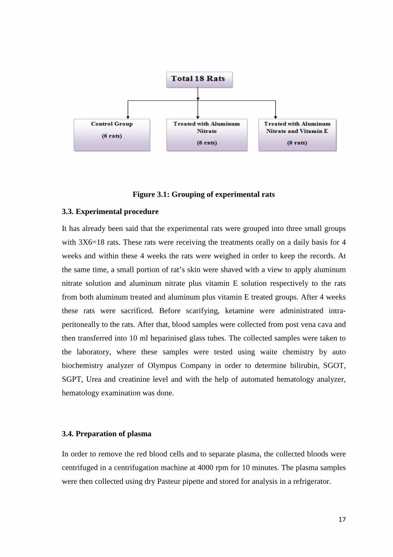
Figure 3.1: Grouping of experimental rats
3.3. Experimental procedure
It has already been said that the experimental rats were grouped into three small groups
with 3X6=18 rats. These rats were receiving the treatments orally on a daily basis for 4
weeks and within these 4 weeks the rats were weighed in order to keep the records. At
the same time, a small portion of rat’s skin were shaved with a view to apply aluminum
nitrate solution and aluminum nitrate plus vitamin E solution respectively to the rats
from both aluminum treated and aluminum plus vitamin E treated groups. After 4 weeks
these rats were sacrificed. Before scarifying, ketamine were administrated intra-
peritoneally to the rats. After that, blood samples were collected from post vena cava and
then transferred into 10 ml heparinised glass tubes. The collected samples were taken to
the laboratory, where these samples were tested using waite chemistry by auto
biochemistry analyzer of Olympus Company in order to determine bilirubin, SGOT,
SGPT, Urea and creatinine level and with the help of automated hematology analyzer,
hematology examination was done.
3.4. Preparation of plasma
In order to remove the red blood cells and to separate plasma, the collected bloods were
centrifuged in a centrifugation machine at 4000 rpm for 10 minutes. The plasma samples
were then collected using dry Pasteur pipette and stored for analysis in a refrigerator.
17

3.5. Histopathology
At first, the livers, kidneys, ovaries and skins of two rats and brain of a rat from each
group were collected. After that the fat of these organs was removed and kept in a
container containing 10% neutral buffered formalin and with the help of graduated
ethanol (50-100%) the organs were dehydrated. Then the organs were cleared in xylene
and embedded in paraffin. After staining with hematoxylin and eosin dye, the organ
tissues were sectioned and examined under the light microscope (Arsad, Esa, & Hamzah,
2014) (Ikawa, et al. 2002).
3.6. Statistical Analysis
Statistical analyses were carried out using the Portable IBM SPSS Statistics version 19.
For continuous metric variables, mean and standard deviation, standard error were used
as descriptive measures, while the independent t-test was used for comparisons when the
data could be shown to be normally distributed
18

CHAPTER 4
RESULTS
4.1. Results
From the collected blood samples, two types of tests were performed. The first test is
hematology test, and another test is clinical biochemistry examination. The liver, kidney,
skin, ovary and brains of the rats were collected in order to conduct the histopathological
test.
4.2. Body weight
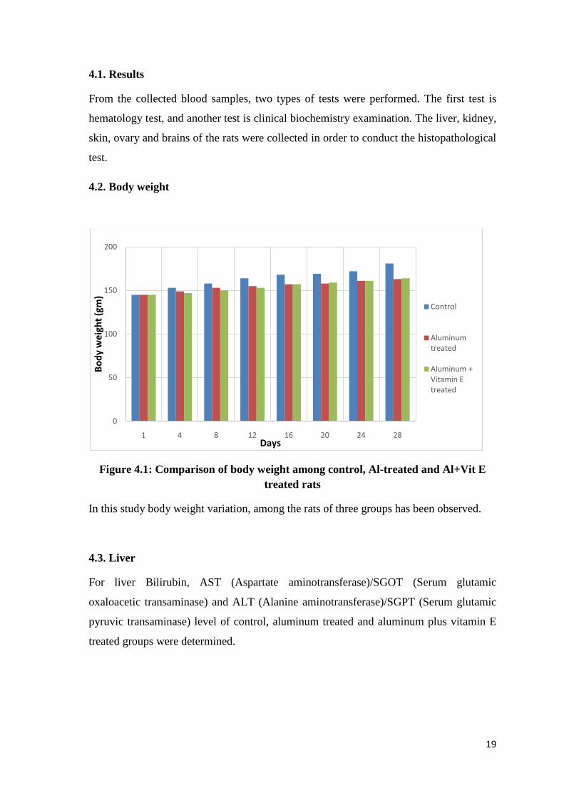
Figure 4.1: Comparison of body weight among control, Al-treated and Al+Vit E treated rats
In this study body weight variation, among the rats of three groups has been observed.
4.3. Liver
For liver Bilirubin, AST (Aspartate aminotransferase)/SGOT (Serum glutamic
oxaloacetic transaminase) and ALT (Alanine aminotransferase)/SGPT (Serum glutamic
pyruvic transaminase) level of control, aluminum treated and aluminum plus vitamin E
treated groups were determined.
0
50
100
150
200
1 4 8 12 16 20 24 28
Body
wei
ght (
gm)
Days
Control
Aluminum treated
Aluminum + Vitamin E treated
19

Table 4.1: Statistical output for liver
In case of liver toxicity, vitamin E showed reduction capacity of aluminium toxicity
significantly (p<0.001 and 0.000).
4.3.1. Histopathology output of liver
*Blue arrow = Normal *Red arrow = Inflamed
Parameters Control Treated with
Al Treated with
Al+Vit E p-value Mean (±SE)
Bilirubin 0.22 (±0.02) 0.24 (±0.02) 0.25 (±0.2) 0.001
SGOT 156.83 (±10.85) 144.40 (±6.95) 169.20 (±6.67) 0.000
SGPT 67.67 (±3.96) 57.20 (±2.63) 47.20 (±2.46) 0.000
Figure 4.2: Histopathological Study of rat liver from control group
Figure 4.3: Histopathological Study of rat liver from Al -
treated group
20

The histopathology tests of liver also proved that vitamin E has the capacity to reduce the
aluminum toxicity in liver.
4.4. Kidney
Since from theory and also from different studies it is found that the two important
parameters for kidney test are the determination of urea and creatinine level, in this study
these to parameters were also observed.
Table 4.2: Statistical output for kidney
Parameters Control Treated with Al
Treated with Al+Vit E
p-value
Mean (±SE) Urea 30.18 (±0.59) 29.94 (±1.85) 28.64 (±0.50) 0.000
Creatinine 0.36 (±0.03) 0.36 (±0.03) 0.35 (±0.02) 0.000
Figure 4.4: Histopathological Study of rat liver from Al + Vit E treated group
21

After analyzing the data, it has been shown that vitamin E can reduce aluminium toxicity
significantly (p<0.000). Here the two utilized parameters were urea and creatinine.
4.4.1. Histopathology output of kidney
Figure 4.5: Histopathological Study of rat kidney from control group
Figure 4.6: Histopathological Study of rat kidney from Al -
treated group
Figure 4.7: Histopathological Study of rat kidney from Al + Vit E group
22
Similar to liver, histopathlogy study of kidney indicated that vitamin E has the capacity
to reduce the aluminium toxicity in kidney.
4.4. Histopathology output of skin, ovary and brain
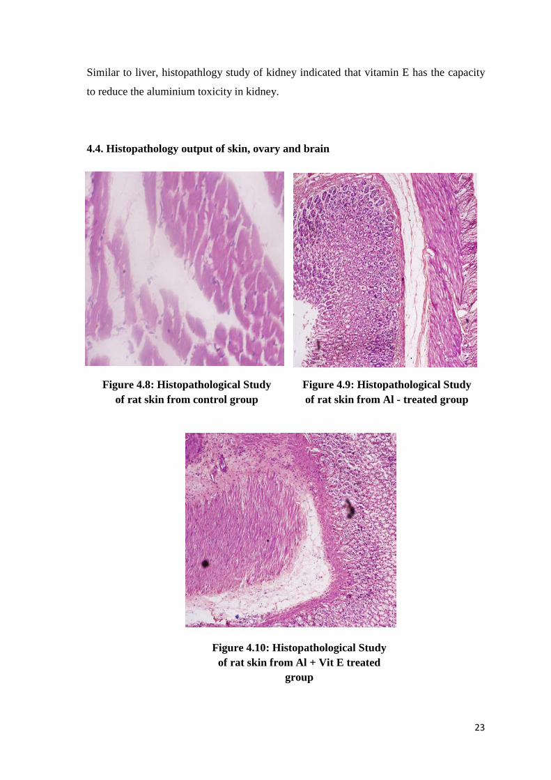
Figure 4.8: Histopathological Study of rat skin from control group
Figure 4.9: Histopathological Study of rat skin from Al - treated group
Figure 4.10: Histopathological Study of rat skin from Al + Vit E treated
group
23

Figure 4.11: Histopathological Study of rat ovary from control group
Figure 4.12: Histopathological Study of rat ovary from Al - treated group
Figure 4.13: Histopathological Study of rat ovary from Al + Vit E treated
group
24

Histopathlogy study of skin, ovary and brains of the rat have not showed any visual
changes.
Figure 4.14: Histopathological study of rat brain from control
group
Figure 4.15: Histopathological study of rat brain from Al-treated
group
Figure 4.16: Histopathological study of rat brain from Al+Vit E treated group
25
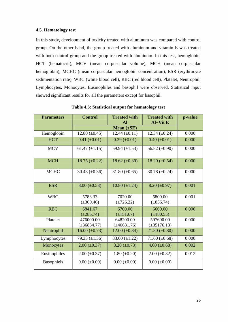
4.5. Hematology test
In this study, development of toxicity treated with aluminum was compared with control
group. On the other hand, the group treated with aluminum and vitamin E was treated
with both control group and the group treated with aluminum. In this test, hemoglobin,
HCT (hematocrit), MCV (mean corpuscular volume), MCH (mean corpuscular
hemoglobin), MCHC (mean corpuscular hemoglobin concentration), ESR (erythrocyte
sedimentation rate), WBC (white blood cell), RBC (red blood cell), Platelet, Neutrophil,
Lymphocytes, Monocytes, Eusinophiles and basophil were observed. Statistical input
showed significant results for all the parameters except for basophil.
Table 4.3: Statistical output for hematology test
Parameters Control Treated with Al
Treated with Al+Vit E
p-value
Mean (±SE) Hemoglobin 12.80 (±0.45) 12.44 (±0.11) 12.34 (±0.24) 0.000
HCT 0.41 (±0.01) 0.39 (±0.01) 0.40 (±0.01) 0.000
MCV 61.47 (±1.15) 59.94 (±1.53) 56.82 (±0.90) 0.000
MCH 18.75 (±0.22) 18.62 (±0.39) 18.20 (±0.54) 0.000
MCHC 30.48 (±0.36) 31.80 (±0.65) 30.78 (±0.24) 0.000
ESR 8.00 (±0.58) 10.80 (±1.24) 8.20 (±0.97) 0.001
WBC 5783.33 (±300.46)
7020.00 (±726.22)
6800.00 (±856.74)
0.001
RBC 6841.67 (±285.74)
6700.00 (±151.67)
6660.00 (±180.55)
0.000
Platelet 476000.00 (±36834.77)
648200.00 (±40631.76)
597600.00 (±35176.13)
0.000
Neutrophil 16.00 (±0.73) 12.00 (±0.84) 21.80 (±0.80) 0.000
Lymphocytes 79.33 (±1.36) 83.00 (±1.22) 71.60 (±0.68) 0.000 Monocytes 2.00 (±0.37) 3.20 (±0.73) 4.60 (±0.68) 0.002
Eusinophiles 2.00 (±0.37) 1.80 (±0.20) 2.00 (±0.32) 0.012
Basophiels 0.00 (±0.00) 0.00 (±0.00) 0.00 (±0.00)
26

CHAPTER 5
DISCUSSION

5.1. Discussion
Among the total mineral components of earth, about 8% contains aluminum (Jiang,
Chen, Han, Tang, & Smith, 2008) and this trivalent cation is found in plant tissues as
well as in most of the animals. Moreover, aluminum is mostly found in combination with
other elements as it is highly reactive in nature (Bernardo, 2015). Since it is abundantly
found in the environment, for different purposes aluminum is being used. However,
universally aluminum has acknowledged as a poison (Adekunle, 2008) and reported to
exert toxic effect particularly when excess amount of aluminum is accumulated in the
body through different sources for instance, water, dietary source, environment etc
(WHO, 2003). The main mechanisms of this toxicity are inhibition of the activity of
enzyme and alternation in nuclic acid function through protein synthesis which leads to
change in cell membrane permeability. As aluminum toxicity has recently become a
matter of concern, several researches have been started to conduct with a view to find
natural dietary sources that can reduce this toxic effect of aluminum on health (Bendich
& Machlin, 1987) (Hodaman, Steer, & Arsenault, 1988)
This present study illustrated that the growth of the adult female Wistar rats of control
group is normal but compare to control, the growth of aluminum treated rats is less. The
same result was also shown with aluminum nitrate conducted by Domingo, et al. (1987).
An interesting finding of this study is that, the average weight of the rats from aluminum
plus vitamin E treated group was less initially. However, the trend of increasing the body
weight has started with time. This finding indicated that if the study could be carried out
for long time, it would give similar effect with control group. Furthermore, the resist of
body weights of the rats was focusing on the fact that the food consumption of the rats
might be affected by aluminum or it might cause any genetically changes responsible for
body weight gain (Domingo, et al. 1987).
Moreover, a previous study was performed to observe the effect of vitamin E in reducing
aluminum hepatotoxicity which was showed positive results (Abubakar, et al. 2003) and
an analogues result has been found from this study. The clinical biochemistry test for
bilirubin (p <0.001), SGOT and SGPT have showed a significant results (p <0.000)
indicating that bilirubin, SGOT and SGPT level were increased in the rat from aluminum
treated groups. Whereas, aluminum plus vitamin E treated group has showed the
decreased level of bilirubin, SGOT and SGPT significantly. These findings focused on
27

the fact that vitamin E has the potentiality to reduce aluminum hepatotoxicity as
bilirubin, SGOT and SGPT are considered as parameters for identifying hepatotoxicity.
Furthermore, liver histopathology has showed that although there were no gross changes
occurred in hepatic cells of control group, some changes have been observed in
aluminum treated group and aluminum plus vitamin E treated group. However, in
aluminum treated rat’s liver it is observed that most of the central veins are injured due
to aluminum toxicity, whereas some of the central veins in aluminum plus vitamin E
treated rat’s liver are present in normal architecture like control group indicating that the
inflammation caused by aluminum toxicity is reduced by the effect of vitamin E.
Besides that, urea and creatinine level of the experimental rats were determined and
compared as these two are considered as important parameters for determining diseases
related to kidney. Like bilirubin, SGOT and SGPT, urea and creatinine level in
aluminum plus vitamin E treated group has exhibited significant with aluminum treated
group (p <0.000). From this study, it can be said that in aluminum treated group, oral
administration of aluminum nitrate have developed nephrotoxicity which is indicated by
the accelerated level of urea. Beside that the result for aluminum plus vitamin E treated
group revealed that the nephrotoxicity developed by aluminum nitrate can be reduced by
vitamin E and similar result was found from a previous study (Abdel-Hamid, 2013).
Moreover, in histopathology study, remarkable changes have been observed for kidneys
of aluminum treated group which were absent in control group and aluminum plus
vitamin E treated group.
Moreover, for analyzing the changes in skin, ovary and brain, histopathology test was
performed. Although aluminum is considered as a reason for Alzheimer disease (Foster,
2000) and even is known as a neurotoxic metal, (Martac, Podgorac, & Sekulic, 2010) no
noticeable changes have been found from histopathology study of the brain. Since
different studies have indicated that aluminum has toxic effect on brain (Krishnan,
McLachlan, Krishnan, Fenton, & Harrison, 1988) (Martac, et al. 2010), the present study
might also show the similar effect if it could be performed for more time. Besides that, as
vitamin E has showed definite result on eliminating aluminum toxicity for liver and
kidney, it can be expected that vitamin E would also show similar result for
neurotoxicity. However, no visual changes have been observed for aluminum treated and
aluminum plus vitamin E treated group in case of skin and ovary.
28
Finally, the hematology test reports of aluminum plus vitamin E treated group have also
showed significant results compare to aluminum treated group especially for hemoglobin
(p< 0.000), HCT (p< 0.000), MCV (p< 0.000), MCH (p< 0.000), MCHC (p< 0.000),
ESR (p< 0.000), WBC (p< 0.001), RBC (p< 0.000), Platelet (p< 0.000), Neutrophil (p<
0.000) and, Lymphocytes (p< 0.000) but in case of monocytes (p< 0.002), eusinophiles
(p< 0.012) and basophiles (p< 0.003) it did not show any specific changes. After
analyzing the hematology report of aluminum treated group, negative changes were
observed in most of the parameters. These changes are the indications for several
diseases, for example, the increased ESR rate indicates inflammation, anemia, infection,
kidney diseases etc. (Holm, 2015). Moreover the significant changes observed in
aluminum plus vitamin E treated group is an evidence that vitamin E is capable of
reducing the blood effects that are developed from aluminum toxicity.
29

CHAPTER 6
CONCLUSION

6.1. Limitations of the study
This study was performed for 28days only. However, if the study could be carried
out for at least three months, it might give better results.
It would be better if the study could be conducted with few more groups having
different doses of aluminum and aluminum with vitamin E.
6.2. Recommendations
In case of food habit, intake of vitamin E should be increased since it may reduce
aluminum toxicity.
Aluminum cookware utensils can be replaced with other cookware that is enamel
coated.
Aluminum foil should not be used to cook or grill the foods especially acidic
foods since it has been estimated that 4 mg aluminum can be deposited to acidic
foods during cooking with aluminum foil or in aluminum cookware (Rae, 2013).
Foods, medicine, cosmetics etc. containing aluminum can be avoided.
Habit of taking natural foods containing potassium, iron etc. (Moon, Davison, &
Bandy, 1992) can be developed in order to diminish the toxic effect of aluminum.
Awareness program can be conducted more in order to prevent aluminum
toxicity.
More study should be performed on aluminum toxicity in Bangladesh.
(For details about aluminum toxicity treatment, see appendix 2)
6.3. Conclusion
Due to gradual increase of the use of metals in several industries, agriculture, domestic
and technological fields, the chance of human exposure to these metals have risen
significantly. Nevertheless, excess metals exposure can be a severe threat for both human
and plant, as they are easily distributed to the biochemical process (Mudgal. et al. 2010).
Aluminum is also extensively present in nature and several reviews on the effect of
aluminum upon human health have been reported. Still according to EFSA and JECFA
there are lacks of specific toxicological data for aluminum (SCCS, 2014). Therefore, this
study was performed to identify whether vitamin E can mitigate the venomous effect of
30

aluminum on biological systems or not. However from this study it is found that, vitamin
E which is best known for its antioxidant effect, has potential to reduce the toxic effect
on biological systems induced by aluminum. In addition to this the findings of this study
regarding aluminum toxicity have led to concern about effect of aluminum in different
organs but the satisfactory fact is that there are some substances like vitamin E that can
reduce this outcome. Although global science has made a huge progression in the area of
health sector, complains about the lacking of initiatives and specification of toxicity are
coming on constant basis. In order to dispel this complain more study need to perform to
identify the prevention of these toxicity.
31

CHAPTER 7
APPENDIX

7.1. Appendix 1: Sources of aluminum in details (Shaw & Tomljenovic, 2013)
Major sources of
Al exposure in
humans
Daily Al
intake
(mg/day)
Weekly Al
intake
(mg/day)
÷ PTWI * (1
mg/kg body
weight; for an
average 70 kg
human,
PTWI = 70
mg)
Amount
delivered daily
into systemic
circulation
(at 0.25 %
absorption
rate*)
Natural food 1–10 7–70 0.1–1 2.5–25 µ g
Food with Al
additives
1–20
(individual
intake can
exceed 100)
7–140 (700) 0.1–2 2.5–50 µ g
(250 µ g)
Water 0.08–0.224 0.56–1.56 0.1–2 0.2–0.56 µ g
Pharmaceuticals
(antacids,
buffered
analgesics, anti-
ulceratives,
anti-diarrheal
drugs)
126–5000 882–35,000
12.6–500 315–12,500 µ g
Vaccines (HepB,
Hib, Td, DTP)
0.51–4.56 NA NA 510–4560 µ
g**
Cosmetics, skin-
care products
and
antiperspirants***
70 490 NA 8.4 µ g (at
0.012 %
absorption rate)
Cooking utensils
and food
packaging
0–2 0–14 0–0.2 0–5 µ g
* PTWI (provisional tolerable weekly intake) is based on orally ingested Al; generally,
only 0.1-0.4 % of Al is absorbed from the gastrointestinal tract; however, Al may form
32

complexes with citrate, fluoride, carbohydrates, phosphates and dietary acids (malic,
oxalic, tartaric, succinic, aspartic and glutamic), which may increase its gastrointestinal
absorption (0.5-5 %). Co-exposure with acidic beverages (lemon juice, tomato juice,
coffee) also increases Al absorption as well as conditions of Ca2+, Mg2+, Cu2+ and Zn2+
deficiency
** A single dose of vaccine delivers the equivalent of 204-1284 mg orally ingested Al
(0.51-4.56 mg), all of which is absorbed into systemic circulation
*** The risk of antiperspirants is both from dermal exposure and inhalation of aerosols.
Inhaled Al is absorbed from the nasal epithelia into olfactory nerves and distributed
directly into the brain
33

7.2. Appendix 2: Treatment of aluminum toxicity
Recognition of aluminum toxicity is considered as important part of immediate medical
treatment on the basis of risks for instance, exposure to aluminum, renal insufficiency
etc. and symptoms like anemia, osteoporosis. It includes elimination of aluminum from
the diet, TPN, dialysate, medications, antiperspirants, and an attempt at the elimination
and chelation of the element from the body's stores which can be attained through the
administration of deferoxamine through any of various routes.
• Serum aluminum level greater than 50-60 µg/L (mcg/dL) suggests aluminum
overload, may correlate with toxicity, and can be used as an indication to start
chelation therapy in symptomatic patients.
• Symptomatic patients with lower serum aluminum levels (eg, greater than 20
mcg/dL) may require chelation therapy.
• It is suggested by Kan et al that a low dose of deferoxamine therapy (2.5
mg/kg/wk) is therapeutically effective as standard dose (5 mg/kg/wk) for the
treatment of aluminum overload.
Chelation therapy with deferoxamine should be initiated in consultation with a
nephrologist and a medical toxicologist, and this can be performed upon admission.
• Deferoxamine, the metal-free ligand of the iron-chelate isolated from the
bacterium Streptomyces pilosus, is used for acute and chronic iron toxicity and
aluminum toxicity.
• It has a high affinity for ferric iron and does not affect iron in hemoglobin or
cytochromes.
No surgical care is applicable to this disorder. Hemodialysis is performed in
conjunction with deferoxamine as therapy for whole-body chelation (Bernardo,
2015).
34

7.3. Appendix 3: Aluminum neurotoxicity in various species
(Tomljenovic & Shaw, 2011).
Aluminum source/compound
Dose & duration
Route Species Neurodevelopmental adverse effects
Standard infant feeding solution
~20 μg/kg/day, >10 days
Intravenous (parenteral)
Human, premature
infants
Reduced developmental attainment at the corrected post-term age of 18 months, as evidenced by significantly lower Bayley Mental Development Index (BMDI) scores (mean loss of one point on the BMDI/day of full intravenous feeding, after adjustment for potentially confounding factors) compared to infants fed with Al-depleted solutions
Al-containing antacids
Chronic Oral Human infants
Craniosynostosis (premature ossification of the skull
and obliteration of the sutures
Al-containing dialysis fluid
(derived from Al-sulphate
treated tap water)
1 ppm, chronic (2-5
years)
Intravenous Human, kidney failure patients (15-61
years old at the start of the dialysis treatment)
Speech impairments (stuttering, dysarthria, dyspraxia, motor aphasia), movement disorders (twitches, tremors, myoclonic jerks, seizures, motor apraxia), cognitive impairments and behavioural changes (progressive dementia, paranoia, confusion,
35

psychosis), death
Al-sulphate (present as
flocculant in potable water
supplies, accidentally
released in high amounts)
500-3000 x the
acceptable limit under European
Union legislation
(0.200 mg/L),
chronic (15 years)
Oral Human adult
(female, 44 years old)
Sporadic early-onset amyloid angiopathy (Alzheimer’s-related disease), difficulty in finding words, progressive dementia, visual hallucinations headache, anxiety, cerebral ischaemia, death
Various dietary Chronic Oral Elderly human subjects
Impaired visuo-motor coordination, poor long-term memory, and increased sensitivity to flicker (correlated with high Al-serum levels
Al-oxide fumes, occupational
exposure
0.13-1.95 mg/m3, chronic
Inhalation Human, adults
(mean age 39
years)
Headache, emotional irritability, concentration difficulty, insomnia, mood liability
Various: Al-chloride,
Alphosphate, Al-powder slurry
Single sub-lethal dose
Intracerebral injection
Cats, rabbits
Decline in memory, impaired learning responses, deterioration in psychomotor control, epileptic seizures and death, neurofibrillary degeneration (resembling Alzheimer’s disease neurofibrillary Tangles
Al-hydroxide 2 injections, 2 weeks
apart
Subcutaneous injection
(behind the neck)
Mice, 3-month old
Motor neuron degeneration and apoptosis, motor function deficits, decrease in strength, cognitive deficits and decreased performance in
36

learning tasks, decrements in spatial memory, activation of microglia
Al-containing food pellets
0.5-1.7 mg/kg/day
(typical human),
chronic (22-32
months)
Oral Rats, 6-month old at the start
of treatment
Cognitive deterioration and impaired performance in learning tasks, impaired concentration, behavioral changes including confusion and repetitive behavior
Al-lactate 500-1000 ppm,
chronic (during
gestation and
lactation)
Oral Mice dams Hind limb paralysis, seizures and death (dams), lower neurobehavioral development and altered performance on a neurobehavioral test battery in pups (foot splay, forelimb and hind limb grip strengths
37

CHAPTER 8
REFERENCES

8.1. References Abdul-Hamid, G. A. (2013). Effect of Vitamin E and Selenium Against Aluminum
Induced Nephrotoxicity. VIA MEDICA , 51 (4), 312-319.
Abubakar, M. G., Taylor, A., and Ferns, G. A. (2003). Aluminum admisintration is associated with enhanced hepatic oxidant stree that may be offset by dietary vitamin E in the rat. Internation Journal of Experimental Pathology , 84 (1), 49-54.
Adekunle, B. A. (2008). Effect of oral administration of aluminum chloride on the hippocampus (brain) of wistar rats. Zaria.
Adler, A. J., and Berlyne, G. M. (1985). Duodenal aluminum absorption in the rat: Effect of Vitamin D. American Journal of Physiology , 249, 209-213.
Alfery, A. C. (1989). Physiology of Aluminum in man. New York: Mercel Dekker.
Aluminum: History. (n.d.). Retrieved February 2 , 2016, from Aluminum: http://nautilus.fis.uc.pt/st2.5/scenes-e/elem/e01300.html
Analytical Research Labs, INC. (2012). Aluminium Toxicity. Retrieved October 15, 2015, from ARL: http://www.arltma.com/Articles/AlumToxDoc.htm
Arsad, S. S., Esa, N. M., and Hamzah, H. (2014). Histopathologic Changes in Liver and Kidney Tissues from Male Sprague Dawley Rats Treated with Rhaphidophora Decursiva (Roxb.) Schott Extract. Journal of Cytology and histology , 4, 1-6.
ATSRD. (2008). Toxicological profile for aluminum. 1-357.
Bassioni, G., Mohammed, F. S., Zubaidy, E. A., and Kobrsi, I. (2012). Risk Assessment of Using Aluminum Foil in Food Preparation. Enternational Journal of Electrochemical Science , 7, 4498-4509.
Bassionil, G., Mohammed, F. S., Zubaidy, E. A., and Kobrsi, I. (2012). Risk Assessment of Using Aluminum Foil in Food Preparation. International Journal of Electrochemical Science , 7, 4498-4509.
Beijer, K., and Jernelov, A. (1986). Sources, transport and transformation of metals in the environment. Handbook on the toxicology of Metals , 1, 68-74.
Bendich, A., and Machlin, L. J. (1987). Safety of Oral Intake of Vitamin E. American Journal of Clinical Nutrition , 48 (3), 612-619.
Bernardo, J. F. (2015, April). Aluminum Toxicity Treatment and Management. Retrieved February 7, 2016, from Medscape : http://emedicine.medscape.com/article/165315-treatment#d6
CKOE. (2011). CKOE Metals. Retrieved February 12, 2016, from Pure Products: http://www.ckoemetals.com/aluminum-shot-aluminum-granules-aluminum-notched-bar-aluminum-pellet.php
Day, J. P., Barker, J., Evans, L. J., Perks, J., and Seabright, P. J. (1991). Aluminum absorption studied by 26Al trace. Lancet , 337, 1340-1345.
DFG. (2013). The Mak-Collection Part I, MAK Value Docomentation 2013.
Domingo, J. L., Llobet, J. M., Gomez, M., Tomas, J. M., and Corbella, J. (1987). Nutritional and toxicological effects of short-term ingestion of aluminum by the
38
rat. Research communications in chemical pathology and pharmacology , 56 (3), 409-419.
Domingo, J. L., Paternain, J. L., Llobet, J. M., and Corbella, J. (1987). The effects of aluminum ingestion on reproduction and postnatal survival in rats. Life sciences , 41, 1127-1131.
Drew, R. T., Gupta, B., and Bend, J. R. (1974). Inhalation studies with a glycol complex of aluminum chloride-hydroxide. Arch. Environ. Health , 28 (6), 321-326.
Foster, H. D. (2000). How Aluminum Causes Alzheimer’s Disease: The Implications for Prevention and Treatment of Foster’s Multiple Antagonist Hypothesis. Retrieved February 7, 2016, from Orthomolecular.org: http://www.orthomolecular.org/library/jom/2000/articles/2000-v15n01-p021.shtml
Geller, T. (2007). Aluminum: Common metal, uncommon past. Retrieved February 3, 2016, from Chemical Heritage Foundation: http://www.chemheritage.org/discover/media/magazine/articles/25-4-aluminum-common-metal-uncommon-past.aspx?page=5
Geyikoqlu, F., Turkez, H., Bakir, T., and Cicek, M. (2013). The genotoxic, hepatotoxic, nephrotoxic, haematotoxic and histopathological effects in rats after aluminium chronic intoxication. Pubmed , 29 (9), 780-791.
Goyer, R. A., and Clarkson, T. W. (2001). Toxic Effects of Metals. (C. D. Klaasen, Ed.) The Basic Science of Poisons , 6, 811-867.
Greger, J. (1992). Dietary and other sources of aluminum intake. Aluminum in Biology and Medicine , 169, 26-49.
Greger, J. L. (1993). Aluminum Metabolism. Annual Reviews Nutrition , 13, 43-63.
Henson, M., and Chedrese, P. (2004). Endocrine disruption by cadmium, a common environmental toxicant with paradoxical effects on reproduction. Exp Biol Med , 229, 383-392.
Hicks, J. S., Hackett, D. S., and Sprague, G. L. (1987). Toxicity and aluminium concentration in bone following dietary administration of two sodium aluminium phosphate formulations in rats. Food and chemical toxicology , 25 (7), 533-538.
Hodaman, A. B., Steer, B. M., and Arsenault, A. L. (1988). Aluminum intoxication in vitamin D-deficient rats: studies of bone aluminum localization and histomorphometry before and after vitamin D repletion. Pubmed , 4, 375-384.
Holm, G. (2015, October 13). ESR Test. Retrieved February 2, 2016, from Healthline: http://www.healthline.com/health/esr
HYDRO. (2012). Aluminum. 3-56.
Ikawa, A., Suzuki, K., Yasoshima, A., Suzuki, N., Nakayama, H., Takahashi, S., et al. (2002). Age-related changes in the dorsal skin histology in Mini and Wistar rats. Histology and Histopathology , 419-426.
Jiang, H. X., Chen, L. S., Han, S., Tang, N., and Smith, B. R. (2008). Aluminum-induced effects on Photosystem II photochemistry in citrus leaves assessed by the chlorophyll a fluorescence transient. Tree Physiology , 28 (12), 1863-1871.
39

Krewski, D., Yokel, R. A., Nieboer, E., Borchelt, D., Cohen, J., Harry, J., et al. (2009). Human health risk assessment for aluminium, aluminium oxide and aluminium oxide. J Toxicol Environ Health B Crit Rev. , 34.
Krishnan, S. S., McLachlan, D. R., Krishnan, B., Fenton, S. S., and Harrison, J. E. (1988). Aluminum Toxicity to the Brain. Pubmed , 71 (1), 59-64.
Lione, A. (1983). The prophylactic reduction of aluminum intake. Food Chem Toxicol , 103-109.
Litaor, M. (1987). Aluminum chemistry: Fractionation, speciation, and mineral equilibria of soil interstitial waters of an alpine watershed, Front Range, Colorado. Geochim Cosmochim Acta , 51, 1285-1295.
Llobet, J. M., Colomina, M. T., Sirvent, J. J., Domingo, J. L., and Corbella, J. (1995). Reproductive toxicology of aluminum in male mice. Fundamental and applied toxicology , 25 (1), 45-51.
Martac, L., Podgorac, J., and Sekulic, S. (2010). Evaluation of the neurotoxic effect of aluminum on the wister rat. Archives of Biological Science Belgrade , 48 (7), 585-588.
McLaughlin, A., Kazantzis, G., and King, E. (1962). Pulmonary fibrosis and encephalopathy associated with the inhalation of aluminum dust. British Journal of International Medicine , 19 (4), 253-263.
Mitchell, J., Manning, G., and Molyneux, M. (1961). Pulmonary fibrosis in workers exposed to finely powdered aluminum. British Journal of International Medicine , 18 (1), 10-20.
Mudgal, V., Madaan, N., Mudgal, A., Singh, R. B., and Mishra, S. (2010). Effect of toxic metals on human health. The Open Nutraceuticals Journal , 3, 94-99.
Priest, N. (2004). The biological behaviour and bioavailability of aluminium in man, with special reference to studies employing aluminium-26 as a tracer: review and study update. J Environ. Monit. , 14, 287-293.
Rae, L. (2013, September 3). Poisonous Metal: How to Avoid Aluminum Toxicity at Home and Eating Out. Retrieved February 7, 2016, from Sunwarrior news: http://www.sunwarrior.com/news/poisonous-metal-avoid-aluminum-toxicity-home-eating/
Rebai, O., and Djebli, N. E. (2008). Chronic Exposure to Aluminum Chloride in Mice: Exploratory Behaviors and Spatial Learning. Advances in Biological Research , 2 (1-2), 26-33.
Reiber, S., Kukull, W., and Standish-Lee, P. (1995). Drinking water aluminum and bioavailability. Journal of Americal Water Work Association , 87 (5), 86-100.
SCCS. (2014). Opinion on the safety of aluminum in cosmetic products. SCCS.
Shaw, C. A., and Tomljenovic, L. (2013). Aluminum In The Central Nervous System (CNS): Toxicity in Humans and Animals, Vaccine Adjuvents, and Autoimmunity. Springer Science , 56 (2).
Silbergeld, E. K. (2012). Encyclopaedia of Occupational Health and Safety. Retrieved February 10, 2016, from Chapter 33- Toxicology: http://www.ilocis.org/documents/chpt33e.htm
40

Spiegel-Ciobanu, V.-E. (2009). Assessment of hazardous substance in particle form welding technology. Welding and cutting , 51, 212-215.
Tomljenovic, L., and Shaw, C. A. (2011). Aluminum Vaccine Adjuvants: Are they safe? Current Medicinal Chemistry , 18, 2630-2637.
Vasudevaraju, B. P., Govindaraju, M., Palanisamy, A. P., Sambamurti, K., and Rao, K. S. (2008). Molecular toxicity of aluminium in relation to neurodegeneration. Indian Journal of Medical Research , 128, 545-556.
Wagner, A., Bleckmann, C., England, E., Hess, K., Hussain, S., and Schlager, J. J. (2001). In Vitro Toxicity of Aluminum Nanoparticles in Rat Alveolar Macrophages. Air Force Research Laboratory , 2.
WHO. (1997). Aluminium. World Health Organization, International Programme on Chemical safety , 2.
WHO. (2003). Aluminum in Drinking-water. Guidline for drinking-water quality , 2, 1-14.
Wilhelm, M. (1994). Metals/Aluminium . In Handbook of Environmental Medicine. Landsberg.
Willey. (2012). Aluminium [MAK Value Documentation, 1991].
Wu, X., Jin, T., Wang, Z., Ye, T., Kong, Q., and Nordberg, G. (2001). Urinary calcium as a biomarker of renal dysfunction in a general population exposed to cadmium. Journal of Occupational and Environmental Medicine , 43, 898-904.
41