“a study on bacterial, fungal and parasitic agents in
TRANSCRIPT
“A STUDY ON BACTERIAL, FUNGAL ANDPARASITIC AGENTS IN INFECTIOUS KERATITIS
PATIENTS DUE TO TRAUMA IN A TERTIARYCARE OPHTHALMIC HOSPITAL”
Dissertation submitted to
THE TAMIL NADU DR. M.G.R. MEDICAL UNIVERSITY,CHENNAI, TAMILNADU
In partial fulfillment of the requirementsfor the degree of
BRANCH – IV – M.D. DEGREE(MICROBIOLOGY)
APRIL 2013.
CERTIFICATE
This is to certify that the dissertation entitled “A STUDY ON
BACTERIAL, FUNGAL AND PARASITIC AGENTS IN
INFECTIOUS KERATITIS PATIENTS DUE TO TRAUMA
IN A TERTIARY CARE OPHTHALMIC HOSPITAL” is the
bonafide work done by Dr.C.SENTHIL VADIVU, during her M.D.
Degree Branch – IV (Microbiology) is a bonafide research work
carried out by her under the direct supervision & guidance.
Prof.V.KANAGASABAIThe DeanMadras Medical College &Rajiv Gandhi Govt. General Hospital,Chennai – 3
DR.G.JAYALAKSHMI.,M.DDirector,Institute of Microbiology,Madras Medical College,Chennai- 600 003.
DECLARATION
I, Dr.C.SENTHILVADIVU, declare that, I carried out this,
work on “A STUDY ON BACTERIAL, FUNGAL AND
PARASITIC AGENTS IN INFECTIOUS KERATITIS
PATIENTS DUE TO TRAUMA IN A TERTIARY CARE
OPHTHALMIC HOSPITAL” at the Institute of Microbiology,
Madras Medical College, I also declare that this bonafide work or a
part of this work was not submitted by me or any other for any
award, degree or diploma to any other University, Board, either in
India or abroad.
This is submitted to the TamilNadu Dr.M.G.R.Medical
University, Chennai in partial fulfillment of the rules and
regulations for the M.D Degree examination in Microbiology.
Place : Chennai Dr. C.SENTHILVADIVU
Date :
ACKNOWLEDGEMENT
I humbly submit this work to the Almighty who has given the
health and ability to pass through all the difficulties in the
compilation and proclamation of this blue print.
I wish to express my sincere thanks to our Dean, Dr.
V.KANAGASABAI M.D., for permitting me to use the resources of
this institutional for my study.
I feel indebted to Prof. Dr.G.JAYALAKSHMI M.D., Director
& Professor, institute of Microbiology for her constant
encouragement, innovative ideas, erudite guidance in my study and
for being a source of inspiration in my endeavours.
My sincere thanks to Dr. K.VASANTHA M.S., Former
Director and Professor, Regional Institute of Opthalmology
government Ophthalmic Hospital, Chennai for permitting to carry
out my study.
I would like to thank My Professors Dr.NIRANJANADEVI
M.D.,D.G.O., Dr.N.S.VASANTHI. M.D., Dr.SHEELA DORRIS M.D.,
Dr.THASNEEM BANU M.D., DR.U. UMADEVI M.D., for their
valuable assistance in my study.
I extend my whole hearted gratitude to our Assistant
professor Dr.LATA SRIRAM M.Sc.,Ph.D., for her valuable guidance
in my study.
I would like thank to our Assistant professor Dr.N.RATHNA
PRIYA M.D., for her valuable guidance in my study.
I would like to thank to Dr.LILLY THERASAE M.D., Chief
Microbiologist, VRF REFERRAL LABORATORY (A unit of
Medical Research Foundation (Sankara Nethralaya Hospital
Chennai) for her valuable guidance in my study.
I would like to thanks to Dr.LALITHA M.D., Chief
Microbiologist Aravind Eye Hospital Madurai for an ideas and
guidance to my study.
I also express my sincere thank to our Assistant Professors
Dr.R.DEEPA M.D., Dr.USHA KRISHNAN M.D., Dr. LAKSHMI
PRIYA M.D., DR.SRIPRIYA M.D., DR K.G.VENKATESH M.D.,
DR. AGATHADAVID M.D., DR. B.NATESAN M.D., for their
supporting in my study.
I express my thanks and gratitude to Dr.S.RAMKUMAR
M.Sc., Ph.D., Department of Microbiology RIOGOH Chennai.
I wish to thanks Mr.ALOCIUS SUKUMAR and
Mrs. S. VIJAYALAKSHMI LAB TECHINNCIANS Microbiology
laboratory RIOGOH Chennai for their Valuable help carrying out of
this study.
I would like to thank all my colleagues and all staff of
institute of Microbiology, Madras Medical College and Chennai – 3
for their help and encouragement.
I would like to thank to Institutional Ethical Committee for
approving my study.
I also extend my thanks to all the patients who participated in
my study.
Finally I am indebted to my family members who have been
solid pillars of everlasting support encouragement and for their
heartfelt blessings.
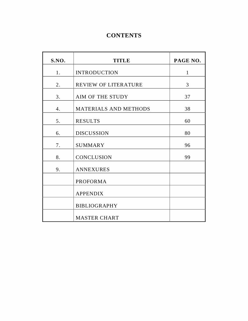
CONTENTS
S.NO. TITLE PAGE NO.
1. INTRODUCTION 1
2. REVIEW OF LITERATURE 3
3. AIM OF THE STUDY 37
4. MATERIALS AND METHODS 38
5. RESULTS 60
6. DISCUSSION 80
7. SUMMARY 96
8. CONCLUSION 99
9. ANNEXURES
PROFORMA
APPENDIX
BIBLIOGRAPHY
MASTER CHART

INTRODUCTION
1
INTRODUCTION
Inflammation of the cornea (keratitis) is characterized by
corneal oedema, cellular infiltration and associated conjunctival
reaction.
Corneal infection or infectious keratitis is one of the most
important cause of preventable blindness in the developing world.
Suppurative keratitis (corneal ulceration) occurs frequently
subsequent to corneal injury. Delay in diagnosing the nature of
infection is one of the paramount factors, which is responsible for
inappropriate initial therapy and poor outcomes. Trauma maybe
initiated by air-borne particles and is especially common in
agricultural workers. Vegetable matter such as rice husks, soil,
sand or dust, getting into the eye can cause damage to the ocular
surface. Other risk factors include contact lens wearer,
contamination of topical medications, lid abnormalities and ocular
surface diseases such as dry eye.
The reported incidence range from 11 per 1,00,000 person
years in the United States to 799 per 1,00,000 person years in the
developing nations like Nepal. In India the annual incidence is
2
reported to be 11.3 per 10,000. Infectious keratitis requires prompt
diagnosis and treatment to prevent blindness or even enucleation.
Left untreated, or treated inappropriately, the patient can go
blind in the affected eye. Corneal ulceration is an important cause
of ocular morbidity. The scarring of the cornea when the ulcer
heals can lead to significant visual impairment.
Infectious keratitis may be caused by bacteria, fungi, viruses
or protozoa. A detailed work up is necessary to arrive at a proper
diagnosis and to initiate appropriate treatment.
Considering the importance of corneal ulceration and its
impact on vision, the present study was conducted to identify the
aetiological agents and their susceptibility profiles in patients
attending a tertiary care Regional Institute of Ophthalmology and
Rajiv Gandhi Government General hospital in Chennai.
REVIEW OF LITERATURE
3
REVIEW OF LITERATURE
Corneal ulceration in the developing world is a silent
epidemic. Corneal infection is a leading cause of ocular morbidity
and blindness worldwide.56
Structure of the cornea:
The cornea consists of five layers namely:
1. The epithelium
2. Bowman’s membrane
3. Substantia propria or stroma
4. Descemet’s membrane
5. The endothelium
Nutrition of the Cornea
Cornea is an avascular structure. It derives nutrition from:
1. Perilimbal blood vessels: Anterior ciliary vessels invade the
periphery of the cornea (limbus) for about 1 mm.
2. Aqueous humor: It supplies glucose and other nutrients by
process of simple diffusion or active transport.
3. Oxygen from atmospheric air is derived directly through the tear
film.3
4
Nerve Supply
The nerve supply is purely sensory. It is derived from the
ophthalmic division of the 5 th cranial nerve through the nasociliary
branch.3
Functions
Two primary functions of the cornea are.
1. It acts as a major refracting medium.
2. It protects the intraocular contents.
This is possible by maintaining corneal transparency and
replacement of its tissues. Transparency is maintained by:
i. Regular arrangement of corneal lamellae (lattice theory of cornea)
ii. Avascularity
iii. Relative state of dehydration.3
DISEASES OF THE CORNEA
Disease of the cornea are of clinical importance as they
often leave permanent opacities, which lowers the visual acuity
and the associated complications may even lead to blindness.56
5
1. Inflammations (Kera`titis)
Bacterial keratitis
Fungal keratitis
Viral keratitis
Parasitic keratitis
2. Degenerations
a) Arcus senilis
b) Arcus juvenilis
c) Band-shaped keratopathy
d) Hereditary corneal dystrophy
e) Reis-Bucklers’ dystrophy
f) Endothelial corneal dystrophy of Fuchs
3. Ectasias
a) Keratoconus
b) Keratoglobus
6
4. Pigmentations
a) Blood Staining
b) Argyrosis
c) Kayser-Fleisher’s ring
Inflammations of the Cornea
Inflammations of the cornea (keratitis) is characterized by
corneal oedema, cellular infiltration and associated conjunctival
reaction.3
1. Exogenous infection e.g. Staphylococcus aureus, Streptococcus
pneumoniae, Pseudomonas aeruginosa, E.coli, Proteus spp,
Klebsiella spp, H.influenzae, etc. common. Usually organisms in
the conjunctival sac, lacrimal sac (dacryocystitis), infected foreign
body, etc. cause inflammation of the cornea.
2. From the ocular tissue
a. Conjunctival diseases spread to the epithelium.
b. Scleral diseases spread to the stroma.
c. Uveal tract diseases spread to the endothelium.
3. Endogenous infection – It is usually due to hypersensitivity
reaction.
7
Predisposing Factors
Keratitis due to microbial aetiology (Bacterial, Fungal, Viral
and Parasitic) has the following predisposing factor. The
following predisposing factors are the same for Bacterial, Fungal,
Viral and Parasitic agents.3,67
1. Epithelial damage due to trauma, e.g. minute foreign body,
misdirected eyelash, vegetative matter such as husks soil, sand,
dust, air-borne particle.
2. Virulent organisms, e.g. Streptococcus pneumoniae, Pseudomonas
spp, Neisseria gonorrohoae, etc.
3. Poor resistance
Xerosis and keratomalacia (vitamin A deficiency)
Protein calorie malnutrition
Corneal oedema leads to desquamation of epithelium
Neuroparalytic keratitis, e.g. Herpes zoster, leprosy
Exposure of the cornea due to proptosis, facial nerve
palsy.67
Cesar et al, in 2008 from UK reported that trauma is the
leading predisposing factor in Infectious keratitis 13 .
8
M.J. Bharathi et al,in 2003 from South India has reported
that the epidemiology and etiology of bacterial keratitis is specific
to the region. Screening patients for predisposing factors, treating
the co-existing ocular diseases, and educating them about proper
lens care and risk of infection may reduce the occurrence of
bacterial keratitis9.
B.H.Jeng et al,in 2003 from UK has concluded that risk
factors for infectious keratitis included contact lens use (55%),
ocular surface disease (16.6%), trauma (11.9%), and bullous
keratopathy (1.3%)47.
Reema nath et al from Upper Assam in 2011 has concluded
that injury with vegetative matter as the most common risk
factor81.
M.Srinivasan et al in 1997 from Madurai studied that
corneal injury (65.4%) was the major predisposing factor in the
aetiology of Infectious keratitis94.
Sadia Seth et al from Peshawar in 2010 has reported that
ocular trauma was the most common cause found in 39% of
patients84.
9
Youhanna HW Ibrahuin et al in 2009 from UK as has
concluded that wearing of the contact lens was the main
predisposing factor (31%)in infectious keratitis patients 109.
Many studies have reported well recognized association of
contact lens wear with Acanthamoeba keratitis and fungal keratitis
97.
Ocular trauma particularly with vegetative matter is a well
known predisposing factor in fungal keratitis54.
Dry, dusty and windy environment have a increased risk of
microtrauma to the cornea, resulting in an increasing incidence of
fungal keratitis during these seasons77.
Stages of Corneal Ulcer
Stages of Corneal Ulcer are categorised into 3 stages namely3
1. Progressive stage
There is grey zone of infiltration by polymorphs.
Localised necrosis and sloughing of sequestrum is
present.
Saucer-shaped ulcer with overhanging edges due to
oedema is characteristic.
10
2. Regressive stage
The dead material is thrown off and the oedema
subsides.
The floor and edges of the ulcer are smooth and
transparent.
3. Healing stage
Minute superficial vessels grow in from the limbus near the
ulcer.
There is formation of fibrous tissue which fills the gap. The
irregular arrangement of fibrous tissue results in opacity, as the
new fibres refract the light irregularly. As Bowman’s
membrane never regenerates, permanent opacity remains, if it
is damaged.
Symptoms
1. Pain - Cornea is richly supplied by ophthalmic division of the
trigeminal nerve.
2. Photophobia – There is undue sensitivity to light.
3. Impairment of visual acuity occurs due to corneal opacity.
4. Lacrimation – There is excessive reflex tear production.
11
Signs
1. Blepharospasm – There is tight closure of the eyelids specially in
children.
2. Corneal opacification occurs due to infiltration and oedema.
3. Ciliary congestion with conjunctival hyperaemia is present.
4. Hypopyon or pus in the anterior chamber may be present.
COMPLICATIONS
1. Corneal opacity
2. Ectatic cicatrix (Keratectasia)
3. Descemetocele (Keratocele)
4. Perforation
EPIDEMIOLOGY
Dr. Rajan K. Anand in 2010 from Bihar studied that corneal
ulcer is a common vision threatening condition among the rural
population, next only to cataract. The annual incidence of corneal
ulcer in India is reported to be 11.3 per 10,000080.
Singh SK et al from Nepal in 2011 studied that the incidence
of corneal ulceration in Nepal is one of the highest reported in the
world. The Bhaktapur Eye study revealed it to be 799 per 100,000
12
population per year. (Upadhyay et al, 2001) which is seven times
higher than in South India (Gonzales et al , 1996) and seventy
times greater than that reported in the USA (Erie JC et al, 1993)93.
M.Srinivasan et al in 1997 from Madurai has concluded
increased incidence of infectious keratitis in males (65%)94.
Sadia Seth et al in 2010 from Peshawar (India) studied
that the incidence of microbial keratitis was high in males
(67%)84.
Youhanna HW Ibrahim et al in 2009 from UK has concluded
predominance of corneal ulcer in female (54%)109.
B.H. Jeng et al in 2003 in US studied that the highest rate of
infectious keratitis was found in females (63%)47,48.
Males are more commonly affected than females, but in the
agricultural population the incidence may be equal or more in
females50.
13
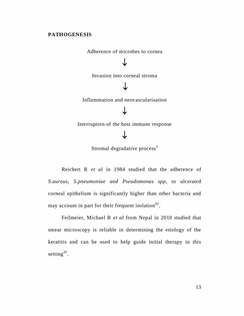
PATHOGENESIS
Adherence of microbes to cornea
Invasion into corneal stroma
Inflammation and neovascularisation
Interruption of the host immune response
Stromal degradative process3
Reichert R et al in 1984 studied that the adherence of
S.aureus, S.pneumoniae and Pseudomonas spp, to ulcerated
corneal epithelium is significantly higher than other bacteria and
may account in part for their frequent isolation82.
Feilmeier, Michael R et al from Nepal in 2010 studied that
smear microscopy is reliable in determining the etiology of the
keratitis and can be used to help guide initial therapy in this
setting26.
14
Acanthamoeba species enter through minor abrasions in the
cornea produced by contact lens or external injury. In the cornea it
elicits inflammation with hypopyon formation. Further
progression of infection leads to perforation75.
BACTERIAL KERATITIS
Bacterial keratitis is a loss in the continuity of the corneal
epithelium associated with tissue infiltration and necrosis.3
Etiology
Bacterial keratitis is always an exogenous infections are
common due to pyogenic organisms which invade the cornea from
outside such as Staphylococcus spp, Streptococcus pneumoniae,
Pseudomonas spp, E.coli, etc.103
The common causative bacterial organisms of corneal ulcer
are as follows:
i. Gram-positive cocci – Staphylococcus aureus, Staphylococcus
epidermidis, Streptococcus hemolyticus, S.pneumoniae.
ii. Gram-negative cocci – Neisseria gonorrheae, N. meningitides.
iii. Gram-positive bacilli – Nocardia asteroides, Corynebacterium
diphtheriae.
15
iv. Gram-negative bacilli – Pseudomonas aeruginosa, Proteus spp,
Klebsiella spp, Moraxella spp,Hemophilus spp, Escherichia coli,
etc.
v. Mycobacteria – Mycobacterium tuberculosis, M.leprae.
Three pathogens can invade normal intact epithelium:
i. Neisseria Gonorrhoeae
ii. Neisseria Meningitides
iii. Corynebacterium Diphtheriae
There has also been a change in the spectrum of bacteria
causing keratitis with time88. Staphylococcus spp, Pseudomonas
spp and Streptococcus spp appear to be the predominant causes of
bacterial corneal ulcer in United States37.
Similarly common bacterial pathogens in most of the studies
in India are Staphylococcus aureus, Staphylococcus epidermidis
and Pseudomonas spp65.
Although relatively uncommon, keratitis caused by
Acinetobacter spp and Serratia marcescens have been
documented60.
16
FUNGAL KERATITIS
Etiology:
Fungal keratitis is commonly caused by Candida albicans,
Aspergillus fumigatus, Fusarium spp, Pencillium spp, Acremonium
spp, curvularia spp, Bipolaris, etc.3
Among the filamentous fungi, Aspergillus spp is the most
common followed by Fusarium spp. Which are most prevalent in
agricultural areas.
Candida albicans is the commonly affecting yeast like
fungas in the immunocompromised host.3
Incidence:
Fungal keratitis is common in rural agricultural areas, and
usually occurs due to ocular trauma involving vegetable matter, e.g.
thorn, sharp wooden stick, wheat and paddy husk, branches of tree,
etc.37
Predisposing Factors
Addition to the common predisposing factor responsible for
keratitis are same as for bacterial keratitis
17
Indiscriminate use of topical or systemic steroids alters host
defence mechanism and hence fungal keratitis is found to be more
common in immunocompromised host.51
Symptoms
Pain, Photophobia, Impairment of vision, Lacrimation are
commonly seen. But they are less prominent than equal-sized
bacterial ulcer.The presence of yellow patch in the cornea is
mainly seen in the fungal keratitis.3
Signs
1. A typical lesion is a yellow-white coloured ulcer with
indistinct margin, with minimum vascularization.
2. Fungal keratitis is dry in appearance with small satellite
lesions around the ulcer due to the stromal infiltration with
delicate feathery, finger-like hyphate edges protruding into
adjacent stroma.
3. Ulcer margin is often elevated above the surface.
4. Massive hypopyon is present commonly, which is dense and
organized.
18
5. Slit-lamp examination – Endothelial plaque and immune
ring may be seen around the ulcer. Some degree of
iridocyclitis is usually present.
Jayahar bharathi et al in 2007 from south India studied that
the incidence of fungal keratitis (66%) was more with agricultural
workers where as the bacterial keratitis (57%) was more common
in non - agricultural workers5.
Jagadish chander et al, from Chandigarh in 2008, reported that
the prevalent organisms involved in microbial keratitis were
Aspergillus spp. (41.18%), Fusarium spp (27%), Candida spp
(8.82%), Curvularia spp(5.88%) and Bipolaris spp (5.88%)40.
Boucier T et al, in 2003 from US has concluded that the
most common causative organisms are bacteria although fungi and
protists are also pathogens12.
M.Srinivasan et al, from Madurai in 1997 has concluded
that S.pneumoniae (44.3%) was the predominant organism
followed by Pseudomonas spp and the most common fungal
pathogen isolated was Fusariuim spp (47. 1%) followed by
Aspergillus spp. (16.1%)94.
19
Usha Arora et al,in 2009 from Amristar has reported that
Aspergillus spp was the most common isolate followed by
Fusariuim spp, Penicillium spp and Curvularia spp102.
Species of Penicillium, Alternaria, Curvularia, Bipolaris,
Acremonium, Aureobasidium were isolated frequently in studies
conducted in various parts of India and Nepal15,52.
Feilmeir et al, in 2010 from Nepal has reported that fungal
organisms are the most common cause of infectious keratitis
inpatient population. Aspergillus spp (35%) among fungus and
S.pneumoniae among bacteria were the most common organisms
responsible for keratitis26.
Lai1a Aktar et al, in 2009 from Bangladesh studied that
Pseudomonas spp (24%), S.pneumoniae (17%), Aspergillus spp
(13%), Fusariuim spp (7 %) and Curvularia spp (6%) were found
as pathogens causing suppurative corneal ulcer58.
Mycotic corneal ulceration although not reported as a major
cause of keratitis in developed countries is an important cause of
ulcerative keratitits in tropical regions including India83.
20
Species of Aspergillus genus especially A.fumigatus,
A.flavus and A.niger were the predominate fungal pathogens in
studies conducted in most parts of India100.
Some studies in South India showed that Fusarium spp to be
more common than Aspergillus spp. Fusarium spp have also been
found to be the principal fungal pathogen in Florida, Paraguay,
Singapore, Nigeria, Tanzania and Hong Kong. This phenomenon
may be explained by difference in climate and the natural
environment23.
Jagadish Chandar et al, in 1993 studied that 8% fungal
corneal ulcer were caused by Acremonium spp in Chandigarh.41
Namrata Kumara et al, in 2002 reported mycotic keratitis in Patna
documented 3.94% isolates as Acremonium spp.71
Verenkar M P et al, in 1998 study in Goa concluded 12.5%
of corneal ulcer shows Penicillium spp103. Namrata kumara et al,
in 2002 reported mycotic keratitis in Patna documented 7.89%
isolates as of Penicillium spp.71
Willi’s eye hospital, Philadelphia in May 2002 reported that
Candida albicans was the most common agent in infectious
keratitis107.
21
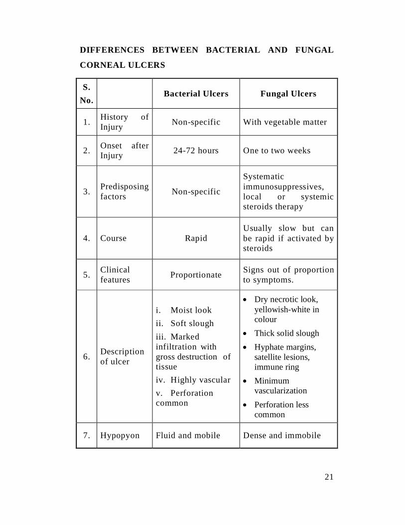
DIFFERENCES BETWEEN BACTERIAL AND FUNGAL
CORNEAL ULCERS
S.No.
Bacterial Ulcers Fungal Ulcers
1. History ofInjury Non-specific With vegetable matter
2. Onset afterInjury 24-72 hours One to two weeks
3. Predisposingfactors Non-specific
Systematicimmunosuppressives,local or systemicsteroids therapy
4. Course RapidUsually slow but canbe rapid if activated bysteroids
5. Clinicalfeatures Proportionate Signs out of proportion
to symptoms.
6. Descriptionof ulcer
i. Moist lookii. Soft sloughiii. Markedinfiltration withgross destruction oftissueiv. Highly vascularv. Perforationcommon
Dry necrotic look,yellowish-white incolourThick solid sloughHyphate margins,satellite lesions,immune ringMinimumvascularizationPerforation lesscommon
7. Hypopyon Fluid and mobile Dense and immobile
22
Liesegang and Foster in South Florida in 1999 studied in six
hundred and sixty three patients (663), the fungal isolates
contribute 20.1%, among the isolates Fusarium spp were the most
common and Aspergillus spp was the next pathogen being
isolated.61
Upadhyay et al, in Nepal reported that Aspergillus spp was
to be predominant fungal pathogen and Fusarium spp were less
commonly isolated101.
Savithri Sharma et al, from Madurai studied that Fusarium
spp showed high prevalence among the isolates88.
ACANTHAMOEBA KERATITIS
Acanthamoeba keratitis has gained importance recently
because of its increasing incidence, difficulty in diagnosis and
unsatisfactory treatment.3
Etiology
Acanthamoeba keratitis is caused by Acanthamoebae, which
are free living protozoans found in air, soil and fresh or brackish
waters. They exist in both active (trophozoite) and dormant
(cystic) forms.97,87
23
Predisposing Factors
1. Acanthamoeba keratitis may occur following a minor corneal
abrasion.
2. Contact lens wearers who use distilled water and salt tablets
instead of commercially prepared saline solutions for their
lens care are at particular risk.
3. The fall of dust particles, trauma due to vegetable matter,
contact with contaminated water etc have been found to be
the predominant risk factors for Acanthamoeba keratitis.3
M.Jayahar Bharath et al,2009 reported that trauma due to
vegetable matter was the major risk factor of Acanthamoeba
keratitis.7 The study on Acanthamoeba keratitis by S.Sharma et al
2000 was similar to the above study.90
Symptoms
Severe pain out of proportion to the degree of inflammation along
with watering, photophobia, blepharospasm and blurred vision, is the
characteristics features of Acanthamoeba keratitis3
24
Signs
Acanthamoeba keratitis evolves over several months as a
gradual worsening keratitis with periods of temporary remissions.3
1. Initial lesions of Acanthamoeba keratitis are in the form of
coarse and opaque streaks. Fine epithelial and subepithelial
opacities are also seen.
2. Advanced cases show a central or paracentral ring-shaped
lesion with stromal infiltrates. There is an overlying
epithelial defect.
3. Severe cases show associated radial keratoneuritis, in the
form of perineural infiltrates along corneal nerves.
Diagnosis
1. Clinical Diagnosis: It is difficult and is usually made by
exclusion and with strong clinical suspicion in non-
responsive patients being treated for viral, bacterial and
fungal keratitis.
2. Laboratory Diagnosis: Staining of Corneal scrapings are
helpful in identification of Acanthamoeba cyst.
25
a. Potassium hydroxide mount is reliable in experienced
hands for recognition of Acanthamoeba cysts.
b. Calcofluor white stain is a chemifluorescent dye which
stains the cysts of Acanthamoeba bright apple green.
c. Lactophenol cotton blue stained film is also useful for
demonstration of Acanthamoeba cysts in corneal
scrapings.
d. Culture on non-nutrient agar (E.coli enriched) showed
trophozoites within 48 hours which gradually become
cysts. E.coli prevents other organisms to grow whereas
Acanthamoeba thrives on it.
3. Confocal Microscopy: Acanthamoebae cysts can be
demonstrated in optically cut parallel sections of cornea
under confocal microscopy.3
DIAGNOSTIC TECHNIQUES
Many fungal ulcers demonstrate no striking morphologic
pattern and often it is not possible to differentiate clinically
between fungal keratitis and bacterial keratitis25.
26
To determine the causative organism meticulous collection
of microbiological specimens is of critical importance. The
corneal ulcer is scraped for microscopy, culture and for further
investigations if indicated74.
MICROSOPIC EVALUATION OF SMEARS
Gram stain
The smear is prepared from corneal scrapings and Direct
Gram staining done to observe the bacteria and yeast like cell51.
Bharathi et al, in 2006 studied 100% sensitivity of Gram
stain procedure in the diagnosis of bacterial keratitis6.
Feilmeier, Michael R et al, from Nepal in 2010 studied that
smear microscopy is reliable in determining the etiology of the
corneal infection and can be used to help guide initial therapy in
this setting26.
Noopur Gupta et al, in 2008 studied that smears prepared by
corneal scraping and Gram staining done to observe the bacteria
and yeast cells74.
Giemsa stain is also useful to distinguish bacteria, fungi and
Acanthamoeba. Chlamydia inclusion bodies can also be identified
27
with Giemsa stain79.
10% Potassium Hydroxide Mount (KOH)
Corneal scrapings were placed on a glass slide with 10%
KOH to see the fungal elements.64
In 1985 Araffa et al, concluded that KOH staining was as
effective, much easier and less expensive than calcofluor white
staining for detection of fungi in corneal tissue2.
In 2007, Bharathi et al, reported that a potassium hydroxide
smear is of greater diagnostic value in the diagnosis of fungal
keratitis, Nocardia keratitis and Acanthamoeba keratitis5.
1988, Sharma et al, reported that KOH preparation
demonstrated fungus in 100 percent of total culture positive
cases.88
Sharma et al, in 2000 concluded that 10% KOH mount could be
used to demonstrate Acanthamoeba cysts in corneal scrapings there
by permitting rapid presumptive diagnosis of Acanthamoeba
Keratitis.90
28
Lactophenol cotton Blue mount (LPCB)
Corneal scraping are placed over a clean glass slide and a
drop of lactophenol cotton blue stain is added over the specimen
and a coverslip is placed taking care to avoid trapping of air
bubbles. It was found to be effective for demonstration of fungal
structures and Acanthamoeba cysts in corneal scrapings.50
Calcofluor white stain
Calcofluor white is a water soluble colourless textile dye and
fluorescent whitener. It selectively binds to chitin and cellulose of
the fungal cell wall. It fluoresces light blue when exposed to ultra
violet light (346-365nm)42.
The corneal scrapings are placed on a clean glass slide and 1
drop of 0.1% calcofluor white with 0.1% Evans blue and 1 drop
10% KOH are added. A coverslip is placed over the specimen and
examined under fluorescent microscope. The morphology of
smaller fungal elements was better appreciated in calcofluor white
mount92.
Acridine orange stain
Acridine orange dye has an affinity for nucleic acid. When
fungi are stained with this dye, RNA component of the cell
29
fluoresces with shades of orange red and DNA component
fluoresces green under fluorescent microscope.42
Acanthamoeba cysts fluoresces bright yellow to orange. This
stain has been used for direct examination of corneal scrapings in
cases of Acanthamoeba Keratitis42.
Culture
Corneal scraping from patients with infectious keratitis due
to trauma were inoculated on to Blood Agar Plate (BAP),
Chocolate Agar Plate (CAP) for identify Bacterial etiology and to
Sabouraud’s Dextrose Agar (SDA) for fungal etiology and to Non
- Nutrient agar (NN) media with lawn culture of E.coli for
parasitic etiology.
Wihelmus et al, in 1994 studied that the culture media
recommended for evaluation of suspected microbial keratitis have
the potential to support the growth of the principal bacteria and
fungi responsible for keratitis.98,107
‘O’Brien et al, in 1994 reported that the SDA agar should
not contain cycloheximide which may inhibit the saprophytic
fungi commonly responsible for ocular infection76.
30
Slide culture technique
The slide culture is used to study morphology without
disturbing, details particularly relationship between reproductive
structure like conidia, conidiophores and hyphae. Fungal slide
culture was performed in cases with doubtful morphology 55.
MOLECULAR DIAGNOSIS:
Polymerase Chain Reaction (PCR)
Sujith venayil et al, in 2009 studied that although PCR has
several advantages due to its rapid and wide spread applicability
to bacteria, fungi and viruses, the technique has various reported
complexities and drawbacks as evidenced from their study also
some of the limitations are logistic and some are technical54,94.
TREATMENT OF BACTERIAL KERATITIS
Principles
1. Control of Infection: Infection is controlled by intensive
local use of antibiotic drops. Broad spectrum antibiotic
drops, ointment, are given frequently 4-6 times a day.
Subconjunctival injections may also be given once or twice
daily. Culture and sensitivity should be done before the
application of antibiotics.
31
2. Cleanliness: Irrigation with warm saline or sodabicarb
lotion is advised to wash away necrotic material, toxin,
secretion and pathogenic organisms.
3. Heat: Heat prevents stasis and encourages repair of the
ulcer. Hot fomentation may be given.
4. Rest: 1% atropine either as drops or ointment is applied 2-3
times a day. It paralyses the ciliary muscles and provides
comfort to the eye by preventing ciliary spasm. There is
associated iritis always in cases of corneal ulcer due to
penetration of endotoxin across the endothelium in the
anterior chamber. It also prevents most of the dangerous
complications of iritis. Pad and bandage give rest to the
eyeball by restricting its movements.
5. Protection: Pad and bandage protect the eye from dust,
wind and harmful external agencies. A shield of dark glasses
are used, if there is associated conjunctival discharge to
avoid retention of secretion, which in turn favours bacterial
growth due to warmth and stasis.
Topical administration is the method of treatment of choice,
because it provides a rapid high level of drug in the cornea and
anterior chamber.96
32
‘O’ Brien TP et al, in 1995 and Panda A et al, in 1991
concluded that initial regimens of fluroquinolone or aminoglycoside
combined with a cephalosporin is effective in approximately 95%
of cases of bacterial keratitis.76,77
Amikacin is a semi synthetic aminoglycoside that is useful
in the treatment of infection due to gram negative infection
resistant to gentamycin.79
In Pseudomonas keratitis ciprofloxacin is the drug of
choice.79
TREATMENT OF FUNGAL KERATITIS
Principles
1. Scraping and debridement of the ulcer is useful in drug
penetration.
2. 1% atropine eyedrops or ointment controls associated iritis
and prevents synechiae formation.
3. Antifungal drugs – The available antifungal drugs are
mainly fungistatic.
33
MEDICAL THERAPY
Antifungal drugs
The role of these drugs is limited due to few approved
antifungal drugs and their poor penetration. Topical antifungals
are to be instilled for a long-time, as the response is often
delayed.3
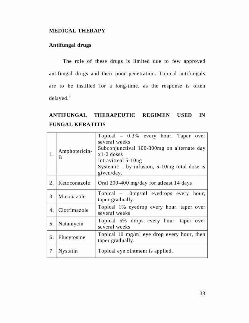
ANTIFUNGAL THERAPEUTIC REGIMEN USED IN
FUNGAL KERATITIS
1. Amphotericin-B
Topical – 0.3% every hour. Taper overseveral weeksSubconjunctival 100-300mg on alternate dayx1-2 dosesIntravitreal 5-10ugSystemic – by infusion, 5-10mg total dose isgiven/day.
2. Ketoconazole Oral 200-400 mg/day for atleast 14 days
3. Miconazole Topical – 10mg/ml eyedrops every hour,taper gradually.
4. Clotrimazole Topical 1% eyedrop every hour. taper overseveral weeks
5. Natamycin Topical 5% drops every hour. taper overseveral weeks
6. Flucytosine Topical 10 mg/ml eye drop every hour, thentaper gradually.
7. Nystatin Topical eye ointment is applied.
34
a. Topical
i. Natamycin (5%) eyedrops is instilled 1 hourly. It is effective
against the most common fungi.
ii. Miconazole (1%) eye ointment is applied 5 times daily.
iii.Nystatin eye ointment is applied 5 times daily. It is only
effective against Candida spp and is less potent.
iv. Topical Amphoterecin B (0.25%) is instilled 1 hourly and is
effective against Aspergillus spp and Candida spp.
SYSTEMIC
Systemic antifungals are indicated, if the infection spreads
to the sclera and there is impending perforation. e.g. oral
ketoconazole or fluconazole 200 mg daily may be given for 2-3
weeks.
Cycloplegics such as atropine is used to prevent posterior
synechiae formation and to control iritis by paralyzing the ciliary
muscle. It also causes vasodilatation.3
35
Corticosteroids are contraindicated as they enhance fungal
growth.
Thomas PA et al, in 2003 from India reported that
Natamycin (5%) (or) Amphotericin B (.15%) remain the drug of
choice for superficial keratitis.99
Pankaj K Agarwal et al, in 2001 from Calcutta concluded
that Itraconazole is effective in treating mycotic corneal ulcers.78
Mohan et al, in 1989, reported success rate of 64.7%, when
1% Miconazole was used to treat smear positive keratitis.68
Newer agents such as triazoles (Posaconazole and Ravuconazole),
Echinocandins, Sodarin derivates and the Nikkomycins might
improve the treatment of fungal keratitis in future.24,27
TREATMENT OF ACANTHAMOEBA KERATITIS
Treatment of acanthamoeba keratitis is usually
unsatisfactory, but the treatment mentioned below is usually
followed.3
1. Non-specific treatment is on the general lines for corneal ulcer.
2. Specific medical treatment includes:
36
a. Topical treatment
1. Propamidine isethionate (Brolene) 0.1% drops are usedhourly.
2. Neomycin drops
3. Polyhexamethylene biguanide (0.01-0.02%) drops areused hourly
4. Chlorhexidine drops
5. Paromomycin drops
6. 1% Clotrimazole drops
7. Polymyxin B drop
b. Systemic treatment
Oral ketoconazole 200mg may be given four times a day for
2-3 weeks.
c. Penetrating ketatoplasty is frequently required in non-
responsive cases.
It is difficult to treat Acanthamoeba Keratitis. Chlorhexidine
and Polyhexamethylene biguanide (PHMB) are the most effective
drugs against trophozoites and cysts and are recommended as the
first line therapy for Acanthamoeba keratitis. Medications have to
be continued for 3-6 months after clinical resolution of infection
to prevent relapses.3
AIM OF THE STUDY
37
AIM OF THE STUDY
To isolate and identify the bacterial, fungal and
parasitic aetiological agents in patient with infectious
keratitis due to trauma.
To analysis the correlation between aetiological
agents of infectious keratitis due to trauma with the
occupation of the patients.
To evaluate the antibiotic susceptibility pattern of the
bacterial isolates and study the beta lactamase
production for their resistant isolates.
To study the antibiotic susceptibility pattern of the
fungal isolates by agar dilution and broth micro
dilution method.
To compare the antifungal susceptibility pattern for
the fungal isolates by agar dilution and broth micro
dilution method.

MATERIALSAND
METHODS
38
MATERIALS AND METHODS
PERIOD OF STUDY
This is a cross sectional study undertaken over a period of
one year from October 2011 to September 2012.
PLACE OF STUDY
This study was carried out at the Institute of Microbiology,
Madras Medical College, Chennai and Regional Institute of
Ophthalmology and Government General Hospital, Chennai.
STUDY GROUP
All patients presenting in the outpatient department with the
history of trauma and signs and symptoms of infectious corneal
ulcer such as pain, redness, watering of the eye, diminished vision
and photophobia were included in the study.
ETHICAL CONSIDERATIONS
Written consent to participate in the study was obtained
from the patients or their guardians after providing full
39
explanation of the study. This study was reviewed and approved
by the Institutional Ethical Committee, Madras Medical College &
Rajiv Gandhi Government General Hospital, Chennai-3. All data
were handled confidentially and anonymously.
COLLECTION OF SPECIMENS:
Written consent from the participants (or) their guardians
included in the study, was obtained after providing full
explanation of the current study in their local language. All the
data collected were kept confidential.
Specimens were collected from patients with infectious
keratitis due to trauma, as per the proforma (Annexure) Informed
consent from the patients and data were collected. Corneal
scrapings were collected for the investigations by the
Ophthalmologist.
Patient was made to lie down comfortably on a couch. The
affected eye was cleansed with sterile normal saline using sterile
swabs. Sterile 2% Xylocaine was applied to the eye, taking care
not to apply too much, as it may inhibit the growth of the
organism. Care was taken to see that the eyelids did not
40
contaminate the specimens. Eye speculum was used whenever
necessary. No.15 and Bard Parker Blades were used to obtain
scrapings from the ulcer.
The scraping were inoculated in a C-streak pattern on
culture media (Blood Agar, Potato Dextrose Agar, Chocolate Agar
and Non-nutrient Agar). The scraping is done by the
Ophthalmologist under an operating microscope.
Direct Gram’s stain and and 10% KOH wet mount were
made on the direct scraping.
Incubate the Blood Agar and Chocolate Agar Plates at 37oC
in the presence of 5% CO2 for 2-7 days.
Sabouraud’s Dextrose Agar slant and Potato dextrose agar
slant were incubated at 250C and 350C aerobically.
The culture plates and slants were observed for the growth
of organisms every day / week. If bacterial growth was observed,
staining (Gram’s, Acid Fast, Modified Acid Fast) was performed.
Biochemical tests were done to identify the bacterial and
fungal pathogens. Antibiotic susceptibility pattern was performed
41
to identify the susceptibility pattern of the isolates to the
antibiotics.
If the fungal growth was observed, Lactophenol Cotton Blue
(LPCB) staining was performed and the fungus was identified
based on the spore morphology.
After 48-72 hours observe non - nutrient agar plate under
low power and high power for the presence of Acanthomoeba cyst.
The cyst present were further confirmed by calcofluor stain.
SPECIMEN PROCESSING
The following test were performed on the scrapings that
were collected.
1. Gram Staining
2. Modified Acid Fast Staining
3. KOH Wet Mount
4. Giemsa Staining
5. Lactophenol Cotton Blue Staining(LPCB)
6. Slide culture method
7. Calcoflour staining

42
1. Gram Staining Procedure
Thin smear of the specimen was prepared on a clean and
sterile glass slide. Then the smear was fixed by heating over a
Bunsen burner flame. The smear was flooded with 1% crystal
violet for 1 minute and washed with distilled water. The smear
was flooded with Gram’s iodine for 1 minute and washed with
distilled water and decolorized with acetone, washed with distilled
water and counter stained with dilute carbol fuschin for 30
seconds.
2. Modified Acid Fast Staining
Thin smear of the specimen was prepared and dried in the
air. The smear was fixed by heating over a Bunsen burner flame.
The smear was flooded with strong carbol fuschin stain for 5
minutes. Washed with distilled water and flooded with 1%
sulphuric acid for 3 minutes. Washed with distilled water and
counter stained with 3% methylene blue for 3 minutes. Washed
with distilled water dried and examined under oil immersion
objective of the microscope.
3. KOH Wet Mount
A clean glass slide was taken. The specimen was placed in
43
the centre of the slide. A drop of 10% KOH was added and a
coverslip was placed over that and observed under the low and
high power of the microscope.
4. Giemsa Staining
Air dried and fixed smear with absolute methanol for 2-3
minutes, was stained with Giemsa stain for 30 minutes, then the
smear was allowed to air dry and observed under the oil
immersion objectives of the microscope.
EXAMINATION OF INOCULATED MEDIA:
Sabouraud’s Dextrose Agar slopes were observed
periodically for growth at 25oC and 35oC and if it was inadequate,
they were reincubated. The Sabouraud’s dextrose agar slopes were
examined daily during first week and twice a week for the next
three weeks. Failure of growth even after six weeks was
considered as negative for fungal growth and were discarded.
LACTOPHENOL COTTON BLUE STAIN:
The fungal growth was taken from Sabouraud’s dextrose
agar slope with spud and transferred onto the clean glass slide and
two to three drops of Lactophenol cotton blue stain was added
over the fungal growth. By using teasing needles the growth was
44
spread over the slide and coverslip was placed without trapping
any air bubbles. The morphology of hyphae, conidia were
observed under microscope and was correlated with macroscopic
features.
RIDDLE’S SLIDE CULTURE METHOD:
This method was used to study the undisturbed
morphological details of fungi particularly relationship between
reproductive structures like conidia, conidiophores and hyphae.
Fungal slide culture was performed in cases with doubtful
morphology.
A round piece of filter paper was placed on the bottom of a
sterile Petri dish. A pair of thin glass rods was placed on top of the
filter paper to serve as support for the 3 inch x 1 inch glass
microscope slide. 3 to 4 coverslips were placed within the
Petridish and sterilized as a whole.
1x1 cm square block of Sabouraud’s Dextrose Agar was cut
from a Petridish by using sterile scalpel and transferred to the
microscope slide.
45
Four sides of the agar block were inoculated with the fungal
colony which is to be studied by using heavy 24 gauge nichrome
wire. The agar block was covered with sterile coverslip in the
Petridish. Moisten the filter paper with sterile water and place the
lid on the Petridish.
The Petridish was incubated at room temperature and
examined periodically for growth. When the growth appeared to
be mature, the coverslip was gently lifted from the surface of the
agar with a pair of forceps taking care not to disturb the mycelium
adhering to the bottom of the coverslip.
The coverslip was placed on a small drop of Lactophenol
cotton blue on a second glass slide. Likewise, the mycelium
adhering to the surface of the original glass slide after the block
was removed, also was stained with Lactophenol cotton blue stain
and a fresh coverslip was overlaid.
The characteristic shape and arrangement of hyphae, conidia
were observed microscopically.
The mycelia which adhere to the glass surface usually show
characteristic microscopic appearance which may be lost if
46
needles are used to tease, as it happens in the routine Lactophenol
cotton blue mounts. The slide culture may also be seen directly by
placing under the low power of the microscope.
The cellophane tape preparation has come into greater use to
overcome the obstacles of time consumption and requirement of
extra equipment to prepare the slide culture. A piece of tape is
gently laid over a portion of the fungal colony and slowly lifted to
remove an area of the colony and placed on a microscope slide
with a drop of Lactophenol cotton blue stain and examined under
the low power objective of the microscope. This preparation
becomes an instant slide culture, revealing relationship of the
various fungal structures.
INTERPRETATION OF BACTERIAL CULTURE :
Bacterial isolates were identified by means of colony
morphology, Gram staining, motility and biochemical reactions by
standard microbiological techniques as recommended by Clinical
and Laboratory Standards Institute (CLSI)
47
Interpretation of Fungal Culture
Inoculated SDA slopes were incubated at 250C and 350C for
a minimum of six weeks before discarding as negative. These
slants were inspected daily during the first week and twice weekly
during the next three weeks for growth.
Identification of filamentous fungi was done by preparing
Lacto Phenol Cotton Blue mount and studying the morphology of
hyphae and conidial arrangement. In difficult, ambiguous cases
where sporulation was inadequate, Riddle’s slide culture technique
was performed.
In case of yeasts, identification was done by Gram’s stain
morphology, germ tube test, morphology on corn meal agar, and
biochemical tests by standard microbiological techniques as
recommended by CLSI.
INTERPRETATION OF PARASITIC CULTURE
Corneal scrapings inoculated on to a non - nutrient agar
plate for the cultivation of free living amoeba, where observed,
after 48-72 hours observed under low power and high power
48
objective of the microscope for the presence of Acanthaomoeba
cyst. The Acanthaomoeba cyst present were further confirmed
by calcofluor stain.
Trophozoites of Acanthamoeba are 14-15 micrometer in size
and actively motile at 37oC and have numerous spiny
acanthopodia. Cysts are smaller 10-25 mm, double walled, with
wrinkled outer wall (ecto cyst) and a stellate polygonal inner wall
(endo cyst)
ANTIMICROBIAL SUSCEPTIBLITY TESTING:
Antibiotic susceptibility testing was performed by the Kirby
Bauer method on Mueller Hinton Agar (MHA) according to the
CLSI protocols. The diameters of zones of inhibition were
interpreted according to CLSI standards for each organism. Media
and discs were tested for quality control using standard strains.
The following standard strains were used
1. Staphylococcus aureus - ATCC 25923
2. Escherichia coli - ATCC 25922
3. Pseudomonas aeruginosa - ATCC 27853
49
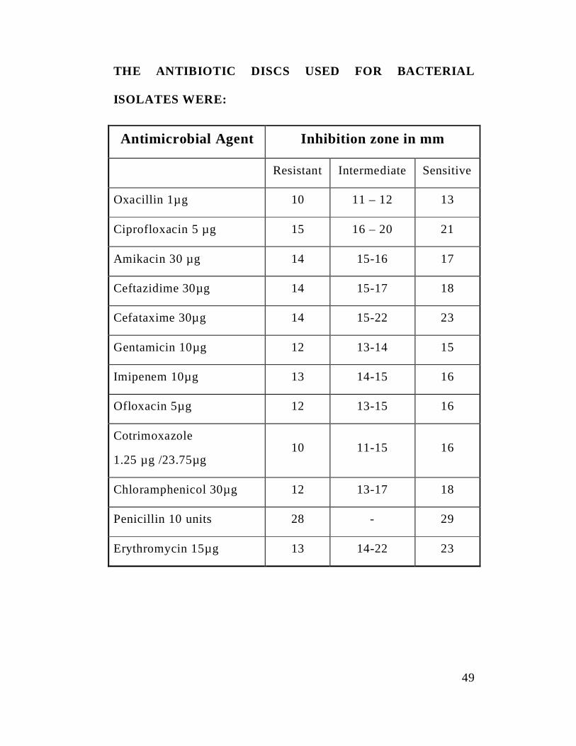
THE ANTIBIOTIC DISCS USED FOR BACTERIAL
ISOLATES WERE:
Antimicrobial Agent Inhibition zone in mm
Resistant Intermediate Sensitive
Oxacillin 1µg 10 11 – 12 13
Ciprofloxacin 5 µg 15 16 – 20 21
Amikacin 30 µg 14 15-16 17
Ceftazidime 30µg 14 15-17 18
Cefataxime 30µg 14 15-22 23
Gentamicin 10µg 12 13-14 15
Imipenem 10µg 13 14-15 16
Ofloxacin 5µg 12 13-15 16
Cotrimoxazole
1.25 µg /23.75µg10 11-15 16
Chloramphenicol 30µg 12 13-17 18
Penicillin 10 units 28 - 29
Erythromycin 15µg 13 14-22 23
50
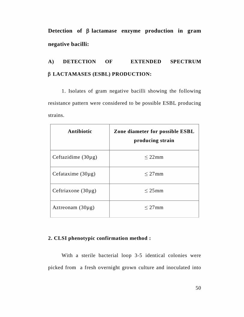
Detection of lactamase enzyme production in gram
negative bacilli:
A) DETECTION OF EXTENDED SPECTRUM
LACTAMASES (ESBL) PRODUCTION:
1. Isolates of gram negative bacilli showing the following
resistance pattern were considered to be possible ESBL producing
strains.
Antibiotic Zone diameter for possible ESBL
producing strain
Ceftazidime (30µg) 22mm
Cefataxime (30µg) 27mm
Ceftriaxone (30µg) 25mm
Aztreonam (30µg) 27mm
2. CLSI phenotypic confirmation method :
With a sterile bacterial loop 3-5 identical colonies were
picked from a fresh overnight grown culture and inoculated into
51
5ml of nutrient broth. The broth was incubated at 350C for 2-4 hrs
and the turbidity matched with 0.5 McFarland’s standard. Lawn
culture of the test organism was made on to MHA plate. Antibiotic
discs like Ceftazidime (CAZ 30µg) and Ceftazidime / Clavulanic
acid (CAZ / CA / 30µg / 10 µg) (Himedia, Mumbai) were placed
onto the plate and incubated at 350C overnight. A 5mm increase
in zone diameter for Ceftazidime tested in combination with
Clavulanic acid than its zone when tested alone confirmed an
ESBL producing organism.
DETECTION OF METHICILLIN RESISTANCE IN
STAPHYLOCOCCUS AUREUS :
1. Disc Diffusion Method
Colonies isolated from agar culture plate were suspended directly
into broth, vortexed to reach 0.5 McFarlands standard. A lawn culture of
the Staphylococcal colonies was made on the MHA plate and Oxacillin
or Cefoxitin disc were applied. Incubation was at 350C for 24 hours in
ambient air. According to CLSI criteria with 1 g Oxacillin disc,
diameters of 10, 11-12, 13mm corresponded to categorization as
resistant, intermediate or susceptible. With 30µg Cefoxitin disc diameter
52
of 19 or 20mm corresponded to resistant or susceptible to Oxacillin.
2. Minimum Inhibitory Concentratin (MIC) for detecting
Vancomycin resistance :
1. Culture media cation adjusted Mueller Hinton Broth (pH 7.2-7.4)
2. Preparation of stock antibiotic solution :
Antibiotic stock solution can be prepared using the formula
= 1000 x V x C = w P
Where p= potency of the antibiotic in relation to the base.
(For vancomycin, p=950/1000 mg : Himedia)
V= Volume of the stock solution to be prepared (10 ml)
C = final concentration of the antibiotic solution
(1024 µg /ml).
W=weight of the antibiotic to be dissolved in the volume
3. Scheme for preparing dilution of antibiotics
Arrange two rows of test tubes in the rack (1 row for the test
& 2nd for ATCC control). Using sterile syringe transfer 2ml of MH
broth to the uricol container containing the working stock solution
53
(128µg/ml concentration) From this transfer 1 ml to the first tube
in each row. Now we have 2ml of the diluted antibiotic in the
uricol container. Using sterile syringe add 2ml of MH broth to the
2ml to the second tube in each row. Repeat this procedure till the
8th tube. Place 1 ml of the antibiotic free broth in the last tube in
each row (growth control). The sterility controls for the antibiotic
solution is kept.
4. Inoculum preparation for the test and ATCC control
and incubation
Take 9.9ml of MH broth in a uricol container. Add 0.1 ml of
0.5 MCFarland turbidity matched test organism broth. Mix well,
transfer 1 ml of inoculum using 2 ml syringe to each tube
containing antibiotic dilutions and also to the control tube.
Similarly, repeat the procedure for ATCC control strain. Incubate
the rack at 370C for overnight. Observe the MIC of ATCC control
strain. If it is out of the sensitive range, the test is invalid. Read
for the test organism. The lowest concentration of the antibiotic in
which there is no visible growth will be the MIC for the drug &
for the test organism.
54
ANTI FUNGAL SUSCEPTIBILITY TESTS
The antifungal susceptibility testing testing was done by two
methods
1.Disc diffusion method
2.Agar dilution method
3.Broth microdilution method
The Clinical Laboratory Standards Institute (CLSI)
subcommittee on Antifungal susceptibility Tests has developed a
reproducible procedure for antifungal susceptibility testing of
filamentous fungi by a broth microdilution. Recently an agar
dilution method has been developed for testing filamentous fungi
by diffusion methodology.
Inoculum preparation :
All organisms were subcultured onto Potato dextrose agar,
incubated at 350C for 7 days. The culture was covered with 1 ml
of sterile 0.85% saline and a suspension prepared by gently
probing the colonies. Addition of 1 drop of Tween 20 will help
dispersion of Aspergillus conidia. The resulting mixture of conidia
55
and hyphal elements was withdrawn and transferred to a sterile
tube and allowed to settle. The uniform suspension was transferred
to a screw capped tube and vortexed. The densities of the conidia
or the sproangiospore suspensions were read and adjusted to a
optical density of 0.09-0.11 for Aspergillus spp.
Disc Diffusion Method :
Disc Diffusion test was performed on Mueller-Hinton agar plates
supplemented with 2% Glucose and 0.5µg/ml of Methylene Blue.
The entire dried agar surface was evenly streaked in three
different directions with a sterile cotton swab dipped into the
inoculums suspension. The plate was allowed to dry for 20
minutes. Using a pair of flame sterilized forceps the antifungal
discs were applied onto the surface of the inoculated plate. The
plates were incubated at 35oC for 48 hours. The plates were read
at 24 hrs and 48 hrs.
The following commercial HI-Media antifungal disks were
used.
56
Amphotericin B 100 units Itraconazole 10µg
Fluconazole 10 µg Nystatin 10 µg
The following standard strains were tested each time to
ensure quality control.
Aspergillus flavus ATCC 204304
Aspergillus fumigates ATCC 204305
INTERPRETATION:
Zone diameters were measured at the point where there was
prominent reduction of growth. The results were compared with
broth microdilution method for respective fungal isolates.
AGAR DILUTION METHOD PROCEDURE
1.8 ml of nutrient agar poured in the test tube and allowed to
cool at 50oc.
From the stock solution, 0.2ml of drug dilution added in the
descending concentration to NA slope
100 l of standardised inoculums added.
57
All the test tubes incubated 300c for 2 days.
Examining macroscopically for growth
Lowest concentration of the drug which permitted no growth
after 2-3 day is taken as MIC.
BROTH MICRODILUTION METHOD:
Amphotericin B and Itraconazole powders and voriconazole
powders were obtained form HiMedia, Mumbai and Pharma
Fabricon respectively. Their essay potency were 750 µg/mg each.
Weight (mg) = volume (ml)x desired concentration (µg/ml)
Assay potency (µg/ml)
Volume (ml) = weight (mg) x assay potency (µg/ml)
Concentration (µg/ml)
STOCK SOLUTION
Solvent used is Dimethyl sulfoxide (DMSO) for
Amphotericin B and Itraconazole and voriconazole. Stock solution
of 1600 (µg/ml) is prepared. A series of dilutions at 100 times the
58
final concentration was prepared from the antifungal stock
solution in the same solvent. Each intermediate solution was then
further diluted to final strength in the test medium. This procedure
was done to avoid dilution artifacts that result from precipitation
of compounds with low solubility in aqueous media.
Media : RPMI 1640 (with glutamine, without bicarbonate
and phenol red as pH indicator) Hi-Media, Mumbai.
INCUBATION :
All microtitre plates were incubated at 350C and examined
for MIC determination at 48 hrs.
INTERPRETATION OF MIC :
By visual examination, MIC was defined as the lowest drug
concentration that showed 100% growth inhibition compared to
the growth control well.
The test was read when the growth control shows adequate
growth, which is typically 24-48 hours for most moulds, but it
could be up to 96 hours. Read MICs the first day that the growths
59
controls showed the visible growth and then 24 hours later.
Scores were given as follows, (1) 0-optically clear (2) 1 + =
slightly hazy (3) 2+ prominent reduction in turbidity compared
with that of the drug-free growth control. 3+ = slight reduction in
turbidity compared with that of the drug-free growth control. 4+=
no reduction in turbidity compared with that of the drug-free
growth control.
STATISTICAL ANALYSIS :
A Statistical analysis was carried out using statistical
package for social science (SPCS) and Epi-info software in a
statistician. The proportional date of the cross sectional study was
tested using pearson’s chi-square analysis and binomial proportion
test.

RESULTS
60
TABLE OF RESULTS
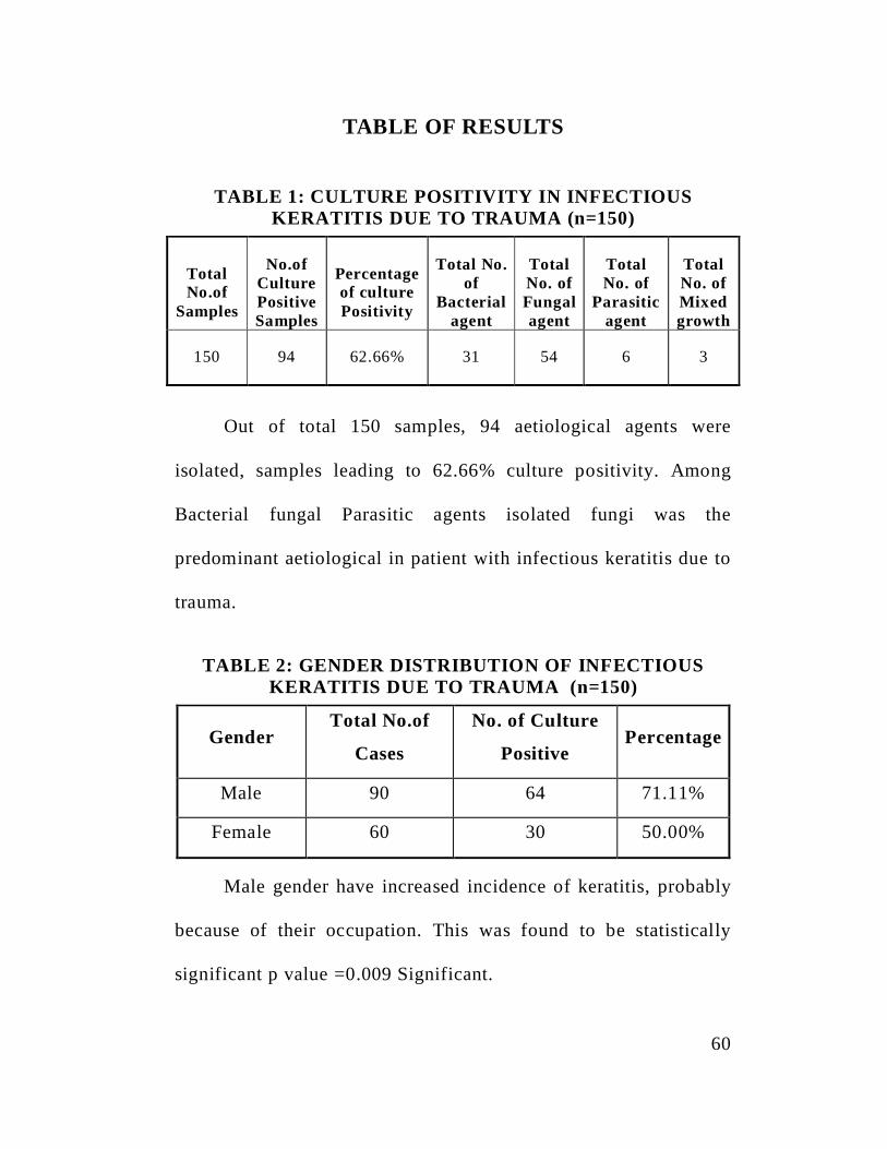
TABLE 1: CULTURE POSITIVITY IN INFECTIOUSKERATITIS DUE TO TRAUMA (n=150)
TotalNo.of
Samples
No.ofCulturePositiveSamples
Percentageof culturePositivity
Total No.of
Bacterialagent
TotalNo. ofFungalagent
TotalNo. of
Parasiticagent
TotalNo. ofMixedgrowth
150 94 62.66% 31 54 6 3
Out of total 150 samples, 94 aetiological agents were
isolated, samples leading to 62.66% culture positivity. Among
Bacterial fungal Parasitic agents isolated fungi was the
predominant aetiological in patient with infectious keratitis due to
trauma.
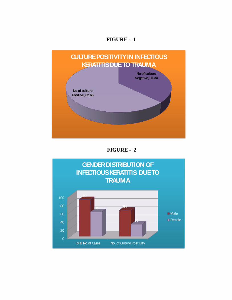
TABLE 2: GENDER DISTRIBUTION OF INFECTIOUSKERATITIS DUE TO TRAUMA (n=150)
GenderTotal No.of
Cases
No. of Culture
PositivePercentage
Male 90 64 71.11%
Female 60 30 50.00%
Male gender have increased incidence of keratitis, probably
because of their occupation. This was found to be statistically
significant p value =0.009 Significant.
No of cultureNegative, 37.34
No of culturePositive, 62.66
CULTURE POSITIVITY IN INFECTIOUSKERATITIS DUE TO TRAUMA
0
20
40
60
80
100
Total No.of Cases No. of Culture Positivity
90
6460
30
GENDER DISTRIBUTION OFINFECTIOUS KERATITIS DUE TO
TRAUMA
Male
Female
FIGURE - 1
FIGURE - 2
61
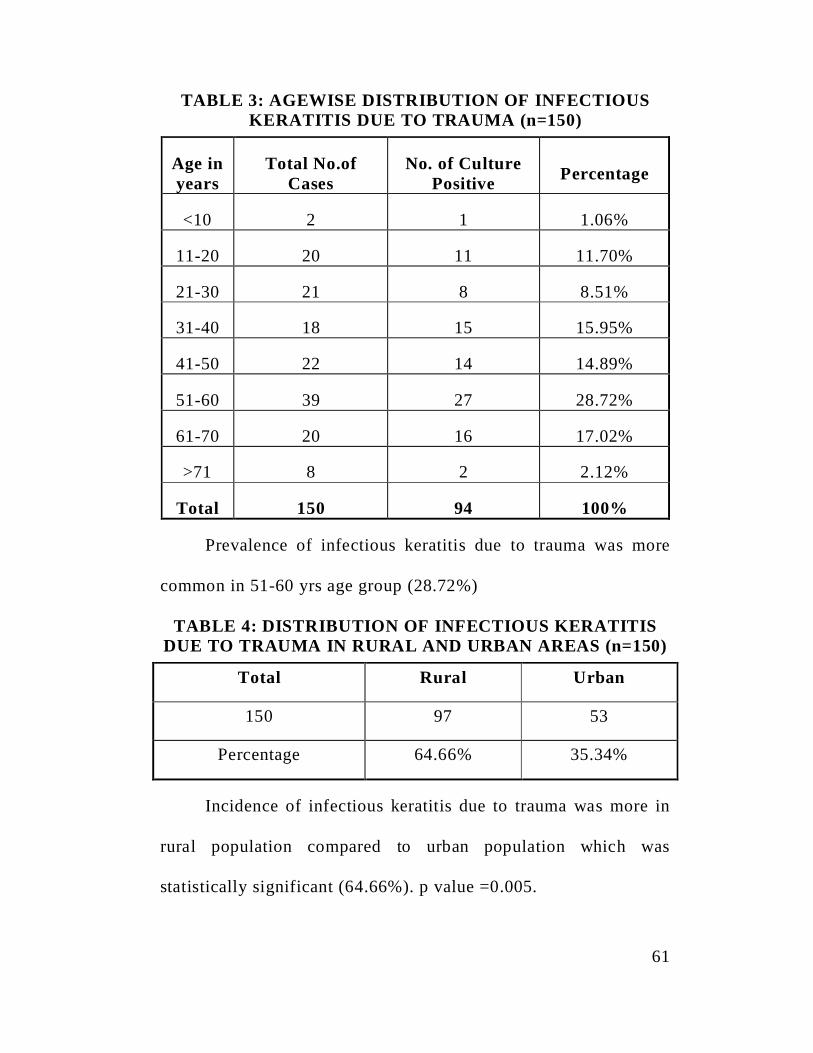
TABLE 3: AGEWISE DISTRIBUTION OF INFECTIOUSKERATITIS DUE TO TRAUMA (n=150)
Age inyears
Total No.ofCases
No. of CulturePositive Percentage
<10 2 1 1.06%
11-20 20 11 11.70%
21-30 21 8 8.51%
31-40 18 15 15.95%
41-50 22 14 14.89%
51-60 39 27 28.72%
61-70 20 16 17.02%
>71 8 2 2.12%
Total 150 94 100%
Prevalence of infectious keratitis due to trauma was more
common in 51-60 yrs age group (28.72%)
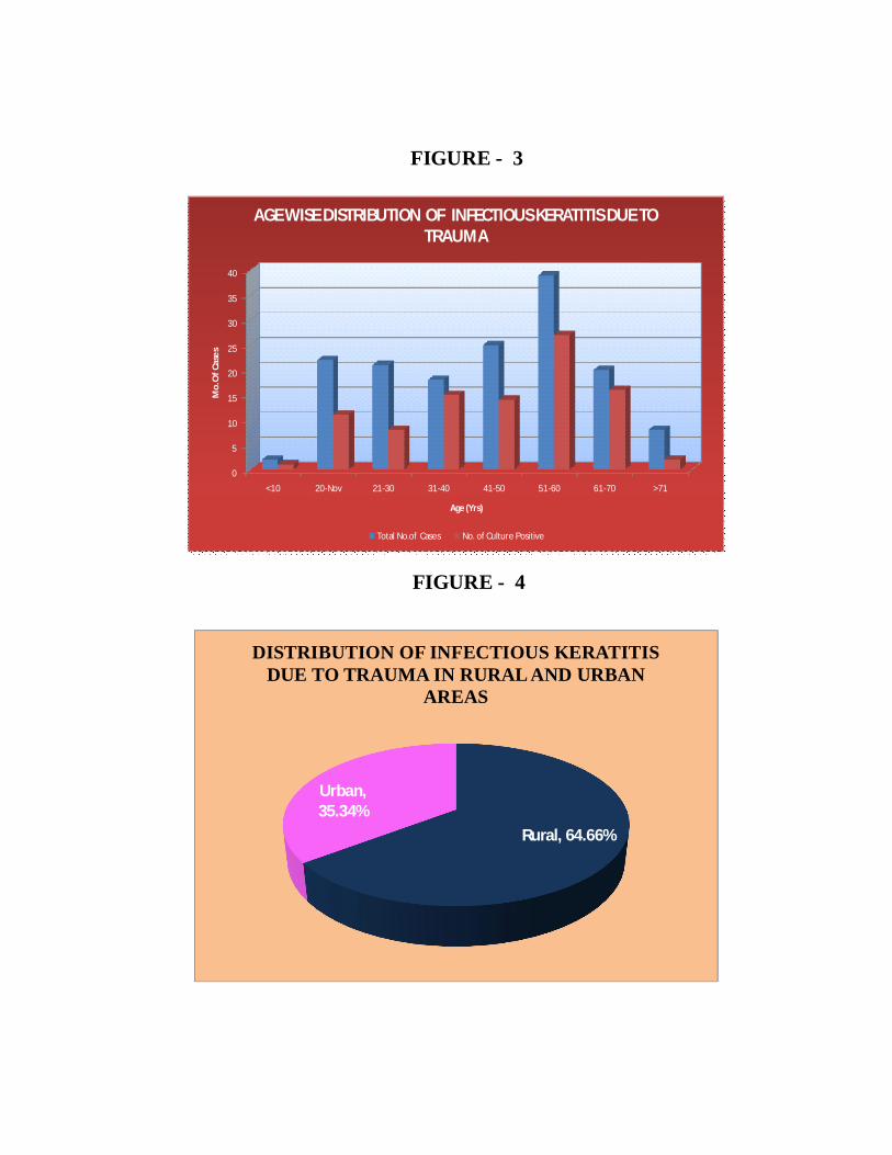
TABLE 4: DISTRIBUTION OF INFECTIOUS KERATITISDUE TO TRAUMA IN RURAL AND URBAN AREAS (n=150)
Total Rural Urban
150 97 53
Percentage 64.66% 35.34%
Incidence of infectious keratitis due to trauma was more in
rural population compared to urban population which was
statistically significant (64.66%). p value =0.005.
0
5
10
15
20
25
30
35
40
<10 20-Nov 21-30 31-40 41-50 51-60 61-70 >71
Mo.
Of C
ases
Age (Yrs)
AGE WISE DISTRIBUTION OF INFECTIOUS KERATITIS DUE TOTRAUMA
Total No.of Cases No. of Culture Positive
Rural, 64.66%
Urban,35.34%
DISTRIBUTION OF INFECTIOUS KERATITISDUE TO TRAUMA IN RURAL AND URBAN
AREAS
FIGURE - 3
FIGURE - 4
62
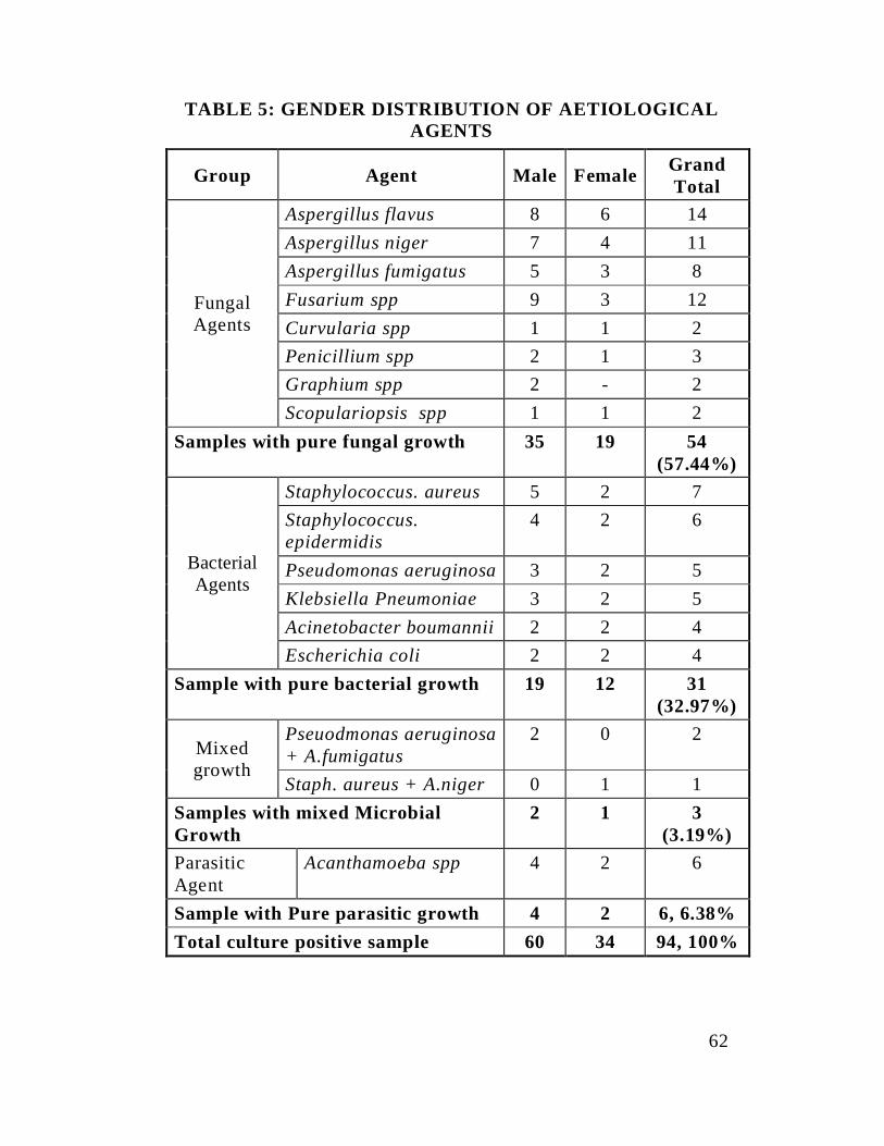
TABLE 5: GENDER DISTRIBUTION OF AETIOLOGICALAGENTS
Group Agent Male Female GrandTotal
FungalAgents
Aspergillus flavus 8 6 14Aspergillus niger 7 4 11Aspergillus fumigatus 5 3 8Fusarium spp 9 3 12Curvularia spp 1 1 2Penicillium spp 2 1 3Graphium spp 2 - 2Scopulariopsis spp 1 1 2
Samples with pure fungal growth 35 19 54(57.44%)
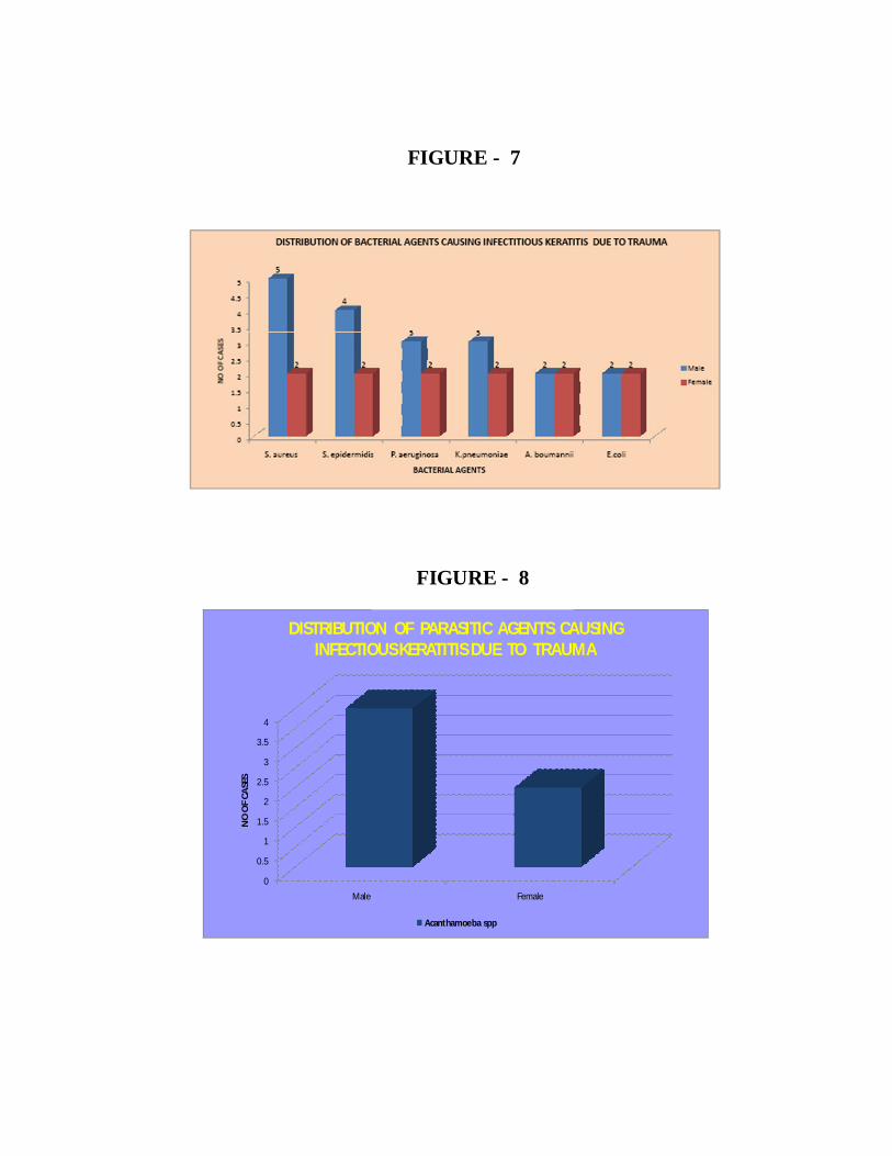
BacterialAgents
Staphylococcus. aureus 5 2 7Staphylococcus.epidermidis
4 2 6
Pseudomonas aeruginosa 3 2 5Klebsiella Pneumoniae 3 2 5Acinetobacter boumannii 2 2 4Escherichia coli 2 2 4
Sample with pure bacterial growth 19 12 31(32.97%)
Mixedgrowth
Pseuodmonas aeruginosa+ A.fumigatus
2 0 2
Staph. aureus + A.niger 0 1 1Samples with mixed MicrobialGrowth
2 1 3(3.19%)
ParasiticAgent
Acanthamoeba spp 4 2 6
Sample with Pure parasitic growth 4 2 6, 6.38%Total culture positive sample 60 34 94, 100%
05
101520253035
NO
OF
CASE
S
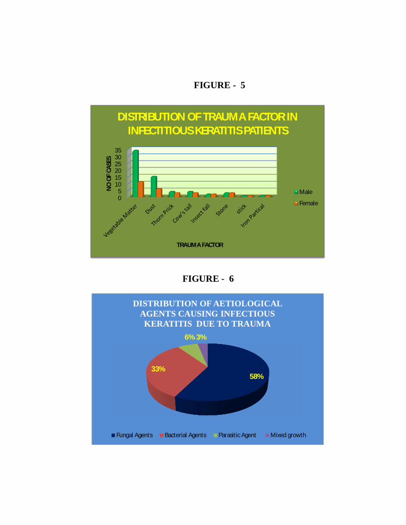
TRAUMA FACTOR
DISTRIBUTION OF TRAUMA FACTOR ININFECTITIOUS KERATITIS PATIENTS
Male
Female
58%33%
6% 3%
DISTRIBUTION OF AETIOLOGICALAGENTS CAUSING INFECTIOUSKERATITIS DUE TO TRAUMA
Fungal Agents Bacterial Agents Parasitic Agent Mixed growth
FIGURE - 5
FIGURE - 6
63
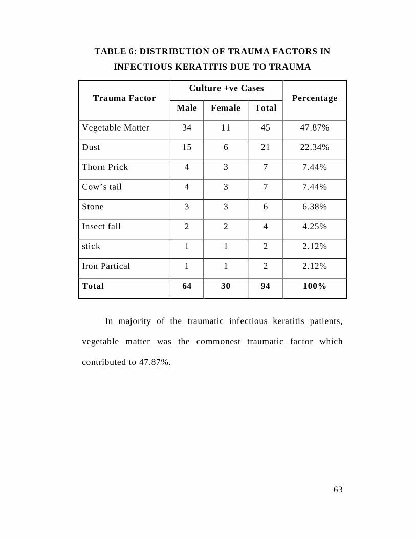
TABLE 6: DISTRIBUTION OF TRAUMA FACTORS IN
INFECTIOUS KERATITIS DUE TO TRAUMA
Trauma FactorCulture +ve Cases
PercentageMale Female Total
Vegetable Matter 34 11 45 47.87%
Dust 15 6 21 22.34%
Thorn Prick 4 3 7 7.44%
Cow’s tail 4 3 7 7.44%
Stone 3 3 6 6.38%
Insect fall 2 2 4 4.25%
stick 1 1 2 2.12%
Iron Partical 1 1 2 2.12%
Total 64 30 94 100%
In majority of the traumatic infectious keratitis patients,
vegetable matter was the commonest traumatic factor which
contributed to 47.87%.
0
0.5
1
1.5
2
2.5
3
3.5
4
Male Female
NO
OF
CASE
S
DISTRIBUTION OF PARASITIC AGENTS CAUSINGINFECTIOUS KERATITIS DUE TO TRAUMA
Acanthamoeba spp
FIGURE - 7
FIGURE - 8
64
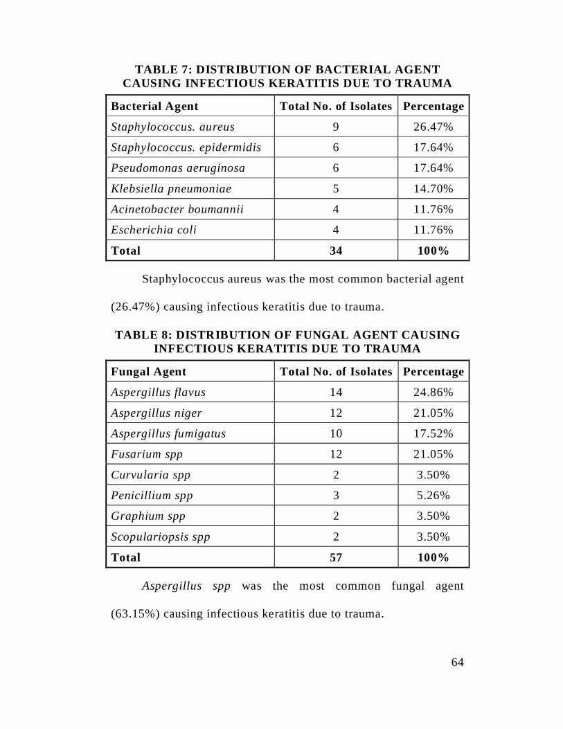
TABLE 7: DISTRIBUTION OF BACTERIAL AGENTCAUSING INFECTIOUS KERATITIS DUE TO TRAUMA
Bacterial Agent Total No. of Isolates Percentage
Staphylococcus. aureus 9 26.47%
Staphylococcus. epidermidis 6 17.64%
Pseudomonas aeruginosa 6 17.64%
Klebsiella pneumoniae 5 14.70%
Acinetobacter boumannii 4 11.76%
Escherichia coli 4 11.76%
Total 34 100%
Staphylococcus aureus was the most common bacterial agent
(26.47%) causing infectious keratitis due to trauma.
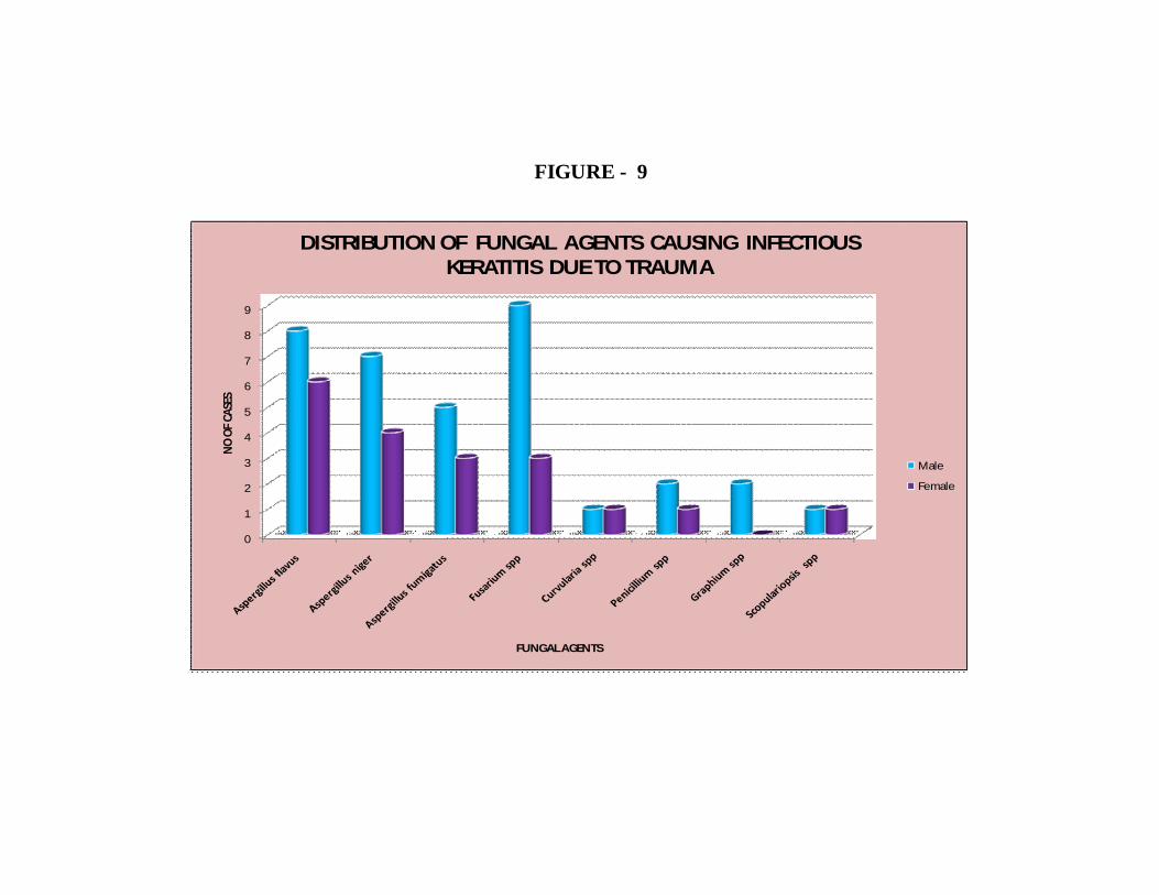
TABLE 8: DISTRIBUTION OF FUNGAL AGENT CAUSINGINFECTIOUS KERATITIS DUE TO TRAUMA
Fungal Agent Total No. of Isolates Percentage
Aspergillus flavus 14 24.86%
Aspergillus niger 12 21.05%
Aspergillus fumigatus 10 17.52%
Fusarium spp 12 21.05%
Curvularia spp 2 3.50%
Penicillium spp 3 5.26%
Graphium spp 2 3.50%
Scopulariopsis spp 2 3.50%
Total 57 100%
Aspergillus spp was the most common fungal agent
(63.15%) causing infectious keratitis due to trauma.
0
1
2
3
4
5
6
7
8
9
NO O
F CAS
ES
FUNGAL AGENTS
DISTRIBUTION OF FUNGAL AGENTS CAUSING INFECTIOUSKERATITIS DUE TO TRAUMA
Male
Female
FIGURE - 9
65
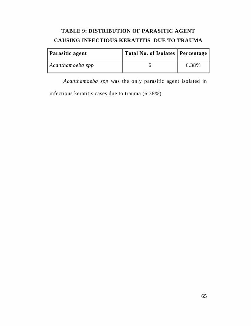
TABLE 9: DISTRIBUTION OF PARASITIC AGENT
CAUSING INFECTIOUS KERATITIS DUE TO TRAUMA
Parasitic agent Total No. of Isolates Percentage
Acanthamoeba spp 6 6.38%
Acanthamoeba spp was the only parasitic agent isolated in
infectious keratitis cases due to trauma (6.38%)
66
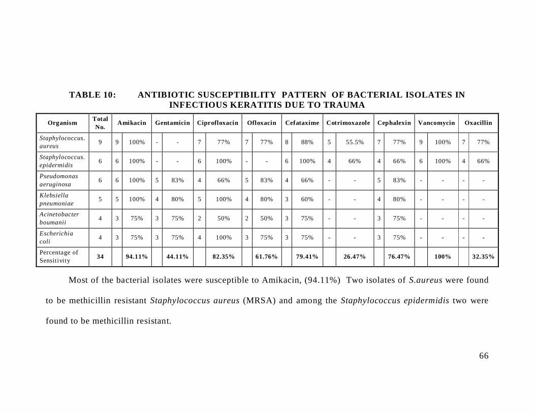
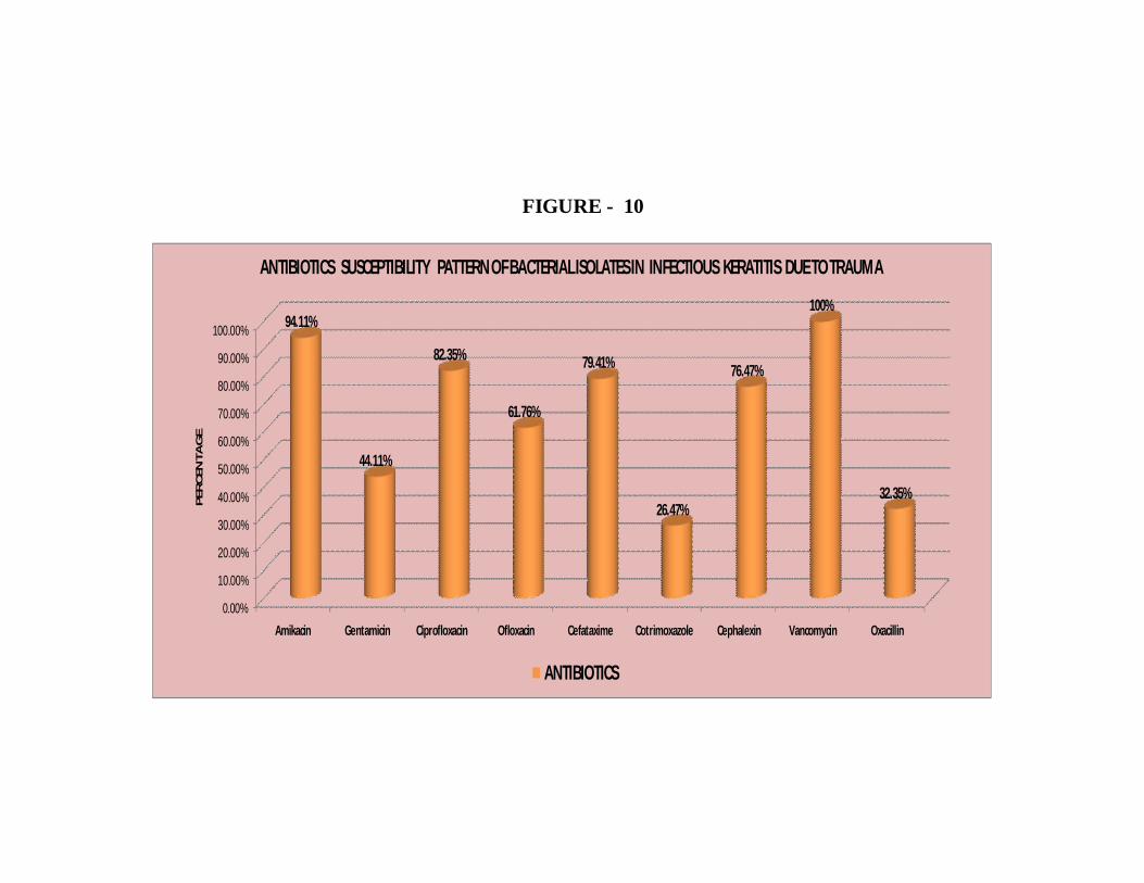
TABLE 10: ANTIBIOTIC SUSCEPTIBILITY PATTERN OF BACTERIAL ISOLATES ININFECTIOUS KERATITIS DUE TO TRAUMA
Organism TotalNo. Amikacin Gentamicin Ciprofloxacin Ofloxacin Cefataxime Cotrimoxazole Cephalexin Vancomycin Oxacillin
Staphylococcus.aureus 9 9 100% - - 7 77% 7 77% 8 88% 5 55.5% 7 77% 9 100% 7 77%
Staphylococcus.epidermidis 6 6 100% - - 6 100% - - 6 100% 4 66% 4 66% 6 100% 4 66%
Pseudomonasaeruginosa 6 6 100% 5 83% 4 66% 5 83% 4 66% - - 5 83% - - - -
Klebsiellapneumoniae 5 5 100% 4 80% 5 100% 4 80% 3 60% - - 4 80% - - - -
Acinetobacterboumanii 4 3 75% 3 75% 2 50% 2 50% 3 75% - - 3 75% - - - -
Escherichiacoli 4 3 75% 3 75% 4 100% 3 75% 3 75% - - 3 75% - - - -
Percentage ofSensitivity 34 94.11% 44.11% 82.35% 61.76% 79.41% 26.47% 76.47% 100% 32.35%
Most of the bacterial isolates were susceptible to Amikacin, (94.11%) Two isolates of S.aureus were found
to be methicillin resistant Staphylococcus aureus (MRSA) and among the Staphylococcus epidermidis two were
found to be methicillin resistant.
0.00%
10.00%
20.00%
30.00%
40.00%
50.00%
60.00%
70.00%
80.00%
90.00%
100.00%
Amikacin Gentamicin Ciprofloxacin Ofloxacin Cefataxime Cotrimoxazole Cephalexin Vancomycin Oxacillin
94.11%
44.11%
82.35%
61.76%
79.41%
26.47%
76.47%
100%
32.35%
PE
RC
EN
TA
GE
ANTIBIOTICS SUSCEPTIBILITY PATTERN OF BACTERIAL ISOLATES IN INFECTIOUS KERATITIS DUE TO TRAUMA
ANTIBIOTICS
FIGURE - 10
67
TABLE 11: MIC OF VANCOMYCIN FOR
STAPHYLOCOCCUS AUREUS
Organism
Minimum inhibitory concentrationbreak- point
0.25g/ml
0.5g/ml
1g/ml
>2g/ml
Staph.auerus (2) 1 1 - -
MIC for the MRSA isolates were studied and found to be
100% sensitive to vancomycin. The break point concentration were
0.25 g/ml and 0.5 g/ml for the isolates.
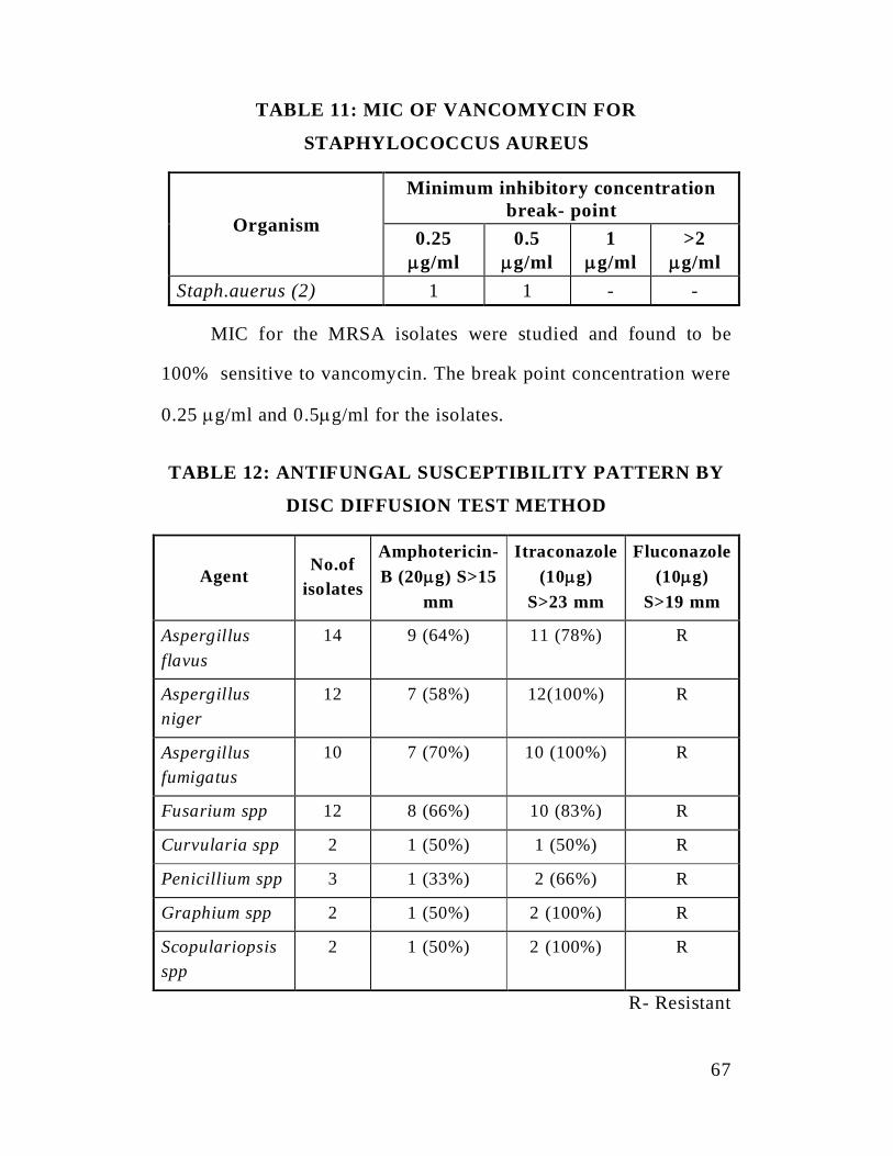
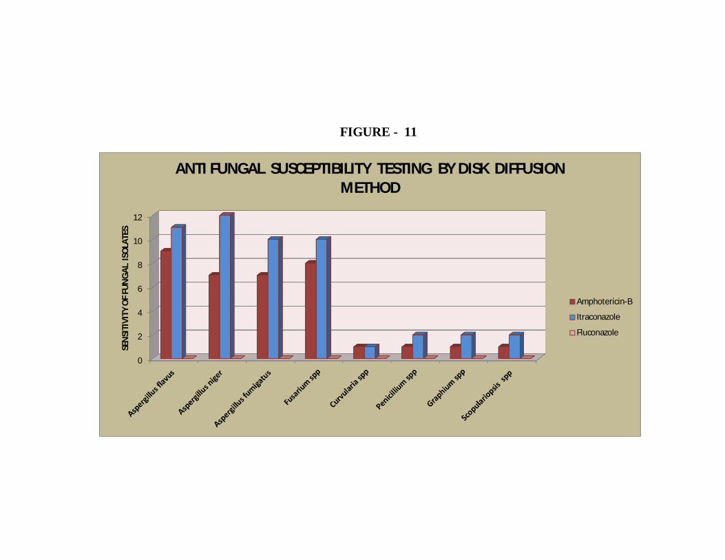
TABLE 12: ANTIFUNGAL SUSCEPTIBILITY PATTERN BY
DISC DIFFUSION TEST METHOD
AgentNo.of
isolates
Amphotericin-B (20 g) S>15
mm
Itraconazole(10 g)
S>23 mm
Fluconazole(10 g)
S>19 mm
Aspergillusflavus
14 9 (64%) 11 (78%) R
Aspergillusniger
12 7 (58%) 12(100%) R
Aspergillusfumigatus
10 7 (70%) 10 (100%) R
Fusarium spp 12 8 (66%) 10 (83%) R
Curvularia spp 2 1 (50%) 1 (50%) R
Penicillium spp 3 1 (33%) 2 (66%) R
Graphium spp 2 1 (50%) 2 (100%) R
Scopulariopsisspp
2 1 (50%) 2 (100%) R
R- Resistant
0
2
4
6
8
10
12
SEN
SITI
VITY
OF
FUN
GAL
ISO
LATE
S
ANTI FUNGAL SUSCEPTIBILITY TESTING BY DISK DIFFUSIONMETHOD
Amphotericin-B
Itraconazole
Fluconazole
FIGURE - 11
68
Itraconzole was found to be more effective against
Aspergillus spp. All fungal isolates were resistant to fluconazole
(<15 mm)
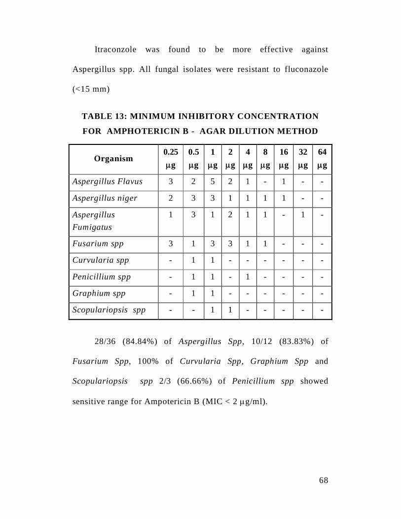
TABLE 13: MINIMUM INHIBITORY CONCENTRATION
FOR AMPHOTERICIN B - AGAR DILUTION METHOD
Organism0.25
g
0.5
g
1
g
2
g
4
g
8
g
16
g
32
g
64
g
Aspergillus Flavus 3 2 5 2 1 - 1 - -
Aspergillus niger 2 3 3 1 1 1 1 - -
AspergillusFumigatus
1 3 1 2 1 1 - 1 -
Fusarium spp 3 1 3 3 1 1 - - -
Curvularia spp - 1 1 - - - - - -
Penicillium spp - 1 1 - 1 - - - -
Graphium spp - 1 1 - - - - - -
Scopulariopsis spp - - 1 1 - - - - -
28/36 (84.84%) of Aspergillus Spp, 10/12 (83.83%) of
Fusarium Spp, 100% of Curvularia Spp, Graphium Spp and
Scopulariopsis spp 2/3 (66.66%) of Penicillium spp showed
sensitive range for Ampotericin B (MIC < 2 g/ml).
69
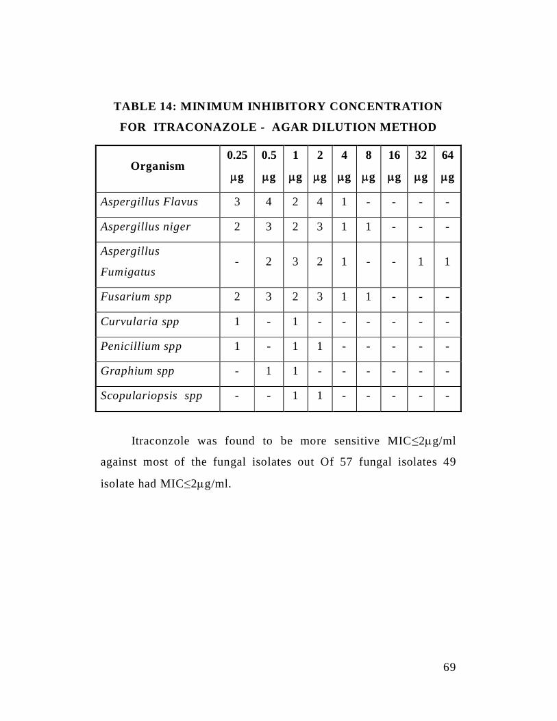
TABLE 14: MINIMUM INHIBITORY CONCENTRATION
FOR ITRACONAZOLE - AGAR DILUTION METHOD
Organism0.25
g
0.5
g
1
g
2
g
4
g
8
g
16
g
32
g
64
g
Aspergillus Flavus 3 4 2 4 1 - - - -
Aspergillus niger 2 3 2 3 1 1 - - -
Aspergillus
Fumigatus- 2 3 2 1 - - 1 1
Fusarium spp 2 3 2 3 1 1 - - -
Curvularia spp 1 - 1 - - - - - -
Penicillium spp 1 - 1 1 - - - - -
Graphium spp - 1 1 - - - - - -
Scopulariopsis spp - - 1 1 - - - - -
Itraconzole was found to be more sensitive MIC 2 g/ml
against most of the fungal isolates out Of 57 fungal isolates 49
isolate had MIC 2 g/ml.
70
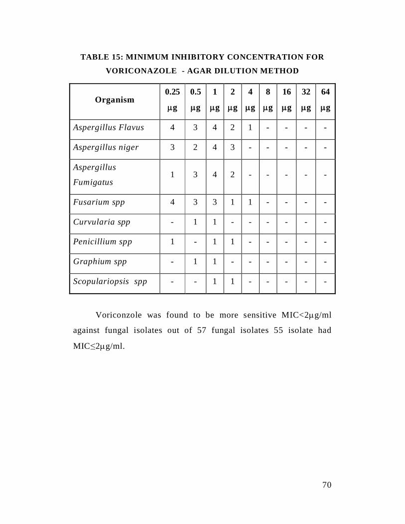
TABLE 15: MINIMUM INHIBITORY CONCENTRATION FOR
VORICONAZOLE - AGAR DILUTION METHOD
Organism0.25
g
0.5
g
1
g
2
g
4
g
8
g
16
g
32
g
64
g
Aspergillus Flavus 4 3 4 2 1 - - - -
Aspergillus niger 3 2 4 3 - - - - -
Aspergillus
Fumigatus1 3 4 2 - - - - -
Fusarium spp 4 3 3 1 1 - - - -
Curvularia spp - 1 1 - - - - - -
Penicillium spp 1 - 1 1 - - - - -
Graphium spp - 1 1 - - - - - -
Scopulariopsis spp - - 1 1 - - - - -
Voriconzole was found to be more sensitive MIC<2 g/ml
against fungal isolates out of 57 fungal isolates 55 isolate had
MIC 2 g/ml.
71
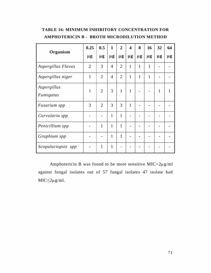
TABLE 16: MINIMUM INHIBITORY CONCENTRATION FOR
AMPHOTERICIN B - BROTH MICRODILUTION METHOD
Organism0.25
g
0.5
g
1
g
2
g
4
g
8
g
16
g
32
g
64
g
Aspergillus Flavus 2 3 4 2 1 1 1 - -
Aspergillus niger 1 2 4 2 1 1 1 - -
Aspergillus
Fumigatus1 2 3 1 1 - - 1 1
Fusarium spp 3 2 3 3 1 - - - -
Curvularia spp - - 1 1 - - - - -
Penicillium spp - 1 1 1 - - - - -
Graphium spp - - 1 1 - - - - -
Scopulariopsis spp - 1 1 - - - - - -
Amphotericin B was found to be more sensitive MIC<2 g/ml
against fungal isolates out of 57 fungal isolates 47 isolate had
MIC 2 g/ml.
72
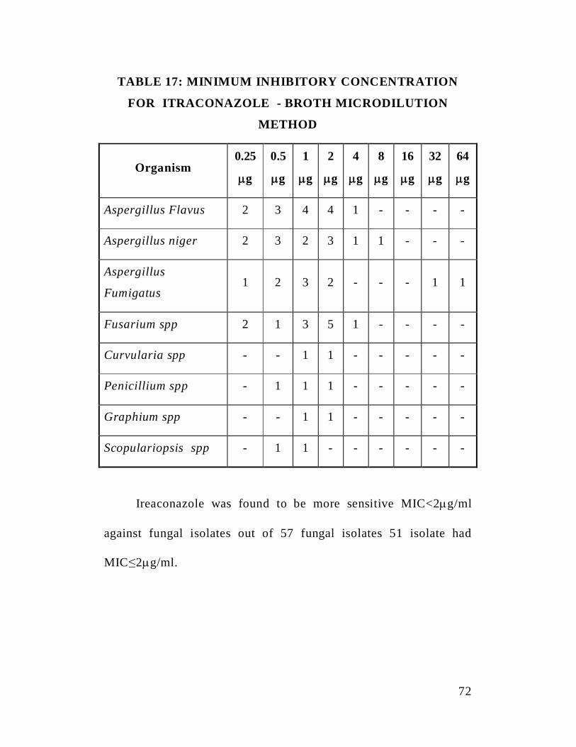
TABLE 17: MINIMUM INHIBITORY CONCENTRATION
FOR ITRACONAZOLE - BROTH MICRODILUTION
METHOD
Organism0.25
g
0.5
g
1
g
2
g
4
g
8
g
16
g
32
g
64
g
Aspergillus Flavus 2 3 4 4 1 - - - -
Aspergillus niger 2 3 2 3 1 1 - - -
Aspergillus
Fumigatus1 2 3 2 - - - 1 1
Fusarium spp 2 1 3 5 1 - - - -
Curvularia spp - - 1 1 - - - - -
Penicillium spp - 1 1 1 - - - - -
Graphium spp - - 1 1 - - - - -
Scopulariopsis spp - 1 1 - - - - - -
Ireaconazole was found to be more sensitive MIC<2 g/ml
against fungal isolates out of 57 fungal isolates 51 isolate had
MIC 2 g/ml.
73
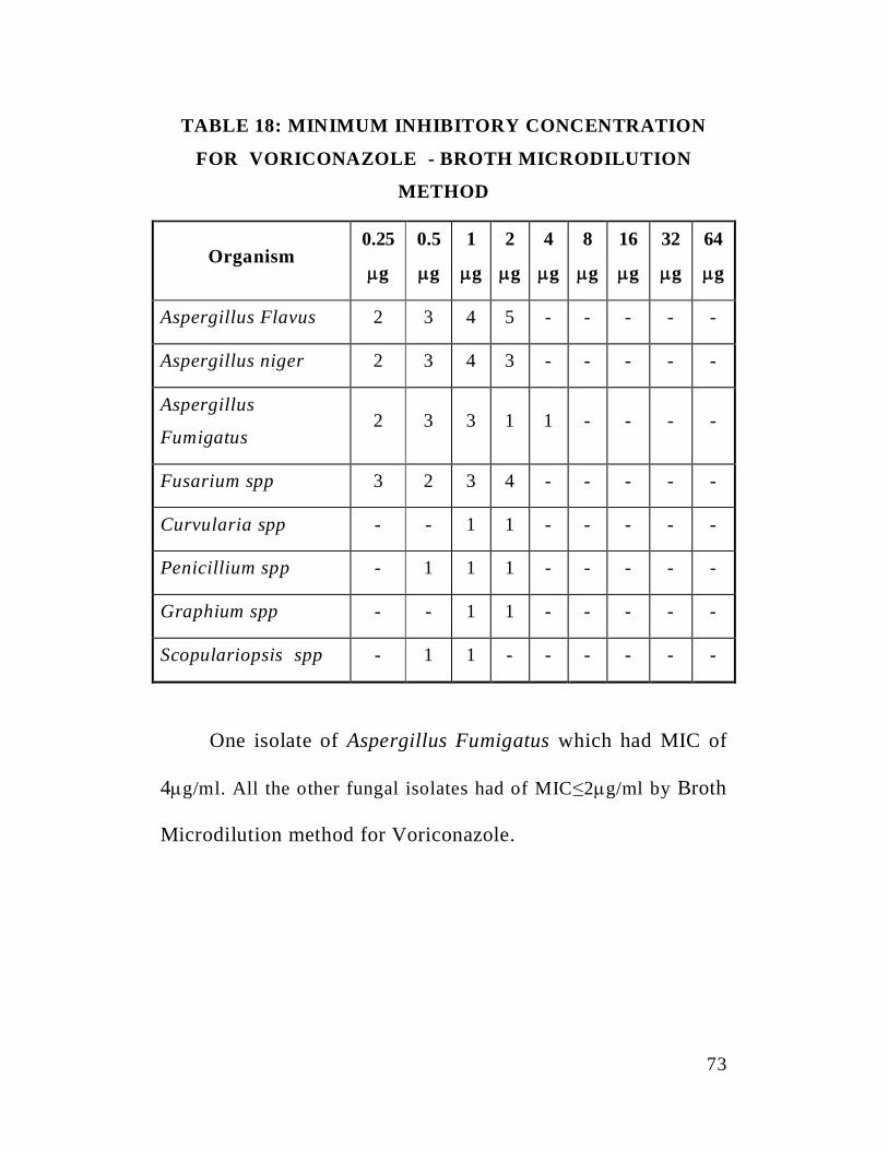
TABLE 18: MINIMUM INHIBITORY CONCENTRATION
FOR VORICONAZOLE - BROTH MICRODILUTION
METHOD
Organism0.25
g
0.5
g
1
g
2
g
4
g
8
g
16
g
32
g
64
g
Aspergillus Flavus 2 3 4 5 - - - - -
Aspergillus niger 2 3 4 3 - - - - -
Aspergillus
Fumigatus2 3 3 1 1 - - - -
Fusarium spp 3 2 3 4 - - - - -
Curvularia spp - - 1 1 - - - - -
Penicillium spp - 1 1 1 - - - - -
Graphium spp - - 1 1 - - - - -
Scopulariopsis spp - 1 1 - - - - - -
One isolate of Aspergillus Fumigatus which had MIC of
4 g/ml. All the other fungal isolates had of MIC 2 g/ml by Broth
Microdilution method for Voriconazole.
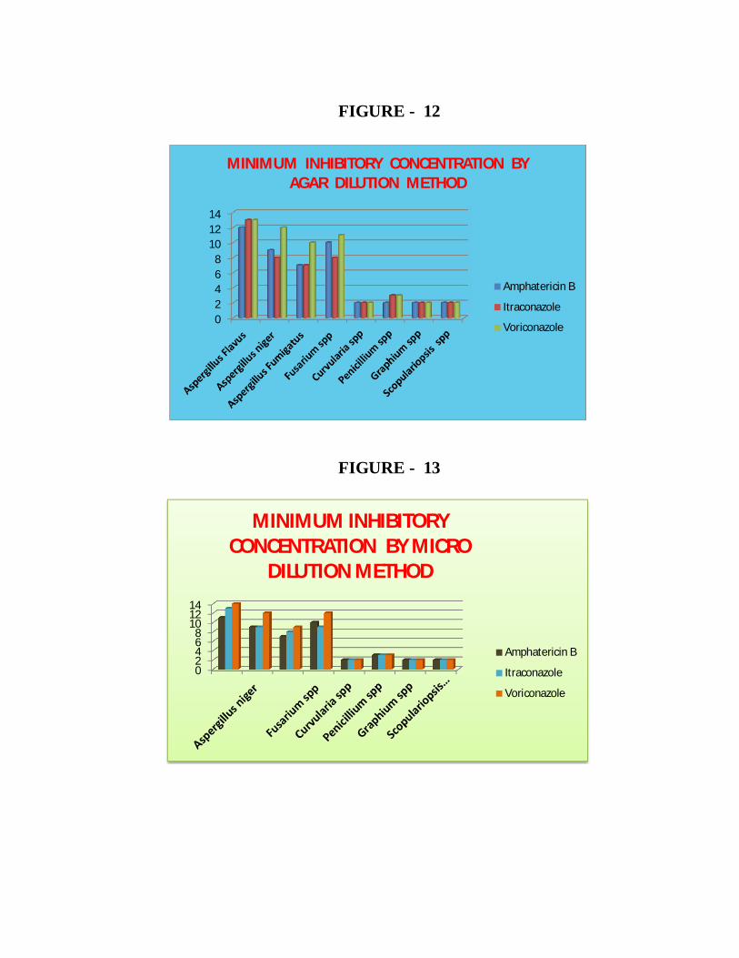
02468
101214
MINIMUM INHIBITORY CONCENTRATION BYAGAR DILUTION METHOD
Amphatericin B
Itraconazole
Voriconazole
02468
101214
MINIMUM INHIBITORYCONCENTRATION BY MICRO
DILUTION METHOD
Amphatericin B
Itraconazole
Voriconazole
FIGURE - 12
FIGURE - 13
74
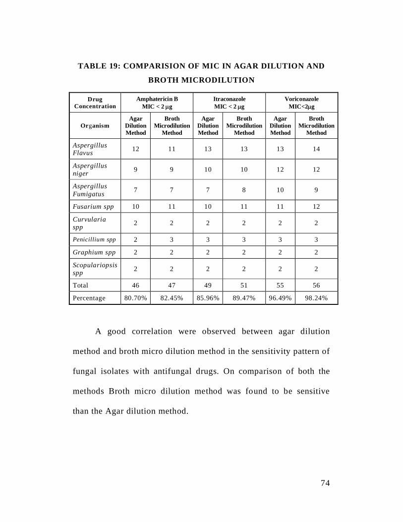
TABLE 19: COMPARISION OF MIC IN AGAR DILUTION AND
BROTH MICRODILUTION
DrugConcentration
Amphatericin BMIC < 2 g
ItraconazoleMIC < 2 g
VoriconazoleMIC<2 g
OrganismAgar
DilutionMethod
BrothMicrodilution
Method
AgarDilutionMethod
BrothMicrodilution
Method
AgarDilutionMethod
BrothMicrodilution
Method
AspergillusFlavus 12 11 13 13 13 14
Aspergillusniger 9 9 10 10 12 12
AspergillusFumigatus 7 7 7 8 10 9
Fusarium spp 10 11 10 11 11 12
Curvulariaspp 2 2 2 2 2 2
Penicillium spp 2 3 3 3 3 3
Graphium spp 2 2 2 2 2 2
Scopulariopsisspp 2 2 2 2 2 2
Total 46 47 49 51 55 56
Percentage 80.70% 82.45% 85.96% 89.47% 96.49% 98.24%
A good correlation were observed between agar dilution
method and broth micro dilution method in the sensitivity pattern of
fungal isolates with antifungal drugs. On comparison of both the
methods Broth micro dilution method was found to be sensitive
than the Agar dilution method.
75
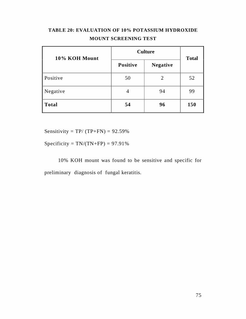
TABLE 20: EVALUATION OF 10% POTASSIUM HYDROXIDE
MOUNT SCREENING TEST
10% KOH MountCulture
TotalPositive Negative
Positive 50 2 52
Negative 4 94 99
Total 54 96 150
Sensitivity = TP/ (TP+FN) = 92.59%
Specificity = TN/(TN+FP) = 97.91%
10% KOH mount was found to be sensitive and specific for
preliminary diagnosis of fungal keratitis.
76
RESULTS
In this study, 150 cases of traumatic infectious keratitis
patients, 94 cases were culture positive (Table 1).
The cases were analyzed under the following parameters.
The age and sex distribution of infectious keratitis was
analysed 28.72% of infectious keratitis case were in 51-60 years of
age group. Extremes of Age group showed low prevalence (<10 and
>70%) (Table 3).
In this study male predominated in all forms of keratitis
71.11% (90/150) were males, and 50% (30/150) were females.
(Table 2)
The urban and rural distribution of cases showed higher
prevalence of infectious keratitis in rural population accounting for
64.66% (Table 4)
In analyzing the contribution of different trauma factors in
infectious keratitis, trauma with vegetable matter like paddy, leaf,
wood were implicated in 47% cases (Table 6).
77
Majority of the aetiological agents were fungi (57.44%)
followed by bacteria (32.97%), mixed bacteria and fungal growth
was observed in (3.19%) of the cases. The parasitic growth was
observed in 6.38% of the cases (Table 5).
Among the fungal isolates, 33 out of 54 (63.15%) cases were
due to Aspergillus spp. The next common agent was Fusarium spp
12 out of 54 (21.05%), followed by Pencillium spp 3 out of 54
(5.26%) Graphium spp 2 out of 54 (3.50%), and Scopulariopsis
spp 2 out of 54 (3.50%).(Table 8)
Staphylococcus aureus was the most common bacterial
isolate, accounting for 26.47%, followed by Pseudomonas
aeruginosa (17.64%) Acinetobacter boumannii was isolated in 4
cases (11.76%), Staphylococcus epidermidis (17.64%), Klebsiella
pneumoniae (16.70%), Escherichia coli (11.76%) (Table 7).
The sensitivity pattern of bacterial isolates revaled sensitivity
to amikacin 96.11%, 44.11 to gentamicin, 82.33 to ciprofloxacin,
61.73% to ofloxacin, 79.41% to cefataxime, 26.47% to
cotrimoxazole, 76.47% to cephalexin, 100% to vancomycin, and
32.35% to oxacillin (Table 10).
78
Two Staphylococus aureus isolates exhibited to oxacillin
resistant and both isolates were sensitive to vancomycin (Table 11)
Antifungal sensitivity pattern of fungal isolates by disc
diffusion test showed that 64% (9) Aspergillus flavus, 58.3% (7)
Aspergillus niger isolates, 70% of (7) Aspergillus fumigatus
isolates and 66% (8) of Fusarium spp, 50% (1) of Curvularia spp,
33% (1) of pencillium spp, 50% (1) of Graphium spp, and 50% (1)
Scopulariopsis spp were sensitive to Amphotericin B (Table 12)
Itraconazole was sensitive in 78% (9) of Aspergillus flavus,
100% (12) of Aspergillus niger isolates, 100% of (10) Aspergillus
fumigatus isolates and 83% (10) of Fusarium spp, 50% (1) of
Curvularia spp, 66% (2) of pencillum spp, 100% (2) of of graphium
spp and 100% (2) of Scopulariopsis spp were sensitive to
Amphotercin B (Table 12)
10% Potassium hydroxide mount preparation used as
screening test for rapid diagnosis of infectious keratitis showed
sensitivity 92.59% and 97.91% specificity (Table 20)
MIC determination by broth microdilution method also
showed that MIC range was comparable with Agar dilution method.
79
84.48% of Aspergillus spp and 81% of Fusarium spp, 100% of
Curvularia, Graphium spp, Scopulariposis spp and 60.66% of
Pencillium spp showed sensitive range of Amphotercin B by Agar
dilution method(Table 12).
Itraconazole (99.9%) of Aspergillus spp and 83.3% of
Fusarium isolates, 100% of Curvularia spp, Graphium spp,
Scopulariopsis spp, Pencillium spp showed MIC<2 g/ml. (Table 14)
Voriconazole was 96.41% sensitive in Agar dilution methods, and
98.2% in Broth Microdilution Method. (Table 19)

COLOURPLATES
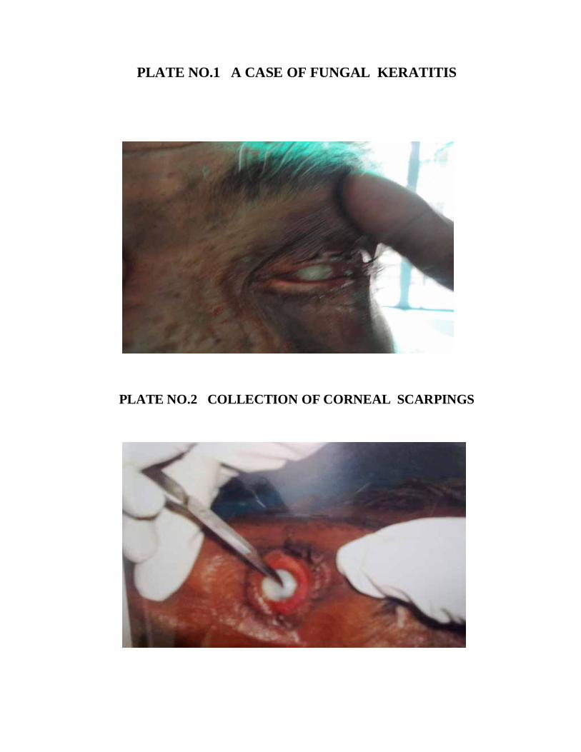
PLATE NO.1 A CASE OF FUNGAL KERATITIS
PLATE NO.2 COLLECTION OF CORNEAL SCARPINGS
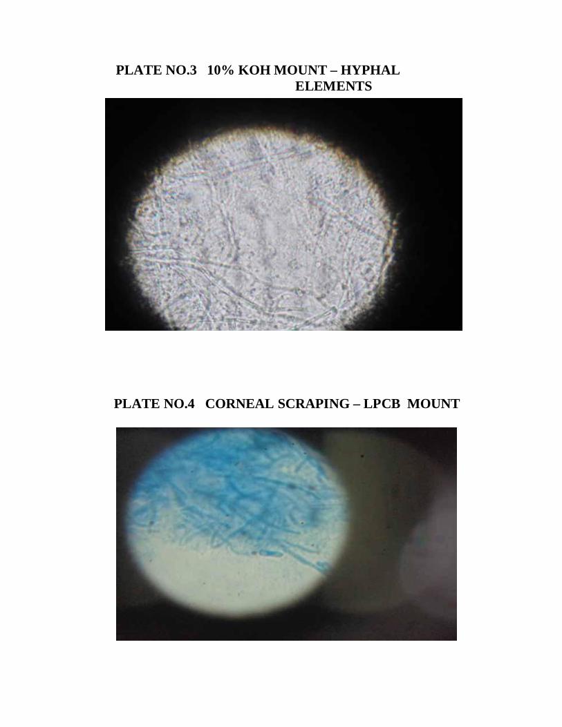
PLATE NO.3 10% KOH MOUNT – HYPHALELEMENTS
PLATE NO.4 CORNEAL SCRAPING – LPCB MOUNT
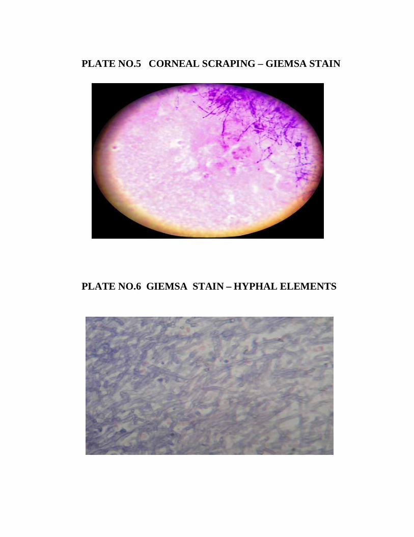
PLATE NO.5 CORNEAL SCRAPING – GIEMSA STAIN
PLATE NO.6 GIEMSA STAIN – HYPHAL ELEMENTS
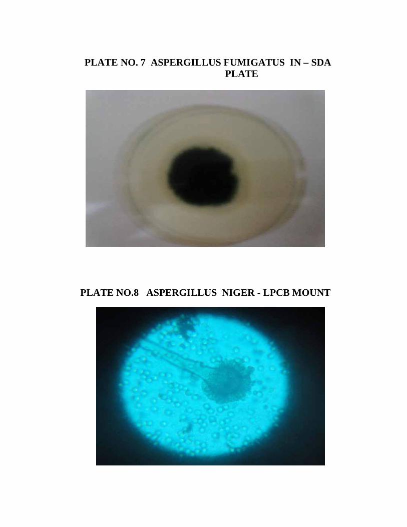
PLATE NO. 7 ASPERGILLUS FUMIGATUS IN – SDAPLATE
PLATE NO.8 ASPERGILLUS NIGER - LPCB MOUNT
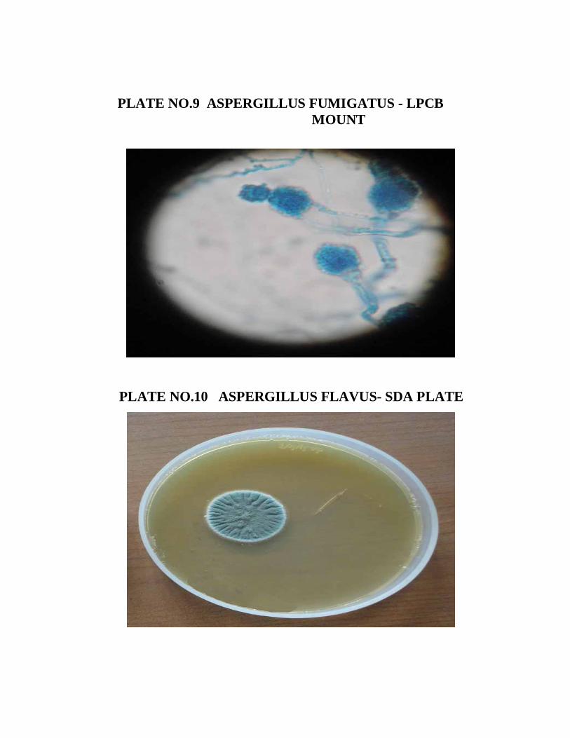
PLATE NO.9 ASPERGILLUS FUMIGATUS - LPCBMOUNT
PLATE NO.10 ASPERGILLUS FLAVUS- SDA PLATE
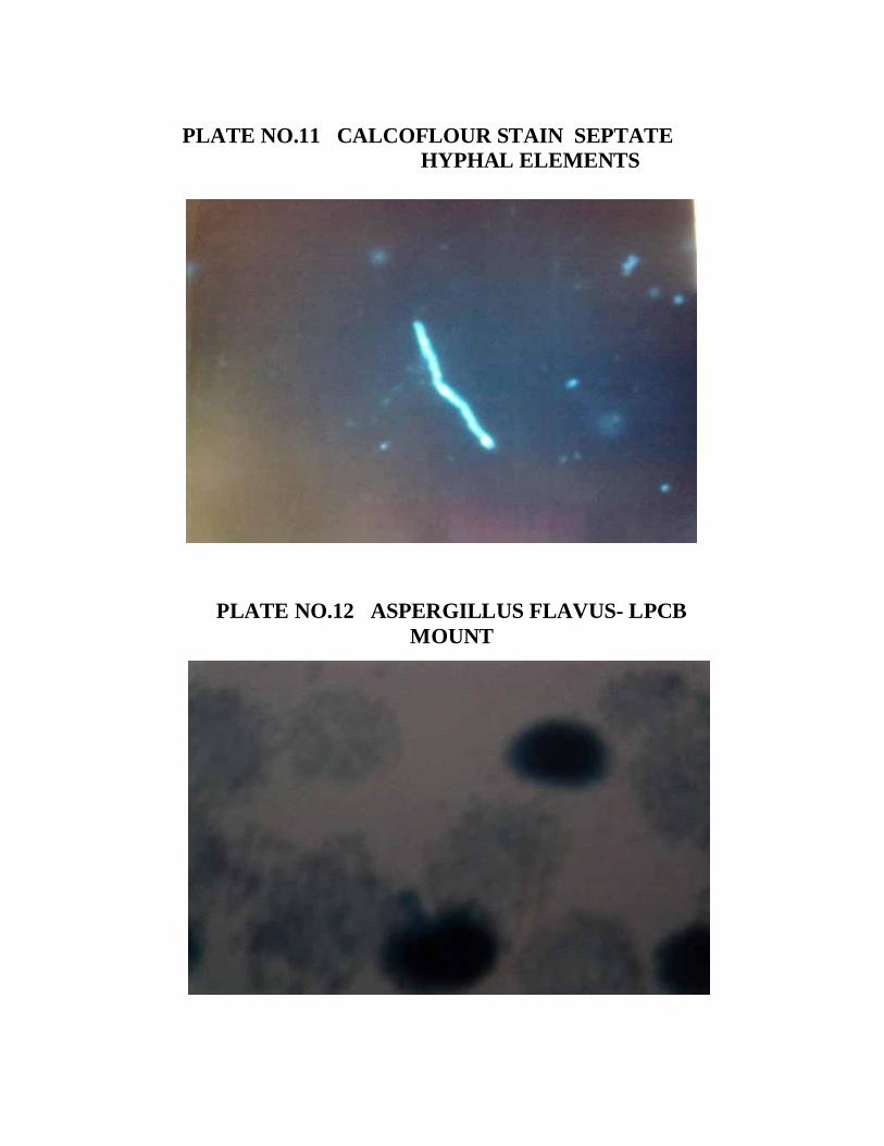
PLATE NO.11 CALCOFLOUR STAIN SEPTATE HYPHAL ELEMENTS
PLATE NO.12 ASPERGILLUS FLAVUS- LPCBMOUNT
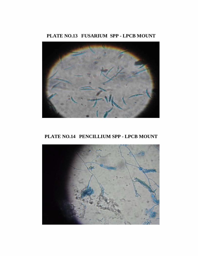
PLATE NO.13 FUSARIUM SPP - LPCB MOUNT
PLATE NO.14 PENCILLIUM SPP - LPCB MOUNT
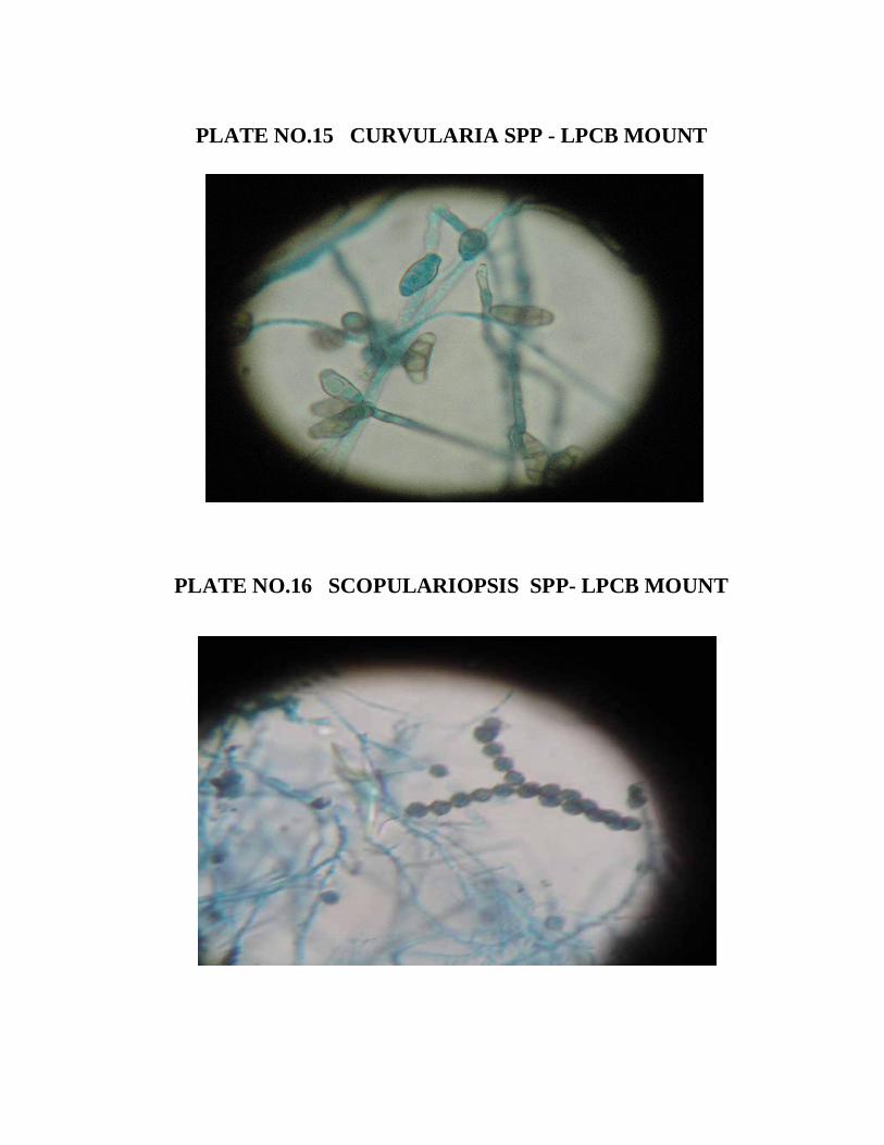
PLATE NO.15 CURVULARIA SPP - LPCB MOUNT
PLATE NO.16 SCOPULARIOPSIS SPP- LPCB MOUNT
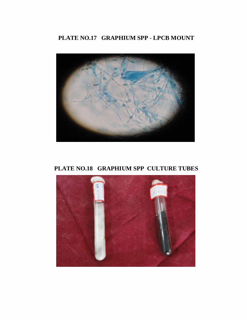
PLATE NO.17 GRAPHIUM SPP - LPCB MOUNT
PLATE NO.18 GRAPHIUM SPP CULTURE TUBES
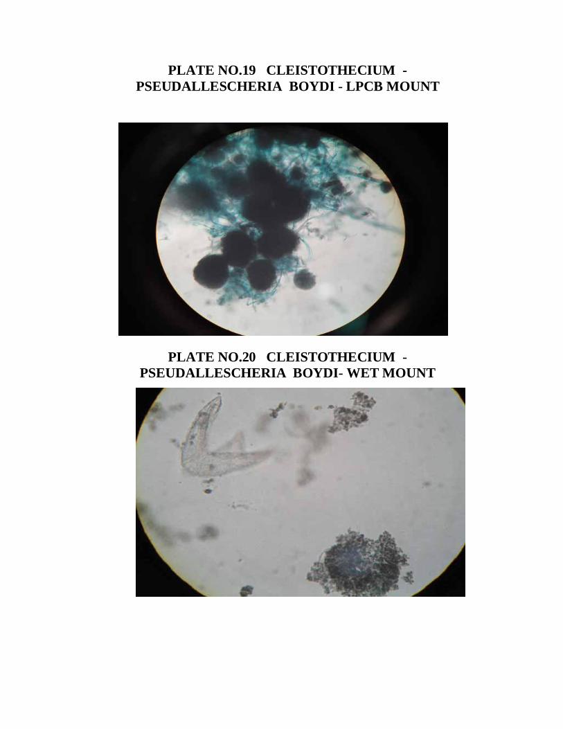
PLATE NO.19 CLEISTOTHECIUM -PSEUDALLESCHERIA BOYDI - LPCB MOUNT
PLATE NO.20 CLEISTOTHECIUM -PSEUDALLESCHERIA BOYDI- WET MOUNT
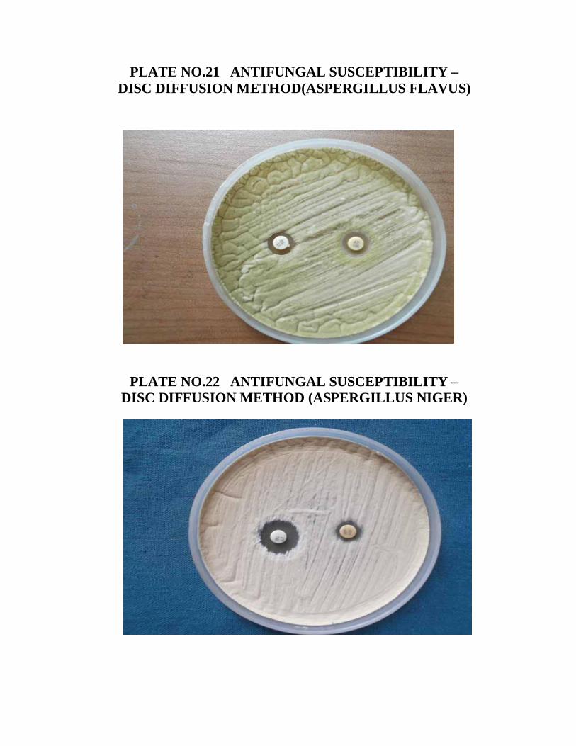
PLATE NO.21 ANTIFUNGAL SUSCEPTIBILITY –DISC DIFFUSION METHOD(ASPERGILLUS FLAVUS)
PLATE NO.22 ANTIFUNGAL SUSCEPTIBILITY –DISC DIFFUSION METHOD (ASPERGILLUS NIGER)
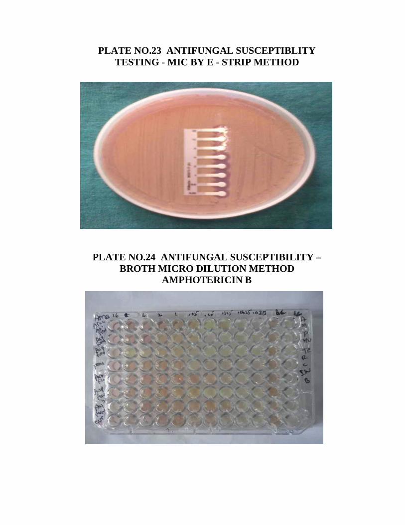
PLATE NO.23 ANTIFUNGAL SUSCEPTIBLITYTESTING - MIC BY E - STRIP METHOD
PLATE NO.24 ANTIFUNGAL SUSCEPTIBILITY –BROTH MICRO DILUTION METHOD
AMPHOTERICIN B
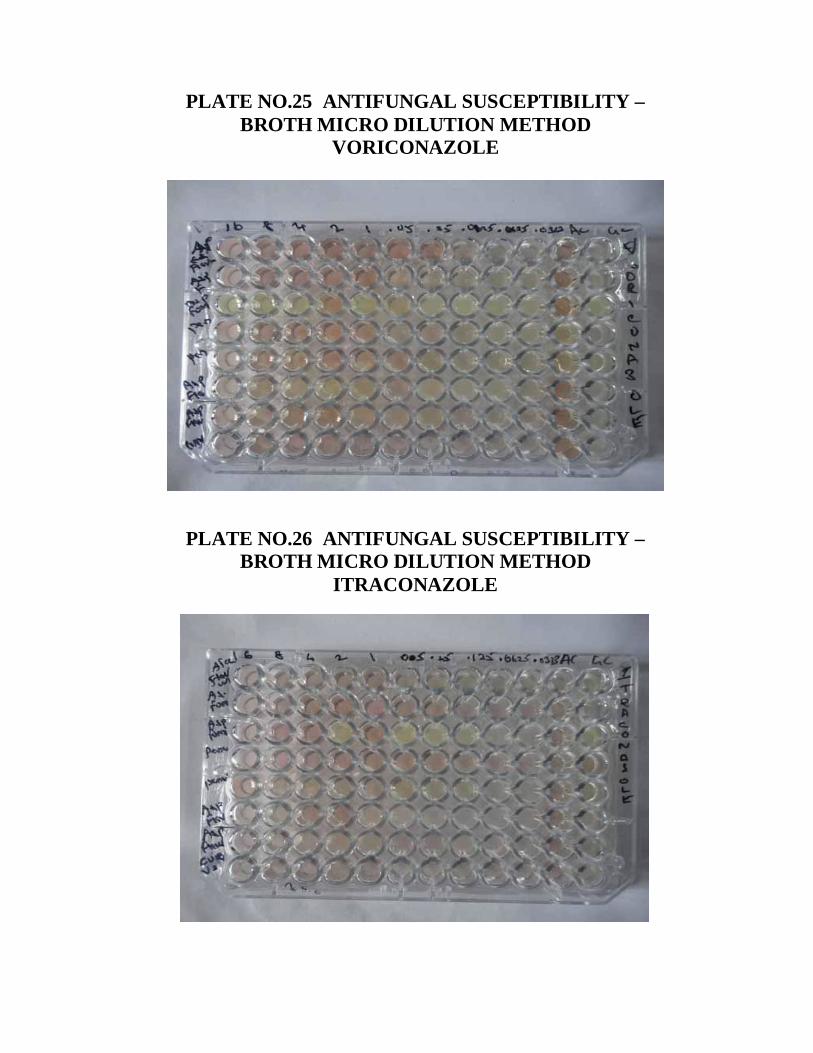
PLATE NO.25 ANTIFUNGAL SUSCEPTIBILITY –BROTH MICRO DILUTION METHOD
VORICONAZOLE
PLATE NO.26 ANTIFUNGAL SUSCEPTIBILITY –BROTH MICRO DILUTION METHOD
ITRACONAZOLE
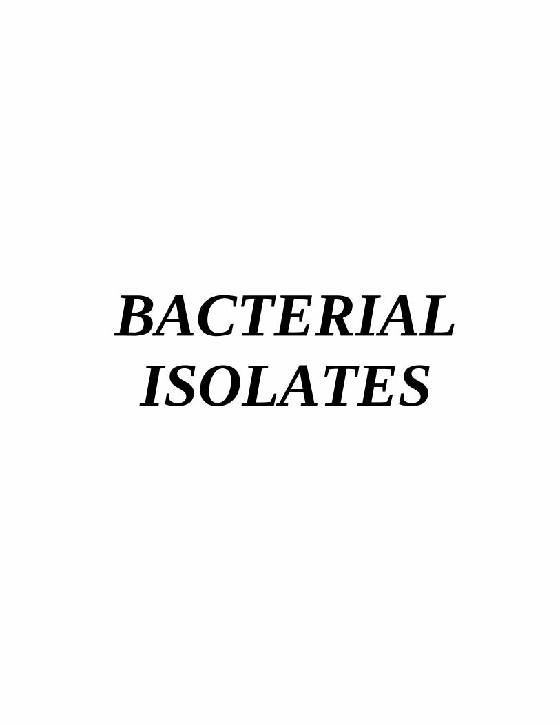
BACTERIALISOLATES
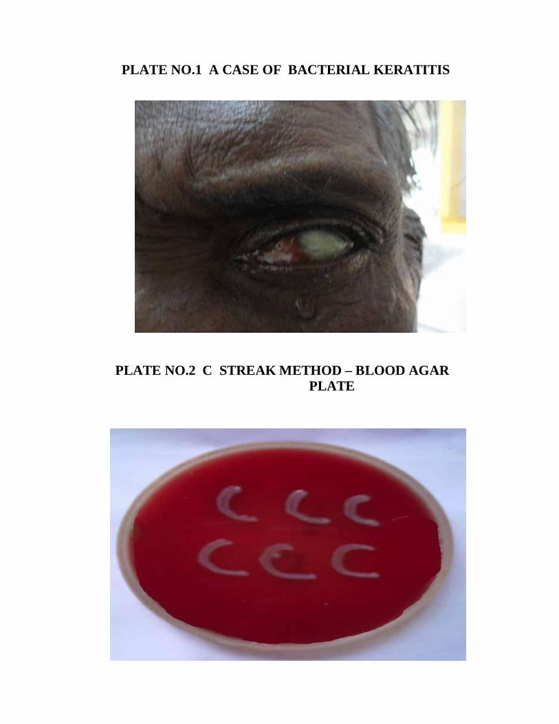
PLATE NO.1 A CASE OF BACTERIAL KERATITIS
PLATE NO.2 C STREAK METHOD – BLOOD AGARPLATE
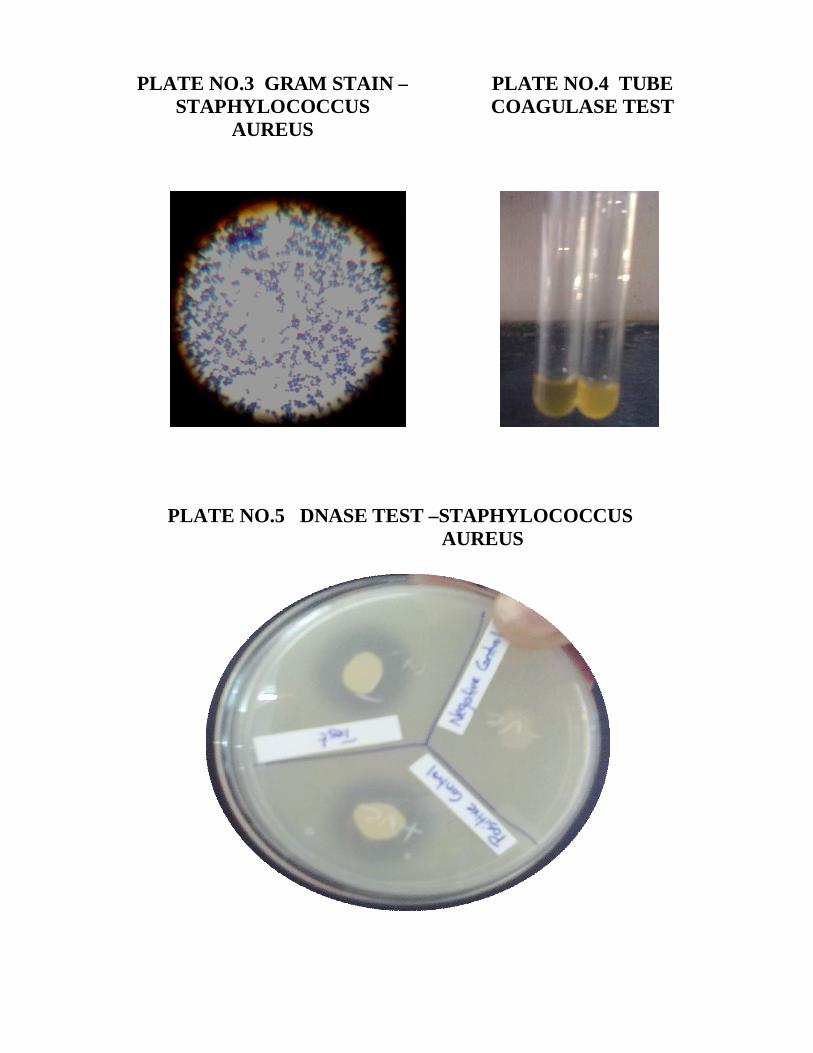
PLATE NO.5 DNASE TEST –STAPHYLOCOCCUSAUREUS
PLATE NO.4 TUBECOAGULASE TEST
PLATE NO.3 GRAM STAIN –STAPHYLOCOCCUS
AUREUS
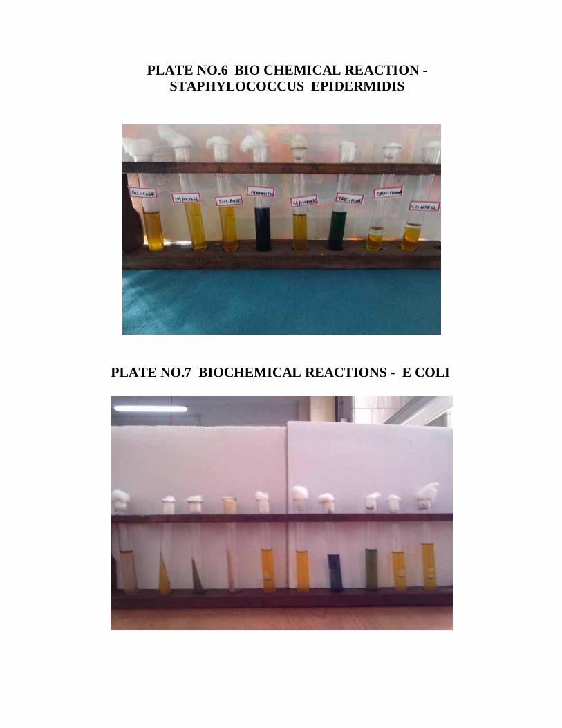
PLATE NO.6 BIO CHEMICAL REACTION -STAPHYLOCOCCUS EPIDERMIDIS
PLATE NO.7 BIOCHEMICAL REACTIONS - E COLI
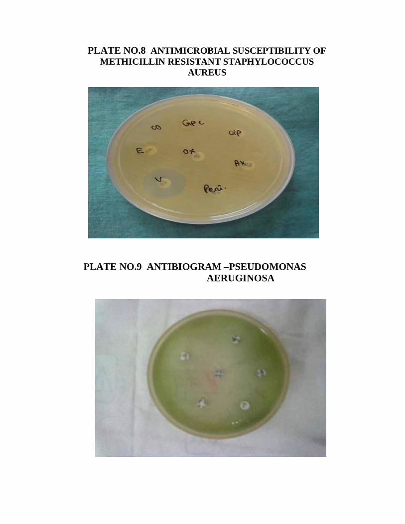
PLATE NO.8 ANTIMICROBIAL SUSCEPTIBILITY OFMETHICILLIN RESISTANT STAPHYLOCOCCUS
AUREUS
PLATE NO.9 ANTIBIOGRAM –PSEUDOMONASAERUGINOSA
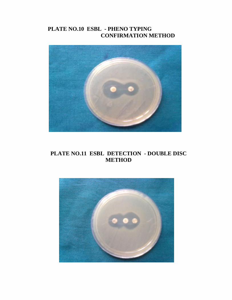
PLATE NO.10 ESBL - PHENO TYPINGCONFIRMATION METHOD
PLATE NO.11 ESBL DETECTION - DOUBLE DISCMETHOD
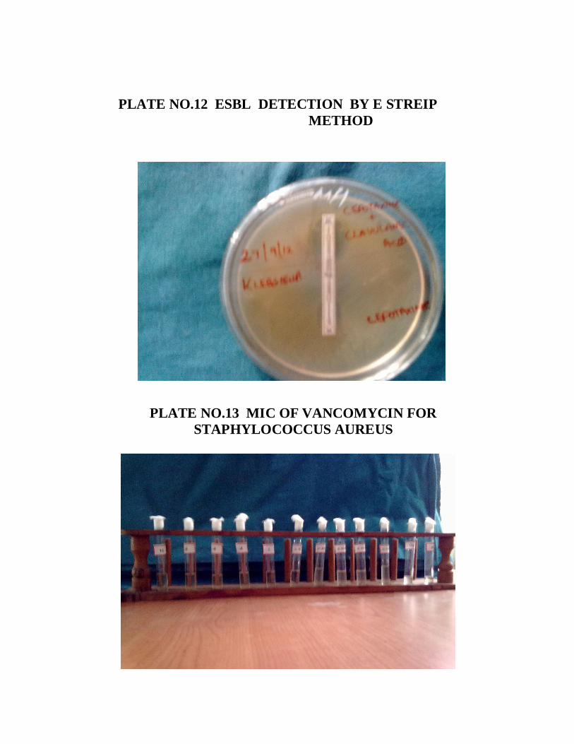
PLATE NO.12 ESBL DETECTION BY E STREIPMETHOD
PLATE NO.13 MIC OF VANCOMYCIN FORSTAPHYLOCOCCUS AUREUS
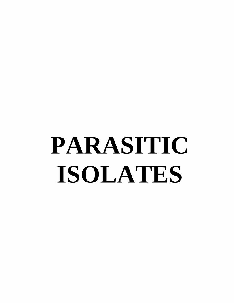
PARASITICISOLATES
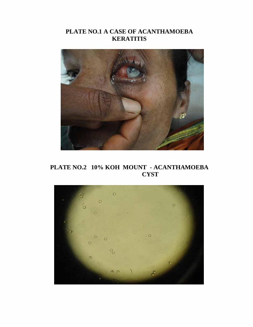
PLATE NO.1 A CASE OF ACANTHAMOEBAKERATITIS
PLATE NO.2 10% KOH MOUNT - ACANTHAMOEBACYST
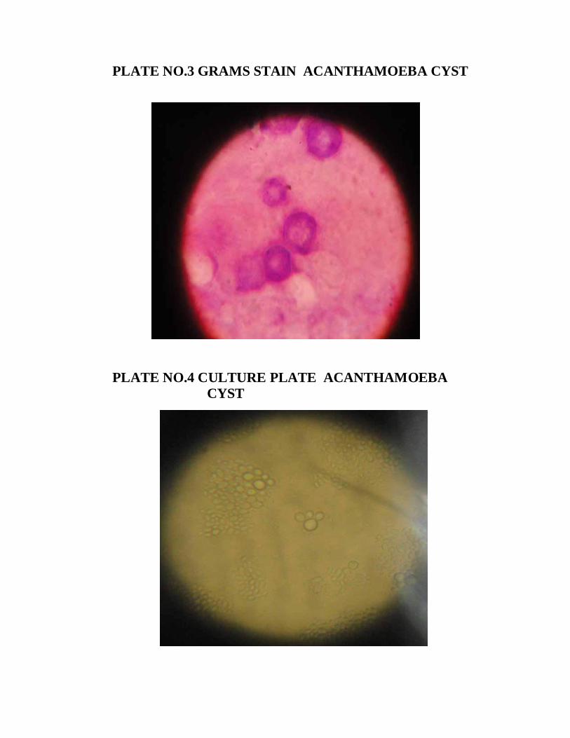
PLATE NO.3 GRAMS STAIN ACANTHAMOEBA CYST
PLATE NO.4 CULTURE PLATE ACANTHAMOEBACYST
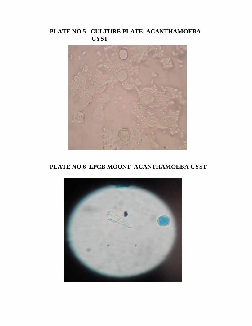
PLATE NO.5 CULTURE PLATE ACANTHAMOEBACYST
PLATE NO.6 LPCB MOUNT ACANTHAMOEBA CYST
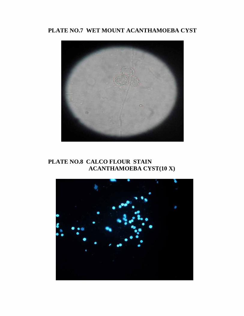
PLATE NO.7 WET MOUNT ACANTHAMOEBA CYST
PLATE NO.8 CALCO FLOUR STAINACANTHAMOEBA CYST(10 X)
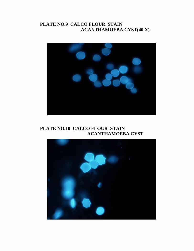
PLATE NO.9 CALCO FLOUR STAINACANTHAMOEBA CYST(40 X)
PLATE NO.10 CALCO FLOUR STAINACANTHAMOEBA CYST

DISCUSSION
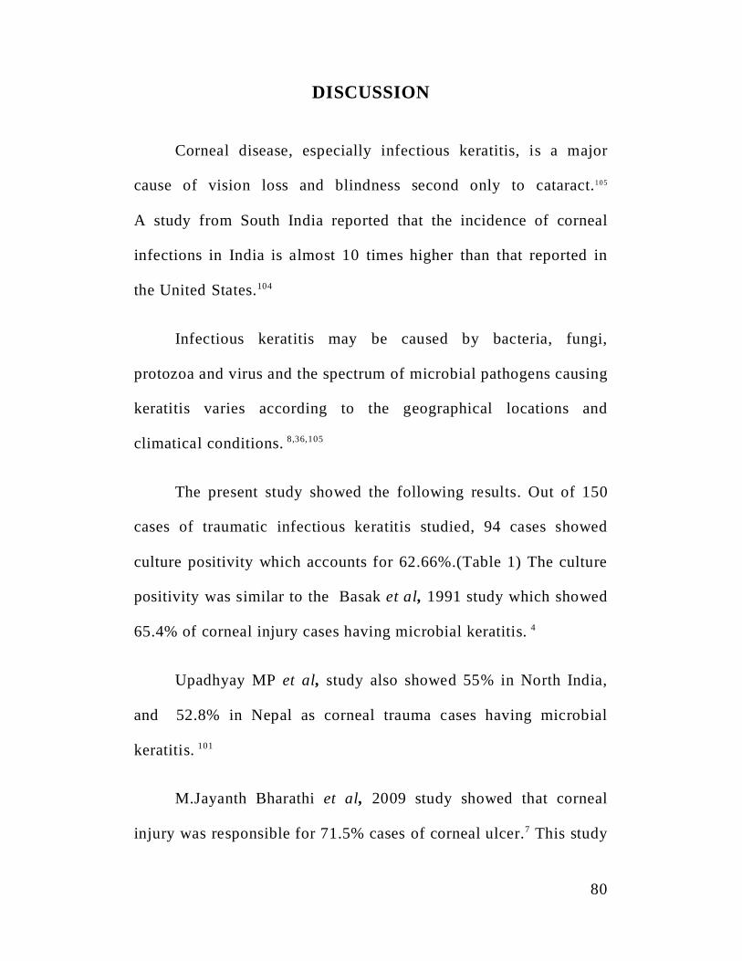
80
DISCUSSION
Corneal disease, especially infectious keratitis, is a major
cause of vision loss and blindness second only to cataract.105
A study from South India reported that the incidence of corneal
infections in India is almost 10 times higher than that reported in
the United States.104
Infectious keratitis may be caused by bacteria, fungi,
protozoa and virus and the spectrum of microbial pathogens causing
keratitis varies according to the geographical locations and
climatical conditions. 8,36,105
The present study showed the following results. Out of 150
cases of traumatic infectious keratitis studied, 94 cases showed
culture positivity which accounts for 62.66%.(Table 1) The culture
positivity was similar to the Basak et al, 1991 study which showed
65.4% of corneal injury cases having microbial keratitis. 4
Upadhyay MP et al, study also showed 55% in North India,
and 52.8% in Nepal as corneal trauma cases having microbial
keratitis. 101
M.Jayanth Bharathi et al, 2009 study showed that corneal
injury was responsible for 71.5% cases of corneal ulcer.7 This study
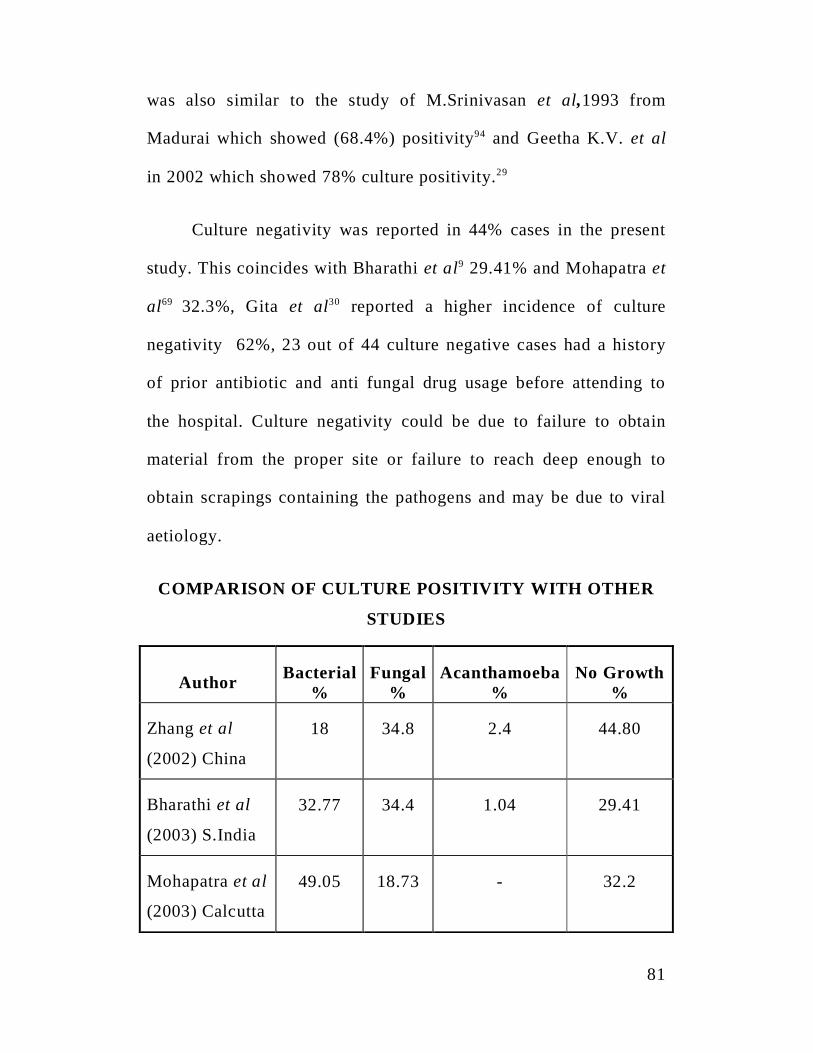
81
was also similar to the study of M.Srinivasan et al,1993 from
Madurai which showed (68.4%) positivity94 and Geetha K.V. et al
in 2002 which showed 78% culture positivity.29
Culture negativity was reported in 44% cases in the present
study. This coincides with Bharathi et al9 29.41% and Mohapatra et
al69 32.3%, Gita et al30 reported a higher incidence of culture
negativity 62%, 23 out of 44 culture negative cases had a history
of prior antibiotic and anti fungal drug usage before attending to
the hospital. Culture negativity could be due to failure to obtain
material from the proper site or failure to reach deep enough to
obtain scrapings containing the pathogens and may be due to viral
aetiology.
COMPARISON OF CULTURE POSITIVITY WITH OTHER
STUDIES
Author Bacterial%
Fungal%
Acanthamoeba%
No Growth%
Zhang et al
(2002) China
18 34.8 2.4 44.80
Bharathi et al
(2003) S.India
32.77 34.4 1.04 29.41
Mohapatra et al
(2003) Calcutta
49.05 18.73 - 32.2
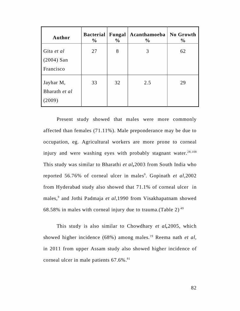
82
Author Bacterial%
Fungal%
Acanthamoeba%
No Growth%
Gita et al
(2004) San
Francisco
27 8 3 62
Jayhar M,
Bharath et al
(2009)
33 32 2.5 29
Present study showed that males were more commonly
affected than females (71.11%). Male preponderance may be due to
occupation, eg. Agricultural workers are more prone to corneal
injury and were washing eyes with probably stagnant water.28,108
This study was similar to Bharathi et al,2003 from South India who
reported 56.76% of corneal ulcer in males9. Gopinath et al,2002
from Hyderabad study also showed that 71.1% of corneal ulcer in
males,9 and Jothi Padmaja et al,1990 from Visakhapatnam showed
68.58% in males with corneal injury due to trauma.(Table 2) 49
This study is also similar to Chowdhary et al,2005, which
showed higher incidence (68%) among males.19 Reema nath et al,
in 2011 from upper Assam study also showed higher incidence of
corneal ulcer in male patients 67.6%.81
83
People of certain age group are affected more in infectious
keratitis.57 In this study, most common age group affected was
between 51-60 years (28.72 % - Table 3). This study was similar
with the study of Bharathi MJ, et al 2003.11 The study of Chander J
et al 1994, also showed more number of patients with infectious
keratitis in the 51-60 years age group.15 Manikandan P et al,2004
study also showed that patients above 50 years of age were more
vulnerable to microbial keratitis.66
In this study, rural population were more vulnerable to
infectious keratitis due to trauma compared to the urban population
(64.66% - Table 4). This present study was similar to Basak samara
et al, in 200585 from West Bengal showed that 78.5% of the patients
were from rural areas.4
Bharathi MJ et al,20038 and Chandar J et al, 16 1994 also
showed higher incidence of infectious keratitis from rural areas.
The present study showed that different agents such as paddy
dust, wood, Insect fall, stick, Cow’s tail, thorn prick, stone, iron
particles were the etiological agent casing corneal injury leading to
infectious keratitis. (Table 6)
In this study corneal injury with vegetable matter was the
84
most common factor casing infectious keratitis (47.87% - Table 6)
This study was similar to Basak Samar et al, in 2005 West Bengal
who reported (59.6%) traumatic cases due to vegetable matter.4
Norina TJ, et al, 2008 and Laspinal F,et al, 2004,59 also showed
history of ocular trauma in 62% and 50% of their patients
respectively.
The above observation clearly show that in developing
countries like India where, agricultural worker were more
common, vegetative matter induced ocular trauma was the major
cause of infectious keratitis in rural population than urban
population. 22,31,80
In this present study 5 (3.3%)patient were found to have
Diabetes mellitus. This study was similar to Basak samara K, et
al,2005, study where 7.6% of patients had Diabetes mellitus.4
Gopinath et al, 2002 from Hyderabad showed Diabetes mellitus as
the predominant systemic disease associated with fungal and
bacterial keratitis.34
In this study, aetioloigal agent was identified and isolated in
94 samples (62.66%). Of these 94 culture positive samples 54
(57%) had pure fungal growth, 31 (32.1%) had pure bacterial
growth and 3 (3.19%) had mixed bacterial and fungal growth, 6
85
(6.38%) had parasitic growth. (Table 5)
In this study, fungal agents were the most common agent
isolated in infectious keratitis patients followed by bacterial and
parasitic agents.
This study correlates with the study of Basak Samar K et al,
2005,4 which showed 62.7% of pure fungal growth, 22.7% of pure
bacterial growth. This study was similar to Khanal B et al, in 2005,
conducted in Nepal which showed 42.7% growth positive for fungi
present.52 This study also similar to Houang et al,2001.35
Aspergillus spp was the predominant isolate, in this present
study (63.15%). This study was similar to Gopinath et al, 2002
from Hyderabad which showed that Aspergillus spp (30.7%) were
the predominant isolate in their study.34
Basak Samar K et al, 2012 and Khanal B et al 2005 studies
also showed that common fungal pathogen was Aspergillus spp,
followed by Fusarium spp.4,52,62
The study done by Zimmerman E L et al, reported Aspergillus
spp was the commonest fungal agent in corneal ulcer.111
In this present study Fusarium spp was found to be the next
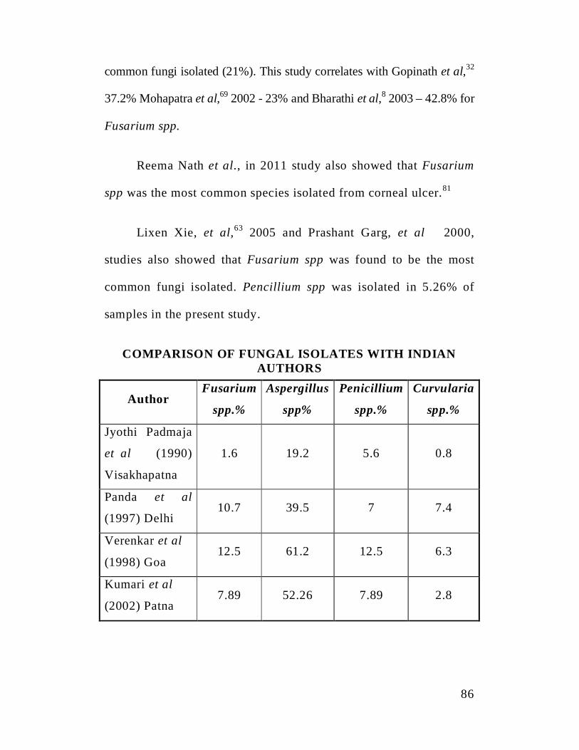
86
common fungi isolated (21%). This study correlates with Gopinath et al,32
37.2% Mohapatra et al,69 2002 - 23% and Bharathi et al,8 2003 – 42.8% for
Fusarium spp.
Reema Nath et al., in 2011 study also showed that Fusarium
spp was the most common species isolated from corneal ulcer.81
Lixen Xie, et al,63 2005 and Prashant Garg, et al 2000,
studies also showed that Fusarium spp was found to be the most
common fungi isolated. Pencillium spp was isolated in 5.26% of
samples in the present study.
COMPARISON OF FUNGAL ISOLATES WITH INDIANAUTHORS
AuthorFusarium
spp.%
Aspergillus
spp%
Penicillium
spp.%
Curvularia
spp.%
Jyothi Padmaja
et al (1990)
Visakhapatna
1.6 19.2 5.6 0.8
Panda et al
(1997) Delhi10.7 39.5 7 7.4
Verenkar et al
(1998) Goa12.5 61.2 12.5 6.3
Kumari et al
(2002) Patna7.89 52.26 7.89 2.8
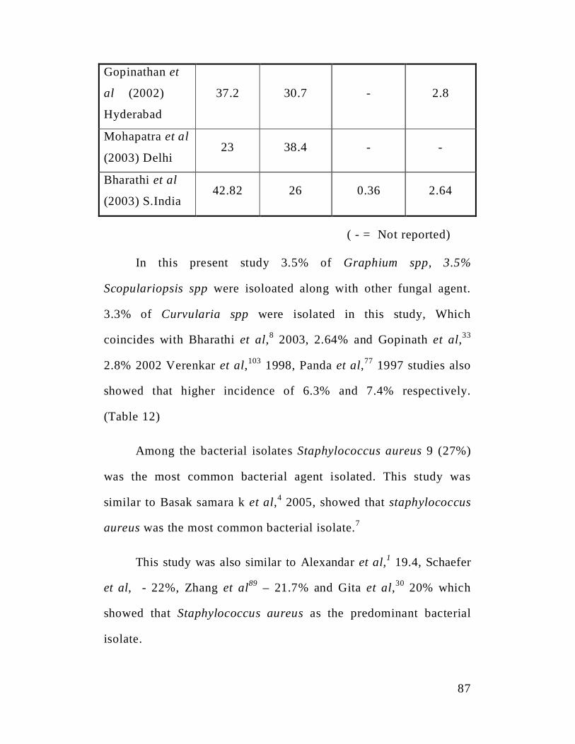
87
Gopinathan et
al (2002)
Hyderabad
37.2 30.7 - 2.8
Mohapatra et al
(2003) Delhi23 38.4 - -
Bharathi et al
(2003) S.India42.82 26 0.36 2.64
( - = Not reported)
In this present study 3.5% of Graphium spp, 3.5%
Scopulariopsis spp were isoloated along with other fungal agent.
3.3% of Curvularia spp were isolated in this study, Which
coincides with Bharathi et al,8 2003, 2.64% and Gopinath et al,33
2.8% 2002 Verenkar et al,103 1998, Panda et al,77 1997 studies also
showed that higher incidence of 6.3% and 7.4% respectively.
(Table 12)
Among the bacterial isolates Staphylococcus aureus 9 (27%)
was the most common bacterial agent isolated. This study was
similar to Basak samara k et al,4 2005, showed that staphylococcus
aureus was the most common bacterial isolate.7
This study was also similar to Alexandar et al,1 19.4, Schaefer
et al, - 22%, Zhang et al89 – 21.7% and Gita et al,30 20% which
showed that Staphylococcus aureus as the predominant bacterial
isolate.
88
Pseudomonas aeruginosa was the next predominant isolate in
the present study (17%). This study was similar to Basak Samar K
et al4 2005 and Bharathi et al6 2003 which also showed 18.05 % for
Pseudomonas spp. M.Srinivasan et al,94 1997 study also showed
that Pseudomonas spp. 14.4% accounted for microbial causes for
infectious keratitis. (Table 7) This study is also similar to
Alexander et al, 2000 USA which showed 25.7% of Pseudomonas
spp in their study.
Staphylococcus epidermidis were isolated in 18.18% cases in
the present study. Alexandar et al,1 2000 and Gita et al,30 also
showed similar isolates.(Table 7)
Klebsiella spp 15.15%, Escherichia coli 12.12% cases were
isolated in the present study,(Table 7) Which correlated with
M.Jayahar Bharathi et al,2009 study which also showed 12% and
14% respectively.7
Among the parasitic agents protozoa like, Acanthamoeba spp
was isolated in 6 patient (6.38%) and this was the only parasitic
agent isolated and identified in infectious keratitis patients due to
trauma.(Table 9)
Acanthamoeba keratitis was more common in agricultural
workers and rural population and this may be due to greater
89
incidence of vegetative corneal injury among rural population
engaged in Agriculture.
In this study corneal injury was the principal risk factor for
fungal and Acanthamoeba keratitis. This study similar to M.Jayahar
Bharthi et al, 2009 which also showed similar report.7Among the
corneal injury with vegetative matter was the more often associated
with fungal keratitis and injury with mud with Acanthamoeba
keratitis.99,107
This study was similar to M.Jayahar Bharathi et al,45 2009,
M.Sharma et al, 2000 studies which showed that 15% of corneal
trauma was the predisposing factor for Acanthamoeba keratitis.90
Ocular infection due to Acanthamoeba was rare before two
decades It was first reported in 1973 in both USA and UK
(Bharathi et al, 2007).5 In our country, first case of Acanthamoeba
keratitis was reported in 1988 by Sharma et al,90 (1990 b). Corneal
infection from Acanthamoeba is believed to result from direct
corneal contact with contaminated material or water. A high level
of clinical suspicion and wet mount examination of specimens from
infected tissue were essential to aid in rapid diagnosis of
Acanthamoeba keratitis (Sharma et al, 1990 b).90
Acanthamoeba keratitis is a growing clinical problem in
90
developed as well as developing countries. Various Indian studies
show prevalence rate of 1 to 4% among culture positive corneal
ulcers. (Sharma et al,90 1990 b, 2000; Manikandan et al,66 2004;
Devamani et al,21 1998). Most of the patient with Acanthamoeba
keratitis (AK) belong to low socio economic group.
In developed countries, the single most important risk factor
is wearing of contact lens. It is associated with 75% to 93% cases
of Acanthamoeba keratitis (Jeanette et al,46 1989; Cherry et al,18
1998; Illingworth et al,38,39 1995). In developing countries, besides
contact lens wearing, fall of dust particles, trauma due to vegetable
matter, contact with contaminated water etc. have been found to be
predominant risk factors of Acanthamoeba keratitis (Bharathi et al,5
2007; Sharma et al,90 1990 a,b, 2000; Manikandan et al,66 2004;
Devamani et al,21 1998).
In this study all the 6 patients gave a definite history of
trauma and immediate washing of the eye with stagnant dirty water.
Various studies have shown increasing prevalence of
Acanthamoeba keratitis due to increased awareness of the clinical
features and easy diagnostic techniques (Manikandan et al,66 2004;
Jeanette et al,46 1989). A characteristic ring infiltrate of the central
cornea was identified on examination of the cornea and 10% KOH
91
mount was done and Acanthamoeba cysts were identified. Similar
clinical feature was described in many of the earlier studies
(Bharathi et al,5 2007; Sharma et al,90 l990b; Nicholson et al,72
1995).
In this study diagnosis of Acanthamoeba keratitis was
suspected on observation of both cyst and motile trophozoites in
normal saline wet mount examination of corneal ulcer scraping on
first day. In most of the reported cases, a primary diagnosis of
Acanthamoeba keratitis was not made and a delay in correct
diagnosis ranged from 7 weeks to 12 months (Sharma et al,90 1990).
Specific drugs like Brolene and Polyhexamethylene biguainide
were not available, so we used combination of Neosporin drops,
Clotrimoxazole drops and Ketaconazole43 tablet. Surprisingly
response to these drugs was good. Corneal infection reduced within
one week of therapy and ulcer completely healed within two
months.7 Corneal transplant may be indicated for medical failure and
for impending or actual corneal perforation (Sharma et al,19 1990b).
The present study showed increasing prevalence of
Acanthamoeba keratitis due to corneal trauma. Which is similar to
Manikandan P et al, 2004.66
Anitbiotic susceptibility was performed for all the bacterial
92
isolate in this study by Kirby Bauer Disc diffusion method 44,70
which showed 94.11% sensitivity to Amikacin, 82.33% to
Ciprofloxacin, and 79.41% to Cefatoxime, 61.76% to Ofloxacin,
76.47% to Cephalexin, 100% to Vancomycin.53,79
Amikacin was found to be the most effective antibiotic
(94.11%) for the isolates in this study. This coincides with Savithri
et al,86,88 1999 and Chien-fan-fong et al,17 2007 and Chalita M.R et
al,14 2004 studies which also showed that Amikacin was found to
be the effective drug against 93% - 95% of isolates.
Staphylococcus aureus exhibited 100% sensitivity to
Amikacin, 77% to ciprofloxacin, 77% to Cephalexin, 88% to
Cefatoxime and 77% to Oxacillin. In this study two isolates of
Staphylococcus aureus were methicilin resistant. Both Methicillin
resistant Staphylococcus aureus (MRSA) strain were 100%
sensitive to Vancomycin. This study was similar to Sotozono C, et
al, 2002. Methicilin being unstabable, oxacillin was used for
studying the resistant pattern.
In this study Pseudomonas aeruoginosa was the second
common bacterial isolates. In this study Pseudomonas aeruoginosa
showed sensitivity pattern of 100% to Amikacin, 83% to
Gentamicin, 66% to ciprofloxacin, 83% to Ofloxacin, 83% to
93
Cephalexin and 66% to Cefatoxime. Which was similar to Chein –
Fen – Fong et al, 17 2003 study.
In this study, Antifungal susceptibility was performed on 57
fungal isolates against Amphotericin B, Itraconazole, Fluconazole
by Disc Diffusion method, Agar dilution method and Broth
microdilution method (CLSI guidelines).20
Antifungal susceptibility pattern for the fungal isolates by
Disc Diffusion method showed that 64% (9) Aspergillus flavus,
58% (7) Aspergillus niger isolates, 70% of (7) Aspergillus
fumigatus isolates and 66% (8) Fusarium isolates, 50%(1)
Curvularia spp, 33% (1) Pencillium spp, 50% (1) Graphium spp
50% (1) Scopulariopsis spp were sensitive to Amphotercin B (Table
12)
By Disc Diffusion method Itraconazole was sensitive in 78% (11) of
Aspergillus flavus, 100% (12) Aspergillus niger isolates, 100% of (10)
Aspergillus fumigatus isolates and 83% (10) Fusarium isolates, 50% (1)
Curvularia spp, 60% (2) pencillium spp, 100% (2) Graphium spp 100% (2)
Scopulariopsis spp were sensitive to Amphotercin B. All the fungal
isolates were resistant to flucunazole by Disc Diffusion method.
(Table 12)
For all fungal isolates MIC was perfomed by agar dilution
94
methods and Broth micro dilution methods for Amhotericin B,
Itraconazole and Voriconazole antifungal drugs as per CLSI
guidelines.20
By Agar dilution method 28/36 (84.84%) of Aspergillus Spp,
10/12 (83.83%) of Fusarium Spp, 100% of Curvularia Spp,
Graphium Spp and Scorpuloriopsis. 2/3 (66.66%) of Penicillium
spp showed sensitive range for Ampotericin B (MIC < 2
g/ml)(Table 13)
For the fungal isolates MIC determination by broth
microdilution method also showed that MIC range was comparable
with Agar dilution method.65 84.74% of Aspergillus spp and 83% of
Fusarium isolates, 100% of Curvularia spp, Graphium spp,
Scopulariopsis spp, 66.60% of Pencillium spp showed sensitive range of
Amphotercin B.
For Itraconaozole, (90.99%) of Aspergillus spp and 83% of
Fusarium isolates, 100% of Curvularia, Graphium spp,
Scopularipsosis spp, Pencillium spp showed MIC < 2 g/ml. (Table 17)
For Voriconazole (96.96%) of Aspergillus spp and 91.66% of
Fusarium isolates, 100% of Curvularia spp, Graphium spp,
Scopulariopsis spp, Pencillium spp showed MIC < 2 g/ml. (Table 18)
95
In this study Broth micro dilution method was found to be better
than Agar dilution method and higher number of fungal isolates were
found to be sensitive in Broth micro dilution method than the Agar
dilution method.(Table 19)
10% Potassium hydroxide mount preparation used as
screening test for rapid diagnosis of infectious keratitis case
showed sensitivity 92.57% and 97.91% specificity (Table 20).
The sensitivity of 10% KOH mount and calcoflour stain in the
diagnosis of Acanthamoeba keratitis was 92.99% and 100%
respectively.
Prevention of Acanthamoeba keratitis by creating awareness
about the predisposing factors among the public, is the prime
responsibility of the clinicians and microbiologists.

SUMMARY
96
SUMMARY
1. A total of 150 patients with infectious keratitis due to trauma
were included in the study, in which etilogical agent were
identified and isolated in 94 cases. (62.66%)
2. Male predominance was seen in the study. (71.11%)
3. The age most commonly affected was between 51-60 years.
(28.72%)
4. Incidence of infectious keratitis was more in the rural population
than urban population. (64.66%) and majority of the patients
were agricultural or manual labourers.
5. Majority of the isolates were fungal agents (57.44%) belonging
to genus Aspergillus spp (63.15%) followed by Fusarium spp
(21.05% ) and Pencillium spp(5.26%)
6. Bacterial infectious keratitis were seen only in 32.97%. The
predominant bacterial pathogen isolated was Staphylococcus
aureus (26.47%) followed by Pseudomonas aeruginosa.
(17.64%)
7. Infectitous keratitis due to parasitic etilology was 6.38%.
Acanthamoeba spp was the only parasitic agent isolated in this
study
97
8. Trauma with vegetative matter was found to be the most
common predisposing factor (47%) in the development of
infectious keratitis.
9. 10% KOH mount was found to be very sensitive (92.59%) rapid
screening test to diagnose fungal keratitis.
10. In anti fungal susceptibility by Disk diffusion Method showed,
80.7% of the fungal isolates were sensitive to Amphotericin B.
82% of the fungal isolates were sensitive to Itraconazole, 96% of
the fungal isolates were sensitive to Voriconazole, and all the
fungal isolates were resistant to Flucanazole by disk diffusion
method.
11. In anti fungal susceptibility by Broth Micro dilution method
showed, 82.45% of the fungal isolates were sensitive to
Amphotericin B, 89.47% of the fungal isolates were sensitive to
Itraconazole, 98.24% of the fungal isolates were sensitive to
Voriconazole.
12. There was a good correlation between antifungal susceptibility
results obtained by Broth microdilution method and Agar dilution
method. In Broth micro dilution method more fungal isolates
were sensitive than by Agar dilution method for Amphotericin B
Itraconazole, and Voricanozole .
98
13. Majority of the Bacterial isolates were sensitive to amikacin.
(94.11%) Two isolates of S.aureus were found to be methicillin
resistant Staphylococcus aureus (MRSA) and among the
Staphylococcus epidermidis two were found to be methicillin
resistant Both the isolates were sensitive to Vancomycin (100%)
14. The sensitivity and specificity of 10% KOH mount was 92.59%
and 97.91%) respectively for identification of fungal and
parasitic keratitis.

CONCLUSION
99
CONCLUSION
Infectious keratitis is one of the most important cause of
preventable blindness in the developing world. Infectious keratitis
was predominant in males of rural background with vegetative
matter induced ocular trauma as the major predisposing factor. A
simple 10% KOH mount preparation was highly beneficial as rapid
screening test for diagnosis. Among the isolates fungal keratitis
were found to be more common than the bacterial and parasitic
keratitis. Aspergillus spp and Staphylococcus aureus were the most
common fungal and bacterial agents causing infectious keratitis.
Among the parasitic agent, Acanthamoeba spp was the only
parasitic agent isolated. From the present study, vital role of
microbiological evaluation in the management of infectious
keratitis is clearly evident, since the clinical features alone are not
adequate to confirm infection. It is important to create awareness
among people especially from rural background with regard to
trauma as a major predisposing factor for Infectious keratitis.
Present study show that diagnostic corneal scraping and culture
(Gold standard) are mandatory in order to identify the causative
organisms when Infectious keratitis is suspected and to choose
100
appropriate antimicrobial therapy. The present study showed that
increasing prevalence of Acanthamoeba keratitis is seen among the
non contact lens wearer. Prevention of Acanthamoeba keratitis by
creating awareness about the predisposing factors among the public
is the prime responsibility of the clinicians and microbiologists.
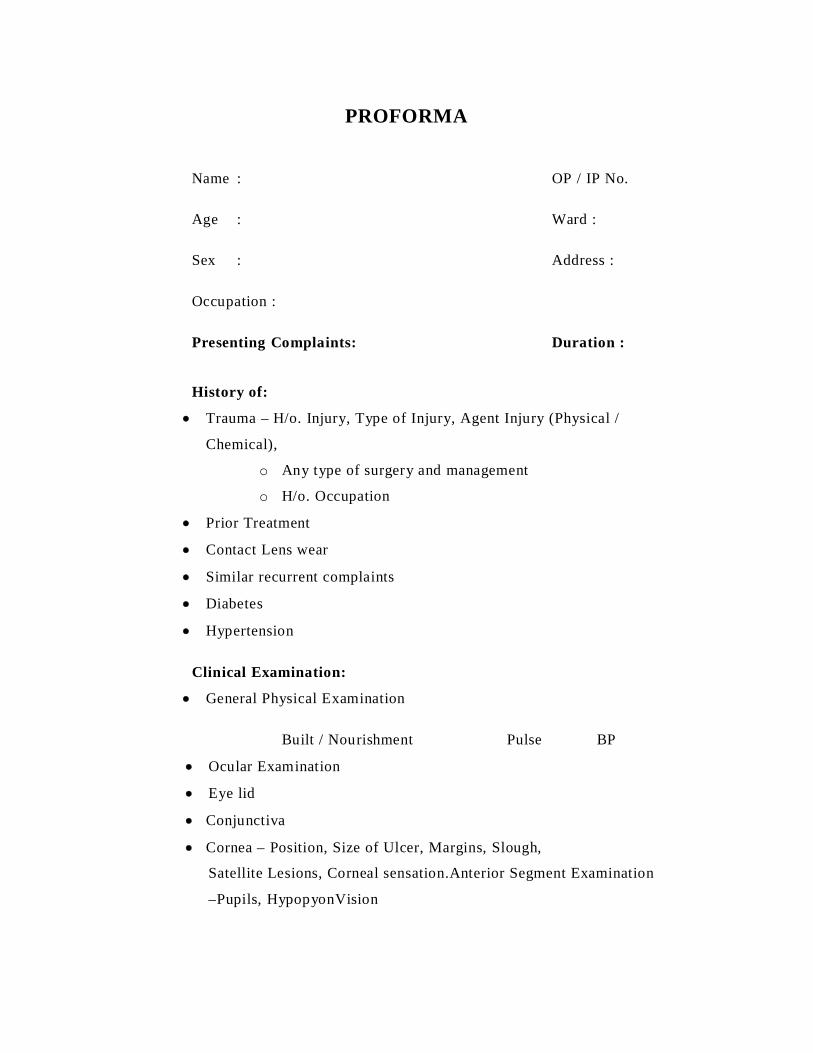
PROFORMA
Name : OP / IP No.
Age : Ward :
Sex : Address :
Occupation :
Presenting Complaints: Duration :
History of:
Trauma – H/o. Injury, Type of Injury, Agent Injury (Physical /
Chemical),
o Any type of surgery and management
o H/o. Occupation
Prior Treatment
Contact Lens wear
Similar recurrent complaints
Diabetes
Hypertension
Clinical Examination:
General Physical Examination
Built / Nourishment Pulse BP
Ocular Examination
Eye lid
Conjunctiva
Cornea – Position, Size of Ulcer, Margins, Slough,
Satellite Lesions, Corneal sensation.Anterior Segment Examination
–Pupils, HypopyonVision
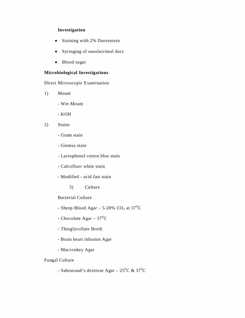
Investigation
Staining with 2% fluorescein
Syringing of nasolacrimal duct
Blood sugar
Microbiological Investigations
Direct Microscopic Examination
1) Mount
- Wet Mount
- KOH
2) Stains
- Gram stain
- Giemsa stain
- Lactophenol cotton blue stain
- Calcofluor white stain
- Modified - acid fast stain
3) Culture
Bacterial Culture
- Sheep Blood Agar – 5-20% CO2 at 370C
- Chocolate Agar – 370C
- Thioglycollate Broth
- Brain heart infusion Agar
- Macconkey Agar
Fungal Culture
- Sabouraud’s dextrose Agar – 250C & 370C
Parasitic Culture
- Agar Agar, Nonnutrient Agar Base, and coat the surface withbacteria. (E.Coli)
4) Parasitic Worms
- Macroscopic appearance and Mount examination
5) Antimicrobial susceptibility for bacterial and Fungal agent.
Bacterial – Disc diffusion method and broth dilution method
Fungal – Disc Diffusion, agar dilution and broth dilution method
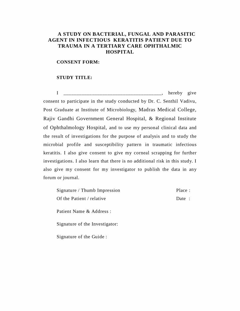
A STUDY ON BACTERIAL, FUNGAL AND PARASITICAGENT IN INFECTIOUS KERATITIS PATIENT DUE TO
TRAUMA IN A TERTIARY CARE OPHTHALMICHOSPITAL
CONSENT FORM:
STUDY TITLE:
I ______________________________________, hereby give
consent to participate in the study conducted by Dr. C. Senthil Vadivu,
Post Graduate at Institute of Microbiology, Madras Medical College,
Rajiv Gandhi Government General Hospital, & Regional Institute
of Ophthalmology Hospital, and to use my personal clinical data and
the result of investigations for the purpose of analysis and to study the
microbial profile and susceptibility pattern in traumatic infectious
keratitis. I also give consent to give my corneal scrapping for further
investigations. I also learn that there is no additional risk in this study. I
also give my consent for my investigator to publish the data in any
forum or journal.
Signature / Thumb Impression Place :
Of the Patient / relative Date :
Patient Name & Address :
Signature of the Investigator:
Signature of the Guide :
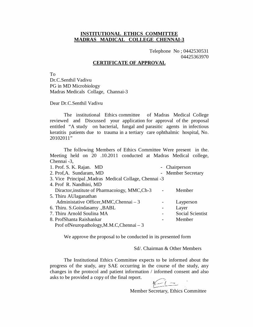
INSTITUTIONAL ETHICS COMMITTEEMADRAS MADICAL COLLEGE CHENNAI-3
Telephone No ; 044253053104425363970
CERTIFICATE OF APPROVAL
ToDr.C.Senthil VadivuPG in MD MicrobiologyMadras Medicals Collage, Channai-3
Dear Dr.C.Senthil Vadivu
The institutional Ethics committee of Madras Medical Collegereviewed and Discussed your application for approval of the proposalentitled “A study on bacterial, fungal and parasitic agents in infectiouskeratitis patients due to trauma in a tertiary care ophthalmic hospital, No.20102011”
The following Members of Ethics Committee Were present in the.Meeting held on 20 .10.2011 conducted at Madras Medical college,Chennai -3,1. Prof. S. K. Rajan. MD - Chairperson2. Prof,A. Sundaram, MD - Member Secretary3. Vice Principal ,Madras Medical Collage, Chennai -34. Prof R. Nandhini, MD Diractor,institute of Pharmacoiogy, MMC,Ch-3 - Member5. Thiru AUlaganathan Administative Officer,MMC,Chennai – 3 - Layperson6. Thiru. S.Goindasamy .,BABL - Layer7. Thiru Arnold Soulina MA - Social Scientist8. ProfShanta Raishankar - Member Prof ofNeuropathology,M.M.C,Chennai – 3
We approve the proposal to be conducted in its presented form
Sd/. Chairman & Other Members
The Institutional Ethics Committee expects to be informed about theprogress of the study, any SAE occurring in the course of the study, anychanges in the protocol and patient information / informed consent and alsoasks to be provided a copy of the final report.
Member Secretary, Ethics Committee
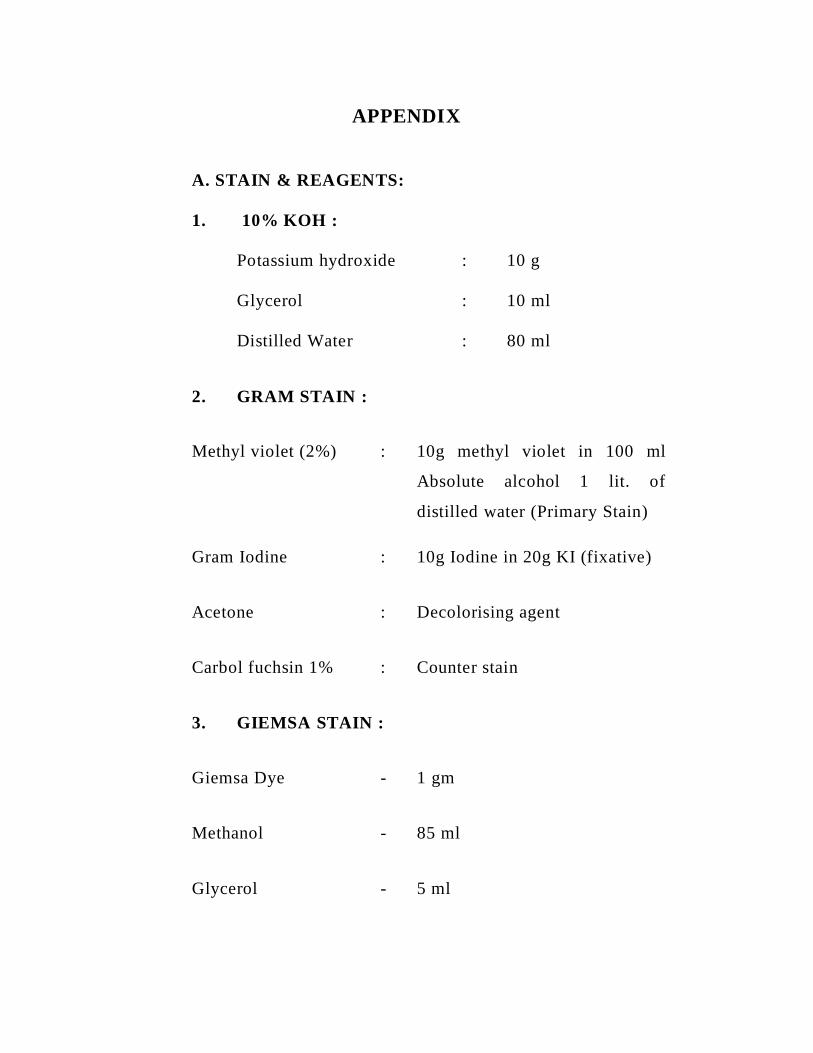
APPENDIX
A. STAIN & REAGENTS:
1. 10% KOH :
Potassium hydroxide : 10 g
Glycerol : 10 ml
Distilled Water : 80 ml
2. GRAM STAIN :
Methyl violet (2%) : 10g methyl violet in 100 ml
Absolute alcohol 1 lit. of
distilled water (Primary Stain)
Gram Iodine : 10g Iodine in 20g KI (fixative)
Acetone : Decolorising agent
Carbol fuchsin 1% : Counter stain
3. GIEMSA STAIN :
Giemsa Dye - 1 gm
Methanol - 85 ml
Glycerol - 5 ml
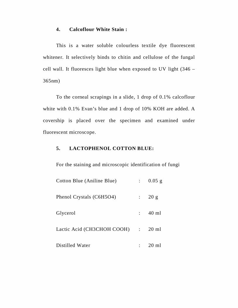
4. Calcoflour White Stain :
This is a water soluble colourless textile dye fluorescent
whitener. It selectively binds to chitin and cellulose of the fungal
cell wall. It fluoresces light blue when exposed to UV light (346 –
365nm)
To the corneal scrapings in a slide, 1 drop of 0.1% calcoflour
white with 0.1% Evan’s blue and 1 drop of 10% KOH are added. A
covership is placed over the specimen and examined under
fluorescent microscope.
5. LACTOPHENOL COTTON BLUE:
For the staining and microscopic identification of fungi
Cotton Blue (Aniline Blue) : 0.05 g
Phenol Crystals (C6H5O4) : 20 g
Glycerol : 40 ml
Lactic Acid (CH3CHOH COOH) : 20 ml
Distilled Water : 20 ml
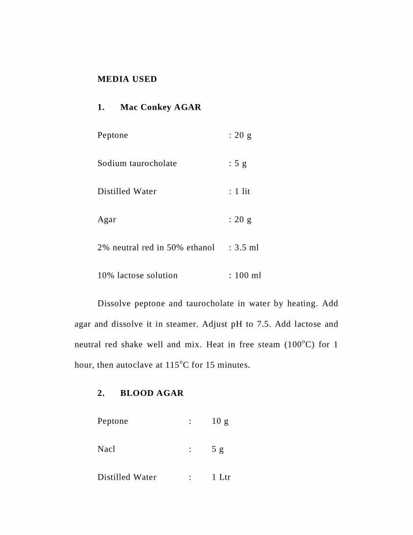
MEDIA USED
1. Mac Conkey AGAR
Peptone : 20 g
Sodium taurocholate : 5 g
Distilled Water : 1 lit
Agar : 20 g
2% neutral red in 50% ethanol : 3.5 ml
10% lactose solution : 100 ml
Dissolve peptone and taurocholate in water by heating. Add
agar and dissolve it in steamer. Adjust pH to 7.5. Add lactose and
neutral red shake well and mix. Heat in free steam (100oC) for 1
hour, then autoclave at 115oC for 15 minutes.
2. BLOOD AGAR
Peptone : 10 g
Nacl : 5 g
Distilled Water : 1 Ltr
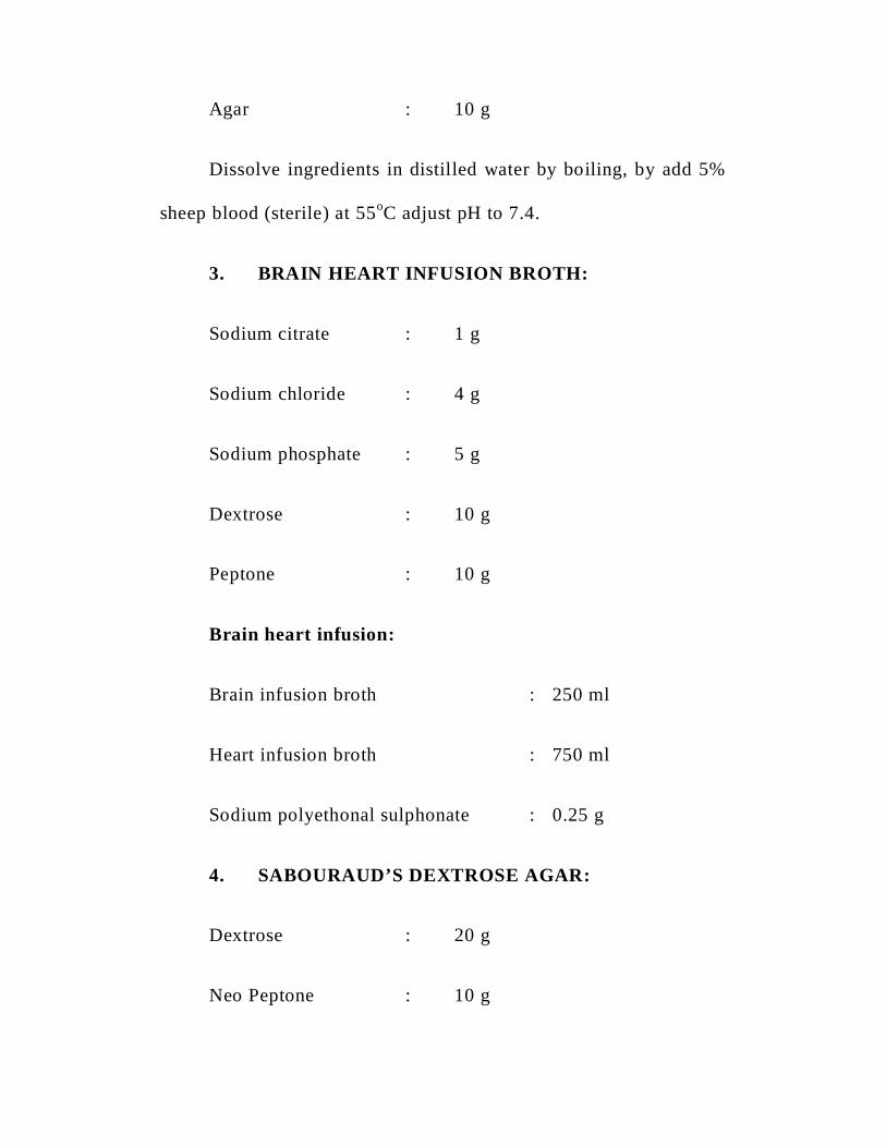
Agar : 10 g
Dissolve ingredients in distilled water by boiling, by add 5%
sheep blood (sterile) at 55oC adjust pH to 7.4.
3. BRAIN HEART INFUSION BROTH:
Sodium citrate : 1 g
Sodium chloride : 4 g
Sodium phosphate : 5 g
Dextrose : 10 g
Peptone : 10 g
Brain heart infusion:
Brain infusion broth : 250 ml
Heart infusion broth : 750 ml
Sodium polyethonal sulphonate : 0.25 g
4. SABOURAUD’S DEXTROSE AGAR:
Dextrose : 20 g
Neo Peptone : 10 g
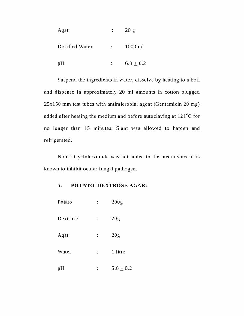
Agar : 20 g
Distilled Water : 1000 ml
pH : 6.8 + 0.2
Suspend the ingredients in water, dissolve by heating to a boil
and dispense in approximately 20 ml amounts in cotton plugged
25x150 mm test tubes with antimicrobial agent (Gentamicin 20 mg)
added after heating the medium and before autoclaving at 121oC for
no longer than 15 minutes. Slant was allowed to harden and
refrigerated.
Note : Cycloheximide was not added to the media since it is
known to inhibit ocular fungal pathogen.
5. POTATO DEXTROSE AGAR:
Potato : 200g
Dextrose : 20g
Agar : 20g
Water : 1 litre
pH : 5.6 + 0.2
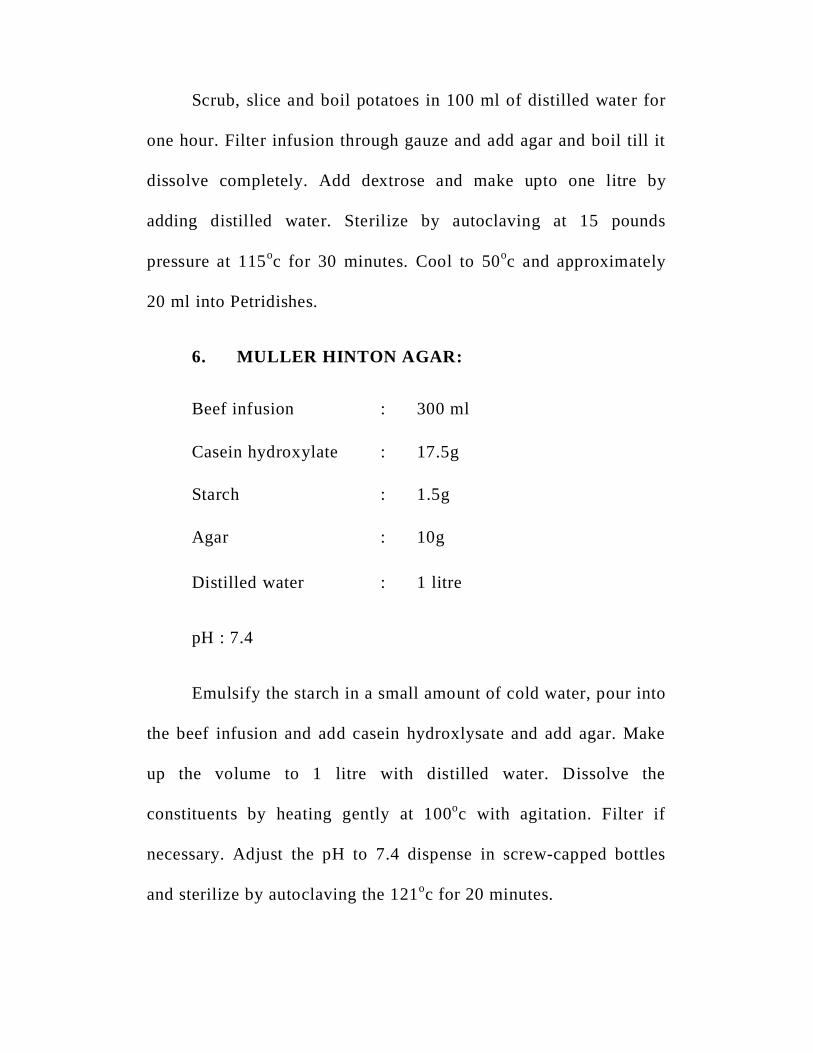
Scrub, slice and boil potatoes in 100 ml of distilled water for
one hour. Filter infusion through gauze and add agar and boil till it
dissolve completely. Add dextrose and make upto one litre by
adding distilled water. Sterilize by autoclaving at 15 pounds
pressure at 115oc for 30 minutes. Cool to 50oc and approximately
20 ml into Petridishes.
6. MULLER HINTON AGAR:
Beef infusion : 300 ml
Casein hydroxylate : 17.5g
Starch : 1.5g
Agar : 10g
Distilled water : 1 litre
pH : 7.4
Emulsify the starch in a small amount of cold water, pour into
the beef infusion and add casein hydroxlysate and add agar. Make
up the volume to 1 litre with distilled water. Dissolve the
constituents by heating gently at 100oc with agitation. Filter if
necessary. Adjust the pH to 7.4 dispense in screw-capped bottles
and sterilize by autoclaving the 121oc for 20 minutes.
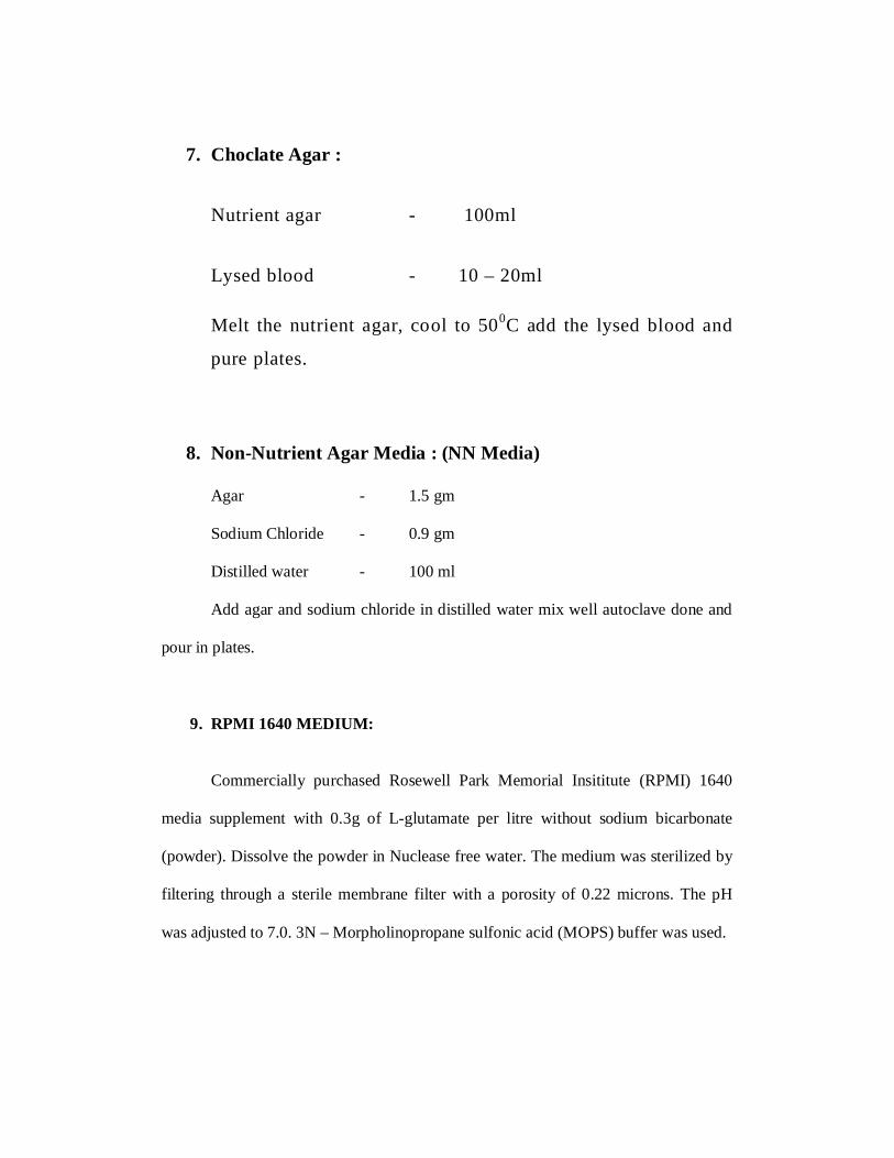
7. Choclate Agar :
Nutrient agar - 100ml
Lysed blood - 10 – 20ml
Melt the nutrient agar, cool to 500C add the lysed blood and
pure plates.
8. Non-Nutrient Agar Media : (NN Media)
Agar - 1.5 gm
Sodium Chloride - 0.9 gm
Distilled water - 100 ml
Add agar and sodium chloride in distilled water mix well autoclave done and
pour in plates.
9. RPMI 1640 MEDIUM:
Commercially purchased Rosewell Park Memorial Insititute (RPMI) 1640
media supplement with 0.3g of L-glutamate per litre without sodium bicarbonate
(powder). Dissolve the powder in Nuclease free water. The medium was sterilized by
filtering through a sterile membrane filter with a porosity of 0.22 microns. The pH
was adjusted to 7.0. 3N – Morpholinopropane sulfonic acid (MOPS) buffer was used.
BIBLIOGRAPHY
1. Alexandrakis. G., Alfonso E.C., Miller D. Shifting trends in bacterial
keratitis in South Florida and emerging resistance to
fluoroquinolones. Ophthalmology. 107 : 8. 1497 to 1502. Aug 2000.
2. Araffa R C, Ami I, lshibashi Y et al. Calcoflour and ink-
Potassium hydroxide preparation for identifying fungi. Am J
Ophthalmol 1985; 100: 719-723.
3. Basics of Ophthalmology Book Renu Johi Chapter 7 Cornea Page
107 – 146.
4. Basak SK, BasakS, MahantaA, BhowmickA. Epideiniological
and microbiological diagnosis of suppurative keratitis in
Gangetic West Bengal, eastern India. indian J Ophihalmol 17-22.
5. Bharathi 3M, Sirinivasan M, Ramakrishan R, Meenakshi R,
Padmavathy 5, Lalitha PN: A study of spectrum of
Acanthoemba keratitis: A three year study at a tertiary eye
care referral center at South India. Indian Journal of
Ophthalmology, 2007; 55(1) :37-42.
6. Bharathi M J, Ramakrishna R, Meenakshi R et al. Microbiological
diagnosis of Infectious keratitis, Comparative evaluation of
direct microscopy and culture results. Br J Ophthalmol 2006;
90: 1271 - 1276.
7. Bharathi M J, Rarnakrishnan R,S, et al. Microbial keratitis in
South India: influence of risk factors, climate, and
geographical variation. Ophthalmic Epidemiol; 14:61-9.
8. Bharathi M.J., Ramakrishna R., Samala Vasu, Meenakshi R., Palani
Appan .R. Epidemiological characteristics and laboratory diagnosis
of fungal keratitis: A three year study (Sep. 99 to Aug.02). Indian
Journal of Ophthalmology. 51:4. 315 to 322. Dec. 2003.
9. Bharathi M.J., Ramakrishnan R., Vasu S., Meenakshi R.
Epidemiology of Bacterial Keratitis at a Referral Centre in
South India. Indian Journal of Medical Microbiology. 21:4.
239 to 245. Oct. 2003.
10. Bharathi M.J., Ramakrishnan.R, Vasu.S., Meenakshi.R,
Shivakumar C, Palaniyappan R. Epidemiology of bacterial
keratitis in a referral centre in South India. Indian J Med
Microbiol 2003 ; 21:239-245.
11. Bharathi Mi, Ramakrishnan R, Vasu 8, Meenakshi R,
Palaniappan R, Epidemiological characteristics and laboratory
diagnosis of fungal keratitis. A three-year study, Indian J
Ophthalmol 2003; 51 :315-21.
12. Bourcier T, Thomas F, Borderie V. Chaumeil C, Laroche L.
Bacterial Keratitis. Predisposing factors, clinical and
microbiological review of 300 cases. Br. J.Ophthalrnol. 2003;
87:834-838.
13. Cesar Espiritu. Treatment and prevention of ocular bacterial
infections in Asia Part 1: The changing landscape of microbial
organisms. Asian J Ophthalmol.2008; 10:383-7.
14. Chalita M R, Hofling-Lima A L, Paranhos A et al., Shifting trends
in invitro antibiotics susceptibilities for common ocular isolates
during a period of 15 years. Am J OphthaImoI2004; 137:43- 51.
15. Chander J, Chakrabarti A, Sharma A et al. Evaluation of
Calcofluor staining in diagnosis of fungal corneal ulcers.
Mycoses 1993: 36: 243 - 245.
16. Chander J, Sharma A, Prevalence of fungal corneal ulcer in
Northern India; Infection 1994; 22; 207-09
17. Chein-Fan Fong, Fung-Rong Hu, Chai-Hui Tseng et al.
Antibiotic
18. Cherry FR, Ordan JL, John KGD: Acanthamoebakeratitis:
multicentric survey in England 1992-96. British Journal of
Ophthalmology, 1998; 82(12); 1387-1392.
19. Chowdhary, Anuradha MD Spectrum of Fungal Keratitis in
North India;Cornea; Jan 2005; Vol 24; Issue 1; pp 8-15.
20. Clinical Laboratory Standards institute (CLSI). 2010.
Reference method for broth dilution antifungal susceptibility
testing of filamentous fungi. Approved standard. NCCLS
document M38-A. National Committee for Clinial Laboratory
Standards, Wayne, Pa, USA.
21. Devamani F, Gananaselvan J, Anandakannan K, Sridhar N,
Sundararaj T: Studies on the prevalence of Acanthamoeba
keratitis in and around Chennai. Indian Journal of
MedicalMicrobiology, 1998: 16(4): 152-153.
22. Donald Armstrong, Janathan Cohen, Infections diseases vol1,
sec, Chapter 10 2/11; 3-11.
23. Erie J C, Nevift M P, Hodge D 0 et al. Incidence of Ulcerative
keratitis in a defined population from 1950 through 1988. Arch
Ophthal, 1993; 111 (12): 1665 -1667.
24. Ernst E J. Investigational antifungal agents. Pharmacotherapy
2001; 21: 165 -174.
25. Espinel-Ingroff A, Arthington-Skaggs B, lqbal N et al.
Multicenter Evaluation of a New Disc Agar Diffusion Method
for Susceptibility Testing of Filamentous Fungi with
Voriconazole, Posaconazole, Itraconazole, Amphotericin B
and Caspofungin. Journal of Clinical Microbiology 2007; 45:
1811 -1820.
26. Feilmeier, Michael R.MD., Sivaraman, Kavitha R. Etiologic
Diagnosis of Comeal ulceration at a tertiary eye care centre in
Kathmandu, Nepal. Cornea, December 2010-Volume 29-issue
12. Page 1380-1385.
27. Fong CF, Tseng CH, Hu FR, Wang IS, Chen WL, Hou YC.
Clinical characteristics of microbial keratitis in a university
hospital in Taiwan. Am J Ophthalmol 2004; 137:329-36.
28. Gebauer A, McGhee CN, Crawford GJ. Severe microbial
keratitis in temperate and tropical western Australia. Eye
1996; 10: 575-80.
29. Geetha Ky. et al. Epidemiology of corneal ulcer. Opthalmic
epidemiology 2002; 9: 87-92.
30. Gita Varaprasthan, Kevin Miller, Thomas Lietman, John W.
Whitcher, Vicky Cevallos. Trends in the etiology of infectious
corneal ulcers at the F.I. Proctor Foundation. Cornea. 23:4.
360 to 364. May 2004.
31. Gonzales CA, Srinivasan M, Whitcher SF, Smolin G.
Incidence of corneal ulceration in Madurai district, south
India, Ophthalmic Epidemiot t996; 3: 159-66.
32. Gopinathan U, Garg F, Fernandes M, Sharina 5, Athmanathan 5,
Rao GN. The epidemiological features and laboratory results
of flingal keratitis: a 10 -year review at a referral eye care
center in south India. Cornea 2002; 21 : 555-9.
33. Gopinathan U., Garg P., Fernandes M., Sharma S.,
Athmanathan S., Rao G.V. The epidemiological features and
laboratory results of Fungal keratitis : A 10 year review at a
referral eye care centre in South India. Cornea. 2 1:6. 555-559.
Aug. 2002.
34. Gopinathan, LJsha, Garg et al. The Epidemiological features
and Laboratory Results of Fungal Keratitis: A 10-Year Review
at a Referral Eye Care center in South India. Cornea 2002; 21:
555 - 559.
35. Hagan M, Wright B, Newman M, Dolin P. Johnson 6. Causes
of supportive keratitis in Ghana. Br J Ophihalmol 1995; 79:
1024-8.
36. Houang B, Lm D, Fan D, Seal D. Microbial keratitis in Hong
Kong: relationship to climate, environment and contact-lens
disinfection. Trans R Soc Trop Mert Hyg 2001; 95 : 36 1-7.
37. Howard M Leibowitz John P Frangie. Infections of the Cornea,
chapter 22 , 613-628.
38. Illingworth CD, Cook SD, Karabatsas CH, Easty DL:
Acanthamoeba keratitis; risk factors and outcome. British
Journalof Ophthalmology, 1995; 79(l2):1078- 1082
39. Illingworth CD, Cook SD. Acanihamoeba keratitis. Sun
Ophthalmol 1998; 42 : 493-508.
40. Jagadhish chander, Nidhi singla Nalini Agnihothri,
Sudheshkumar Aa, Antariksh deep. Keratornycosis in and
around Chandigarh; A 5 year study from North Indian tertiary
care hospital. Indian Journal of Pathology and Microbiology.
Year 2008/Volume:l/issue:2/Page : 304-306. Fayahar Bharathi
. R. Ramakrishnan, R. Meenakshi, S. Padmavathy.
41. Jagadish chandar et al, Dept of Medical Micro&ophthal PGI
Chandigarh IJMM 1993,11(3), 218-222
42. Jagadish Chander. Text book of Medical Mycology. Mehta
Publications. 2nd edition. Chapter 28 - Oculomycoses. 310 to
319. 2002.
43. Jayahar Bharathi M, Ramakrishnan, S.Vasu or Meenakshi invitro
efficacy of Antibacterial against bacterial isolates form corneal
isolates from Indian journal of Ophthmalogy 2002.
44. Etiology and Antibacterial suceptlibility pattern of community
acquired bacterial Occular infections in a tertiaray care Indian
journal of Ophthmalogy 2010.
45. Jayahar Bharathi M, Ramakrishnan R, Meenkshi R, Shivakumar C
& Lional Raj et al Analysis of the risk factor predisposing to
fungal, bacterial & Acanthamoeba keratitis in South India,
Indian J Med Res 130, December 2009, pp 749-757
46. Jeanette JS, Theodore MB, Visvesvara GS: The epidemiology
of Acanthamoeba keratitis in the United States. American
Journal of Ophthalmology, 1989; 107;331-336.
47. Jeng BH, MC Leod SD. Microbial keratitis. Br.J.Ophthalmol.
2003; 87:805-806.
48. Jeng BH. D.S.Holsdew, A.B. Kumar. Invest ophthalmol vis Sci
2003; 44: E abstract 805.
49. Jyothi Padmaja I, Saroja Venugopal M.A. Study of
keratomycosis. The Indian Practitioner. XLIII: 1. 19-22. 1990.
50. Kaufman HE and Wood RM. Mycotic keratitis. Am. J.
Ophthalmology, 1965, 59; 993 -1000.
51. Khalid.F. Tabbara et al., Infections of the eye . 1st edition.
52. Khanal B , Deb M , Panda A et al. Laboratory diagnosis in
Ulcerative keratitis. International journal in Experimental and
Clinical Ophthalmology. 2005, vol. 37, No. 3, page 123 - 127.
53. Khauf H.P Silvery R. Southern P.M Risser R.C. Wilson S.E
Susceptibility of cornea and conjuctival pathogen to
ciprofloxin cornea Jan 1996.
54. Knox C M, Cvellos V and Den D. 16s ribosomal DNA typing
for identification of pathogens in patients with bacterial
keratitis. J. Clini. Microbiol 1998; 36 : 3492 - 3496.
55. Koneman, E.W., and G. D. Roberts. 1985. Practical laboratory
mycology, 3rd edition., p. 64-65. The Williams & Wilkins Co.,
Baltimore.
56. Krachrner’s text book of cornea and external disease - clinical
diagnosis and management, 9th Edition page 1139 - 1263.
57. Kunimoto, D.Y., Sharma .S., Garg P., Gopinathan U., Miller
D., Rao. G.N. Corneal ulcers in elderly in Hyderabad, South
India. British Journal of Ophthalmology. 84:1. 54 to 59. Jan.
2000.
58. Laila Akter, M.A. Salam, Bulbul Hasan, Nurjahan Begam,
Iftikhar Ahmed. Etiological agents of supportive corneal ulcer:
study of 56.
59. Laspina F, Samudio M, Cibils D et at. Epidemiological
characteristics of microbiological results on patients with
infectious corneal ulcer: a 13 year survey in Paraguay. Graefes
Arch Clin Exp Qphthalmol 2004; 24: 204—209.
60. Leck A K , Thomas P A , Hagan M et at. Aetiology of
suppurative corneal ulcer in Ghana and South India and
Epidemiology of fungal keratitis. BrJ Ophthalmol 2002; 86:
1211-1215.
61. Liesegang .T.J and Forster R.K Spectrum of microbial keratitis
in south florida; American Jour of Ophthal; 1980; 90; 38-47
62. Lisa J. keay Phd Ermily W.gower, Altonasa, Lovienu, clinical
and microbiological characatersitic of fungal keratitis in the
united states 2001 – 2007 Opthamology volume 110.
63. Lixin Xie, Wenxian Zhong, Weiyun Shi, Shiying Sun.
Spectrum of Fungal keratitis in North China. Ophthalmology
2006; 113: 1943 - 1948.
64. Mackie & McCartney, Practical Medical Microbiology 14th
edition 2007, Chapter 6 p 125.
65. Mahajan V M. Ulcerative keratitis; An analysis of lab data in
674 cases J Ocul. Ther. Surg. 1985; 4, 138 - 141.
66. Manikandan P, Baskar M, Revathy R et al. Acanthamoeba
keratitis A Six year epidemiological review from a tertiary
care eye hospital in Southindia. IJMM 2006, 22; 226-230.
67. Microbial Keratitis in South India: Influence of risk factors,
climate and geographical variation. Ophthalmic epidemiology,
14:2 , 61-62
68. Mohan M, Panda A, Gupta S K. Management of human
keratomycosis. Aust NZ J Ophthamol 1985; 17: 295.
69. Mohapatra .D, Mallick B, Panda P.L., Sarangi G, Chayani N.,
Panda .B, Patra G.C. XXVII National Conference of Indian
Association of Medical Microbiologists souveneigr. A study of
infective keratitis with special reference to mycotic etiology.
85. Nov. 2003.
70. Murray, Manual of Clinical Microbiology, 9th edition 2006,
Volume II, Chapter 131, Antifungal agents and Susceptibility
test methods. P 1980.
71. Namrata kumara et ci dept of micro, Indira Gandhi institute of
medical sciences, Patna; Indian journal of Micro & Path 2002,
45(3) 299-302
72. Nicholson AD, Motwane 5, Gogate A: Acanthamoeba keratitis:
Jounal of Postgraduate Medicines, 1995; 41(3): 81-82.
73. Noopur Gupta,Radhika Tandon.Investigative modalities in
infective keratitis.Indian Journal Opthalmol2008;56 ;209 -213.
74. Noopur Gupta., Radhika Tandon., Investigative modalities in
infective Keratitis. Indian J ophthalmol 2008;56:209-213.
75. Norma T J, Raihan S, Bakiah S et al. Microbial keratitis:
aetiological diagnosis and clinical features in patients admitted
to Hospital Universiti Sains Malaysia. Singapore Med J 2008;
49: 67 — 71.
76. Obrien T.P., Maguine . MG., Fink NE. Efficacy of ofloxacin
vs cefazolin and tobramycin in the therapy for bacterial
keratitis. Report from the bacterial keratitis study research
group. Arch ophthalmol 1995;1 13:1257-65.
77. Panda A., Sharma N., Das G., Kumar N. Sripathy G. Mycotic
keratitis in children. Epideniiological and microbiologic
evaluation. Cornea 16:3. 295 to 299. May 1997.
78. Panda A.Ahuja R.Sastry S.S. Comparison of topical 0.3% of
ofloxacin with fortified tobramycin plus cefazolin in the
treatment of bacterial keratitis. Eye: 1991; 13: 744-77.
79. Parmer P. Seleman A. Kalavathy C.M. Et al Comparision of
Topical Gaitfloxacin and Ciprofloxacin AMJ Opthamology
2001.
80. Rajan K Anand, Double trouble: A study of injury and steroids
as predisposing factors in comeal ulcers among rural patients
of koshi region of Bihar. AIOC 2010, Community and
ophthalmology session, 158—160.
81. Reemanath, Syamanta Baruah, Lahari saikia, Banu Devi,
Mycotic Corneal ulcer in upper Assam. Indian Journal of
Ophtholmology year 201 1/Volume:59/Issue :5/Page: 367-371.
82. Reichert R, Stern GA: Quantitative adherence of bacteria to
human corneal epi cells; Arch ophthalmol 102: 1394, 1984.
83. Rondeau N., Bourcier T. Chaumeil. C., Borderie V., Tonzeau.
Fungal keratitis at the central hospitalier National d’ Ophthal
mologie des Quinze-Vingts. A retrospective study of 19 cases.
Journal of France Ophthalmology. 29:9. 890 to 896. Nov.
2002.
84. Sadia Sethi, Mohammed Junaid Sethi, Rashid Iqbal. Gomal
Journal of Medical Sciences January - June 2010, Vol. 8. No.1.
85. Samar K Basak, Sukumar Basak, Ayan Mohanta, Amp
Bhowmick. Epidemiological and microbiological diagnosis of
suppurative keratitis in Gangetic West Benghal, Eastern India.
Indian Journal of Opthalmology, year: 2005/ Volume:53/issue
ii / page: l7-22.
86. Santha Ram V.S., Satyavani .M. Dissertation on
Epidemiological study of Keratomycosis. Rangaraya Medical
College, Kakinada 1987.
87. Saroj Gupta, Atul Aher et al A case report Acanthamoeba
keratitis, Vol 2, July 2009, people’s journal of scientific
research
88. Savithri Sharma.et al., Infrequent etiological agents of corneal
ulcers Journal of MSOA. 1989;26(32):19 -21
89. Schaefer F., Bruttin., Zografos L., Guex Crosier Y. Bacterial
keratitis : A prospective clinical and microbiological study.
British Journal of Ophthalmoloy. 85:7. July 2001.
90. Sharma 5, Garg P, Rao GN. Patient characteristics, diagnosis,
and treatment of non-contact lens related Acanthamoeba
keratitis. BrlOphthalmol 2000; 84: 1103-8.
91. Sharma S, Garg P, Rao GN. Patient characteristics, diagnosis
and treatment of non-contact lens related Acanthamoeba
keratitis. Brithish Journal of Ophthalmology, 2000; 84(10):
1103-1108.
92. Sharma S.L. Mycotic keratitis - Indian Journal of
Ophthalmology. 29. 443. 1981.
93. Singh S.K Comeal Ulcer. Nephal J Ophthalmol 2011; 3(5): 1-2.
94. Srinivasan M. Gonzales CA, George C et al, Epidemiology and
aetiological diagnosis of corneal ulceration in Madurai, South
India. Br. J. Ophthalmol.1997; 8 1:965-971.
95. Sujith Vengayil, Anita Panda, Gita Satpathy. Niranjan Nayak.
PCR guided diagnosis of mycotic keratitis : A prospective
evaluation of its efficacy and limitations. Invest ophthalrnol.
vis. sd. January 2009 Vol 5ONo.1 152-156.
96. Susceptibility of Bacterial isolates from Bacterial keratitis
cases in a University Hospital in Taiwan. Am J Ophthalmol
2007; 144 : 682 - 689.
97. Tara K. Beattie, Alan Tomlison et al, attachement of
acanthamoeba to first and second generation silicon hydrogen
contact lenses Opthamology 2006.
98. Tenure MA, Cohen F.J, Sudesh S et al Spectrum of fungal
keratitis in Willes eye hospital, Philadelphia, Pennsylvania.
Cornea 2000; 19(3); 307-12.
99. Thomas PA. current perspective in Ophthalmic mycoses. din
microbio Rev 2003; 16:730-97.
100. Upadhyay M P, Karmacharya P C, Koirala S et al.
Epidemiology Characteristics predisposing factors and
aetiologic diagnosis of corneal ulceration in Nepal Ameican
Jour Ophtal 1991; 11: 92-99
101. Upadhyay M.P., Rai N.C., Brandt F., Shrestha R.]3. Corneal
ulcers in Nepal. Graefes Arch Clinical experimental
Ophthalmology. 219:2. 55 to 59. 1982.
102. Usha Gopinathan, Ph.D., Prashant Garg, M.S., Merle
Fernandes M.S., Savithri Sharma M.D., The epidemiological
features and laboratory results of fungal keratitis. A 10 year
review at a referral eye care centre in South India. Indian J
ophthalmol.2009.July - Aug: 57(4) 273-279.
103. Verenkar M.P., Borkar Shubhangi, Pinto M.J.W., Naik
Pradeep. A study of mycotic keratitis in Goa. Indian Journal of
Medical Microbiology. 16:2. 56 to 60. 1998.
104. Wang A.G., Wu. C.C., Liu J.H. Bacterial corneal ulcer : A
multivariate study. Ophthalmologica. 212 : 2. 126 to 132.
1998.
105. Whitcher.IP, Srinivasan M, Upadhyay MP. Corneal blindness:
a global perspective. Bull World Health Organ 2001; 79:
214- 21.
106. Whitcher JP, Srinivasan M, Upadhyay MP. Microbial keratitis.
In: Johnson 65, Minassian DC, Weale RA, West 8K, editors.
The epidemiology of eye diseases. 2” ed. London: Arnold;
2003. p.190-5.
107. Wilhelmus KR. ,Liesegang TG., Osato MS., Jones DB.,
cumitech 13A: Lab diagnosis of ocular infection. Washington
DC, 1994, American Society of Microbiology.
108. Wong T, Ormonde 5, Gamble G, McGhee CN. Severe infective
keratitis leading to hospital admission in New Zealand. Br I
Ophihalmol 2003; 87: 1103-8.
109. Y W Ibrahirn, D L Boase, I A Cree, Epidemiologic
charactestics, predisposing factors and microbiological profile
of infectious corneal ulcers. The ports mouth corneal ulcer
study. British journal of ophthalmology 93, 10 (2009)
110. Zhang W., Pau. Z., Jin-X., Luo.S., Luo.Y., Wu.Y., Li-R. The
variance of pathogenic organisms of purulent ulcerative
keratitis. Zhoghua yan ke Za zhi. 38. 8 to 12. January 2002.
111. Zirnrnerman, E.L. 1962, Mycotic keratitis. Lab Investigations.
2: 1151.
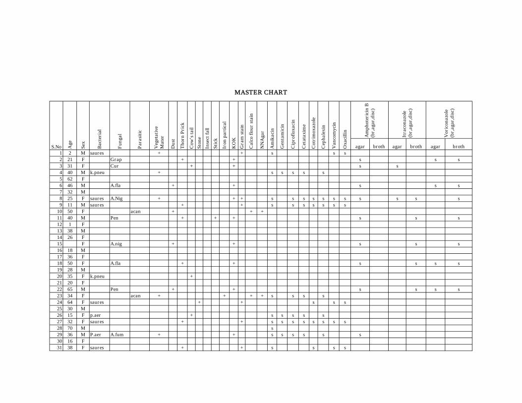
agar broth agar broth agar broth1 2 M saures + + s s s2 21 F Grap + + s s s3 31 F Cur + + s s4 40 M k.pneu + s s s s s5 62 F6 46 M A.fla + + s s s7 32 M8 25 F saures A.Nig + + + s s s s s s s s s s s9 11 M saures + + s s s s s s s
10 50 F acan + + +11 40 M Pen + + + s s s12 1 F13 38 M14 26 F15 F A.nig + + s s s16 18 M17 36 F18 50 F A.fla + + s s s s19 28 M20 35 F k.pneu +21 20 F22 65 M Pen + + s s s s23 34 F acan + + + + s s s s24 64 F saures + + s s s25 30 M26 15 F p.aer + s s s s s27 32 F saures + + s s s s s s s s28 70 M s29 36 M P.aer A.fum + + s s s s s s30 16 F31 38 F saures + + s s s s
Itra
con
azol
e(b
r,ag
ar,d
isc)
Vor
icon
azol
e(b
r,ag
ar,d
isc)
Age
Sex
Bac
teri
al
Fun
gal
Par
asit
ic
Gen
tam
icin
Cip
rofl
oxac
in
Sti
ck
Iron
par
tica
l
KO
K
Gra
m s
tain
Cet
atax
ime
Cot
rim
oxaz
ole
Cep
hale
xin
Van
com
ycin
Oxa
cilli
n
MASTER CHART
S.No Cal
co f
lou
r st
ain
NN
Aga
r
Am
ikac
in
Veg
etat
ive
Mat
er
Du
st
Th
orn
Pri
ck
Cow
's t
ail
Sto
ne
Inse
ct f
all
Am
ph
oter
icin
B(b
r,ag
ar,d
isc)
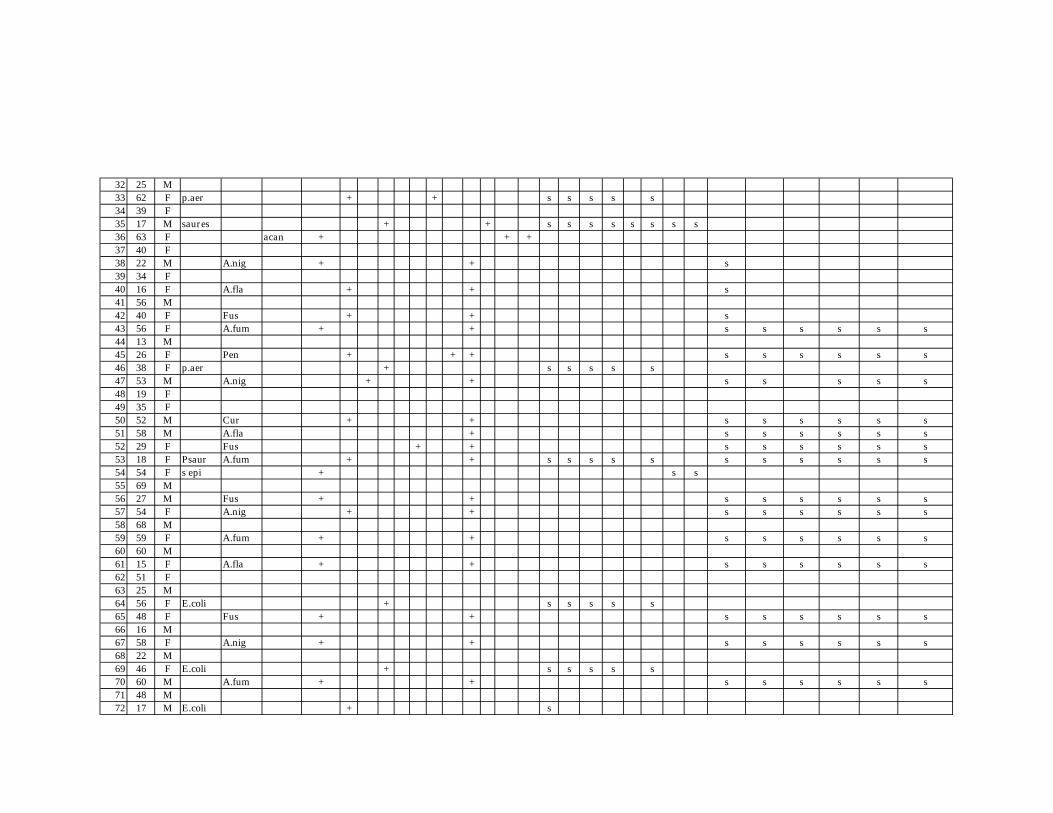
32 25 M33 62 F p.aer + + s s s s s34 39 F35 17 M saures + + s s s s s s s s36 63 F acan + + +37 40 F38 22 M A.nig + + s39 34 F40 16 F A.fla + + s41 56 M42 40 F Fus + + s43 56 F A.fum + + s s s s s s44 13 M45 26 F Pen + + + s s s s s s46 38 F p.aer + s s s s s47 53 M A.nig + + s s s s s48 19 F49 35 F50 52 M Cur + + s s s s s s51 58 M A.fla + s s s s s s52 29 F Fus + + s s s s s s53 18 F Psaur A.fum + + s s s s s s s s s s s54 54 F s epi + s s55 69 M56 27 M Fus + + s s s s s s57 54 F A.nig + + s s s s s s58 68 M59 59 F A.fum + + s s s s s s60 60 M61 15 F A.fla + + s s s s s s62 51 F63 25 M64 56 F E.coli + s s s s s65 48 F Fus + + s s s s s s66 16 M67 58 F A.nig + + s s s s s s68 22 M69 46 F E.coli + s s s s s70 60 M A.fum + + s s s s s s71 48 M72 17 M E.coli + s
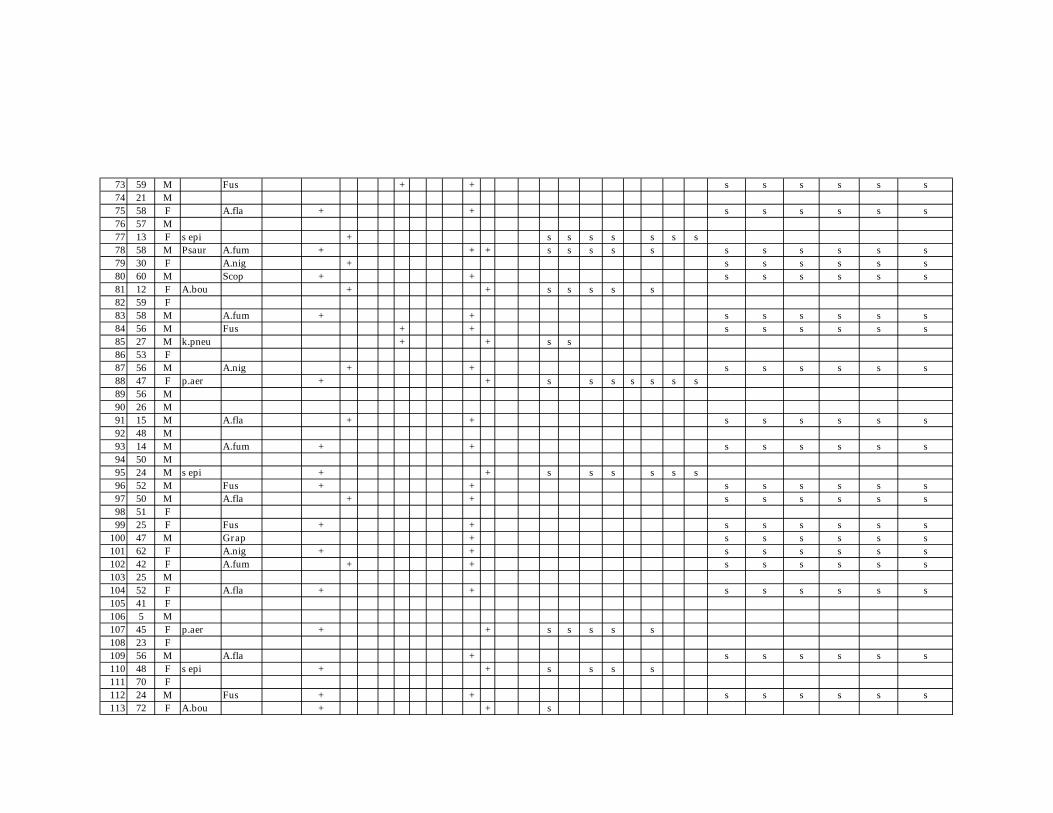
73 59 M Fus + + s s s s s s74 21 M75 58 F A.fla + + s s s s s s76 57 M77 13 F s epi + s s s s s s s78 58 M Psaur A.fum + + + s s s s s s s s s s s79 30 F A.nig + s s s s s s80 60 M Scop + + s s s s s s81 12 F A.bou + + s s s s s82 59 F83 58 M A.fum + + s s s s s s84 56 M Fus + + s s s s s s85 27 M k.pneu + + s s86 53 F87 56 M A.nig + + s s s s s s88 47 F p.aer + + s s s s s s s89 56 M90 26 M91 15 M A.fla + + s s s s s s92 48 M93 14 M A.fum + + s s s s s s94 50 M95 24 M s epi + + s s s s s s96 52 M Fus + + s s s s s s97 50 M A.fla + + s s s s s s98 51 F99 25 F Fus + + s s s s s s
100 47 M Grap + s s s s s s101 62 F A.nig + + s s s s s s102 42 F A.fum + + s s s s s s103 25 M104 52 F A.fla + + s s s s s s105 41 F106 5 M107 45 F p.aer + + s s s s s108 23 F109 56 M A.fla + s s s s s s110 48 F s epi + + s s s s111 70 F112 24 M Fus + + s s s s s s113 72 F A.bou + + s
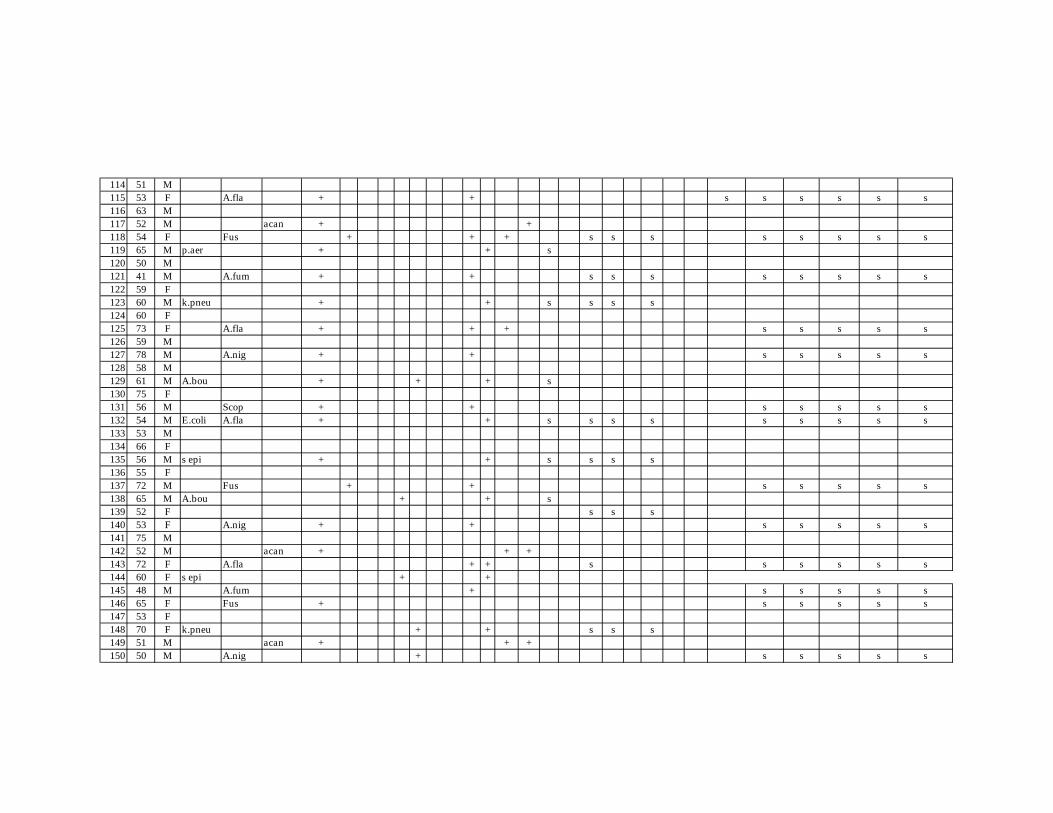
114 51 M115 53 F A.fla + + s s s s s s116 63 M117 52 M acan + +118 54 F Fus + + + s s s s s s s s119 65 M p.aer + + s120 50 M121 41 M A.fum + + s s s s s s s s122 59 F123 60 M k.pneu + + s s s s124 60 F125 73 F A.fla + + + s s s s s126 59 M127 78 M A.nig + + s s s s s128 58 M129 61 M A.bou + + + s130 75 F131 56 M Scop + + s s s s s132 54 M E.coli A.fla + + s s s s s s s s s133 53 M134 66 F135 56 M s epi + + s s s s136 55 F137 72 M Fus + + s s s s s138 65 M A.bou + + s139 52 F s s s140 53 F A.nig + + s s s s s141 75 M142 52 M acan + + +143 72 F A.fla + + s s s s s s144 60 F s epi + +145 48 M A.fum + s s s s s146 65 F Fus + s s s s s147 53 F148 70 F k.pneu + + s s s149 51 M acan + + +150 50 M A.nig + s s s s s