a survey of mycobionts of federally threatened platanthera
TRANSCRIPT

Symbiosis, 34 (2003) 145-155 Balaban, Philadelphia/Rehovot
A Survey of Mycobionts of Federally Threatened Platanthera praeclara (Orchidaceae)
JYOTSNA SHARMA~,~', LAWRENCE W. ZETIZER~, and J.W. VAN SAMBEEK~
* l ~ l a n t Science Unit, University of Missouri-Columbia, MO 65211, USA; 2Current Address: Department of Horticulture, Iowa State University,
I Ames, liZ 5001 1, USA, Tel. +1-515-294-5075, Fax. +I -51 5-294-0730, 8 i E-mail. [email protected];
3~epar tment of Biology, Illinois College, Jacksonville, IL 62650, USA, Tel. +I-217-245-3479, Fax. +1-217-245-3008, E-mail. [email protected]; 4North Central Research Station, USDA Forest Service, Columbia, MO 65211, USA, Tel. +I-573-875-5341 (ext. 233), Fax. +I-573-882-1977, E-mail. [email protected]
Received December 23,2002; Accepted February 11,2003
Abstract Terrestrial orchids require mycobionts for critical nutritional support during
germination and growth. Despite the importance of such fungi, little is known of their identity and ecological roles. In the United States, the destruction of midwestern prairie ecosystems has resulted in the decline of the native Platanthera praeclara Sheviak and Bowles and its associated mycobionts. Mycobionts of P. praeclara from six populations across Minnesota and Missouri were isolated from protocorms and mature plants and were identified to the genus level. Hyphal morphology, colony appearance, rate of growth, and monilioid cell morphology including septa1 pore ultrastructure were examined to characterize the isolates. Results indicate that P. praeclara is primarily associated with Ceratorhiza isolates at various growth stages. Few Epulorhiza isolates were recovered from roots and protocorms indicating this genus may be less critical for the orchid. Worldwide, Epulorhim have been documented as orchid mycobionts more frequently but species
The author to whom correspondence should be sent.
0334-51 14/2003/$05.50 02003 Balaban
146 J. SHARMA ET AL.
of Ceraforhiur seem to be more prevalent in the North American prairie ecosystems. Preservation of prairies with special attention to conserving mycobionts of P. praeclara is needed if viable populations of both organisms are to persist.
Keywords: Terrestrial orchids, Ceratorhiza, Epulorhiza, fungi
d
1. Introduction I
Orchidaceous plants form a unique symbiosis with saprophytic fungi in \ which coils of fungal hyphae, i.e., pelotons, are digested by the host plant to acquire carbon and other nutrients (Smith, 1967). In terrestrial orchids, mycobionts are indispensable particularly during seedling development and establishment in the wild. Platanthera praeclara, a terrestrial orchid native to midwestern prairies of the United States, has experienced steady decline in recent years and is listed as federally threatened [U.S. Fish and Wildlife Service (USFWS), 19961. Simultaneous loss of suitable fungal associates could possibly accelerate the decline of this federally listed species. Because the conservation of orchids will ultimately depend on availability of suitable fungi in the habitat to generate new plants, cataloguing and preserving natural mycobionts of rare orchids is vital for use in conservation projects (e.g., in vitro symbiotic germination and seedling re-introduction).
Mycobionts of wild orchids from Australia (Warcup, 1971, 1973, 1981, 1985, 1991), Canada (Zelmer and Currah, 1995; Zelmer et al., 1996), Europe (Andersen, 1996) and Italy (Marchisio et al., 1985) have been studied to some extent with regard to conservation of orchid fungi; however, fungi of American orchids, especially those native to the prairies, have received little attention. Given that the North American prairies are a unique and vulnerable ecosystem, studies aimed at identifying mycobionts of prairie orchids are warranted.
The current state of fungal taxonomy remains largely unsettled and confusing. Nomenclature and identity of orchid fungi before Moore's (1987) segregation of the form genus Rhizoctonia is not compatible with the modern taxonomy of orchid fungi (Parmeter and Whitney, 1970; Currah and Zelrner, 1992). Moore (1987) based his system on septa1 ultrastructure, nuclear condition of hyphal cells, and association with a known teleomorphic state; he proposed three anamorphic genera of mostly rhizotrophic orchidaceous fungi namely, Cera torhiza (teleomorphs in Ceratobasidium), Epulorhiza (teleomorphs in Tulasnella or Sebacina), and Monoliopsis (teleomorphs in Thana tephorus or Waitea). However, teleomorphs are rarely produced in culture, and therefore,

MYCOBIONTS OF PLAMNTHERA PRAECLARA 147
identification of their associated anamorphs is largely based on morphological characteristics of mycelia, hyphae, and monilioid cells and confirmed by examining the septal ultrastructure (Currah and Zelmer, 1992; Zelmer and Currah, 1995).
Based on limited studies to date, North American orchids appear to be most commonly associated with Ceratorhiza and Epulorhiza (Currah and Zelmer, 1992; Sen et al., 1999), with Epulorhiza being particularly common in southern orchids, e.g., species of Spiranthes, Platanthera, and Epidendrum. Zelmer and
@ Currah (1995) isolated an endophyte of Platanthera praeclara from Canada and described it as Ceratorhiza pernacatena. In another study, Zelmer et al.
I (1996) isolated strains of Epulorhiza and Cera t orhiza (including C . li pernacatena) from roots of adult P. praeclara, but field-incubated seeds only
yielded an Epulorhiza isolate. In this investigation, fungal isolates from roots of P. praeclara, collected from its native habitat in midwestern U.S., were surveyed. The objectives of this study were to: (1) isolate, characterize, and identify the endophytic, naturally-occurring fungi of P, praeclara in its native habitat, and (2) examine septal pore ultrastructure to corroborate genus identification in selected isolates.
2. Materials and Methods
The host orchid
Often found in calcareous wet prairies and sedge meadows, Platanthera praeclara mostly occurs in association with Carex lanuginose, Calarnagrostis stricta, and Juncus balticus in wet-mesic sedge meadows. Prairie swales with
1
Poa pratensis, Euphorbia esula, Spartina pec tinata, Salix exigua, and Sa 1 ix bebbiana are also reported to harbour the orchid (Seig and King, 1995; Wolken
b et al., 2001). The perennial root system consists of a fusiform tuber with several thick adventitious roots.
Fungal isolation and characterization
A limited number of plants were collected for this study because of the threatened status of the orchid (Table 1). Adult plants and seedlings were collected with the root system intact and transported on ice to the laboratory. The collection included an intact protocorm attached to a strap-leaved seedling. All roots and the protocorm were processed within 2-3 days. Tissue segments were rinsed with deionised (DI) water, surface-sterilised in 1:l:l V / V / V 5.25% NaOC1, 95% ethanol and sterile DI water for 1 minute and finally, rinsed twice in sterile DI water. The inner cortex was macerated in ca.

148 J. SHARMA ET AL.
0.5 ml sterile water after the cortical layer was removed with a sterile scalpel. Macerated tissue was suspended in molten Modified Melin Norkran's agar (MMN; Marx, 1969) and incubated at 23OC to allow fungal growth from pelotons released from the cortical cells, Pure fungal cultures were obtained by harvesting and transferring hyphal tips to potato dextrose agar (PDA). Of the 350+ cultures, 87 were selected based on visual evaluation to obtain a set of orchidaceous fungi. These selected isolates were cultured on oat meal agar (OMA) (2.5 oat meal and 7 gl-I agar) and stored long-term at 5OC. Another set of cultures growing on OMA was covered with sterile mineral oil and stored at 23°C.
Fungal characterization and identification was based on previously published keys and other literature (see Moore, 1987; Currah et al., 1989; Currah and Zelmer, 1992). Identification to genus level was made by analysing cultural morphology and septal ultrastructure of the fungal isolates. A 1 cm3 piece of fungal inoculum was placed in the centre of the Petri plate containing corn meal agar (CMA, Sigma Chemical Co., USA), OMA, or PDA and plates were incubated at 23°C. Each isolate and medium combination was represented by three replicates. Mycelium colour was assessed by visual comparison of the underside of the fungal colony with the standards in Methuen Handbook of Colour (Kornerup and Wanscher, 1983). Width of hyphae and appearance of mycelia growing on CMA and PDA were measured using light microscopy. Formation of monilioid cells was not consistent on all media, therefore dimensions of monilioid cells were measured on OMA, CMA, or PDA. Based on these features, isolates were assigned to orchid fungal genera. To ascertain the reliability of morphological measures, septal ultrastructure was examined for five selected isolates. Voucher specimens were deposited at the University of Alberta Microfungus Collection and Herbarium (UAMH) for safekeeping and future use.
For TEM work, thin sections of 7-day-old mycelia were fixed in 2% paraformaldehyde in cacodylate buffer (pH 7.35) for 2 hours at ambient temperature followed by a 2-hour secondary fixation in 1% 0 s 0 4 and a 2-hour tertiary fixation in 1% aqueous uranyl acetate. Washed samples were dehydrated in graded ethanol series and in propylene oxide (PO). Dehydrated specimens were infiltrated with 1:2, 1:1, and 2:l mixture of Epon-Spur:PO for 2 hrs, 2 hrs, and 8 hrs, respectively. After two additional 8-hour incubation periods, samples were transferred to embedding capsules filled with fresh, pure Epon-Spur. Capsules containing the specimens were polymerised in a 55°C oven for 2 days. Thin sections were cut with a diamond knife on a LKB Ultratome I11 ultramicrotome, placed on grids, stained in uranyl acetate, and then subjected to lead citrate staining. Sections were washed in DI water and then examined in a Hitachi H-600 electron microscope.

MYCOBIONTS OF PLATANTHERA PRAECLARA 149
Table 1. Number of mycobionts belonging to either Ceratorhiza or Epulorhiza from all isolates recovered from below-ground organs of Platanthera praeclara plants at various stages of growth. Samples were collected from several locations in midwestern United States.
Site Location Developmental stage Mycobiont genus of source plant1 (# of isolates)
Bicentennial Clay County, MN a
Dalby Norman County, MN
4
Bluestern Clay County, MN
Pennbina Polk County, MN
Hwy 56 Mower County, MN
Helton Harrison County, MO
Protocorm; vegetative; flowering Protocorm
Vegetative; flowering Vegetative
Vegetative; flowering Vegetative
Vegetative Vegetative
Vegetative; flowering
Flowering
Ceratorhiza (25) Epulorhiza (1)
Ceratorhiza (29) Epulorhiza (1)
Ceratorhiza (14) Epulorhiza (2)
Ceratorhiza (4) Epulorhiza (3)
Ceratorhiza ( 6 )
Epulorhiza (2)
1Total number of plants collected from each site: Bicentennial (4); Dalby (3); Bluestem (4); Pembina (4); Hwy 56 (4); Helton (1).
3, Results and Discussion
Of the 87 isolates considered in this study, most (89%) were assignable to Cera torhiza (Table 1); remaining isolates were assigned to Epu lorhiza. Recovery of Ceratorhiza and Epulorhiza isolates from several Minnesota and Missouri populations suggests that these two genera may constitute the primary mycobionts of Platanthera praeclara. At each study site, Ceratorhiza isolates
I
were consistently recovered in larger numbers from roots of plants spanning several different phenological stages (Table 1).
Ceratorhiza isolates appear cream, yellow, or tan in colour, and have cottony, aerial hyphae on PDA; in comparison, Epulorhiza mycelia are often submerged in the medium with an overall waxy appearance and pale greyish- cream colour (Zettler et al., 2003). Mycelial growth is slow in Epulorhiza compared to the growth rate in Ceratorhiza, which commonly colonise a 9 cm Petri plate within one week at ca. 23OG. Hyphae in Epulorhiza range from 1 to 4 pm in width and are mostly wider than 4 pm in Ceratorhiza (Currah and Zelmer, 1992). W e n examined through TEM, isolates of Epulorhiza have entire or imperforate parenthesomes, whereas the parenthesomes in Ceratorhiza species are perforate (Moore, 1987).

Tab
le 2
. M
orph
omet
ric
data
and
sep
ta1
ultr
astr
uctu
re c
ondi
tion
of
sele
cted
myc
obio
nts
reco
vere
d fr
om b
elow
-gro
und
orga
ns o
f P
lata
nthe
ra p
raec
lara
. C
ultu
res
wer
e gr
own
at 2
3OC
.
Isol
ate
App
eara
nce
on C
MA
l A
ppea
ranc
e on
PD
A a
nd
Gro
wth
H
ypha
l wid
th
Mon
ilio
id
Pare
nthe
som
es
Met
huen
Col
our2
.3
rate
(c
urs
cell
wid
th
(m
kl
>
and
leng
th
PDA
C
MA
PD
A
(p)
He1
166
- E
pulo
rhiz
a U
AM
H 9
846
Blu
61 -
Epu
lorh
iza
UA
MH
984
5
Blu
86 -
Cer
ator
hiza
U
AM
H 9
848
Bic
68 -
Epu
lorh
iza
UA
MH
984
4
Bic
70 -
Cer
ator
hiza
U
AM
H 9
847
Loo
se m
argi
n; u
nifo
rm
subm
erge
d m
ycel
ium
Loo
se m
argi
n; u
nifo
rm
subm
erge
d m
ycel
ium
Loo
se m
argi
n; s
catt
ered
sm
all t
ufts
of
aeri
al
myc
eliu
m; c
once
ntri
c zo
nati
on t
owar
d th
e ou
ter e
dges
of c
olon
y
Lm
se m
argi
n; u
nifo
rm
subm
erge
d m
ycel
ium
Ver
y lo
ose
mar
gin;
tu
fts o
f ae
rial
myc
eliu
m
in c
entr
e
Loo
se ja
gged
mar
gin;
0.
10
unif
orm
mat
of s
ubm
erge
d m
ycel
ium
; no
conc
entr
ic
zona
tion
; 2A
2
Ent
ire
mar
gin;
uni
form
0.
10
dens
e mat
of s
ubm
erge
d m
ycel
ium
; 2A2
Som
ewha
t ent
ire
mar
gin;
0.
20
tuft
s of
aer
ial m
ycel
ium
in
cen
tre;
con
cent
ric
zona
tion
tow
ard
the
oute
r ed
ges
of c
olon
y; 5B
5
Ent
ire
mar
gin;
uni
form
0.
10
subm
erge
d m
ycel
ium
; 3A
2
Loo
se m
argi
n; s
catt
ered
0.
25
tuft
s of
aeri
al m
ycel
ium
; 4B
6
4.0
3.5
9.0
x 15
.0;
Ent
ire
PDA
4.0
4.0
8.5
x 13
.0;
Ent
ire
PDA
7.0
6.0
20.0
x 3
0.0;
P
erfo
rate
O
MA
3.5
3.0
8.0
x 12
.0;
Ent
ire
PDA
6.0
6.0
18.0
x 3
0.5;
P
erfo
rate
C
MA
~C
MA
- C
orn
mea
l ag
ar;
PD
A - P
otat
o de
xtro
se a
gar;
OM
A - O
at m
eal
agar
; 2K
orne
rup,
A.
and
Wan
sche
r, J
.H. (
1983
). M
ethu
en
Han
dboo
k of
Col
our,
3rd
edi
tion
, Met
huen
, Lon
don;
3C
olou
r as
appe
arin
g on
the
unde
rsid
e of
myc
eliu
m.
* rP
I

EXYCOBIONTS OF PLATANTHERA PRAECLARA 151
Of the five endophytes selected for examination with TEM, two were confirmed as Ceratorhiza and three as Epulorhiza based on the condition of septa1 ultrastructure (Table 2). Ceratorhiza isolates contained perforate parenthesomes (Sharma, 2002). Mycelia of Ceratorhiza endophytes had loose margins and grew rapidly on PDA incubated at 23OC; cottony tufts of aerial hyphae were initially scattered throughout the upper surface on PDA and CMA and eventually covered the entire surface (Table 2; Fig, la). Vegetative hyphae were thin-walled and hyaline, averaging in width from 6.0 pm on PDA to 6.5 pm on CMA. Pale yellowish-brown, granular sclerotia formed readily on OMA or CMA, most often appearing first near the point of inoculation. An isthmus connected adjacent monilioid cells borne in loose branching chains (Fig. lb). Average width and length of elliptical thick- walled monilioid cells on CMA or OMA was 19.0 pm x 30.3 pm, respectively.
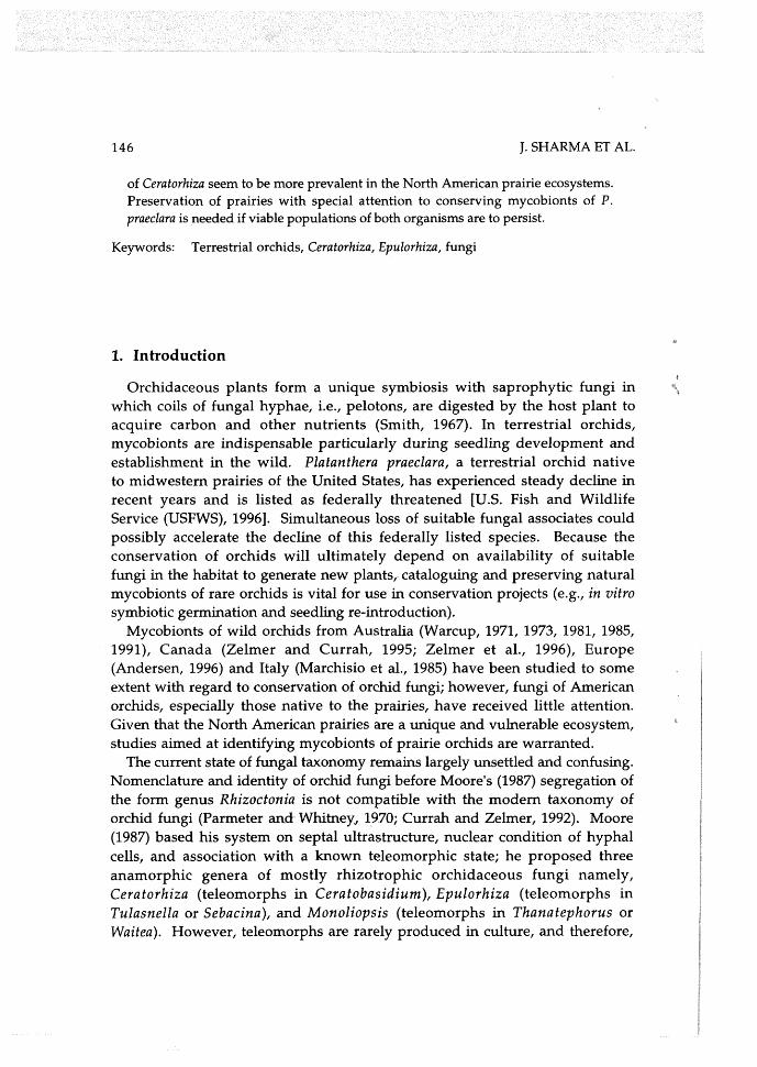
In comparison, monilioid cells of Epulorhiza averaged 8.5 pm x 13.0 pm (Table 2). Hyphal width ranged from 3 pm on PDA to 4 pm on CMA. Dolipore septa in Epulorhiza had entire or imperforate parenthesomes (Figs. 2a and 3a). Mycelium on PDA and CMA was submerged in the medium and had uniform, entire margins; greyish-cream in colour, the colonies appeared waxy throughout (Figs. 2b and 3b). Sclerotia appeared greyish-brown and had short chains of ellipsoidal to nearly spherical thin-walled monilioid cells without isthmus connections (Figs. 2c and 3c).
Isolates of both genera were recovered from a protocorm and from roots of adult plants in Minnesota. However, an adult plant or field-incubated seeds in Missouri are yet to yield an isolate assignable to Ceratorhiza implying that mycobionts may be distributed differently in different regions (Sharma, 2002). Although sampling was limited in Missouri and no Ceratorhiza strains were obtained, results from several disjunct populations in Minnesota indicate that Platanthera praeclara preferentially associates with Ceratorhiza. Results from this study support observations of Zelmer et al. (1996) who documented 15 Ceratorhiza strains and only one Epulorhiza isolate from P. praeclara in Canada. In the same study, the ratio of Ceratorhiza:Epulorhiza was ca. 2:l among all mycobionts isolated from 25 central Canadian orchid species. Some Ceratorhiza strains were also isolated from Platanthera leucophaea which is closely related to P. praeclara and occurs in midwestern prairies of America (Zettler et al., 2001). Currah et al. (1989) identified Cera to basidium cornigerum (teleomorph of Ceratorhiza goodyearae-repentis) as a common species throughout the northern hemisphere with the anamorph colnmonly found in mycorrhizae of mature temperate orchids. Although Epulorhiza strains have thus far been recovered more often from orchids worldwide (Zettler et al., 2003), Ceratorhiza species appear to be the dominant orchid mycobionts in midwestern prairies of North America. Besides physiological preference for Ceratorhiza, this phenomenon could possibly result from the

152 J. SHARMA ET AL.
Figure 1. Blu 86 (UAMH 9848) recovered from Platanthera praeclara: (a) mycelium of Blu 86, a Ceratorhiza strain, growing on potato dextrose agar (PDA) at 23OC; (b) monilioid cells growing on oat meal agar (OMA).
Figure2. Bic 68 (UAMH 9844), an Epulorhiza strain isolated from a protocorm of Platanthera praeclara: (a) septa1 ultrastructure showing entire parenthesomes (arrow); (b) mycelium of Bic 68 appeared as a waxy submerged colony; (c) monilioid cells growing on potato dextrose agar (PDA).
M,YCOBIONTS OF PLATANTHERA PRAECLARA 153
Figure 3. An Epulorhiza isolate, He1 166 (UAMH 9846), recovered from roots of mature Platanthera praeclara from a site in Missouri: (a) a micrograph showing an entire parenthesome (arrow); (b) mycelial growth is typical of Epulorhiza with a waxy, submerged colony; (c) nearly spherical monilioid cells borne in short chains growing on potato dextrose agar (PDA).
t
more aggressive attributes (fast growth rate and dense mycelia) of Ceratorhiza strains. Ceratorhiza mycelia were observed to overgrow and out-compete Epulorhiza isolates in co-inoculation experiments in vitro (Sharma, 2002).
In a symbiotic seed propagation study Ceratorhiza isolates promoted best overall in vitro germination of Platanthera praeclara, and leaf bearing seedlings developed only when seeds were cultured with a Ceratorhiza strain (UAMH 9847) (Shanna et al., 2003). Inclusion of suitable mycobionts to in vitro culture of rare orchids is preferable for use in transplant projects because the technique allows reintroduction of critical endophytes along with the orchids (Anderson, 1996; Zettler et al., 2003). Characterization and preservation of mycobionts of rare orchids, therefore, is especially necessary to ensure long- term success of orchid conservation programs.

154 J. SHARMA ET AL.
Acknowledgments
Sincere gratitude is extended to Minnesota Department of Natural Resources, The Nature Conservancy of Minnesota, and the Missouri Department of Conservation for allowing access to sites and collection of plant material (MN Special Permit nos. 9416 and 1999-13R). Nancy Sather (MNDNR), Brian Winter (TNC), and Dave Ashley (Missouri Western State College) assisted with locating plants. Thanks are also extended to USDA Forest Service for allowing use of the microscopy laboratory. Randy Tindall (University of Missouri-Columbia) supervised the TEM work.
REFERENCES
Anderson, A. B. 1996. The reintroduction of Platanthera ciliaris in Canada. In: Proceedings of the North American Native Terrestrial Orchid - Propagation and Production Conference. C. Allen, ed. National Arboretum, Washington DC, pp. 73-76.
Andersen, T.F. 1996. A comparative taxonomic study of Rhizoctonia sensu lato employing morphological ultrastructural and molecular methods. Mycological Research 100: 11 17- 1128.
Currah, R.S. and Zelmer, C.D. 1992. A key and notes for the genera of fungi mycorrhizal with orchids and a new species in the genus Epulorhiza. Reports of the Tottori Mycological lnstitute 30: 43-59.
Currah, R.S., Smreciu, E.A., and Hambleton, S. 1989. Mycorrhizae and mycorrhizal fungi of boreal species of Platanthera and Coeloglossum (Orchidaceae). Canadian Journal of Botany 68: 1171-1181.
Kornerup, A. and Wanscher, J.H. 1983. Methuen Handbook of Colour. 3rd edition. Methuen, London.
Marchisio, V.P., Berta, G., Fontana, A., and Mannina, F.M. 1985. Endophytes of wild orchids native to Italy: their morphology, caryology, ultrastructure and cytochemical characterization. New Phytologist 100: 623-641.
Marx, D.H. 1969. Antagonism of mycorrhizal fungi to root pathogenic fungi and soil bacteria. Phytopathology 59: 153-163.
Moore, R.T. 1987. The genera of Rhizoctonia-like fungi: Ascorhizoctonia, Ceratorhiza gen. nov., Epulorhiza gen. nov., Moniliopsis and Rhizoctonia. Mycotaxon 29: 91-99.
Parmeter, J.R., Jr. and Whitney, H.S. 1970. Taxonomy and nomenclature of the imperfect state. In: Rhizoctonia solani, Biology and Pathology. J.R. Parmeter, ed. University of California Press, Berkeley, Los Angeles, pp. 7-31.
Seig, C.H. and King, R.M. 1995. Influence of environmental factors and preliminary demographic analyses of a threatened orchid, Platanthera praeclara. American Midland Naturalist 134: 307-323.
Sen, R., Hietala, A.M., and Zelmer, C.D. 1999. Common anastomosis and internal transcribed spacer RFLP groupings in binucleate Rhizoctonia isolates representing root endophytes of Pinus sylvestris, Ceratorhiza spp. from orchid mycorrhizas and a phytopathogenic anastomosis group. New Phytologist 144: 331-341.

MYCOBIONTS OF PLATANTHERA PRAECLARA 155
Sharma, J. 2002. Mycobionts, germination, and conservation genetics of federally threatened Platanthera praeclara (Orchidaceae). Ph.D. Dissertation. University of Missouri-Columbia, USA.
Sharma, J., Zettler, L.W., Van Sambeek, J.W., Ellersieck, M.R., and Starbuck, C.J. 2003. Symbiotic seed germination and mycorrhizae of federally threatened Platanthera praeclara (Orchidaceae). American Midland Naturalist 149: 104-120.
Smith, S.E. 1967. Carbohydrate translocation in orchid mycorrhizal fungi. N e w Phytologist 66: 371-378.
U.S. Fish and Wildlife Service. 1996. Platanthera praeclara (western prairie fringed orchid) Recovery Plan. U.S. Fish and Wildlife Service, Ft. Snelling, Minnesota, USA, p.
d 101.
Warcup, J.H. 1971. Specificity of mycorrhizal association in some Australian terrestrial
* orchids. New Phytologist 70: 41-46. Warcup, J.H. 1973. Symbiotic germination of some Australian terrestrial orchids. New
Phytologist 72: 387-392. Warcup, J.H. 1981. The mycorrhizal relationships of Australian orchids. New Phytologist 87: 371-381.
Warcup, J.H. 1985. Rhizanthella gardneri (Orchidaceae), its Rhizoctonia endophyte and close association with Melaleuca uncinata (Myrtaceae) in Western Australia. N e w Phytologist 99: 273-280.
Warcup, J.H. 1991. The Rhizoctonia endophytes of Rhizanthella (Orchidaceae). Mycological Research 95: 656-659.
Wolken, P.M., Sieg, C.H., and Williams, S.E. 2001. Quantifying suitable habitat of the threatened western prairie fringed orchid. Journal of Range Management 54: 611-616.
Zelmer, C.D. and Currah, R.S. 1995. Ceratorhiza pernacatena and Epulorhiza calendulina spp. nov.: mycorrhizal fungi of terrestrial orchids. Canadian Journal of Botany 73: 1981- 1985.
Zelmer, C.D., Cuthbertson, L., and Currah, R.S. 1996. Fungi associated with terrestrial orchid mycorrhizas, seeds and protocorms. Mycoscience 37: 439-448.
B Zettler, L.W., Sharma, J., and Rasmussen, F.N. 2003. Mycorrhizal diversity. In: Orchid
Conservation. K.W. Dixon, S.P. Kell, R.L. Barrett, and P.J. Cribb, eds. Natural History 6- Publications, Kota Kinabalu, Sabah, pp. 205-226.
Zettler, L.W., Stewart, S.L., Bowles, M.L., and Jacobs, K.A. 2001. Mycorrhizal fungi and cold-assisted symbiotic germination of federally threatened eastern prairie fringed orchid, Platanthera leucophaea (Nuttall) Lindley. American Midland Naturalist 145: 168-175.