a synthetic ion channel with anisotropic ligand response
TRANSCRIPT
ARTICLE
A synthetic ion channel with anisotropicligand responseTakahiro Muraoka 1,2✉, Daiki Noguchi3, Rinshi S. Kasai 4, Kohei Sato 1, Ryo Sasaki 1,
Kazuhito V. Tabata 5, Toru Ekimoto6, Mitsunori Ikeguchi 6,7, Kiyoto Kamagata3, Norihisa Hoshino 3,
Hiroyuki Noji 5, Tomoyuki Akutagawa3, Kazuaki Ichimura1 & Kazushi Kinbara 1,3✉
Biological membranes play pivotal roles in the cellular activities. Transmembrane proteins are
the central molecules that conduct membrane-mediated biochemical functions such as signal
transduction and substance transportation. Not only the molecular functions but also the
supramolecular properties of the transmembrane proteins such as self-assembly, delocali-
zation, orientation and signal response are essential for controlling cellular activities. Here we
report anisotropic ligand responses of a synthetic multipass transmembrane ion channel. An
unsymmetrical molecular structure allows for oriented insertion of the synthetic amphiphile
to a bilayer by addition to a pre-formed membrane. Complexation with a ligand prompts ion
transportation by forming a supramolecular channel, and removal of the ligand deactivates
the transportation function. Biomimetic regulation of the synthetic channel by agonistic and
antagonistic ligands is also demonstrated not only in an artificial membrane but also in a
biological membrane of a living cell.
https://doi.org/10.1038/s41467-020-16770-z OPEN
1 School of Life Science and Technology, Tokyo Institute of Technology, 4259 Nagatsuta-cho, Midori-ku, Yokohama 226-8503, Japan. 2 Precursory Researchfor Embryonic Science and Technology, Japan Science and Technology Agency, 4-1-8 Honcho, Kawaguchi, Saitama 332-0012, Japan. 3 Institute ofMultidisciplinary Research for Advanced Materials, Tohoku University, 2-1-1 Katahira, Aoba-ku, Sendai 980-8577, Japan. 4 Institute for Frontier Life andMedical Sciences, Kyoto University, Shougoin, Kyoto 606-8507, Japan. 5 Department of Applied Chemistry, School of Engineering, The University of Tokyo,Bunkyo-ku, Tokyo 113-8656, Japan. 6Graduate School of Medical Life Science, Yokohama City University, 1-7-29 Suehiro-cho, Tsurumi-ku, Yokohama 230-0045, Japan. 7Medical Sciences Innovation Hub Program RIKEN, 1-7-22 Suehiro-cho, Tsurumi-ku, Yokohama 230-0045, Japan. ✉email: [email protected]; [email protected]
NATURE COMMUNICATIONS | (2020) 11:2924 | https://doi.org/10.1038/s41467-020-16770-z | www.nature.com/naturecommunications 1
1234
5678
90():,;
Cellular and organelle membranes play pivotal roles incontrolling and maintaining biological activities, includingenvironmental sensing, energy conversion, signal trans-
duction and substance transportation. A significant part of thesefunctions is realized by a series of proteins so-called transmem-brane proteins, and their structure–function relationships havebeen attracting interest not only in biology and medicine, but alsoin chemistry and materials science for developing functionalmolecules and nanodevices1–3. Inspired by the proteinic channels,for example, numerous types of synthetic supramolecular ionchannels have been developed4–6, and ion transportation throughbiological membranes have been demonstrated7,8. On the otherhand, it has also been recognized that not only the structure of theproteins itself, but also other features such as self-assembly,delocalization and orientation of the molecules in the membraneas well as their dynamic properties like stimuli-responsiveness,are also responsible for their functions. In this context, control ofthese features for synthetic molecules remains important chal-lenges, and draws increasing interest to develop sophisticatedstimuli-responsive systems like ligand-, light-, voltage- andtension-gated ion channels9–11. Indeed, only a few successfulexamples to control the orientation of the synthetic transmem-brane molecules have been demonstrated, which critically limitsthe applicability of the synthetic molecules to sensing andseparation devices12,13.
In this study, we report a totally synthetic multipass trans-membrane channel that can be introduced in lipid bilayers uni-directionally, and shows anisotropic responses to ligands allowingreversible regulation of ion transportation through lipid bilayers.This synthetic transmembrane molecule shows agonistic andantagonistic responses to different ligands, and functions not onlyin an artificial membrane but also in a plasma membrane of aliving cell.
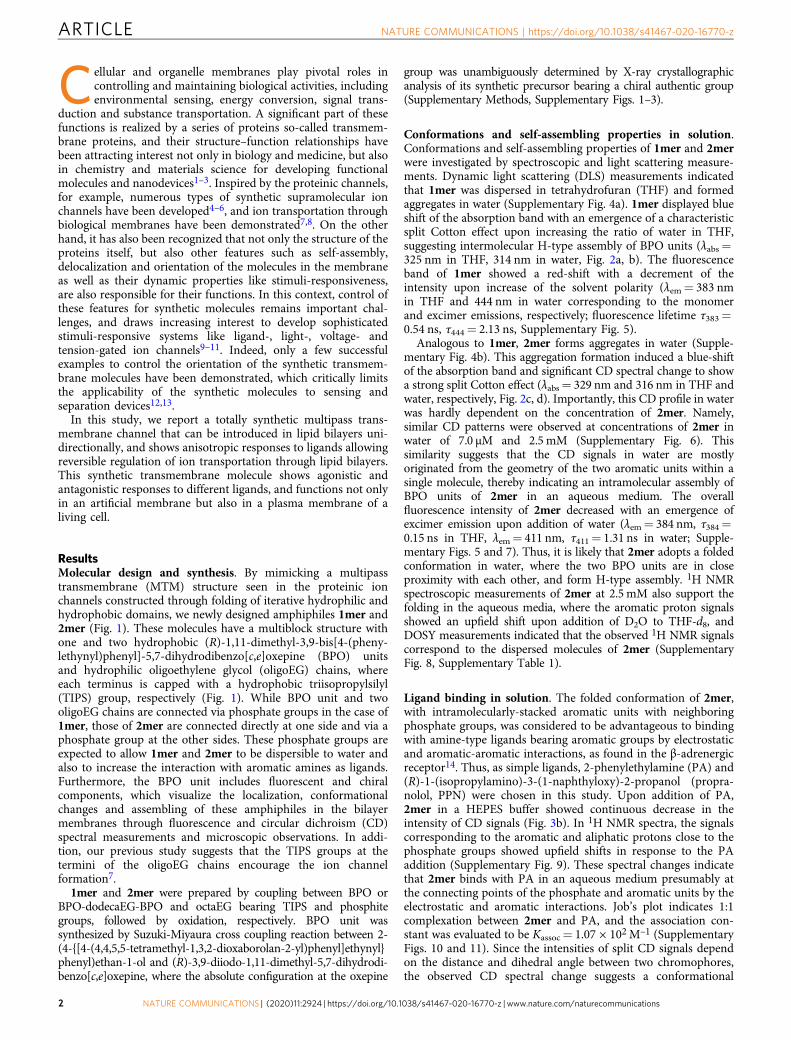
ResultsMolecular design and synthesis. By mimicking a multipasstransmembrane (MTM) structure seen in the proteinic ionchannels constructed through folding of iterative hydrophilic andhydrophobic domains, we newly designed amphiphiles 1mer and2mer (Fig. 1). These molecules have a multiblock structure withone and two hydrophobic (R)-1,11-dimethyl-3,9-bis[4-(pheny-lethynyl)phenyl]-5,7-dihydrodibenzo[c,e]oxepine (BPO) unitsand hydrophilic oligoethylene glycol (oligoEG) chains, whereeach terminus is capped with a hydrophobic triisopropylsilyl(TIPS) group, respectively (Fig. 1). While BPO unit and twooligoEG chains are connected via phosphate groups in the case of1mer, those of 2mer are connected directly at one side and via aphosphate group at the other sides. These phosphate groups areexpected to allow 1mer and 2mer to be dispersible to water andalso to increase the interaction with aromatic amines as ligands.Furthermore, the BPO unit includes fluorescent and chiralcomponents, which visualize the localization, conformationalchanges and assembling of these amphiphiles in the bilayermembranes through fluorescence and circular dichroism (CD)spectral measurements and microscopic observations. In addi-tion, our previous study suggests that the TIPS groups at thetermini of the oligoEG chains encourage the ion channelformation7.
1mer and 2mer were prepared by coupling between BPO orBPO-dodecaEG-BPO and octaEG bearing TIPS and phosphitegroups, followed by oxidation, respectively. BPO unit wassynthesized by Suzuki-Miyaura cross coupling reaction between 2-(4-{[4-(4,4,5,5-tetramethyl-1,3,2-dioxaborolan-2-yl)phenyl]ethynyl}phenyl)ethan-1-ol and (R)-3,9-diiodo-1,11-dimethyl-5,7-dihydrodi-benzo[c,e]oxepine, where the absolute configuration at the oxepine
group was unambiguously determined by X-ray crystallographicanalysis of its synthetic precursor bearing a chiral authentic group(Supplementary Methods, Supplementary Figs. 1–3).
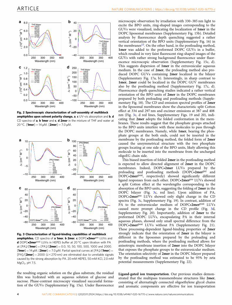
Conformations and self-assembling properties in solution.Conformations and self-assembling properties of 1mer and 2merwere investigated by spectroscopic and light scattering measure-ments. Dynamic light scattering (DLS) measurements indicatedthat 1mer was dispersed in tetrahydrofuran (THF) and formedaggregates in water (Supplementary Fig. 4a). 1mer displayed blueshift of the absorption band with an emergence of a characteristicsplit Cotton effect upon increasing the ratio of water in THF,suggesting intermolecular H-type assembly of BPO units (λabs=325 nm in THF, 314 nm in water, Fig. 2a, b). The fluorescenceband of 1mer showed a red-shift with a decrement of theintensity upon increase of the solvent polarity (λem= 383 nmin THF and 444 nm in water corresponding to the monomerand excimer emissions, respectively; fluorescence lifetime τ383=0.54 ns, τ444= 2.13 ns, Supplementary Fig. 5).
Analogous to 1mer, 2mer forms aggregates in water (Supple-mentary Fig. 4b). This aggregation formation induced a blue-shiftof the absorption band and significant CD spectral change to showa strong split Cotton effect (λabs= 329 nm and 316 nm in THF andwater, respectively, Fig. 2c, d). Importantly, this CD profile in waterwas hardly dependent on the concentration of 2mer. Namely,similar CD patterns were observed at concentrations of 2mer inwater of 7.0 μM and 2.5 mM (Supplementary Fig. 6). Thissimilarity suggests that the CD signals in water are mostlyoriginated from the geometry of the two aromatic units within asingle molecule, thereby indicating an intramolecular assembly ofBPO units of 2mer in an aqueous medium. The overallfluorescence intensity of 2mer decreased with an emergence ofexcimer emission upon addition of water (λem= 384 nm, τ384=0.15 ns in THF, λem= 411 nm, τ411= 1.31 ns in water; Supple-mentary Figs. 5 and 7). Thus, it is likely that 2mer adopts a foldedconformation in water, where the two BPO units are in closeproximity with each other, and form H-type assembly. 1H NMRspectroscopic measurements of 2mer at 2.5 mM also support thefolding in the aqueous media, where the aromatic proton signalsshowed an upfield shift upon addition of D2O to THF-d8, andDOSY measurements indicated that the observed 1H NMR signalscorrespond to the dispersed molecules of 2mer (SupplementaryFig. 8, Supplementary Table 1).
Ligand binding in solution. The folded conformation of 2mer,with intramolecularly-stacked aromatic units with neighboringphosphate groups, was considered to be advantageous to bindingwith amine-type ligands bearing aromatic groups by electrostaticand aromatic-aromatic interactions, as found in the β-adrenergicreceptor14. Thus, as simple ligands, 2-phenylethylamine (PA) and(R)-1-(isopropylamino)-3-(1-naphthyloxy)-2-propanol (propra-nolol, PPN) were chosen in this study. Upon addition of PA,2mer in a HEPES buffer showed continuous decrease in theintensity of CD signals (Fig. 3b). In 1H NMR spectra, the signalscorresponding to the aromatic and aliphatic protons close to thephosphate groups showed upfield shifts in response to the PAaddition (Supplementary Fig. 9). These spectral changes indicatethat 2mer binds with PA in an aqueous medium presumably atthe connecting points of the phosphate and aromatic units by theelectrostatic and aromatic interactions. Job’s plot indicates 1:1complexation between 2mer and PA, and the association con-stant was evaluated to be Kassoc= 1.07 × 10²M–1 (SupplementaryFigs. 10 and 11). Since the intensities of split CD signals dependon the distance and dihedral angle between two chromophores,the observed CD spectral change suggests a conformational
ARTICLE NATURE COMMUNICATIONS | https://doi.org/10.1038/s41467-020-16770-z
2 NATURE COMMUNICATIONS | (2020) 11:2924 | https://doi.org/10.1038/s41467-020-16770-z | www.nature.com/naturecommunications
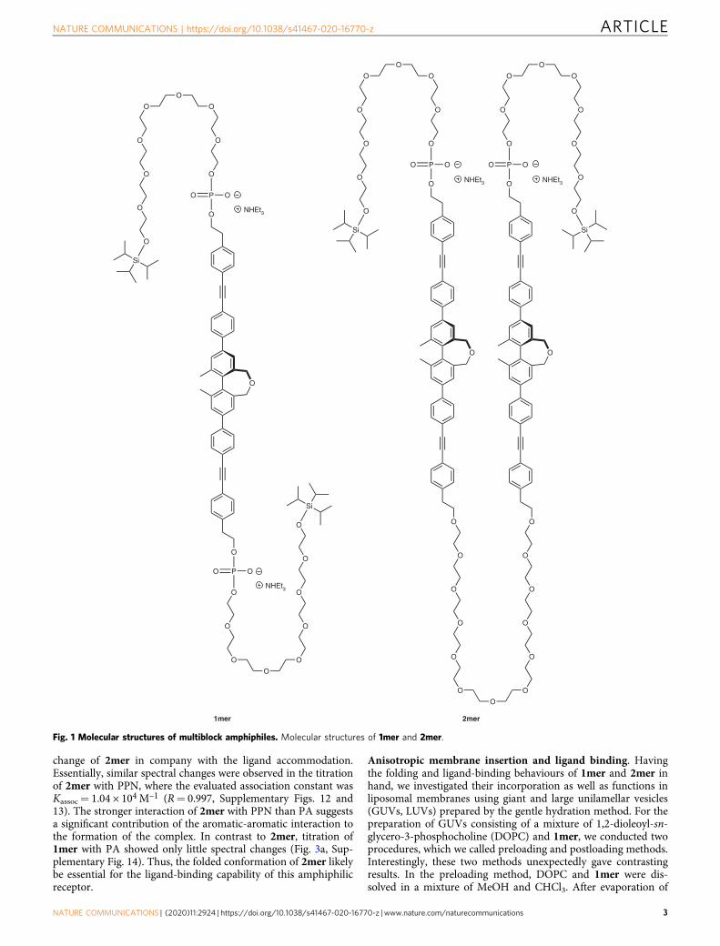
change of 2mer in company with the ligand accommodation.Essentially, similar spectral changes were observed in the titrationof 2mer with PPN, where the evaluated association constant wasKassoc= 1.04 × 10⁴M–1 (R= 0.997, Supplementary Figs. 12 and13). The stronger interaction of 2mer with PPN than PA suggestsa significant contribution of the aromatic-aromatic interaction tothe formation of the complex. In contrast to 2mer, titration of1mer with PA showed only little spectral changes (Fig. 3a, Sup-plementary Fig. 14). Thus, the folded conformation of 2mer likelybe essential for the ligand-binding capability of this amphiphilicreceptor.
Anisotropic membrane insertion and ligand binding. Havingthe folding and ligand-binding behaviours of 1mer and 2mer inhand, we investigated their incorporation as well as functions inliposomal membranes using giant and large unilamellar vesicles(GUVs, LUVs) prepared by the gentle hydration method. For thepreparation of GUVs consisting of a mixture of 1,2-dioleoyl-sn-glycero-3-phosphocholine (DOPC) and 1mer, we conducted twoprocedures, which we called preloading and postloading methods.Interestingly, these two methods unexpectedly gave contrastingresults. In the preloading method, DOPC and 1mer were dis-solved in a mixture of MeOH and CHCl3. After evaporation of
O
O
O
PO O
O
O
O
O
O
PO O
O
O
OO
O
O
O
O
O
2mer
O
O
PO O
NHEt3
O
O
O
PO O
O
O
O
OO
O
O
Si
O
O
O
O
O
OO
O
O
Si
O
O
O
O
O
O O
O
O
Si
O
O
O
O
O
O O
O
O
Si
1mer
NHEt3
NHEt3 NHEt3
Fig. 1 Molecular structures of multiblock amphiphiles. Molecular structures of 1mer and 2mer.
NATURE COMMUNICATIONS | https://doi.org/10.1038/s41467-020-16770-z ARTICLE
NATURE COMMUNICATIONS | (2020) 11:2924 | https://doi.org/10.1038/s41467-020-16770-z | www.nature.com/naturecommunications 3
the resulting organic solution on the glass substrate, the residualfilm was hydrated with an aqueous solution of glucose andsucrose. Phase-contrast microscopy visualized successful forma-tion of the GUVs (Supplementary Fig. 15a). Under fluorescence
microscopic observation by irradiation with 330–385 nm light toexcite the BPO units, ring-shaped images corresponding to theGUVs were visualized, indicating the localization of 1mer in theDOPC liposomal membranes (Supplementary Fig. 15b). Detailedanalysis by fluorescence depth quenching suggested a rathervertical orientation of the BPO units (Supplementary Fig. 16) inthe membranes15. On the other hand, in the postloading method,1mer was added to the preformed DOPC GUVs in a buffer,which resulted in very faint fluorescent ring-shaped images of theGUVs with rather strong background fluorescence under fluor-escence microscopic observation (Supplementary Fig. 15c, d).This suggests dispersion of 1mer in the extravesicular aqueousmedium. In the case of 2mer, the preloading method also pro-duced DOPC GUVs containing 2mer localized in the bilayer(Supplementary Fig. 17a, b). Interestingly, in sharp contrast to1mer, 2mer could be localized in the DOPC GUV membranesalso by the postloading method (Supplementary Fig. 17c, d).Fluorescence depth quenching studies indicated a rather verticalorientation of the BPO units of 2mer in the DOPC membranesprepared by both preloading and postloading methods (Supple-mentary Fig. 18). The CD and emission spectral profiles of 2merin the liposomal membranes show the characteristic split Cottoneffect at 334 and 297 nm and excimer emissions at 387 and 408nm (Fig. 3c, d red lines, Supplementary Figs. 19 and 20), indi-cating that 2mer adopts the folded conformation in the mem-branes. These results suggest that the phosphate groups attachedto the BPO units interfere with these molecules to pass throughthe DOPC membranes. Namely, while 1mer, bearing the phos-phate groups at the both ends, could not be inserted in themembrane by the postloading method, the folded form of 2mercaused the unsymmetrical structure with the two phosphategroups locating at one side of the BPO units, likely allowing thismolecule to be inserted into the membrane from the unchargedoligoEG chain side.
This biased insertion of folded 2mer in the postloading methodis expected to allow directed alignment of 2mer in the DOPCmembranes. Indeed, DOPC•2mer LUVs prepared by thepreloading and postloading methods (DOPC•2merpre andDOPC•2merpost, respectively) showed significantly differentligand responses from each other. DOPC•2merpre LUVs showeda split Cotton effect at the wavelengths corresponding to theabsorption of the BPO units, suggesting the folding of 2mer in theDOPC bilayer (Fig. 3c, red line). Upon addition of PA,DOPC•2merpre LUVs showed only slight change in the CDspectra (Fig. 3c, Supplementary Fig. 19). In contrast, addition ofPA to the extravesicular medium of DOPC•2merpost LUVscaused more prompt change in the CD profile (Fig. 3d,Supplementary Fig. 20). Importantly, addition of 2mer to thepreformed DOPC LUVs, encapsulating PA in their internalaqueous phases, showed only small spectral difference from theDOPC•2merpost LUVs without PA (Supplementary Fig. 21).These processing-dependent ligand-binding properties of 2merstrongly indicate that the orientation of 2mer in the bilayer isdifferent in the liposomes prepared by the preloading andpostloading methods, where the postloading method allows foranisotropic membrane insertion of 2mer into the DOPC bilayerthat exposes the phosphate groups to the extravesicular medium.The orientation selectivity of 2mer in the DOPC bilayer preparedby the postloading method was estimated to be 95% by zetapotential measurements (Supplementary Fig. 22).
Ligand-gated ion transportation. Our previous studies demon-strated that the multipass transmembrane structures like 2mer,consisting of alternatingly connected oligoethylene glycol chainsand aromatic components are effective for ion transportation
Δ� (
cm–1
M–1
)Δ�
(cm
–1 M
–1)
Δ� (
cm–1
M–1
)
250
DOPC•2merpre LUVs DOPC•2merpost LUVs
10a
c
b
d
100
80
60
40
20
0
–20
–40
5
0
0105010050010002000
[PA]/[1mer]
0105010050010002000
[PA]/[2mer]
0105010050010002000
[PA]/[2mer]0105010050010002000
[PA]/[2mer]
–5
–10
60
40
20
0
–20
–40
–60
Δ� (
cm–1
M–1
)
60
40
20
0
–20
–40
–60
300Wavelength (nm)
350 400 250 300Wavelength (nm)
350 400
250 300Wavelength (nm)
350 400250 300Wavelength (nm)
350 400
Fig. 3 Characterization of ligand-binding capabilities of multiblockamphiphiles. CD spectra of a 1mer, b 2mer, c DOPC•2merpre LUVs andd DOPC•2merpost LUVs in HEPES buffer at 20 °C upon titration with PAat [PA]/[1mer]= [PA]/[2mer]=0.0, 10, 50, 100, 500, 1000 and 2000.[1mer]= 14 μM. [2mer]= 7.0 μM. Partial spectral curves at [PA]/[1mer]=[PA]/[2mer]= 2000 (λ < 270 nm) are eliminated due to unreliable signalscaused by the strong absorption by PA. 20mM HEPES, 50mM KCl, 2.0mMMgCl2, pH 7.5.
8.0 40
20
0
–20
–40
a
c
b
d
THF/Water100/090/1075/2550/5025/7510/900/100
THF/Water100/090/1075/2550/5025/7510/900/100
THF/Water100/090/1075/2550/5025/7510/900/100
THF/Water100/090/1075/2550/5025/7510/900/100
6.0
4.0
2.0
0.0
1.2 250
200
150
100
50
0
–50
–100
1.0
0.8
0.6
0.4
0.2
0.0
� (1
04 cm
–1 M
–1)
� (1
04 cm
–1 M
–1)
Δ� (
cm–1
M–1
)Δ�
(cm
–1 M
–1)
250 300Wavelength (nm)
350 400 250 300Wavelength (nm)
350 400
250 300Wavelength (nm)
350 400250 300Wavelength (nm)
350 400
Fig. 2 Spectroscopic characterization of self-assembly of multiblockamphiphiles upon solvent polarity change. a, c UV-vis absorption and b, dCD spectra of a, b 1mer and c, d 2mer in the mixture of THF and water at20 °C. [1mer]= 14 μM. [2mer]= 7.0 μM.
ARTICLE NATURE COMMUNICATIONS | https://doi.org/10.1038/s41467-020-16770-z
4 NATURE COMMUNICATIONS | (2020) 11:2924 | https://doi.org/10.1038/s41467-020-16770-z | www.nature.com/naturecommunications
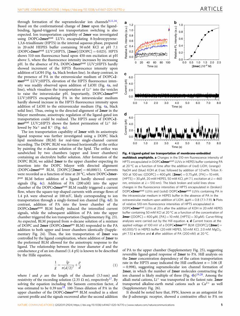
through formation of the supramolecular ion channels9,11,16.Based on the conformational change of 2mer upon the ligand-binding, ligand-triggered ion transportation switching is alsoexpected. Ion transportation capability of 2mer was investigatedusing DOPC•2merpost LUVs encapsulating 8-hydroxypyrene-1,3,6-trisulfonate (HPTS) in the internal aqueous phase, preparedin 20 mM HEPES buffer containing 50 mM KCl at pH 7.1(DOPC•2merpost LUV⊃HPTS, [2mer]/[DOPC]= 0.025). HPTSshows 510-nm fluorescence band upon 450-nm excitation at pHabove 5, where the fluorescence intensity increases by increasingpH. In the absence of PA, DOPC•2merpost LUV⊃HPTS hardlyshowed increment of the HPTS fluorescence intensity uponaddition of LiOH (Fig. 4a, black broken line). In sharp contrast, inthe presence of PA in the extravesicular medium of DOPC•2-merpost LUV⊃HPTS, elevation of the HPTS fluorescence inten-sity was readily observed upon addition of LiOH (Fig. 4a, redline), which visualizes the transportation of Li+ into the vesiclesto raise the intravesicular pH. Importantly, DOPC•2merpost
LUV⊃HPTS encapsulating PA in the intravesicular mediumhardly showed increase in the HPTS fluorescence intensity uponaddition of LiOH to the extravesicular medium (Fig. 4a, blacksolid line). Thus, owing to the directed alignment of 2mer in thebilayer membrane, anisotropic regulation of the ligand-gated iontransportation could be realized. The HPTS assay of DOPC•2-merpost LUV⊃HPTS shows the fastest permeation of Li+ fol-lowed by Na+ and K+ (Fig. 4a).
The ion transportation capability of 2mer with its anisotropicligand response was further investigated using a DOPC blacklipid membrane (BLM) for real-time single-channel currentrecording. The DOPC BLM was formed horizontally at the orificeby painting the n-decane solution of the lipid. The orifice wassandwiched by two chambers (upper and lower chambers)containing an electrolyte buffer solution. After formation of theDOPC BLM, we added 2mer to the upper chamber expecting itsinsertion into the DOPC bilayer with directed alignment(DOPC•2merpost BLM, [DOPC]/[2mer]= 60,000/1). Currentswere recorded as a function of time at 20 °C, where DOPC•2mer-post BLM before addition of a ligand hardly showed currentsignals (Fig. 4c). Addition of PA (200 nM) into the upperchamber of the DOPC•2merpost BLM readily triggered a currentflow, where the square-top-shaped currents with average flows of1.4 pA were observed at 100 mV, likely corresponding to iontransportation through a singly-formed ion channel (Fig. 4d). Incontrast, addition of PA into the lower chamber of theDOPC•2merpost BLM hardly induced the intensified currentsignals, while the subsequent addition of PA into the upperchamber triggered the ion transportation (Supplementary Fig. 23).As expected, BLM prepared from n-decane solution of a mixtureof DOPC and 2mer (DOPC•2merpre BLM) responded to the PAaddition to both upper and lower chambers identically (Supple-mentary Fig. 24). Thus, the ion transportation of 2mer wascontrolled by the ligand complexation, where addition of 2mer tothe preformed BLM allowed for the anisotropic response to theligand. The relationship between the inner diameter d and theconductance g of an ion channel (1.4 pS) is known to be describedby the Hille equation,
1g¼ lρ
π d=2ð Þ2 þρ
dð1Þ
where l and ρ are the length of the channel (3.5 nm) andresistivity of the recording solution (2.35 Ω m), respectively17. Bysolving the equation including the Sansom correction factor, dwas estimated to be 0.39 nm18. 100-Times dilution of PA in theupper chamber of the DOPC•2merpost BLM resulted in a silentcurrent profile and the signals recovered after the second addition
of PA to the upper chamber (Supplementary Fig. 25), suggestingreversible ligand-gated response of 2mer to PA. Hill analysis onthe 2mer concentration dependency of the cation transportationrate in the HPTS assay indicated the Hill coefficient n= 3.06 (R= 0.998), suggesting supramolecular ion channel formation of2mer, in which the number of 2mer molecules constructing theion channel is likely multiple of three (Fig. 4b)19,20. Among thealkali metal cations, Li+ was transported in the fastest rate. 2mertransported alkaline-earth metal cations such as Ca2+ as well(Supplementary Fig. 26).
It should be noted here that, PPN, known as an antagonist forthe β-adrenergic receptor, showed a contrastive effect to PA on
a
c
b1.2 1.0
0.8
0.6
0.4
0.2
0.00.0 0.5 1.0 1.5 2.0 2.5
1.0
0.8
0.6
0.4
0.2
0.0
6.0
4.0
2.0
0.0
0.0 1.0 2.0 3.0 4.0Time (s)
5.0 6.0 7.0 8.0
0.0 1.0 2.0 3.0 4.0Time (s)
5.0 6.0 7.0 8.0
Cur
rent
(pA
)
d6.0
4.0
2.0
0.0
Cur
rent
(pA
)
0 20 40Time (s) [2mer] (10–6 M)
60 80 100
Rel
ativ
e F
L in
tens
ity (
a.u.
)
Rel
ativ
e F
L in
tens
ity (
a.u.
)
Fig. 4 Ligand-gated ion transportation of membrane-embeddedmultiblock amphiphile. a Changes in the 510-nm fluorescence intensity ofHPTS encapsulated in DOPC•2merpost LUVs in HEPES buffer containing PAat 20 °C as a function of time after the addition of (red) LiOH, (orange)NaOH and (blue) KOH at 0 sec followed by addition of 1.0 wt% Triton X-100 at 100 sec ([DOPC]= 400 μM, [2mer]= 0.75 μM, [PA]= 10mM,[HPTS]= 30 μM, 20mM HEPES, 50mM KCl, pH 7.1, excitation at λ= 460nm, emission at λ= 510 nm). The black broken and solid lines showchanges in the fluorescence intensities of HPTS encapsulated in (broken)DOPC•2merpost LUVs and (solid) DOPC•2merpost LUVs containing PA inthe intravesicular medium in HEPES buffer in the absence of PA in theextravesicular medium upon addition of LiOH. ΔpH= 0.8 (7.1–7.9). b Plotsof relative 510-nm fluorescence intensities of HPTS encapsulated inDOPC•2merpost LUVs at 20 s after the addition of KOH in 20mM HEPESbuffer containing 50mM KCl at 20 °C as a function of the concentration of2mer ([DOPC]= 400 μM, [PA]= 10 mM, [HPTS]= 30 μM). Curve-fittinganalyses were carried out by the Hill equation. c, d Current traces at theapplied voltage of 100mV of a DOPC•2merpost BLM ([DOPC]/[2mer]=60,000/1) in HEPES buffer (20mM HEPES, 50mM KCl, 2.0 mM MgCl2,pH 7.5) c before and d after addition of PA (200 nM) at 20 °C.
NATURE COMMUNICATIONS | https://doi.org/10.1038/s41467-020-16770-z ARTICLE
NATURE COMMUNICATIONS | (2020) 11:2924 | https://doi.org/10.1038/s41467-020-16770-z | www.nature.com/naturecommunications 5
the ion transportation of 2mer. In fact, addition of PPN (50 nM)to the upper chamber of DOPC•2merpost BLM hardly promptedcurrent flow, and even subsequent addition of PA (200 nM) didnot trigger the current flow (Supplementary Fig. 27a, b).Furthermore, subsequent addition of PPN to the upper chamberof the DOPC•2merpost BLM, after addition of 2mer in thepresence of PA, deactivated the current flow (SupplementaryFig. 27c, d). Thus, analogous to the agonistic and antagonisticeffects of PA and PPN to β-adrenergic receptor, PA and PPNapparently act to 2mer like an agonist and an antagonist,respectively. NMR and modelling studies of 2mer–ligandcomplexes indicated that PA and PPN bind with 2mer throughdifferent interacting modes (Supplementary Figs. 28 and 29).Namely, electrostatic interaction between the phosphate andammonium groups is likely dominant between PA and 2mer, andPA is located at the rim of the channel. Meanwhile, hydrophobicinteraction is preferred between 2mer and PPN, therefore PPN isinserted into the hydrophobic cavity of the channel to infillthe pore.
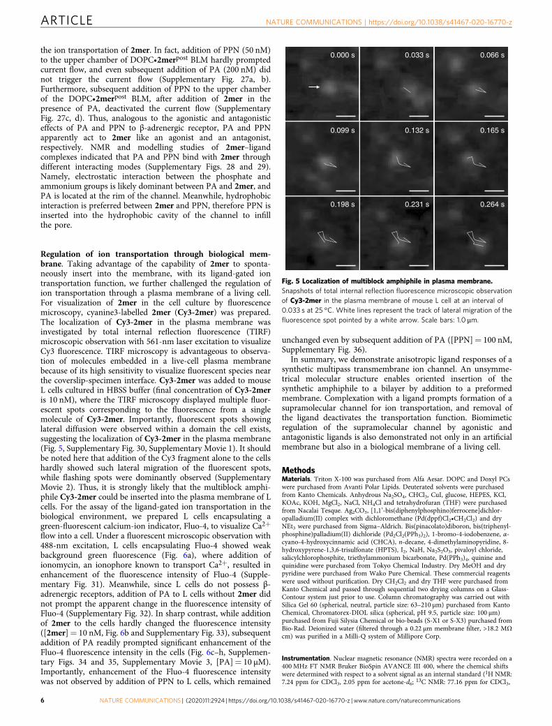
Regulation of ion transportation through biological mem-brane. Taking advantage of the capability of 2mer to sponta-neously insert into the membrane, with its ligand-gated iontransportation function, we further challenged the regulation ofion transportation through a plasma membrane of a living cell.For visualization of 2mer in the cell culture by fluorescencemicroscopy, cyanine3-labelled 2mer (Cy3-2mer) was prepared.The localization of Cy3-2mer in the plasma membrane wasinvestigated by total internal reflection fluorescence (TIRF)microscopic observation with 561-nm laser excitation to visualizeCy3 fluorescence. TIRF microscopy is advantageous to observa-tion of molecules embedded in a live-cell plasma membranebecause of its high sensitivity to visualize fluorescent species nearthe coverslip-specimen interface. Cy3-2mer was added to mouseL cells cultured in HBSS buffer (final concentration of Cy3-2meris 10 nM), where the TIRF microscopy displayed multiple fluor-escent spots corresponding to the fluorescence from a singlemolecule of Cy3-2mer. Importantly, fluorescent spots showinglateral diffusion were observed within a domain the cell exists,suggesting the localization of Cy3-2mer in the plasma membrane(Fig. 5, Supplementary Fig. 30, Supplementary Movie 1). It shouldbe noted here that addition of the Cy3 fragment alone to the cellshardly showed such lateral migration of the fluorescent spots,while flashing spots were dominantly observed (SupplementaryMovie 2). Thus, it is strongly likely that the multiblock amphi-phile Cy3-2mer could be inserted into the plasma membrane of Lcells. For the assay of the ligand-gated ion transportation in thebiological environment, we prepared L cells encapsulating agreen-fluorescent calcium-ion indicator, Fluo-4, to visualize Ca2+
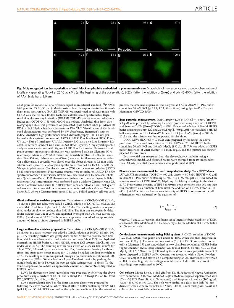
flow into a cell. Under a fluorescent microscopic observation with488-nm excitation, L cells encapsulating Fluo-4 showed weakbackground green fluorescence (Fig. 6a), where addition ofionomycin, an ionophore known to transport Ca2+, resulted inenhancement of the fluorescence intensity of Fluo-4 (Supple-mentary Fig. 31). Meanwhile, since L cells do not possess β-adrenergic receptors, addition of PA to L cells without 2mer didnot prompt the apparent change in the fluorescence intensity ofFluo-4 (Supplementary Fig. 32). In sharp contrast, while additionof 2mer to the cells hardly changed the fluorescence intensity([2mer]= 10 nM, Fig. 6b and Supplementary Fig. 33), subsequentaddition of PA readily prompted significant enhancement of theFluo-4 fluorescence intensity in the cells (Fig. 6c–h, Supplemen-tary Figs. 34 and 35, Supplementary Movie 3, [PA]= 10 μM).Importantly, enhancement of the Fluo-4 fluorescence intensitywas not observed by addition of PPN to L cells, which remained
unchanged even by subsequent addition of PA ([PPN]= 100 nM,Supplementary Fig. 36).
In summary, we demonstrate anisotropic ligand responses of asynthetic multipass transmembrane ion channel. An unsymme-trical molecular structure enables oriented insertion of thesynthetic amphiphile to a bilayer by addition to a preformedmembrane. Complexation with a ligand prompts formation of asupramolecular channel for ion transportation, and removal ofthe ligand deactivates the transportation function. Biomimeticregulation of the supramolecular channel by agonistic andantagonistic ligands is also demonstrated not only in an artificialmembrane but also in a biological membrane of a living cell.
MethodsMaterials. Triton X-100 was purchased from Alfa Aesar. DOPC and Doxyl PCswere purchased from Avanti Polar Lipids. Deuterated solvents were purchasedfrom Kanto Chemicals. Anhydrous Na2SO4, CHCl3, CuI, glucose, HEPES, KCl,KOAc, KOH, MgCl2, NaCl, NH4Cl and tetrahydrofuran (THF) were purchasedfrom Nacalai Tesque. Ag2CO3, [1,1’-bis(diphenylphosphino)ferrocene]dichlor-opalladium(II) complex with dichloromethane (Pd(dppf)Cl2•CH2Cl2) and dryNEt3 were purchased from Sigma–Aldrich. Bis(pinacolato)diboron, bis(triphenyl-phosphine)palladium(II) dichloride (Pd2Cl2(PPh3)2), 1-bromo-4-iodobenzene, α-cyano-4-hydroxycinnamic acid (CHCA), n-decane, 4-dimethylaminopyridine, 8-hydroxypyrene-1,3,6-trisulfonate (HPTS), I2, NaH, Na2S2O3, pivaloyl chloride,salicylchlorophosphite, triethylammonium bicarbonate, Pd(PPh3)4, quinine andquinidine were purchased from Tokyo Chemical Industry. Dry MeOH and drypyridine were purchased from Wako Pure Chemical. These commercial reagentswere used without purification. Dry CH2Cl2 and dry THF were purchased fromKanto Chemical and passed through sequential two drying columns on a Glass-Contour system just prior to use. Column chromatography was carried out withSilica Gel 60 (spherical, neutral, particle size: 63–210 μm) purchased from KantoChemical, Chromatorex-DIOL silica (spherical, pH 9.5, particle size: 100 μm)purchased from Fuji Silysia Chemical or bio-beads (S-X1 or S-X3) purchased fromBio-Rad. Deionized water (filtered through a 0.22 μm membrane filter, >18.2 MΩcm) was purified in a Milli-Q system of Millipore Corp.
Instrumentation. Nuclear magnetic resonance (NMR) spectra were recorded on a400MHz FT NMR Bruker BioSpin AVANCE III 400, where the chemical shiftswere determined with respect to a solvent signal as an internal standard (1H NMR:7.24 ppm for CDCl3, 2.05 ppm for acetone-d6; 13C NMR: 77.16 ppm for CDCl3,
0.000 s 0.033 s 0.066 s
0.099 s 0.132 s 0.165 s
0.198 s 0.231 s 0.264 s
Fig. 5 Localization of multiblock amphiphile in plasma membrane.Snapshots of total internal reflection fluorescence microscopic observationof Cy3-2mer in the plasma membrane of mouse L cell at an interval of0.033 s at 25 °C. White lines represent the track of lateral migration of thefluorescence spot pointed by a white arrow. Scale bars: 1.0 μm.
ARTICLE NATURE COMMUNICATIONS | https://doi.org/10.1038/s41467-020-16770-z
6 NATURE COMMUNICATIONS | (2020) 11:2924 | https://doi.org/10.1038/s41467-020-16770-z | www.nature.com/naturecommunications
28.98 ppm for acetone-d6) or a reference signal as an external standard (31P NMR:0.00 ppm for 6% H3PO4 aq.). Matrix-assisted laser desorption/ionization time-of-flight mass spectrometry (MALDI-TOF MS) was performed in reflector mode withCHCA as a matrix on a Bruker Daltonics autoflex speed spectrometer. High-resolution electrospray ionization (HR ESI) TOF MS spectra were recorded on aBruker micrOTOF-Q II-S1 with MeOH as a solvent. Analytical thin layer chro-matography (TLC) was performed on precoated, glass-backed silica gel Merck 60F254 or Fuji Silysia Chemical Chromatorex Diol TLC. Visualization of the devel-oped chromatogram was performed by UV absorbance, Hanessian’s stain oriodine. Analytical high-performance liquid chromatography (HPLC) was per-formed with a system composed of JASCO PU-2080 Plus Intelligent HPLC Pump,UV-2077 Plus 4-λ Intelligent UV/VIS Detector, DG-2080-53 3-Line Degasser, LG-2080-02 Ternary Gradient Unit and LC-Net II/APC system. X-ray crystallographicanalyses were carried out with Rigaku RAPID II refractometer. Fluorescent andphase-contrast microscopic observation was performed with an Olympus IX-71microscope, where a U-MWU2 mirror unit (excitation filter: 330–385 nm, emis-sion filter: 420 nm, dichroic mirror: 400 nm) was used for fluorescence observation.On a slide glass, a coverslip was placed over the object through a 0.1-mm thicksilicon-based spacer. UV absorption spectra were recorded on JASCO V-530 UV-Vis spectrophotometer. Circular dichroism (CD) spectra were recorded on JASCOJ-820 spectropolarimeter. Fluorescence spectra were recorded on JASCO FP-6500spectrofluorometer. Fluorescence lifetime was measured with Hamamatsu Photo-nics Quantaurus-Tau C11367 fluorescence lifetime spectrometer. Dynamic lightscattering (DLS) measurement was performed with a Malvern Zetasizer Nano ZS,where a Zetasizer nano series DTS 1060 Folded capillary cell or a 1-cm-thick quartzcell was used. Zeta potential measurement was performed with a Malvern ZetasizerNano ZSP, where a Zetasizer nano series DTS 1070 Folded capillary cell was used.
Giant unilamellar vesicles preparation. To a mixture of CHCl3/MeOH (2/1 v/v,10 μL) in a glass test tube, were added a CHCl3 solution of DOPC (2.0 mM, 20 μL)and a MeOH solution of glucose (10 mM, 12 μL). The resulting mixture was gentlydried under Ar flow to produce thin lipid film. The film was subsequently driedunder vacuum over 3 h at 25 °C and hydrated overnight with 200 mM sucrose aq.(200 μL) under Ar at 37 °C. To the vesicle suspension was added an appropriateamount of 1mer or 2mer dispersed in HEPES buffer.
Large unilamellar vesicles preparation. To a mixture of CHCl3/MeOH (2/1 v/v,50 μL) put in a glass test tube, was added a CHCl3 solution of DOPC (2.0 mM, 100μL). The resulting mixture was gently dried under Ar flow to produce thin lipidfilm, which was subsequently dried under vacuum over 3 h at 25 °C and hydratedovernight in HEPES buffer (20 mM HEPES, 50 mM KCl, 2.0 mM MgCl2, pH 7.5)under Ar at 37 °C. The resulting mixture was stirred on a shaker (203 min–1) for1 h at 37 °C, followed by vortex mixing for 10 s, freezing-and-thawing for threetimes, and subsequent vortex mixing for 10 s. After being left standing overnight at37 °C, the resulting mixture was passed through a polycarbonate membrane of 100-nm pore size (LFM-100) attached in a LiposoFast-Basic device by pushing thesample back and forth between the two gas-tight syringes over 11 times. To thevesicle suspension was added an appropriate amount of 1mer or 2mer dispersed inHEPES buffer.
LUVs for fluorescence depth quenching were prepared by following the aboveprocedure using a mixture of DOPC and 5-Doxyl PC, 12-Doxyl PC, or 16-DoxylPC ([DOPC]/[Doxyl PC]= 90/10).
LUVs encapsulating HPTS in the inner aqueous phase were prepared byfollowing the above procedure, where 20 mM HEPES buffer containing 50 mM KCl(pH 7.1) and 30 μM HPTS was used as the hydration medium. After the extrusion
process, the obtained suspension was dialyzed at 4 °C in 20 mM HEPES buffercontaining 50 mM KCl (pH 7.1, 1.0 L, three times) using Spectra/Por DialysisMembrane (MWCO 3500).
Zeta potential measurement. DOPC•2merpre LUVs ([DOPC]= 10mM, [2mer]=500 μM) were prepared by following the above procedure using a mixture of DOPCand 2mer in CHCl3 ([2mer]/[DOPC]= 1/20). To a stirred solution of 20mM HEPESbuffer containing 50mM KCl and 2.0mMMgCl2 (980 μL, pH 7.5) was added a HEPESbuffer suspension of DOPC•2merpre LUVs ([DOPC]= 10mM, [2mer] = 500 μM,20 μL), and the mixture was further pipetted for five times.
DOPC LUVs ([DOPC]= 10 mM) were prepared by following the aboveprocedure. To a stirred suspension of DOPC LUVs in 20 mM HEPES buffercontaining 50 mM KCl and 2.0 mM MgCl2 (980 μL, pH 7.5) was added a HEPESbuffer dispersion of 2mer ([2mer]= 1 mM, 20 μL), and the mixture was furtherpipetted for five times.
Zeta potential was measured from the electrophoretic mobility using aSmoluchowski model, and obtained values were averaged from 10 independentruns. All zeta potential measurements were performed at 20 °C.
Fluorescence measurement for ion transportation study. To a DOPC•2merLUV⊃HPTS suspension ([DOPC]= 400 μM, [2mer]= 0.75 μM, [HPTS]= 30 μM)in 20 mM HEPES buffer containing 50 mM KCl (1.99 mL, pH 7.1) was added anaqueous solution of KOH (0.60 M, 10 μL, ΔpH= 0.8) by a syringe in the dark at20 °C. Fluorescence intensity of HPTS at 510 nm upon excitation with 460 nm-lightwas monitored as a function of time until the addition of 1.0 wt% Triton X-100(40 μL) at 100 s. Relative fluorescence intensity of HPTS in response to the pHenhancement was evaluated by the equation of
I ¼ It � I0I1yzed � I0
ð2Þ
where I0, It and Ilyzed represent the fluorescence intensities before addition of KOH,at t seconds after addition of KOH, and after lysis by the addition of 1.0 wt% TritonX-100, respectively.
Conductance measurements using BLM system. A CHCl3 solution of DOPC(12.7 mM, 100 μL) was gently dried under N2 flow, which was then dispersed inn-decane (100 μL). The n-decane suspension (5 μL) of DOPC was painted on anorifice (diameter 150 μm) sandwiched by two chambers containing HEPES buffer(upper chamber: trans, lower chamber: cis, 20 mM HEPES, 50 mM KCl, 2.0 mMMgCl2, pH 7.5, 0.30 mL each) followed by the addition of an appropriate amount of2mer dispersed in HEPES buffer. Current was measured with a Nihon KohdenCEZ2400 amplifier and stored on a computer using an AD Instruments PowerLabat 40 kHz sampling rate. Recordings were filtered at 1 kHz. All the currentrecordings were performed at 20 °C.
Cell culture. Mouse L cells, a kind gift from Dr. H. Fujisawa of Nagoya University,were cultured in Dulbecco’s Modified Eagle’s Medium (Sigma) supplemented with10% (v/v) FBS, Penicillin G (100 units/mL) and Streptomycin sulfate (100 µg/mL,Wako) at 37 °C in 5% CO2. The cells were seeded in a glass-base dish (35-mmdiameter with a window diameter of 12 mm, 0.12~0.17-mm thick glass; Iwaki) andcultured for at least 24 h before observation.
a 0 s
2mer added PA added
22 s 45 s 47 s
49 s 51 s 55 s 100 se
b
f
c
g
d
h
Fig. 6 Ligand-gated ion transportation of multiblock amphiphile embedded in plasma membrane. Snapshots of fluorescence microscopic observation ofL cells encapsulating Fluo-4 at 25 °C at a 0 s (at the beginning of the observation), b 22 s (after the addition of 2mer) and c–h 45–100 s (after the additionof PA). Scale bars: 5.0 μm.
NATURE COMMUNICATIONS | https://doi.org/10.1038/s41467-020-16770-z ARTICLE
NATURE COMMUNICATIONS | (2020) 11:2924 | https://doi.org/10.1038/s41467-020-16770-z | www.nature.com/naturecommunications 7
Single-molecule fluorescence tracking by TIRF. Single fluorescent-moleculeobservation was performed as previously described21. To briefly explain, anobjective lens-type TIRF (Total Internal Reflection fluorescence) microscope basedon an Olympus IX-81 was employed. Mouse L cells were observed at room tem-perature of 25 °C after addition of Cy3-2mer (10 nM) in HBSS-HEPES buffer(HBSS supplemented with 10 mM HEPES, adjusted to pH 7.4 with NaOH). Thebottom plasma membrane was locally illuminated with an evanescent field (anOlympus ×100, 1.49 NA objective lens; excitation at 561 nm). The fluorescentimage of Cy3-2mer was projected onto a two-stage microchannel plate intensifier(C8600-03; Hamamatsu Photonics), and its output image was then lens coupled toan electron bombardment charge-coupled device camera (C7190-23; HamamatsuPhotonics). The obtained images were recorded at 30 Hz on a digital video deck forthe following analysis. These instruments were also employed for fluorescenceimaging of intracellular Ca2+ mobilization (described in the next section).
Fluorescence microscopic observation of L cells. The mouse L cells wereincubated with HBSS-HEPES buffer in the presence of Fluo-4-AM (4.6 μM),Probenecid (1.25 mM) and Pluronic F127 (0.04%) for 1 h at 37 °C, and washedthree times with HBSS-HEPES buffer, and then fresh HBSS-HEPES buffer wereadded. During the observation, 2mer (10 nM) was added, followed by addition ofionomycin (0.9 μM) as a positive control of Ca2+ mobilization. The cells wereilluminated with laser light with a wavelength of 488 nm, and observed at roomtemperature with the same Olympus IX-81.
Modelling of 2mer and ligand complexes. A three dimensional structure of a2mer was built by 2D Sketcher and Minimize-Selected-Atoms modules inMAESTRO (Shrödinger release 2018-2). The silicon atom at the TIPS group wasreplaced with a carbon atom. The folded structure of the 2mer was set to the M-shape as shown in Fig. 1. After building the one 2mer structure, other two 2merstructures were further built, and a three-2mer complex was prepared, because itwas suggested that the supramolecular ion channel formation of 2mer estimated bythe Hille equation was achieved by multiple of three. The initial formation of thethree-2mer complex was set to a symmetric triangle form, and the directions of thethree 2mer were aligned (Supplementary Fig. 29a). The three dimensional struc-tures of PA and PPN were built by the same protocol for 2mer. A ligand waslocated between the two BPO units for one 2mer, and the three ligands werearranged one-to-one with the three 2mer. The position of ligand was located at theposition of benzene near the phosphorus atom in 2mer. To reduce steric hin-drances among TIPS groups for each 2mer or among 2mer and PA, the structuresaround the phosphate part near PA and the positions of the TIPS group weremodified manually. Using the modified structure of the three-2mer complex, eachPA was replaced with PPN, and these positions of PA (Supplementary Fig. 29b) orPPN (Supplementary Fig. 29c) were matched as possible.
The three-2mer and ligand complex were embedded in DOPC membrane andwater molecules using the Membrane Builder implemented in CHARMM-GUI22–27.The orientation of 2mer relative to a lipid bilayer was set vertically, and the buriedarea of 2mer was determined so that the phosphate part of 2mer and DOPCmolecules were close to each other. The molecular dynamics unit cell was set to arectangular cell. In the center of the cell, the complex was embedded in a 70 Å × 70 ÅDOPC bilayer at the x–y plane, and the number of DOPC molecules at the upperand lower leaflets was 56 and 65, respectively. Along the z-axis of the unit cell, watermolecules were added, and the water thickness was set to 22.5 Å. Counterions and150mM KCl were included. A three-2mer and PA complex embedded in DOPCmembrane and water molecules is illustrated in Supplementary Fig. 29d.
The complex embedded in the membrane-water system was equilibrated by all-atom molecular dynamics (MD) simulations. MD simulations were performedusing the MD program package GROMACS ver. 2016.328–30 under periodicboundary conditions. The CHARMM36m force field was used for membranes31–35
and the TIP3P water model36, and the CHARMM General Force Field (CGenFF)37
was used for 2mer and ligands. The CGenFF parameters were assigned byParamChem through the Membrane Builder. For CGenFF parameters of 2mer, theparameters for each part of the oligoEG including the TIPS group, the BPO unit,the PEG were assigned piece by piece, and then these parameters were assembled.The electrostatic interaction was handled by the smooth particle mesh Ewaldmethod38, and the van del Waals interaction was truncated by the switchingfunction with the range of 10–12 Å. Bond lengths involving hydrogen atoms wereconstrained by the P-LINKS algorithm39. The temperature and pressure were 300K and 1 atm, respectively. According to the default setup in the Membrane Builder,an energy minimization by the steepest descent method and six equilibrationsimulations, written as EQ1-EQ6 hereafter, were performed sequentially before theproduction run. The ensemble adopted in the simulations was NVT ensemble inEQ1 and EQ2 and NPT ensemble in EQ3 to EQ6 and the production run. Thethermostat in the equilibration runs was the weak-coupling scheme of Berendsen40,and that in the production run was the Nosé–Hoover scheme41,42. The barostat inEQ3 to EQ6 was the semi-isotropic Berendsen algorithm40, and that in theproduction run was the semi-isotropic Parrinello–Rahman approach43,44. The timestep was set to 1 fs in EQ1 to EQ3, and 2 fs in EQ4 to EQ6 and the production runs.The simulation length of each simulation was 125 ps for EQ1 to EQ3, 500 ps forEQ4 to EQ6, and 500 ps for a production run.
In the equilibration processes, structural restraints were imposed on 2mer,ligand, and DOPC molecules according to the default setup of the MembraneBuilder. The position harmonic restraints were imposed on heavy atoms of 2merand ligand. During the equilibration runs, the strength of the force constant wasreduced gradually from 4000 to 50 kJ·mol–2·nm–2. In DOPC molecules, the z-axisof the phosphorus atom was restrained by the position harmonic restraints, and itsforce constant was gradually reduced as 1000–0 kJ·mol–2·nm–2. In addition, threedihedral angles were restrained. One was the dihedral angle among three carbonatoms at the sn-1, -2, -3 positions and an adjacent oxygen atom at the sn-2position. The angle was restrained at 120 degree. The other was the two dihedralangles corresponding to the double bond between carbon atoms in the oleic acidpart in the sn-2 and -3 positions, and the angle was restrained at the cisconformation. The force constants of these dihedral angles were reduced graduallyfrom 1000 to 0 kJ·mol–2·rad–2.
After the equilibration runs, a 500 ps production run was carried out for eachthe three-2mer and PA complex and the three-2mer and PPN complex. In theproduction runs, no restraints were imposed.
Reporting summary. Further information on research design is available inthe Nature Research Reporting Summary linked to this article.
Data availabilityThe authors declare that the data that support the findings of this study are availablewithin the paper and its Supplementary Information file. All other information isavailable from the corresponding authors upon reasonable request.
Received: 14 October 2019; Accepted: 26 May 2020;
References1. Das, G., Talukdar, P. & Matile, S. Enzyme activity with synthetic
supramolecular pores. Science 298, 1600–1602 (2002).2. Reiner, J. E. et al. Disease detection and management via single nanopore-
based sensors. Chem. Rev. 112, 6431–6451 (2012).3. Tan, C. S., Fleming, A. M., Ren, H., Burrows, C. J. & White, H. S. γ-Hemolysin
nanopore is sensitive to guanine-to-inosine substitutions in double-strandedDNA at the single-molecule level. J. Am. Chem. Soc. 140, 14224–14234 (2018).
4. Hayata, A., Itoh, H. & Inoue, M. Solid-phase total synthesis and dualmechanism of action of the channel-forming 48-mer peptide polytheonamideB. J. Am. Chem. Soc. 140, 10602–10611 (2018).
5. Schneider, S. et al. Columnar self-assemblies of triarylamines as scaffolds forartificial biomimetic channels for ion and for water transport. J. Am. Chem.Soc. 139, 3721–3727 (2017).
6. Sakai, N. & Matile, S. Metal-organic scaffolds: heavy-metal approaches tosynthetic ion channels and pores. Angew. Chem. Int. Ed. 47, 9603–9607(2008).
7. Gokel, G. W. & Negin, S. Synthetic ion channels: from pores to biologicalapplications. Acc. Chem. Res. 46, 2824–2833 (2013).
8. Saha, T., Gautam, A., Mukherjee, A., Lahiri, M. & Talukdar, P. Chloridetransport through supramolecular barrel-rosette ion channels: lipophiliccontrol and apoptosis-inducing activity. J. Am. Chem. Soc. 138, 16443–16451(2016).
9. Muraoka, T. et al. Reversible ion transportation switch by a ligand-gatedsynthetic supramolecular ion channel. J. Am. Chem. Soc. 136, 15584–15595(2014).
10. Gilles, A. & Barboiu, M. Highly selective artificial K+ channels: an example ofselectivity-induced transmembrane potential. J. Am. Chem. Soc. 138, 426–432(2016).
11. Muraoka, T. et al. Mechano-sensitive synthetic ion channels. J. Am. Chem.Soc. 139, 18016–18023 (2017).
12. Macrae, M. X., Blake, S., Mayer, M. & Yang, J. Nanoscale ionic diodes withtunable and switchable rectifying behavior. J. Am. Chem. Soc. 132, 1766–1767(2010).
13. Su, G., Zhang, M., Si, W., Li, Z.-T. & Hou, J.-L. Directional potassiumtransport through a unimolecular peptide channel. Angew. Chem. Int. Ed. 55,14678–14682 (2016).
14. Herm, M., Molt, O. & Schrader, T. Towards synthetic adrenalinereceptors–shape-selective adrenaline recognition in water. Angew. Chem. Int.Ed. 40, 3148–3151 (2001).
15. Ladokhin, A. S. Distribution analysis of depth-dependent fluorescencequenching in membranes: a practical guide. Methods Enzymol. 278, 462–473(1997).
16. Muraoka, T. et al. Ion permeation by a folded multiblock amphiphilicoligomer achieved by hierarchical construction of self-assembled nanopores. J.Am. Chem. Soc. 134, 19788–19794 (2012).
ARTICLE NATURE COMMUNICATIONS | https://doi.org/10.1038/s41467-020-16770-z
8 NATURE COMMUNICATIONS | (2020) 11:2924 | https://doi.org/10.1038/s41467-020-16770-z | www.nature.com/naturecommunications

17. Hille, B. Ion Channels of Excitable Membranes 3rd edn. (Sinauer Associates:Sunderland, 2001).
18. Smart, O. S., Breed, J., Smith, G. R. & Sansom, M. S. P. A novel method forstructure-based prediction of ion channel conductance properties. Biophys. J.72, 1109–1126 (1997).
19. Hill, A. V. The combinations of haemoglobin with oxygen and with carbonmonoxide. I. Biochem. J. 7, 471–480 (1913).
20. Kano, K. & Fendler, J. H. Pyranine as a sensitive pH probe for liposomeinteriors and surfaces. pH gradients across phospholipid vesicles. Biochim.Biophys. Acta 509, 289–299 (1978).
21. Kasai, R. S. et al. Full characterization of GPCR monomer-dimer dynamicequilibrium by single molecule imaging. J. Cell Biol. 192, 463–480 (2011).
22. Jo, S., Kim, T. & Im, W. Automated builder and database of protein/membrane complexes for molecular dynamics simulations. PLoS ONE 2, e880(2007).
23. Jo, S., Kim, T., Iyer, V. G. & Im, W. CHARMM-GUI: a web-based graphicaluser interface for CHARMM. J. Comput. Chem. 29, 1859–1865 (2008).
24. Jo, S., Lim, J. B., Klauda, J. B. & Im, W. CHARMM-GUI Membrane Builderfor mixed bilayers and its application to yeast membranes. Biophys. J. 97,50–58 (2009).
25. Wu, L. E. et al. CHARMM‐GUI Membrane Builder toward realistic biologicalmembrane simulations. J. Comput. Chem. 35, 1997–2004 (2014).
26. Lee, J. et al. CHARMM-GUI input generator for NAMD, GROMACS,AMBER, OpenMM, and CHARMM/OpenMM simulations using theCHARMM36 additive force field. J. Chem. Theory Comput. 12, 405–413(2016).
27. Lee, J. et al. CHARMM-GUI Membrane Builder for complex biologicalmembrane simulations with glycolipids and pipoglycans. J. Chem. TheoryComput. 15, 775–786 (2019).
28. Abraham, M. J. et al. GROMACS: High performance molecular simulationsthrough multi-level parallelism from laptops to supercomputers. SoftwareX 1,19–25 (2015).
29. Páll, S., Abraham, M. J., Kutzner, C., Hess, B. & Lindahl, E. in Solving SoftwareChallenges for Exascale (eds Markidis, S. & Laure, E.) 3–27 (Springer,2015).
30. Pronk, S. et al. GROMACS 4.5: a high-throughput and highly parallel opensource molecular simulation toolkit. Bioinformatics 29, 845–854 (2013).
31. Huang, J. et al. CHARMM36m: an improved force field for folded andintrinsically disordered proteins. Nat. Methods 14, 71–73 (2017).
32. MacKerell, A. D. et al. All-atom empirical potential for molecular modelingand dynamics studies of proteins. J. Phys. Chem. B 102, 3586–3616(1998).
33. MacKerell, A. D. Jr, Feig, M. & Brooks, C. L. III Improved treatment of theprotein backbone in empirical force fields. J. Am. Chem. Soc. 126, 698–699(2004).
34. Klauda, J. B. et al. Update of the CHARMM all-atom additive force field forlipids: validation on six lipid types. J. Phys. Chem. B 114, 7830–7843 (2010).
35. Venable, R. M. et al. CHARMM all-atom additive force field forsphingomyelin: elucidation of hydrogen bonding and of positive curvature.Biophys. J. 107, 134–145 (2014).
36. Jorgensen, W. L., Chandrasekhar, J. & Madura, J. D. Comparison of simplepotential functions for simulating liquid water. J. Chem. Phys. 79, 926–935(1983).
37. Yu, W., He, X., Vanommeslaeghe, K. & MacKerell, A. D. Jr. Extension of theCHARMM general force field to sulfonyl-containing compounds and itsutility in biomolecular simulations. J. Comput. Chem. 33, 2451–2468 (2012).
38. Essmann, U., Perera, L. & Berkowitz, M. L. A smooth particle mesh Ewaldmethod. J. Chem. Phys. 103, 8577–8592 (1995).
39. Hess, B. J. P.-L. I. N. C. S. A parallel linear constraint solver for molecularsimulation. J. Chem. Theory Comput. 4, 116–122 (2008).
40. Berendsen, H. J. C., Postma, J. P. M., van Gunsteren, W. F., DiNola, A. &Haak, J. R. Molecular dynamics with coupling to an external bath. J. Chem.Phys. 81, 3684–3690 (1984).
41. Nosé, S. A molecular dynamics method for simulations in the canonicalensemble. Mol. Phys. 52, 255–268 (1984).
42. Hoover, W. G. Canonical dynamics: equilibrium phase-space distributions.Phys. Rev. A 31, 1965–1967 (1985).
43. Parrinello, M. & Rahman, A. Polymorphic transitions in single crystals: a newmolecular dynamics method. J. Appl. Phys. 52, 7182–7190 (1981).
44. Nosé, S. & Klein, M. L. Constant pressure molecular dynamics for molecularsystems. Mol. Phys. 50, 1055–1076 (1983).
AcknowledgementsThis work was partially supported by Grant-in-Aid for Scientific Research on InnovativeAreas Molecular Engine (No. 8006) (18H05418 and 18H05419 to K.Ki., 18H05424 toR.S.K., 18H05426 to M.I.), Grant-in-Aid for Scientific Research on Innovative Areas π-System Figuration (No. 2601) (17H05147 to T.M.), JST PRESTO program MolecularTechnology and Creation of New Functions (JPMJPR13KH to T.M.), and the Manage-ment Expenses Grants for National Universities Corporations from MEXT. This workwas performed under the Cooperative Research Program of Network Joint ResearchCenter for Materials and Devices.
Author contributionsT.M. and K.Ki. conceived the study, supervised the experimental work and data analysisand wrote the manuscript. T.M., D.N., K.S., R.S. and K.I. performed the synthesis anddata collection. R.S.K. contributed to the cell experiments. K.V.T. and H.N. contributedto the conductance measurements. T.E. and M.I. contributed to the modelling studies.K.Ka. contributed to the data analyses. N.H. and T.A. contributed to the X-ray crystal-lographic analyses. All authors were involved in the composition of the manuscript.
Competing interestsThe authors declare no competing interests.
Additional informationSupplementary information is available for this paper at https://doi.org/10.1038/s41467-020-16770-z.
Correspondence and requests for materials should be addressed to T.M. or K.K.
Peer review information Nature Communications thanks Jun-Li Hou, Mihail Barboiuand the other anonymous reviewer(s) for their contribution to the peer review ofthis work.
Reprints and permission information is available at http://www.nature.com/reprints
Publisher’s note Springer Nature remains neutral with regard to jurisdictional claims inpublished maps and institutional affiliations.
Open Access This article is licensed under a Creative CommonsAttribution 4.0 International License, which permits use, sharing,
adaptation, distribution and reproduction in any medium or format, as long as you giveappropriate credit to the original author(s) and the source, provide a link to the CreativeCommons license, and indicate if changes were made. The images or other third partymaterial in this article are included in the article’s Creative Commons license, unlessindicated otherwise in a credit line to the material. If material is not included in thearticle’s Creative Commons license and your intended use is not permitted by statutoryregulation or exceeds the permitted use, you will need to obtain permission directly fromthe copyright holder. To view a copy of this license, visit http://creativecommons.org/licenses/by/4.0/.
© The Author(s) 2020
NATURE COMMUNICATIONS | https://doi.org/10.1038/s41467-020-16770-z ARTICLE
NATURE COMMUNICATIONS | (2020) 11:2924 | https://doi.org/10.1038/s41467-020-16770-z | www.nature.com/naturecommunications 9