a tecpr1-dependent selective autophagy pathway targets ... · a tecpr1-dependent selective...
TRANSCRIPT

Cell Host & Microbe, Volume 9
Supplemental Information
A Tecpr1-Dependent Selective Autophagy
Pathway Targets Bacterial Pathogens
Michinaga Ogawa, Yuko Yoshikawa, Taira Kobayashi, Hitomi Mimuro, Makoto Fukumatsu, Kotaro Kiga, Zhenzi
Piao, Hiroshi Ashida, Mitsutaka Yoshida, Shigeru Kakuta, Tomohiro Koyama, Yoshiyuki Goto, Takahiro Nagatake,
Shinya Nagai, Hiroshi Kiyono, Magdalena Kawalec, Jean-Marc Reichhart, and Chihiro Sasakawa

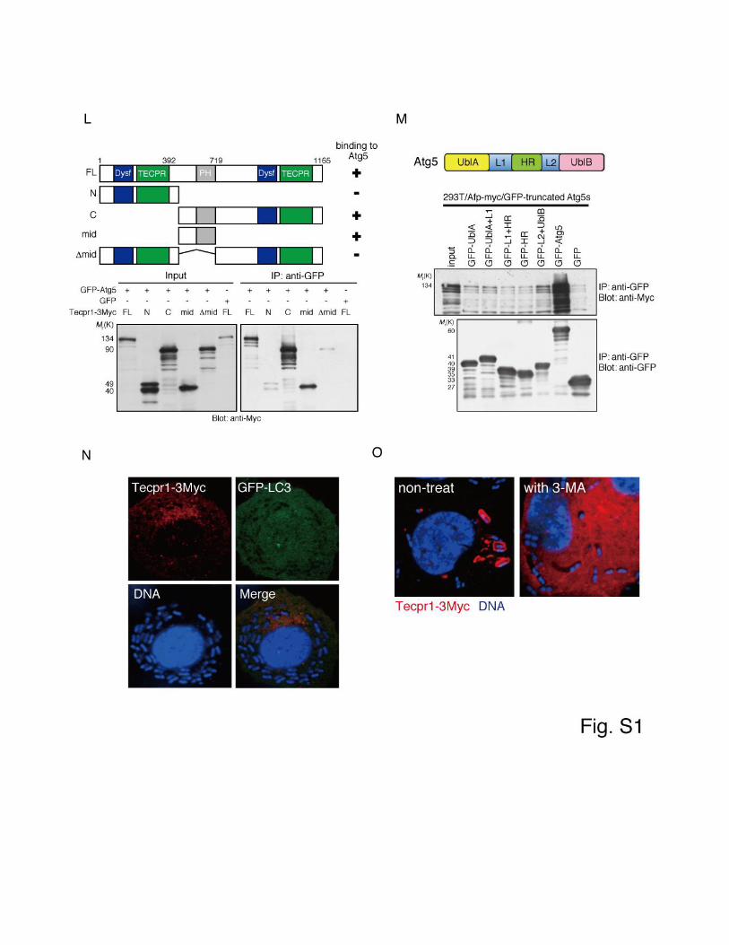

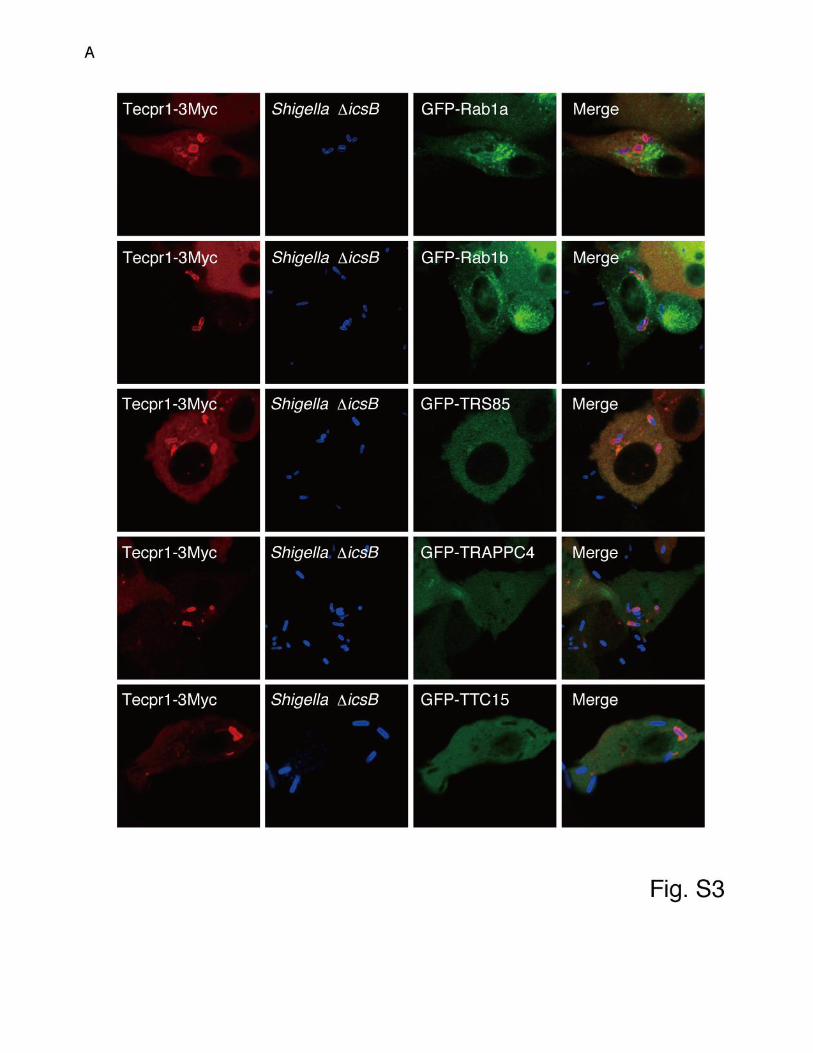


Figure S1, Related to Figure 1. Characterization of Tecpr1 as an Atg5-Binding
Protein
(A) The Atg5 binding candidates identified by yeast two-hybrid screening are listed.
(B) Each truncated protein was purified as a GST-fused protein (CBB), and His-Atg5
was pulled down with beads bound to each protein and immunoblotted with anti-His
antibody.
(C) MCC23918-3Myc was expressed in BHK cells and then stained with anti-Myc
antibody. MCC23918-3Myc was exclusively localized in the nucleus.
(D) BHK/GFP-LC3 cells expressing Tecpr1-3Myc or Atg16L2-3Myc were infected
with Shigella icsB, and 2 h later they were stained with anti-Myc antibody and
DAPI.
(E) The level of tecpr1 expression in human tissues was evaluated by real-time
quantitative RT-PCR. The gapdh gene was used as an internal control for
normalization. (n= 3). Data are the means ± S. E. M.
(F) Schematic diagram of the domains of Tecpr1. Tecpr1 contains several distinct
domains, including Dysferlin N-terminal motifs (DysF-N), Dysferlin C-terminal motifs
(DysF-C), a Tachylectin-II-like beta-propeller (TECPR), and putative Pleckstrin
homology (PH) domains.
(G) Lysates of 293T/Atg5WT or Atg5K130R-Myc cells expressing GFP-Tecpr1 or GFP
were immunoprecipitated with an anti-GFP antibody, and the bound proteins were

immunoblotted with an anti-Myc antibody.
(H) Lysates of 293T/HA-Atg12 cells expressing GFP-Tecpr1 or GFP with or without
Atg5-Myc were immunoprecipitated, and the bound proteins were immunoblotted
with an anti-HA antibody.
(I) Lysates of HeLa/Tecpr1-3Myc cells expressing the autophagy proteins indicated
were immunoprecipitated with an anti-GFP antibody, and the bound proteins were
immunoblotted with anti-Myc or anti-GFP antibodies.
(J) Lysates of 293T cells expressing Tecpr1-3Myc or Atg5-Myc were
immunoprecipitated with an anti-Myc antibody, and the bound proteins were
immunoblotted with anti-Atg16L1 or anti-Myc antibodies.
(K) Lysates of 293T cells expressing Tecpr1-3Myc or Tecpr2-3Myc were
immunoprecipitated with an anti-Myc antibody, and the bound proteins were
immunoblotted with an anti-Atg5 antibody.
(L) Schematic diagram of the domains of Tecpr1 and the Tecpr1 truncation mutants
(top), and identification of the Tecpr1 domains involved in Atg5 binding (bottom).
Lysates of 293T cells expressing Tecpr1-3Myc derivatives and GFP-Atg5 or GFP
were immunoprecipitated with an anti-GFP antibody, and the bound proteins were
immunoblotted with an anti-Myc antibody.
(M) Schematic diagram of the domains of Atg5 (top) and identification of the Atg5
domains involved in Tecpr1 binding (bottom). Lysates of 293T cells expressing

Atg5-3Myc derivatives and GFP-Tecpr1 or GFP were immunoprecipitated with an
anti-GFP antibody, and the bound proteins were immunoblotted with antibodies
against Myc or GFP.
(N) BHK/Tecpr1-3Myc/GFP-LC3 cells were infected with Shigella icsB/ virG, and
2 h later they were stained with an anti-Myc antibody and DAPI.
(O) BHK/Tecpr1-3Myc cells were infected with Shigella icsB in the presence or
absence of 3-methyladenine and then stained with an anti-Myc antibody and DAPI.
Figure S2, Related to Figure 4. Effect of siRNA-Mediated Knockdown of Tecpr1 on
Its Production and LC3-II Formation
(A) Lysates of HeLa cells that were treated with the siRNAs indicated for 48 h
subjected to SDS-PAGE and Western blot analysis with anti-Tecpr1 antibody (top
left). mRNA was extracted from HeLa cells (top right), 293T cells, or MEFs (bottom)
treated with the siRNAs indicated for 48 h, and the tecpr1 and -actin expression
levels were evaluated by RT-PCR.
(B and C) 293T cells treated with the siRNAs indicated were cultured under the
conditions indicated. Lysates were subjected to immunoblotting with antibodies
against LC3 or actin (B), and the LC3-II/LC3-I ratios were quantified (C).

Figure S3, Related to Figure 6. Examination of the Ability of Tecpr1 to Bind to Class
III PI3K Complex, Phospholipids, Sugars, and WIPI-2
(A) BHK/Tecpr1-3Myc expressing the Tecpr1 binding candidates tagged with GFP
indicated were infected with Shigella icsB, and 90 min later they were stained with
an anti-Myc antibody and DAPI.
(B) Lysates of 293T cells expressing GFP-Tecpr1, GFP-Vps34 (positive control), or
GFP were immunoprecipitated with an anti-GFP antibody, and the bound proteins
were immunoblotted with an anti-Vps15 antibody (left). Cell lysates of 293T cells
expressing Tecpr1-3Myc and the GFP derivatives indicated were
immunoprecipitated with an anti-GFP antibody, and the bound proteins were
immunoblotted with an anti-Myc antibody (right).
(C) Lysates of 293T cells expressing GFP-2FYVE (positive control), Tecpr1-GFP, or
GFP were incubated with PI3P-immobilized beads (Echelon), and the bound protein
was immunoblotted with an anti-GFP antibody.
(D and E) Lysates of 293T cells expressing Tecpr1-GFP, GFP-galectin3 (positive
control), or GFP were incubated with (D) lactose- or (E) GlcNAc-immobilized beads
(EY Lab.) and the eluted protein was immunoblotted with an anti-GFP antibody.
(F) Identification of the Tecpr1 domains involved in WIPI-2 binding (top) and
schematic diagram of the domains of Tecpr1 and the Tecpr1 truncation mutants

(bottom). Lysates of 293T cells expressing 2HA-WIPI-2 and GFP-Tecpr1 derivatives
or GFP were immunoprecipitated with an anti-GFP antibody, and the bound proteins
were immunoblotted with antibodies against HA or GFP.
(G) Lysates of 293T cells expressing wild-type or FKKG-mutant 2HA-WIPI-2 and
Tecpr1-GFP or GFP were immunoprecipitated with an anti-GFP antibody, and the
bound proteins were immunoblotted with antibodies against HA or GFP.
(H) mRNA was extracted from HeLa cells that had been treated with the siRNAs
indicated for 48 h, and the levels of wipi-2 and -actin expression were evaluated by
RT-PCR.
(I) HeLa/Atg5-Myc cells treated with the siRNAs indicated for 48 h were subjected to
SDS-PAGE and Western blot analysis with an anti-Myc antibody. The
Atg12-Atg5/Atg5 levels are shown.
(J) 293T cells were treated with the siRNAs indicated for 48 h and cultured under
the conditions indicated. Whole-cell lysates were subjected to SDS-PAGE and
Western blot analysis with antibodies against LC3 or actin.
Figure S4, Related to Figure 7. Characterization of Tecpr1 Knockout Mice-Derived
MEF Cells
Tecpr1 knock-out mice were born healthy at the expected Mendelian ratio and had
no gross phenotypic abnormalities.

(A) Structure of the mouse tecpr1 (2210010N04Rik) locus (WT allele), the tecpr1
targeting construct (Targeting construct), and the predicted mutant tecpr1 gene
(Targeted allele). Exons are represented by boxes. Exons 2 to 5 in the tecpr1 gene
were replaced by a neo resistance gene. A diphtheria toxin A (DT) gene was
attached to the 5’ end of the genomic fragment for negative selection. Southern blot
analysis for targeted clone screening was performed after EcoRI digestion.
(B) Correct targeting of the tecpr1 locus was confirmed by genomic Southern blot
analysis. The endogenous (10.3 kb) and/or mutant (7.5 kb) bands were detected by
using a 3’ probe and EcoRI-digested tail DNA from tecpr1 WT (+/+), heterozygous
(+/-), and mutant (-/-) littermates.
(C) The genotype was confirmed by genomic PCR. Tail DNA from tecpr1 WT (+/+),
heterozygous (+/-), and mutant (-/-) littermates was used as a template, and the
genotype was determined by genomic PCR.
(D) Expression of tecpr1 mRNA. RNA was isolated from Tecpr1-/- or Tecpr1+/+MEFs,
and the tecpr1 mRNA levels were determined by RT-PCR.
(E) Expression of Tecpr1 protein. Lysates from Tecpr1-/- or Tecpr1+/+MEFs were
subjected to SDS-PAGE and Western blot analysis with antibodies against Tecpr1
or actin.
(F and G) Lysates of Tecpr1-/- or Tecpr1+/+MEFs cultured under the conditions
indicated were subjected to immunoblotting with antibodies against LC3 or actin (F),

and the ratios of LC3-II/LC3-I were quantified (G).
(H and I) Lysates of Tecpr1-/- or Tecpr1+/+MEFs were subjected to immunoblotting
with antibodies against Tecpr1, p62, Atg5, Atg16L1, or actin (H), and the level of
each protein was quantified (I).
(J) Tecpr1-/- or Tecpr1+/+MEFs were stained with anti-mtHSP70antibodies, and
tangled and condensed mitochondria containing cells were counted (>300cells, n =
3). Data are the means ± S. E. M. *p<0.001.
(K) To measure the mass of depolarized mitochondria, MEFs cultured in the
presence or absence of CCCP were stained with MitoTracker Green FM and
MitoTracker Red CMXRos and analyzed by FACS.
Figure S5. Proposed Model for the Role of Tecpr1 in Autophagy
When autophagy is induced, PI(3)P is generated by the type III PI3K complex.
PI(3)P targets WIPI-2 to the phagophore. WIPI-2 then recruits Tecpr1 to the
phagophore, and Atg5 is recruited to the phagophore by Tecpr1-Atg5 binding. The
interaction between Atg5 and Tecpr1 promotes Atg12-Atg5 conjugation, leading to
the generation of LC3-II, elongation of the phagophore, and autophagosome
formation. During intracellular growth the Shigella surface protein VirG is localized
at one pole of intracellular Shigella. The VirG binds to Atg5 on the phagophore, and
intracellular Shigella is recognized by the WIPI-2-Tecpr1-Atg5 pathway, which leads

to engulfment of Shigella by autophagosomes.
SUPPLEMENTAL EXPERIMENTAL PROCEDURES
Plasmids
Human Tecpr1 cDNA was PCR amplified by using the DKFZP434B0335 cDNA
clone as a template, and cloned into the pEGFP-C3 (Clontech), pEGFP-N3
(Clontech), pcDNA-3Myc (C-terminal tagged), pMal-C2X (NEB), or pGEX-6p (GE
Healthcare) vectors. Human Atg5, Rab1a, Rab1b, TRAPP4, Rab24, WIPI-2,
galectin-3, Lamp1, Atg10, and Atg7 cDNA were amplified by PCR from Caco-2 total
mRNA and cloned into the pcDNA3.1-2HA, pEGFP, pGEX6P, and pTB101-Tp-His
vectors(Ogawa et al., 2003). Human Vps15, Atg14, TTC15, and mouse Rubicon
cDNAs were amplified by PCR by using IMAGE cDNA clones (Clone ID: 5492292,
40034574, 5097580 and 30432960, respectively) as the template, and cloned into
the pEGFP vector. Human Trs85cDNA was amplified by PCR by using Flexi clones
(Clone ID: pF1KA1012) as the template, and cloned into the pEGFP vector. Human
Atg12 cDNA was PCR amplified by using pHA-Atg12 as the template, and cloned
into the pMal-C2X vector. The expression vectors for GFP-LC3, GFP-Atg5,
GFP-Beclin1, GFP-SKD1, and GFP-Vps34 were a generous gift from Dr. Tamotsu
Yoshimori (Osaka University). The expression vectors for Atg5-Myc and
GFP-ATg16L1 were a generous gift from Dr. Noboru Mizushima (Tokyo Medical
and Dental University). The expression vector for HA-Atg12 was kindly provided by

Dr. Fumihiko Takeshita (Yokohama City University). The expression vector
encoding YFP-PARK2 was a generous gift from Dr. Richard J. Youle (N. I. H.) and
mCherry-PARK2 was constructed by cloning of PARK2 amplified by PCR from
YFP-PARK2 into pmCherry-C1 (Clontech). GFP-170*, a nonpoly-Q protein that
consists of GFP fused to an internal segment of the Golgi membrane protein 170,
was kindly provided by Dr. Elizabeth Sztul (University of Alabama at Birmingham).
The expression vector encoding GFP-2FYVE (Hrs derived)was a generous gift from
Dr. Suetsugu (The University of Tokyo). RT-PCR was performed by using a
SuperScript One-Step RT-PCR System and Platinum Taq DNA Polymerase
(Invitrogen). Site-directed mutagenesis of Tecpr1, Atg5 and WIPI-2 was performed
by using a QuickChange site-directed mutagenesis kit (Stratagene).
Antibodies, Reagents, Immunoprecipitation, and Immunostaining
A polyclonal rabbit anti-Tecpr1 antibody was generated against recombinant
maltose-binding protein (MBP)-Tecpr1. The anti-Shigella LPS and anti-VirG
antibodies were prepared as previously described(Ogawa et al., 2005). To prepare
the anti-LC3 antibody for immnoblotting, rabbits were immunized with a synthetic
peptide containing the N-terminal amino acids of LC3 and an additional cysteine
(PSDRPFKQRRSFADC), and the serum was purified as previously described.
Anti-Myc (9B11, Cell Signaling), anti-HA (Cell Signaling), anti-MBP (MBL), anti-Atg5
(Nanotools), anti-Atg16L1 (MBL), anti-multi-ubiquitin (FK2, MBL), anti-Vps15
(Abnova), anti-LBPA (Echelon), anti-mtHSP70 (Enzo), and anti-actin (Chemicon)
mouse monoclonal antibodies, anti-Myc (A14, Santa Cruz), anti-GFP (MBL, Roche,
or Clontech), anti-LC3 (MBL) and anti-multi-ubiquitin (Biomol) rabbit polyclonal
antibodies, anti-p62/SQSTM1 guinea pig polyclonal antibody (Acris), and
HRP-conjugated anti-HA rat monoclonal antibody(Roche) were used as the primary
antibodies for immunostaining, immunoblotting, and immunoprecipitation. A mouse
monoclonal anti-Myc antibody (9B11) or agarose-conjugated anti-GFP rat
monoclonal antibody (MBL) was used for immunoprecipitation. Immunoprecipitation
was performed by using IP buffer (50 mM Tris-HCl pH 7.4, 150 mM NaCl, 1 mM
EDTA, 10% glycerol, Complete protease inhibitor cocktail-EDTA [Roche]). An
HRP-conjugated goat anti-rabbit (Jackson Laboratories), anti-mouse IgG
(Sigma),or anti-guinea pig (Sigma) antibodies were used as secondary antibodies
for immunoblotting. Cy5 (GE Healthcare)-, FITC-, or TRITC (Sigma)-conjugated
goat anti-rabbit or anti-mouse IgG antibodies were used as secondary antibodies
for immunostaining. Rhodamine-labeled phalloidin (Sigma) was used for F-actin
staining. DAPI (for DNA staining) and TexasRed-conjugated transferrin were
purchased from Molecular Probes. Rapamycin was purchased from Sigma and LC
laboratories. Earle's balanced salt solution (EBSS) for amino acid starvation,
monodansylcadaverine (MDC), 3-methyladenine (3-MA), bafilomycin A1, pepstatin

A, and carbonyl cyanide m-chlorophenyl hydrazone (CCCP) were purchased from
Sigma. E-64d was purchased from the Peptide Institute, and vinblastine was
purchased from Calbiochem. ISOGEN for mRNA extraction and purification was
purchased from NIPPON GENE. Phosphate buffer containing 4%
paraformaldehyde was purchased from Wako.
Cell Cultures and Transfections
HeLa (human cervical epithelial) cells and BHK (baby hamster kidney) cells were
cultured in Eagle’s medium (MEM, Sigma) supplemented with 10% heat-inactivated
fetal bovine serum (FCS, Invitrogen), 100 g/ml of gentamicin (Wako), and 60 g/ml
of kanamycin (Wako). 293T (Human Embryonic Kidney) cells and MEFs (mouse
embryonic fibroblasts) were cultured in Dulbecco’s modified Eagle medium (DMEM,
Sigma) supplemented with 10% FCS, 100 g/ml of gentamicin (Wako), and 60
g/ml of kanamycin (Wako). HeLa cells stably expressing EGFP-LC3 were cultured
in DMEM supplemented with 10% FCS and 2 g/ml of puromycin. BHK cells stably
expressing EGFP-LC3 were cultured in MEM supplemented with 10% FCS and
1000 g/ml of G418 (Roche). HeLa cells and BHK cells stably expressing
Tecpr1-GFP were cultured in MEM supplemented with 10% FCS and 1000 g/ml of
G418. Transfections were performed by using Lipofectamine LTX (Invitrogen) for
293T cells and Fugene 6 (Roche) for HeLa and BHK cells according to the

manufacturers’ protocols. siRNA transfections were performed by using
Lipofectamine RNAi MAX (Invitrogen) according to the manufacturer’s protocol.
GST Pull-Down Assay
The GST-pull down assay was performed as previously described (Ogawa et al.,
2003; Ogawa et al., 2005). Briefly, GST derivatives were bound to Glutathione
Sepharose 4B beads and then mixed with the recombinant proteins for 2 h at 4 C.
After centrifugation, the beads were washed five times with 1% Triton X-100-PBS
and subjected to Western blot analysis.
Quantification of Bacteria Associated with GFP-LC3
The number of bacteria associated with GFP-LC3 or Tecpr1-3Myc was counted
visually by examination with a fluorescence microscope.
Assay for Intracellular Bacterial Multiplication
siRNA-transfected HeLa cells or MEFs were seeded into 24-well plates and infected
with the Shigella icsB strain as previously described (Ogawa et al., 2009; Ogawa
et al., 2005). Briefly, the cells were infected with the Shigella icsB strain at an MOI
of 100, centrifuged for 10 min at 700 x g, and incubated at 37 C for 20 min to allow
bacterial uptake and invasion. The infected cells were then washed three times with

PBS, and fresh medium supplemented with 200 g/ml of gentamicin was added to
kill the remaining extracellular bacteria. After incubating at 37 C for 15 min, the
medium was replaced with fresh medium supplemented with 100 g/ml of
gentamicin, and the cells were cultured for the time periods indicated. At each time
point, the cells were washed three times with PBS and then lysed with PBS
containing 0.5% Triton X-100. The lysates were diluted with PBS, plated onto
LB-agar plates, and the number of intracellular bacteria was counted as cfu
(colony-forming units).
Expression and Purification of Recombinant Proteins
To express GST fusion proteins, the E. coli BL21 (DE3) strain harboring pGEX-6P-1
derivatives was cultured for 10 h at 20 C in L-broth supplemented with ampicillin
(50 g/ml). Protein expression was induced by adding 1 mM IPTG and then
incubating the cells for 14 h at 20 C. Bacteria were disrupted by sonication and
lysozyme treatment. The GST fusion proteins were purified by performing affinity
chromatography with Glutathione Sepharose 4B (GE Healthcare) according to the
manufacturer’s protocol. The GST tag was removed by using PreScission protease
(GE Healthcare) to treat beads that were bound to the GST fusion protein. For
His6-tagged protein purification, the E. coli BL21 (DE3) strain harboring
pTB101-Tp-His derivatives was cultured, and protein expression was induced as

described above. The His6-tagged proteins were purified by using a Ni-ATA
super-flow resin (Qiagen) according to the manufacturer’s protocol. For MBP-fusion
protein purification, the E. coli BL21 (DE3) strain harboring pMal-C2X derivatives
was cultured, and expression was induced as described above. The proteins were
purified by using a maltose resin (NEB) according to the manufacturer’s protocol.
Evaluation of Tecpr1 Expression Levels in Human Tissues
The level of Tecpr1 expression in human tissues was evaluated by quantitative
real-time RT-PCR using the First Choice Human Total RNA Survey Panel (Ambion)
as a template. The following primer pairs were used: tecpr1, 5’-
AAACGGGGTCATGCACATCTCG -3’ and 5’- TGTGAGAGTTGACGCCTCTTCG
-3’;gapdh, 5’- TGCCCTCAACGACCACTTTG -3’ and 5’-
TTCCTCTTGTGCTCTTGCTGGG -3’.
siRNA
siRNAs were synthesized and duplexed by RNAi Co., Ltd. The siRNA sequences
were: human tecpr1, 5'-CAC ACG GCC UGG GUA UAC ACA -3' and 5’- UGU AUA
CCC AGG CCG UGU GGU-3’; mouse tecpr1, 5'-GGU UCC GGA GGG GUG UCA
ACU-3' and 5’-UUG ACA CCC CUC CGG AAC CAC-3’; and human wipi-2, 5'-GGG
ACC GUG AUU AGG GUA UUU-3' and 5’-AUA CCC UAA UCA CGG UCC CCU-3’.

The siRNAs were transfected into cells by reverse transfection using Lipofectamine
RNAi MAX. siRNA-mediated gene knockdown was confirmed by Western blot
analysis of endogenous Tecpr1 levels in HeLa cells and of GFP-Tecpr1 levels in
BHK cells stably expressing GFP-Tecpr1. The wipi-2 mRNA level was confirmed by
RT-PCR with the following primer pair: 5’-AGCGCGACCATCGGAGAGGT-3’ and
5’-TGTGGCCGCAGAATGGCAGG -3.
Yeast Two-Hybrid Screening
The yeast two-hybrid screen was performed with Pro-Quest (Invitrogen). cDNA
encoding full-length human atg5 was cloned in-frame into the GAL-4 DNA-binding
domain vector pDB-Leu (Invitrogen). The resulting plasmid, pDB-Leu-hAtg5, was
used as the bait in the yeast two-hybrid screening. A human brain cDNA library
(Invitrogen) was screened for interacting proteins according to the manufacturer’s
protocol.
Retroviral Infection
To construct HeLa cells stably expressing GFP-LC3, gfp-lc3 cDNA was subcloned
into the pMX-puro expression vectors. The vectors obtained were transfected into
Plat-E cells to generate recombinant mouse retroviruses. MEFs or HeLa cells stably
expressing the mouse ecotropic retroviral receptor were infected with the
recombinant mouse retroviruses and then selected in DMEM containing 10% FCS

and 2 g/ml puromycin (Sigma).
Electron Microscopy
Cells seeded on 35 mm dishes were fixed with 2% glutaraldehyde in PBS for 1 h,
post-fixed in 2% OsO4, dehydrated with a graded ethanol series, and embedded in
epoxy resin. Ultra-thin sections were stained with both uranyl acetate and lead
citrate.
Generation of tecpr1 Knockout Mice
The tecpr1 gene in J1 ES cells was disrupted by homologous recombination with a
targeting vector in which exons 2 5 were replaced by a neo cassette. A diphtheria
toxin A (DT) gene was attached to the 5’ end of the genomic fragment for negative
selection. One ES cell clone that carried the targeted allele was used to generate
chimeric mice that passed the mutant allele to their offspring. Southern blot analysis
for targeted clone screening was performed by using EcoRI. The primers used to
amplify the genomic probe for Southern blot analysis were
5’-AATTGGTAATACAGCTACATCTCTGGCGCC-3’ and
5’-GGAAGCTACTCTCAGAGGGGTGTGGTCTGG-3’. Genotyping was also
performed by genomic PCR with lysed mouse tails and the following genomic PCR
primers: WT/KO-forward 5’-AGGTGCACATGAGGGTTAGC-3’, WT-reverse

5’-CCTTCCACGAGCTCATCTCT-3’ and KO-reverse
5’-CATTATACGAAGTTATCTCGAGTCGC-3’. The tecpr1 mRNA level was also
determined by RT-PCR with the following primer pairs: tecpr1 exons 1 6,
5’-TCGAGAGGAGGCCTATGAGA-3’ and 5’-CCACAGGGGTTGTGAGAGTT-3’;
and -actin, 5’-GCTGGTCGTCGACAACGGCT-3’ and
5’-CAAACATGATCTGGGTCATCTTTTC-3’. To generate MEFs, fibroblasts were
prepared from day 13.5 Tecpr1-/- and wild-type embryos. Animal experiments were
performed in accordance with the guidelines for animal use issued by the
Committee of Animal Experiments, The Institute of Medical Science, The University
of Tokyo.
Statistical Analysis
The density of each band was quantified by measuring the mean intensity with the
NIH image software version 1.43. The expression levels were normalized to the
-actin or gapdh levels.

SUPPLEMENTAL REFERENCES
Ogawa, M., Suzuki, T., Tatsuno, I., Abe, H., and Sasakawa, C. (2003). IcsB, secreted
via the type III secretion system, is chaperoned by IpgA and required at the
post-invasion stage of Shigella pathogenicity. Mol Microbiol 48, 913-931.
Ogawa, M., Yoshimori, T., Suzuki, T., Sagara, H., Mizushima, N., and Sasakawa, C.
(2005). Escape of intracellular Shigella from autophagy. Science 307, 727-731.