a tour of the cell - nauset public schools
TRANSCRIPT

A Tour of the Cell

By the end of this presentation, you should be able to:
Distinguish between prokaryotic and eukaryotic cells
Explain why cells are small (surface area to volume ratio)
Explain the function of the cell parts
Explain how organelles work together to carry out specific cellular functions

Cells Are the Fundamental Units of Life
All organisms are made of cells
Cells are descended from other cells
Cells share common features

Biologists use microscopes and the tools of biochemistry to study cells
Most cells are between 1 and 100 m in diameter, too small to be seen by the unaided eye
Electron microscopes can be used to view structures within cells

© 2014 Pearson Education, Inc.
Figure 4.3
Scanning electron microscopy (SEM)
Transmission electron microscopy (TEM)
Longitudinal section of cilium
Cross section of cilium
Cilia
2 m
2 m
50
m
10
m
50
m
Brightfield (unstained specimen)
Electron Microscopy (EM)
Fluorescence
Brightfield (stained specimen)
Differential-interference contrast (Nomarski)
Phase-contrast
Confocal
Light Microscopy (LM)

Prokaryotic and Eukaryotic Cells
All living organisms are composed of one of two types of cells: prokaryotic or eukaryotic
Prokaryotic: Domains Bacteria and Archaea
Eukaryotic: Protists, fungi, animals, and plants
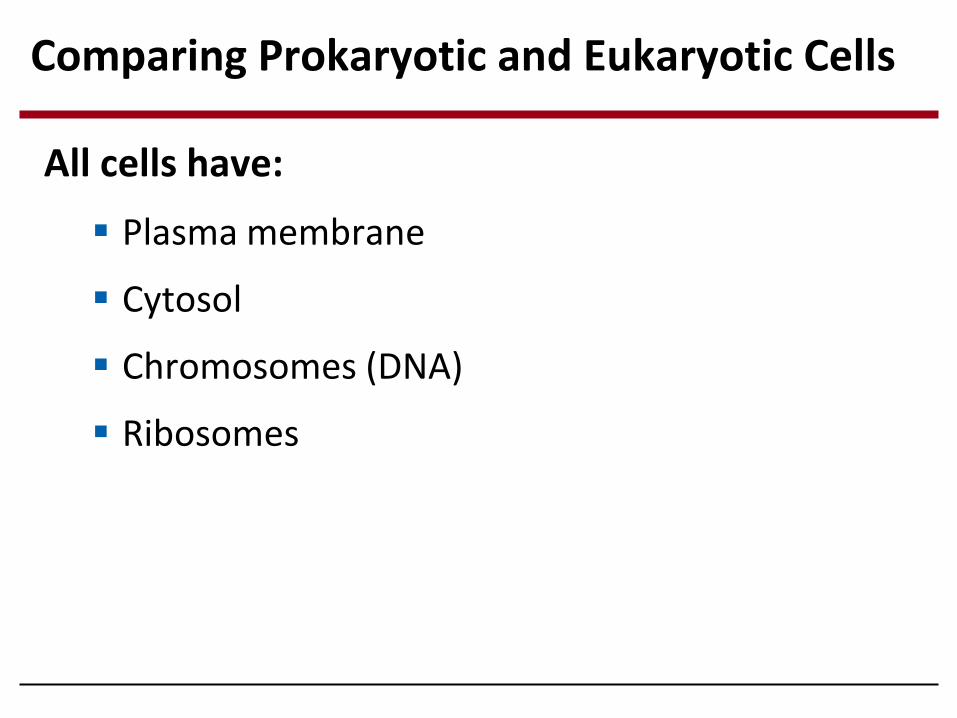
Comparing Prokaryotic and Eukaryotic Cells
All cells have:
Plasma membrane
Cytosol
Chromosomes (DNA)
Ribosomes

Prokaryotic cells are characterized by having
No nucleus
DNA is located in a region called the nucleoid
No membrane-bound organelles
© 2014 Pearson Education, Inc.
Prokaryotic Cells

© 2014 Pearson Education, Inc.
(a) A typical rod-shaped bacterium
0.5 m
(b) A thin section through the bacterium Bacillus coagulans (TEM)
Bacterial chromosome
Nucleoid
Ribosomes
Cell wall
Plasma membrane
Capsule
Flagella
Prokaryotic Cells

Eukaryotic cells are characterized by having
DNA in a membrane-bound nucleus
Membrane-bound organelles
Eukaryotic cells are generally much larger than prokaryotic cells
Eukaryotic Cells

Metabolic requirements of the cell set limits on cell size
Increase in volume = increase in demand for material resources
Smaller cells have a more favorable surface area-to-volume ratio
Why are cells so small?


© 2014 Pearson Education, Inc.
Figure 4.7a
CYTOSKELETON:
NUCLEUS
ENDOPLASMIC RETICULUM (ER)
Smooth ER
Rough ER Flagellum
Centrosome
Microfilaments
Intermediate filaments
Microvilli
Microtubules
Mitochondrion
Peroxisome Golgi apparatus
Lysosome
Plasma membrane
Ribosomes
Nucleolus
Nuclear envelope
Chromatin

© 2014 Pearson Education, Inc.
Figure 4.7e
1 m
A single yeast cell (colorized TEM)
Mitochondrion
Nucleus
Vacuole
Cell wall

© 2014 Pearson Education, Inc.
Figure 4.7b
CYTO- SKELETON
NUCLEUS
Smooth endoplasmic reticulum
Chloroplast
Central vacuole
Microfilaments
Intermediate filaments
Cell wall
Microtubules
Mitochondrion
Peroxisome
Golgi apparatus
Plasmodesmata
Plasma membrane
Ribosomes
Nucleolus
Nuclear envelope
Chromatin
Wall of adjacent cell
Rough endoplasmic reticulum
© 2014 Pearson Education, Inc.
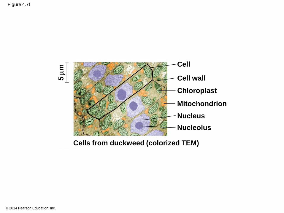
Figure 4.7f
5
m
Cell wall
Cell
Chloroplast
Mitochondrion
Nucleus
Nucleolus
Cells from duckweed (colorized TEM)

Selective barrier that allows sufficient passage of oxygen, nutrients, and waste to meet the needs of the cell
Consists of a double layer of phospholipids and embedded proteins
Plasma Membrane

The Nucleus: Information Central
Contains most of the cells genetic information (genes)
Enclosed by a nuclear envelope (contains pores to allow materials to pass in and out of the nucleus)

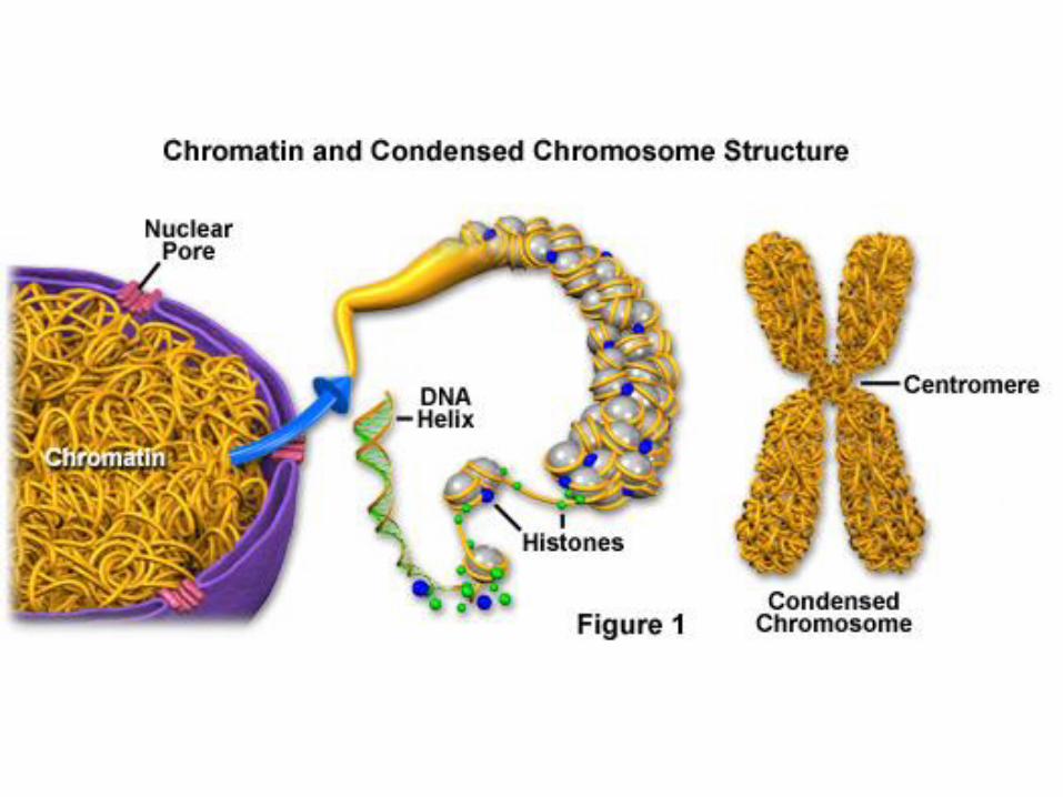
DNA is organized into chromosomes
Chromosomes consist of chromatin: DNA and proteins
Chromatin condenses to form chromosomes during cell division
The nucleolus is located within the nucleus and is the site of ribosomal RNA (rRNA) synthesis
Within the Nucleus

Ribosomes: Protein Factories
Ribosomes are composed of ribosomal RNA and protein
Ribosomes carry out protein synthesis

© 2014 Pearson Education, Inc.
Figure 4.9
TEM showing ER and ribosomes Diagram of a ribosome
Ribosomes bound to ER
Free ribosomes in cytosol
Endoplasmic reticulum (ER)
Ribosomes ER
0.25 m
Large subunit
Small subunit

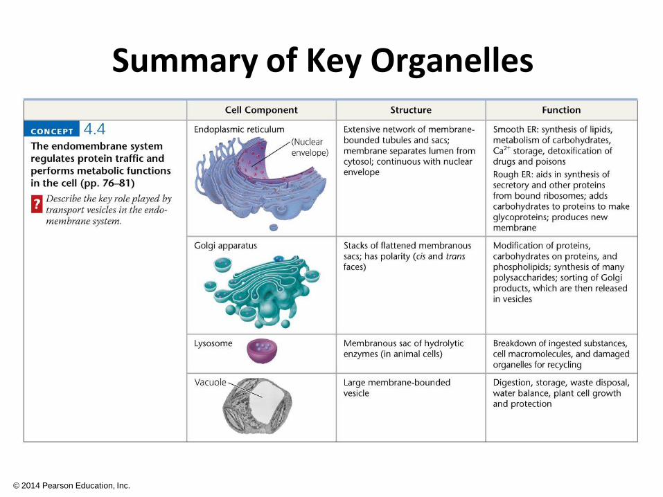
Endomembrane System
Regulates protein traffic and performs metabolic functions in the cell
Components of the endomembrane system: nuclear envelope, ER, Golgi apparatus, lysosome, vacuoles, plasma membrane
Components are either continuous or connected through transfer by vesicles

Endoplasmic Reticulum: Biosynthetic Factory
The ER membrane is continuous with the nuclear envelope
There are two distinct regions of ER
Smooth ER: lacks ribosomes
Rough ER: ribosomes on surface

Functions of Smooth ER
Synthesizes lipids
Metabolizes carbohydrates
Detoxifies drugs and poisons
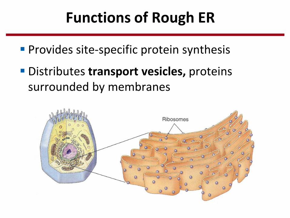
Functions of Rough ER
Provides site-specific protein synthesis
Distributes transport vesicles, proteins surrounded by membranes

Consists of a series of flattened membrane sacs (cisternae)
Functions of the Golgi apparatus
Modifies products of the ER
Manufactures certain macromolecules
Sorts and packages materials into transport vesicles
Golgi Apparatus: Shipping and Receiving Center

Lysosomes: Digestive Compartments
Contains hydrolytic enzymes that digest macromolecules
Recycles old organelles (autophagy), digests molecules within food vacuoles (phagocytosis)

Lysosomes carry out intracellular digestion in a variety of ways

Contain oxidative enzymes
Performs essential metabolic functions, including:
Decomposition of long fatty acids
Decomposition of hydrogen peroxide into water and oxygen
Peroxisomes: Oxidation

Vacuoles: Diverse Maintenance Compartments
Vacuoles are large vesicles – three types:
Food vacuoles formed by phagocytosis
Contractile vacuoles in protists (pump water0
Central vacuoles in plant cells

Mitochondria and chloroplasts change energy from one form to another
Mitochondria - sites of cellular respiration, a metabolic process that uses oxygen to generate ATP
Chloroplasts - found in plants and algae, sites of photosynthesis

Mitochondria and chloroplasts have similarities with bacteria
Enveloped by a double membrane
Contain free ribosomes and circular DNA molecules
Grow and reproduce somewhat independently in cells
The Evolutionary Origins of Mitochondria and Chloroplasts

An early ancestor of eukaryotic cells engulfed a nonphotosynthetic prokaryotic cell, which formed an endosymbiont relationship with its host
The host cell and endosymbiont merged into a single organism, a eukaryotic cell with a mitochondrion
At least one of these cells may have taken up a photosynthetic prokaryote, becoming the ancestor of cells that contain chloroplasts
© 2014 Pearson Education, Inc.
Endosymbiont Theory

© 2014 Pearson Education, Inc.
Figure 4.16
Mitochondrion
Mitochondrion
Nonphotosynthetic eukaryote
Photosynthetic eukaryote
At least one cell Chloroplast
Engulfing of photosynthetic prokaryote
Nucleus
Nuclear envelope
Endoplasmic reticulum
Ancestor of eukaryotic cells (host cell)
Engulfing of oxygen- using nonphotosynthetic prokaryote, which becomes a mitochondrion

Mitochondria: Chemical Energy Conversion
Smooth outer membrane and an inner membrane folded into cristae
Cristae present a large surface area for enzymes that synthesize ATP
The inner membrane creates two compartments: intermembrane space and mitochondrial matrix

© 2014 Pearson Education, Inc.
Figure 4.17
Free ribosomes in the mitochondrial matrix
Mitochondrion
Intermembrane space
Matrix
Cristae
DNA
Outer membrane
Inner membrane
0.1 m

Chloroplasts: Capture of Light Energy
Contain the green pigment chlorophyll
Function in photosynthesis
Found in plants, algae and other photosynthetic protists
Chloroplasts are a type of plant plastid

Thylakoids, membranous sacs, stacked to form a granum (light reactions of photosynthesis)
Stroma, the internal fluid (Calvin-Benson cycle of photosynthesis (“dark” reactions))
Structure of a Chloroplast
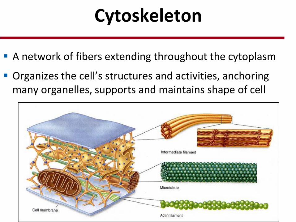
Cytoskeleton
A network of fibers extending throughout the cytoplasm
Organizes the cell’s structures and activities, anchoring many organelles, supports and maintains shape of cell

Roles of the Cytoskeleton
Interacts with motor proteins to transport vesicles and other organelles along the “tracks” of the cytoskeleton

© 2014 Pearson Education, Inc.
Table 4.1

Centrosome is a “microtubule-organizing center”
The centrosome has a pair of centrioles which plays a role in cell division
Centrioles and Centrosomes

Cell projections that are composed of microtubules
Flagella are limited to one or a few per cell, while cilia occur in large numbers on cell surfaces
Cilia and Flagella


Cell Walls of Plants
An extracellular structure that distinguishes plant cells from animal cells
Prokaryotes, fungi, and some protists also have cell walls
Protects the plant cell, maintains its shape, and prevents excessive uptake of water
Plant cell walls are composed mostly of cellulose

The Extracellular Matrix (ECM) of Animal Cells
Animal cells lack cell walls but are covered by an elaborate extracellular matrix (ECM)

Cell Junctions
Neighboring cells in an animal or plant often adhere, interact, and communicate through direct physical contact
Types of intercellular junctions:
Plasmodesmata (plants)
Tight junctions
Desmosomes
Gap junctions
© 2014 Pearson Education, Inc.

Channels between adjacent plant cells
Allow for the exchange of materials (water, solutes) between cells
Plasmodesmata of Plant Cells (Cell Walls)

Tight Junctions, Desmosomes, and Gap Junctions in Animal Cells
Animal cells have three main types of cell junctions
Tight junctions
Desmosomes
Gap junctions
All are especially common in epithelial tissue (skin, lines organs and body cavities)
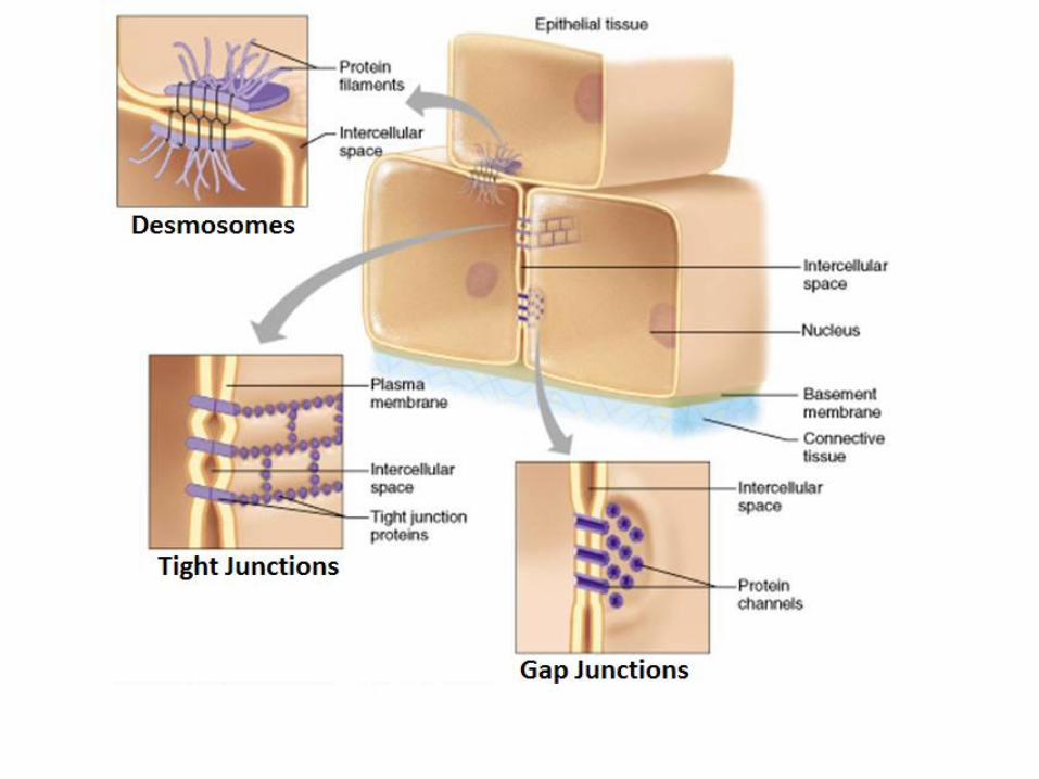

Tight Junctions
Between the cell membranes of adjacent animal cells
Form continuous seals around cells to prevent leakage of extracellular fluid
Example: between skin cells

Desmosomes
“Anchoring junctions”, fasten cells together in strong sheets
Composed of tough keratin proteins
Example: muscle cell attachments

Gap Junctions
“Communicating” junctions
Channels that directly connect the cytoplasm of two cells
Allow for the passage of small molecules such as ions, sugars and amino acids
Example: between heart muscle cells

The Cell: A Living Unit Greater Than the Sum of Its Parts
Cellular functions arise from cellular order
Example: a macrophage’s ability to destroy bacteria involves the whole cell, coordinating components such as the cytoskeleton, lysosomes, and plasma membrane

Macrophage (red) Consuming Bacteria (green)

© 2014 Pearson Education, Inc.
Summary of Key Organelles
© 2014 Pearson Education, Inc.
Summary of Key Organelles

© 2014 Pearson Education, Inc.
Summary of Key Organelles