a vesicle superpool spans multiple presynaptic terminals ... · neuron report a vesicle superpool...
TRANSCRIPT
Neuron
Report
A Vesicle Superpool Spans MultiplePresynaptic Terminals in Hippocampal NeuronsKevin Staras,1,* Tiago Branco,2,3 Jemima J. Burden,4 Karine Pozo,4 Kevin Darcy,4 Vincenzo Marra,1 Arjuna Ratnayaka,1
and Yukiko Goda3,4
1School of Life Sciences, University of Sussex, Brighton BN1 9QG, UK2Wolfson Institute for Biomedical Research3Department of Neuroscience, Physiology and Pharmacology4Medical Research Council Laboratory for Molecular Cell Biology and Cell Biology Unit
University College London, Gower Street, London WC1E 6BT, UK
*Correspondence: [email protected] 10.1016/j.neuron.2010.03.020
Open access under CC BY license.
SUMMARY
Synapse-specific vesicle pools have been widelycharacterized at central terminals. Here, we demon-strate a vesicle pool that is not confined to a syn-apse but spans multiple terminals. Using fluores-cence imaging, correlative electron microscopy,and modeling of vesicle dynamics, we show thatsome recycling pool vesicles at synapses form partof a larger vesicle ‘‘superpool.’’ The vesicles withinthis superpool are highly mobile and are rapidlyexchanged between terminals (turnover: �4% oftotal pool/min), significantly changing vesicular com-position at synapses over time. In acute hippo-campal slices we show that the mobile vesicle poolis also a feature of native brain tissue. We alsodemonstrate that superpool vesicles are availableto synapses during stimulation, providing an exten-sion of the classical recycling pool. Experimentsusing focal BDNF application suggest the involve-ment of a local TrkB-receptor-dependent mecha-nism for synapse-specific regulation of presynapticvesicle pools through control of vesicle release andcapture to or from the extrasynaptic pool.
INTRODUCTION
Presynaptic terminals in hippocampal neurons harbor defined
vesicle pools, which are major determinants of synaptic perfor-
mance (Rizzoli and Betz, 2005; Sudhof, 2004). In conventional
models of synaptic transmission, these pools are synapse-
specific, with vesicles being locally recycled after exocytosis at
the same terminal (Ceccarelli et al., 1973; Heuser and Reese,
1973). As such, presynaptic function is characterized by the
number and properties of vesicles within an individual terminal.
Recent experimental evidence, however, shows that some
synaptic vesicles (SVs) can move between adjacent release sites
in mature neurons (Chen et al., 2008; Darcy et al., 2006a; Fernan-
dez-Alfonso and Ryan, 2008; Krueger et al., 2003; Westphal
et al., 2008), raising the possibility that vesicles arising from
outside a synaptic terminal might contribute to its presynaptic
function. For example, if vesicles were trafficked at high rates
across multiple terminals and were readily available to all
neighboring synapses, this would represent a common vesicle
pool that could underlie axonal synapse-synapse interactions.
To directly test this possibility, we characterized the spatiotem-
poral organization of vesicle sharing in hippocampal neurons
using fluorescence imaging and correlative light and electron
microscopy (EM). Our findings, in dissociated cultures and acute
hippocampal slices, strongly support the existence of a large
vesicle resource or ‘‘superpool’’ composed of some of the recy-
cling pool vesicles from many adjacent terminals that can be
rapidly and directly accessed by individual synapses. Such an
arrangement provides a unique perspective on presynaptic
organization at central terminals.
RESULTS
A Vesicle Pool Common to Multiple Synaptic TerminalsStudies characterizing lateral vesicle traffic (Chen et al., 2008;
Darcy et al., 2006a; Fernandez-Alfonso and Ryan, 2008; Hopf
et al., 2002; Krueger et al., 2003; Westphal et al., 2008) have
mainly relied on single-color vesicle markers, but these probes
offer limited information about the origins and fates of mobile
vesicles across multiple synapses over time (Figure S1 available
online). To explore spatiotemporal dynamics of SV traffic in
detail, we designed a vesicle probe using a photoswitchable
fluorochrome, Dendra2, which can be rapidly and irreversibly
photoswitched from a green- to a red-emitting form following
brief intense exposure to 488 nm light (Gurskaya et al., 2006).
We fused Dendra2 to the C terminus of Synaptophysin I (Taka-
mori et al., 2006) and expressed the resulting fusion protein
(SypI-Dendra2) in hippocampal cultures (Figure 1A). SypI-Den-
dra2 showed punctate distribution that colocalized with the
activity-dependent vesicle marker FM4-64 (Figures 1A and 1B)
and was closely apposed to the postsynaptic marker PSD-95
and the dendritic marker MAP2 (Figure S2), confirming its
expression at functional presynaptic terminals. Focal 488 nm
laser illumination selectively photoswitched synapses in the
target area, with typically a >40-fold increase in red fluorescence
intensity and a 12-fold decrease in green fluorescence intensity
(Figure 1C).
Neuron 66, 37–44, April 15, 2010 ª2010 Elsevier Inc. 37
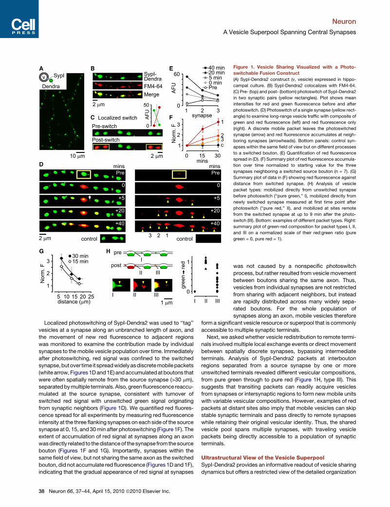
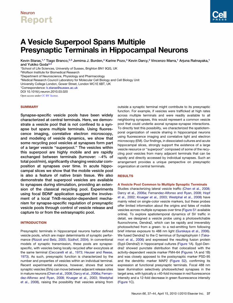
Figure 1. Vesicle Sharing Visualized with a Photo-
switchable Fusion Construct
(A) SypI-Dendra2 construct (v, vesicle) expressed in hippo-
campal culture. (B) SypI-Dendra2 colocalizes with FM4-64.
(C) Pre- (top) and post- (bottom) photoswitch of SypI-Dendra2
in two synaptic pairs (yellow rectangles). Plot shows mean
intensities for red and green fluorescence before and after
photoswitch. (D) Photoswitch of a single synapse (yellow rect-
angle) to examine long-range vesicle traffic with composite of
green and red fluorescence (left) and red fluorescence only
(right). A discrete mobile packet leaves the photoswitched
synapse (arrow) and red fluorescence accumulates at neigh-
boring synapses (arrowheads). Bottom panels: control syn-
apses within the same field of view but on different processes
to a switched bouton. (E) Quantification of red fluorescence
spread in (D). (F) Summary plot of red fluorescence accumula-
tion over time normalized to starting value for the three
synapses neighboring a switched source bouton (n = 7). (G)
Summary plot of data in (F) showing red fluorescence against
distance from switched synapse. (H) Analysis of vesicle
packet types: mobilized directly from unswitched synapse
before photoswitch (‘‘pure green,’’ I), mobilized directly from
newly switched synapse measured at first time point after
photoswitch (‘‘pure red,’’ II), and mobilized at sites remote
from the switched synapse at up to 9 min after the photo-
switch (III). Bottom: examples of different packet types. Right:
summary plot of green-red composition for packet types I, II,
and III on a normalized scale of their red:green ratio (pure
green = 0, pure red = 1).
Neuron
A Vesicle Superpool Spanning Central Synapses
Localized photoswitching of SypI-Dendra2 was used to ‘‘tag’’
vesicles at a synapse along an unbranched length of axon, and
the movement of new red fluorescence to adjacent regions
was monitored to examine the contribution made by individual
synapses to the mobile vesicle population over time. Immediately
after photoswitching, red signal was confined to the switched
synapse,butover time it spreadwidelyas discrete mobilepackets
(white arrow, Figures 1D and 1E) and accumulated at boutons that
were often spatially remote from the source synapse (>30 mm),
separated by multiple terminals. Also, green fluorescence reaccu-
mulated at the source synapse, consistent with turnover of
switched red signal with unswitched green signal originating
from synaptic neighbors (Figure 1D). We quantified red fluores-
cence spread for all experiments by measuring red fluorescence
intensity at the three flanking synapses on each side of the source
synapse at 0, 15, and 30 min after photoswitching (Figure 1F). The
extent of accumulation of red signal at synapses along an axon
was directly related to the distance of the synapse fromthe source
bouton (Figures 1F and 1G). Importantly, synapses within the
same field of view, but not sharing the same axon as the switched
bouton, did not accumulate red fluorescence (Figures 1D and 1F),
indicating that the gradual appearance of red signal at synapses
38 Neuron 66, 37–44, April 15, 2010 ª2010 Elsevier Inc.
was not caused by a nonspecific photoswitch
process, but rather resulted from vesicle movement
between boutons sharing the same axon. Thus,
vesicles from individual synapses are not restricted
from sharing with adjacent neighbors, but instead
are rapidly distributed across many widely sepa-
rated boutons. For the whole population of
synapses along an axon, mobile vesicles therefore
form a significant vesicle resource or superpool that is commonly
accessible to multiple synaptic terminals.
Next, we asked whether vesicle redistribution to remote termi-
nals involved multiple local exchange events or direct movement
between spatially discrete synapses, bypassing intermediate
terminals. Analysis of SypI-Dendra2 packets at interbouton
regions separated from a source synapse by one or more
unswitched terminals revealed different vesicular compositions,
from pure green through to pure red (Figure 1H, type III). This
suggests that transiting packets can readily acquire vesicles
from synapses or intersynaptic regions to form new mobile units
with variable vesicular compositions. However, examples of red
packets at distant sites also imply that mobile vesicles can skip
stable synaptic terminals and pass directly to remote synapses
while retaining their original vesicular identity. Thus, the shared
vesicle pool spans multiple synapses, with traveling vesicle
packets being directly accessible to a population of synaptic
terminals.
Ultrastructural View of the Vesicle SuperpoolSypI-Dendra2 provides an informative readout of vesicle sharing
dynamics but offers a restricted view of the detailed organization
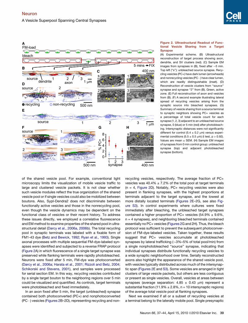
Figure 2. Ultrastructural Readout of Func-
tional Vesicle Sharing from a Target
Synapse
(A) Experimental scheme. (B) Ultrastructural
reconstruction of target process showing axon,
dendrite, and SV clusters (red). (C) Sample EM
images from synapses in (B), fixed after �5 min.
Top left (‘‘s’’): unbleached source synapse. Recy-
cling vesicles (PC+) have dark lumen (arrowheads)
and nonrecycling vesicles (PC�) have clear lumen,
which are readily distinguishable (inset). (D)
Reconstruction of vesicle clusters from ‘‘source’’
synapse and synapse ‘‘2’’ from (B). Green, active
zone. (E) Full reconstruction of axon and vesicles
from (B). (F) A second example illustrating lateral
spread of recycling vesicles arising from the
synaptic source into bleached synapses. (G)
Summary of vesicle sharing from a source terminal
to synaptic neighbors showing PC+ vesicles as
a percentage of total vesicle count for each
synapse (1, 2, 3) adjacent to an unbleached source
synapse, 0 (blue) or 5 min (red) after photobleach-
ing. Intersynaptic distances were not significantly
different for control (5.4 ± 0.2 mm) versus experi-
mental conditions (5.5 ± 0.5 mm) (t test, p > 0.92).
Values are mean ± SEM. (H) Sample EM images
of synapses from 0 min control group: unbleached
synapse (top) and adjacent photobleached
synapse (bottom).
Neuron
A Vesicle Superpool Spanning Central Synapses
of the shared vesicle pool. For example, conventional light
microscopy limits the visualization of mobile vesicle traffic to
large and clustered vesicle packets. It is not clear whether
such vesicle modules reflect the true organization of the shared
vesicle pool or if single vesicles could also be mobilized between
boutons. Also, SypI-Dendra2 does not discriminate between
functionally active vesicles and those in the nonrecycling pool,
even though the vesicle dynamics may be dependent on the
functional class of vesicles or their recent history. To address
these issues directly, we employed a correlative fluorescence
and EM method to examine properties of the shared pool in ultra-
structural detail (Darcy et al., 2006a, 2006b). The total recycling
pool in synaptic terminals was labeled with a fixable form of
FM1-43 dye (Betz and Bewick, 1992; Ryan et al., 1993). Single
axonal processes with multiple sequential FM-dye-labeled syn-
apses were identified and subjected to a reverse FRAP protocol
(Figure 2A) in which fluorescence of a single target synapse was
preserved while flanking terminals were rapidly photobleached.
Neurons were fixed after 5 min, FM-dye was photoconverted
(Darcy et al., 2006a; Harata et al., 2001; Rizzoli and Betz, 2004;
Schikorski and Stevens, 2001), and samples were processed
for serial section EM. In this way, recycling vesicles contributed
by a single target bouton to the neighboring regions over 5 min
could be visualized and quantified. As controls, target terminals
were photobleached and fixed immediately.
In an axon fixed after 5 min, the target (unbleached) synapse
contained both photoconverted (PC+) and nonphotoconverted
(PC�) vesicles (Figures 2B–2D), representing recycling and non-
recycling vesicles, respectively. The average fraction of PC+
vesicles was 40.4% ± 7.2% of the total pool at target terminals
(n = 4, Figure 2D). Notably, PC+ recycling vesicles were also
present in flanking synapses, with the highest proportions at
terminals adjacent to the target synapse, and the lowest at
more distally located terminals (Figures 2E–2G, see also Fig-
ure S3). In control experiments where cultures were fixed
immediately after bleaching, the unbleached target synapses
contained a higher proportion of PC+ vesicles (54.9% ± 9.6%,
n = 4 synapses), and neighboring bleached terminals contained
essentially no PC+ vesicles (Figures 2G and 2H). Thus, the bleach
protocol was sufficient to prevent the subsequent photoconver-
sion of FM-dye-labeled vesicles. Taken together, these results
suggest that PC+ vesicles accumulate at photobleached
synapses by lateral trafficking (�3%–5% of total pool/min) from
a single nonphotobleached ‘‘source’’ synapse, indicating that
individual synapses distribute functionally recycling vesicles to
a wide synaptic neighborhood over time. Serially reconstructed
axons also highlight the appearance of the shared vesicle pool,
with vesicles typically distributed across much of the intersynap-
tic span (Figures 2E and S3). Some vesicles are arranged in tight
clusters of large vesicle packets, but others are less contiguous
or present as single vesicles. Overall, vesicles at areas between
synapses (average separation: 4.85 ± 0.43 mm) represent a
substantial fraction (11.9% ± 2.8%, n = 10 intersynaptic regions)
of the average total vesicle pool at flanking synapses.
Next we examined if all or a subset of recycling vesicles at
a terminal belong to the laterally mobile pool. Single presynaptic
Neuron 66, 37–44, April 15, 2010 ª2010 Elsevier Inc. 39
Figure 3. Lateral Sharing of Recycling Vesi-
cles in Native Hippocampal Tissue
(A) FM1-43-labeled synapses (examples shown
with arrowheads) in CA1 region imaged using
two-photon microscopy. (B) Top left: schematic.
Right: destaining of FM puncta (arrowheads)
by local 20 Hz stimulation at 0, 20, and 180 s.
Bottom left: plot showing stimulation-evoked
fluorescence loss for 26 puncta. (C) Sample time-
lapse sequence (left) and corresponding line scan
plots (right) showing multiple trafficking events
(arrowheads) along an axon between stable
puncta (red arrows). (D) A discrete trafficking
event in which fluorescent packet (arrowhead)
passes through a stable terminal. (E) Cumulative
fluorescence intensity change plot for n = 39
boutons.
Neuron
A Vesicle Superpool Spanning Central Synapses
terminals (n = 9) in FM-dye-loaded neurons were photobleached
and, after a 1 hr recovery period, prepared for ultrastructural
analysis as above (Figure S4). Whereas large numbers of new
recycling vesicles were seen at photobleached synapses after
1 hr compared to numbers in newly bleached control terminals
(n = 6, Figure S4), these still represented a subset (�40%) of
the total recycling pool that we measured at unbleached syn-
apses after the same 1 hr period (n = 4, Figure S4). Given the
high rate of vesicle mobility we observe over short timescales,
the incomplete longer-term recovery suggests that recycling
vesicle pools may include both highly mobile and more stable
(i.e., those likely to be retained) vesicle fractions, implying
a possible heterogeneity in whether vesicles are associated
with (or belong to) the superpool.
Vesicle Sharing in Native Hippocampal TissueTo date, the characterization of intersynaptic vesicle movement
has been limited to work in cultured neurons (Chen et al., 2008;
Darcy et al., 2006a; Fernandez-Alfonso and Ryan, 2008; Krueger
et al., 2003; Westphal et al., 2008), and the relevance of this phe-
nomenon to presynaptic organization in native tissue remains
unclear. We addressed this question in acute hippocampal
slices using two-photon microscopy to image presynaptic
terminals labeled with FM1-43. After dye-loading, we observed
discrete fluorescent puncta corresponding to presynaptic
terminals in region CA1 as reported previously (Zakharenko
et al., 2001) (Figure 3A). These labeled terminals were release
competent because their fluorescence destained upon stimula-
tion (Figure 3B). Axonal regions between stable puncta showed
bidirectional trafficking of many fluorescent packets, large and
small, with both merging and shedding events (Figures 3C and
3D), analogous to vesicle movement in culture (Figure S1) (Darcy
et al., 2006a). To quantify vesicle flux at stable synapses, we
monitored changes in fluorescence levels of single terminals
over time. The cumulative fluorescence change corrected for
imaging noise shows a linear profile (n = 39: Figure 3E), indicating
that at most synapses fluorescence intensity fluctuates con-
40 Neuron 66, 37–44, April 15, 2010 ª2010 Elsevier Inc.
tinuously, implying a constant vesicle flux through terminals.
Our findings strongly support the idea that a shared pool of func-
tional vesicles is a feature of native hippocampal tissue.
A Shared Vesicle Pool as an Extensionof the Recycling PoolMobile populations of extrasynaptic vesicles that are adjacent to
stable presynaptic terminals might serve as additional vesicle
reservoirs for presynaptic release. While previous work has
shown that mobile vesicles enter synaptic terminals and undergo
fusion alongside native vesicles (Darcy et al., 2006a), whether
incorporation and fusion are sufficiently rapid to contribute to
release during sustained transmission has not been considered.
We investigated this issue in culture using FM-dye-loaded
neurons combined with field stimulation. Mobile vesicles that
became newly incorporated into terminals could readily partici-
pate in vesicle fusion (Figure 4A). A similar observation was
also made in an acute slice preparation (Figure 4B). Complemen-
tary to this idea of rapid fusion-competence, we also observed
examples of mobile vesicle clusters that underwent FM-dye
loss while moving (Figure 4C). Next, we examined the conse-
quence of synaptic incorporation of mobile vesicles during
continuous stimulation. A synapse along a process with high
vesicle mobility continually received new consignments of fluo-
rescent vesicles that, during stimulation, were released along-
side native vesicles (Figure 4D, left). This lateral draining of
mobile vesicles into stable synapses can be observed directly
in a kymograph plot (Figure 4E), and in this example, resulted
in a delayed stimulation-evoked FM-dye loss compared with
a synapse on a process that showed low levels of mobile vesicle
traffic (Figures 4E and 4F). Quantifying the fate of mobile vesicle
packets during activity by measuring stimulus-evoked fluores-
cence changes in intersynaptic axonal segments (n = 23 from
three cultures) revealed a net loss of FM-dye fluorescence sig-
nal (39% ± 2.2%: Figure 4G and 4I). This indicates substantial
activity-dependent fusion of mobile vesicles originating from
axonal regions. To establish what fraction of the packets
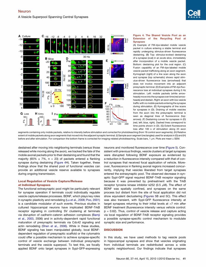
Figure 4. The Shared Vesicle Pool as an
Extension of the Recycling Pool at
Synapses
(A) Example of FM-dye-labeled mobile vesicle
packet in culture entering a stable terminal and
rapidly undergoing stimulus-driven fluorescence
destaining. (B) Top: stimulus-evoked destaining
of a synapse (oval) in an acute slice, immediately
after incorporation of a mobile vesicle packet.
Bottom: destaining plot for the oval region. (C)
Fusion capability of an FM-dye-labeled mobile
vesicle packet trafficking along an axon segment.
Kymograph (right) of a line scan along the axon
and synapse (top schematic) shows rapid stim-
ulus-driven fluorescence loss (arrowhead) that
does not involve movement into an adjacent
presynaptic terminal. (D) Examples of FM-dye fluo-
rescence loss at individual synapses during 5 Hz
stimulation. Left: mobile packets (white arrow-
heads) move into the synapse over time (red arrow-
heads) and destain. Right: an axon with low vesicle
traffic with no mobile packets entering the synapse
during stimulation. (E) Kymographs of line scans
for synapses in (D). Draining of mobile vesicles
from the axon into the presynaptic terminal is
seen as diagonal lines of fluorescence (top:
arrows). (F) Destaining curves for synapses in (D)
(red, left; blue, right). Dashed lines correspond to
time points shown in (D). (G) Extent fluorescence
loss after 100 s of stimulation along 23 axon
segments containing only mobile packets, relative to intensity before stimulation and corrected for photobleaching (from 18 control axon segments). (H) Relative
extent of mobile packets along axon segments that moved into the adjacent synaptic terminal. (I) Sample axon segment (rectangles) used for analysis in (G) and (H)
before and after stimulation. For comparison the bottom frame is corrected for imaging-related photobleaching. Scale bars, 1 mm. Plots are mean ± SEM.
Neuron
A Vesicle Superpool Spanning Central Synapses
destained after moving into neighboring terminals (versus those
released while moving along the axon), we tracked the fate of the
mobile axonal packets prior to their destaining and found that the
majority (65% ± 7%, n = 23) of packets entered a flanking
synapse during destaining (Figure 4H). Taken together, these
findings show that the shared pool of functional vesicles can
provide an additional vesicle reserve available to synapses
during ongoing transmission.
Local Regulation of Vesicle Capture/Releaseat Individual SynapsesThe functional extrasynaptic pool might be particularly relevant
for synapse operation if terminals could individually regulate
vesicle capture/release processes. BDNF, which plays key roles
in synaptic plasticity and remodeling (Lu et al., 2008; Poo, 2001)
is a candidate modulator of such events. Previous studies in
cultured hippocampal neurons have implicated BDNF-TrkB
receptor signaling in controlling SV clustering at terminals
via disruption of cadherin-catenin adhesion complexes (Bamji
et al., 2003, 2006) and in activity-dependent rapid functional
maturation of presynaptic terminals via Cdc42 signaling and
actin remodeling (Shen et al., 2006). While in these studies,
BDNF signaling has been manipulated globally, local BDNF-
dependent regulation of presynaptic scaffold or the cytomatrix
could offer a possible mechanism to achieve synapse-specific
control of vesicle exchange between individual presynaptic
terminals and the vesicle superpool. To test this, we focally
applied BDNF onto target synapses in SypI-GFP-expressing
neurons and monitored fluorescence over time (Figure 5). Con-
sistent with previous findings, vesicle clusters at target synapses
were disrupted following BDNF exposure as evidenced by
a reduction in fluorescence intensity compared with that of con-
trol synapses that received focal application of vehicle. More-
over, fluorescence in flanking axonal regions increased concur-
rently, implying that vesicles liberated from target synapses
entered the extrasynaptic pool. The observed decrease in syn-
aptic SypI-GFP signal required BDNF-TrkB receptor signaling
because it was prevented by pretreatment with the TrkB
receptor tyrosine kinase inhibitor k252 (0.5 mM). The effect of
BDNF was spatially confined, and synapses on the same
process but distant from the site of focal application did not
show equivalent declustering (Figures 5B and 5C). This effect
was also transient, with SypI-GFP fluorescence intensity at
target synapses returning to their initial levels at >7 min after
BDNF treatment (fluorescence intensity versus vehicle control:
p = 0.62). Thus, control of vesicle release and capture acting
via local regulation of BDNF-TrkB receptor signaling provides
a possible synapse-specific control mechanism to modulate
synaptic size and performance.
DISCUSSION
In this study, we have used methods to tag vesicle pools
in hippocampal synapses and show that vesicles originating
from individual terminals are redistributed across a wide
synaptic neighborhood. Our findings indicate that synapses
Neuron 66, 37–44, April 15, 2010 ª2010 Elsevier Inc. 41
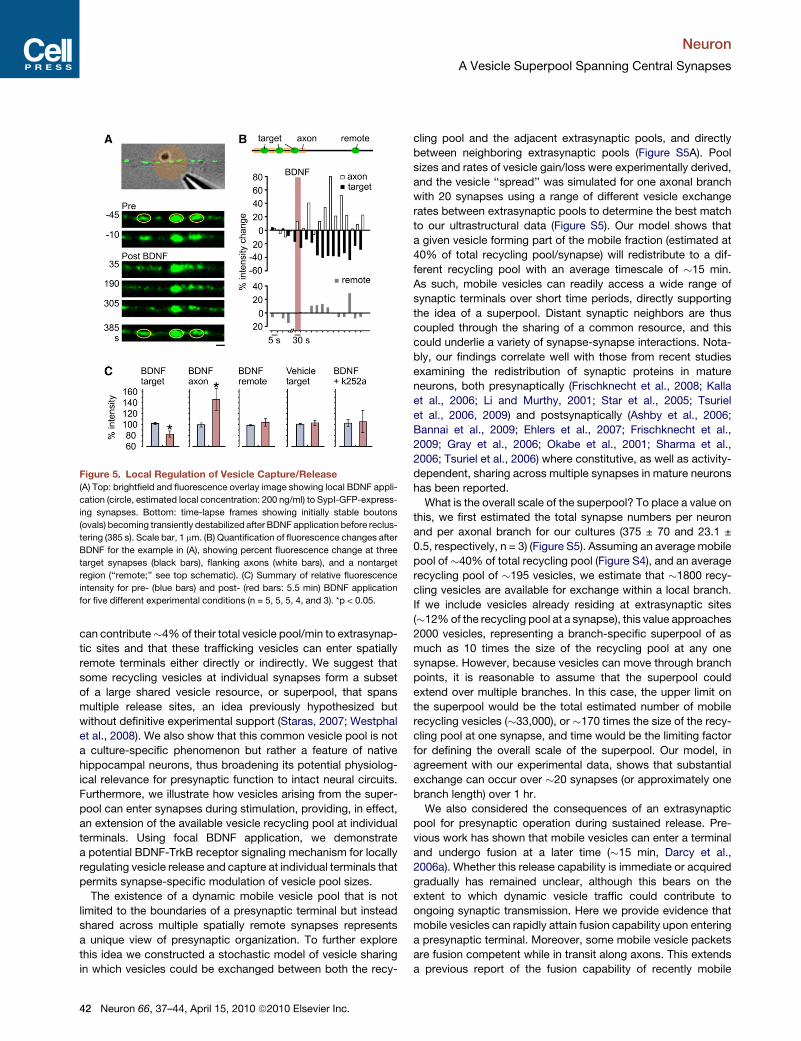
Figure 5. Local Regulation of Vesicle Capture/Release
(A) Top: brightfield and fluorescence overlay image showing local BDNF appli-
cation (circle, estimated local concentration: 200 ng/ml) to SypI-GFP-express-
ing synapses. Bottom: time-lapse frames showing initially stable boutons
(ovals) becoming transiently destabilized after BDNF application before reclus-
tering (385 s). Scale bar, 1 mm. (B) Quantification of fluorescence changes after
BDNF for the example in (A), showing percent fluorescence change at three
target synapses (black bars), flanking axons (white bars), and a nontarget
region (‘‘remote;’’ see top schematic). (C) Summary of relative fluorescence
intensity for pre- (blue bars) and post- (red bars: 5.5 min) BDNF application
for five different experimental conditions (n = 5, 5, 5, 4, and 3). *p < 0.05.
Neuron
A Vesicle Superpool Spanning Central Synapses
can contribute�4% of their total vesicle pool/min to extrasynap-
tic sites and that these trafficking vesicles can enter spatially
remote terminals either directly or indirectly. We suggest that
some recycling vesicles at individual synapses form a subset
of a large shared vesicle resource, or superpool, that spans
multiple release sites, an idea previously hypothesized but
without definitive experimental support (Staras, 2007; Westphal
et al., 2008). We also show that this common vesicle pool is not
a culture-specific phenomenon but rather a feature of native
hippocampal neurons, thus broadening its potential physiolog-
ical relevance for presynaptic function to intact neural circuits.
Furthermore, we illustrate how vesicles arising from the super-
pool can enter synapses during stimulation, providing, in effect,
an extension of the available vesicle recycling pool at individual
terminals. Using focal BDNF application, we demonstrate
a potential BDNF-TrkB receptor signaling mechanism for locally
regulating vesicle release and capture at individual terminals that
permits synapse-specific modulation of vesicle pool sizes.
The existence of a dynamic mobile vesicle pool that is not
limited to the boundaries of a presynaptic terminal but instead
shared across multiple spatially remote synapses represents
a unique view of presynaptic organization. To further explore
this idea we constructed a stochastic model of vesicle sharing
in which vesicles could be exchanged between both the recy-
42 Neuron 66, 37–44, April 15, 2010 ª2010 Elsevier Inc.
cling pool and the adjacent extrasynaptic pools, and directly
between neighboring extrasynaptic pools (Figure S5A). Pool
sizes and rates of vesicle gain/loss were experimentally derived,
and the vesicle ‘‘spread’’ was simulated for one axonal branch
with 20 synapses using a range of different vesicle exchange
rates between extrasynaptic pools to determine the best match
to our ultrastructural data (Figure S5). Our model shows that
a given vesicle forming part of the mobile fraction (estimated at
40% of total recycling pool/synapse) will redistribute to a dif-
ferent recycling pool with an average timescale of �15 min.
As such, mobile vesicles can readily access a wide range of
synaptic terminals over short time periods, directly supporting
the idea of a superpool. Distant synaptic neighbors are thus
coupled through the sharing of a common resource, and this
could underlie a variety of synapse-synapse interactions. Nota-
bly, our findings correlate well with those from recent studies
examining the redistribution of synaptic proteins in mature
neurons, both presynaptically (Frischknecht et al., 2008; Kalla
et al., 2006; Li and Murthy, 2001; Star et al., 2005; Tsuriel
et al., 2006, 2009) and postsynaptically (Ashby et al., 2006;
Bannai et al., 2009; Ehlers et al., 2007; Frischknecht et al.,
2009; Gray et al., 2006; Okabe et al., 2001; Sharma et al.,
2006; Tsuriel et al., 2006) where constitutive, as well as activity-
dependent, sharing across multiple synapses in mature neurons
has been reported.
What is the overall scale of the superpool? To place a value on
this, we first estimated the total synapse numbers per neuron
and per axonal branch for our cultures (375 ± 70 and 23.1 ±
0.5, respectively, n = 3) (Figure S5). Assuming an average mobile
pool of �40% of total recycling pool (Figure S4), and an average
recycling pool of �195 vesicles, we estimate that �1800 recy-
cling vesicles are available for exchange within a local branch.
If we include vesicles already residing at extrasynaptic sites
(�12% of the recycling pool at a synapse), this value approaches
2000 vesicles, representing a branch-specific superpool of as
much as 10 times the size of the recycling pool at any one
synapse. However, because vesicles can move through branch
points, it is reasonable to assume that the superpool could
extend over multiple branches. In this case, the upper limit on
the superpool would be the total estimated number of mobile
recycling vesicles (�33,000), or �170 times the size of the recy-
cling pool at one synapse, and time would be the limiting factor
for defining the overall scale of the superpool. Our model, in
agreement with our experimental data, shows that substantial
exchange can occur over �20 synapses (or approximately one
branch length) over 1 hr.
We also considered the consequences of an extrasynaptic
pool for presynaptic operation during sustained release. Pre-
vious work has shown that mobile vesicles can enter a terminal
and undergo fusion at a later time (�15 min, Darcy et al.,
2006a). Whether this release capability is immediate or acquired
gradually has remained unclear, although this bears on the
extent to which dynamic vesicle traffic could contribute to
ongoing synaptic transmission. Here we provide evidence that
mobile vesicles can rapidly attain fusion capability upon entering
a presynaptic terminal. Moreover, some mobile vesicle packets
are fusion competent while in transit along axons. This extends
a previous report of the fusion capability of recently mobile
Neuron
A Vesicle Superpool Spanning Central Synapses
orphan synapses (Krueger et al., 2003) by demonstrating
that trafficking vesicles can move and destain simultaneously.
What determines whether an individual vesicle packet enters
into a synapse or fuses at axonal regions remains unclear,
although the former seems to predominate (Figures 4F–4H).
Together these observations imply that mobile vesicles could
be of considerable relevance to presynaptic terminals during
sustained transmission, providing an additional functional SV
reserve that extends beyond the conventional boundaries of
the synapse.
One key aspect of vesicle sharing is its potential importance
for regulating presynaptic performance over time. For example,
given that populations of vesicles can have different release
modes (e.g., Fredj and Burrone, 2009; Goda and Stevens,
1994; Sara et al., 2005; Sun et al., 2007), vesicles might be
functionally heterogeneous. The trafficking of vesicles across
multiple synaptic neighbors would provide a means for reallocat-
ing functionally distinct vesicles to specific terminals, and could
represent a potential mechanism for achieving rapid changes in
synaptic properties. Since vesicle redistribution occurs quite
rapidly, vesicle sharing could also be relevant for modulating
synaptic weights through the resizing of SV pools. Given that
release probability (pr) is known to be directly correlated with
recycling pool size (Murthy et al., 1997), changes in the regula-
tory mechanisms that control the size of SV pools (see below)
could therefore profoundly affect synaptic performance. The fact
that synapses draw on a pool of shared vesicles from a wide
synaptic neighborhood, a substantial fraction of which lie outside
the boundaries of a presynaptic terminal, suggests that such
resizing of an SV pool at a single synapse could be readily
achieved without significantly impacting individual adjacent
synaptic neighbors. Such a rapid mechanism for presynaptic
strength adjustments could participate in the fast synapse-
specific homeostatic changes in pr and synaptic pool sizes
observed in hippocampal synapses (Branco et al., 2008), and
in turn, changes in rates of vesicle flux at individual terminals
could contribute to intersynaptic variability of pr (Branco and Sta-
ras, 2009; Branco et al., 2009).
How compatible is the wide-scale sharing of vesicular
resources with the established concept of synapse specificity?
In our experiments only a subset of the total vesicle pool is later-
ally mobile, suggesting that the identity and specificity of indi-
vidual synapses can still be preserved. Also, we would favor
the argument that synapse specificity is conferred mainly by
stable, structural elements of the presynaptic terminal, which
also govern the size of SV pools at individual synapses.
In support of this, bassoon, an active zone scaffold protein, is
very stable and exchanged between boutons over a timescale
of hours (Tsuriel et al., 2009). Furthermore, impairing key struc-
tural/scaffolding protein complexes of the synaptic junction,
such as the cadherin-catenin complex or the MALS proteins,
perturbs presynaptic organization by reducing the size of
vesicle clusters (Bamji et al., 2003, 2006; Olsen et al., 2005).
In this study we demonstrate how vesicle clusters at presyn-
aptic terminals can be directly and individually regulated by
focal application of BDNF, a known modulator of vesicle pool
organization and release at synapses (Bamji et al., 2003,
2006; Shen et al., 2006; Tyler et al., 2006). Such synapse-
specific regulation provides a mechanism to control release/
capture of vesicles at individual boutons and thus could play
a role in maintaining and/or modulating individual presynaptic
terminals. An additional level of regulation could be provided
by postsynaptic targets, acting through either structural com-
ponents or other retrograde messengers to shape presynaptic
properties according to the state of postsynaptic activity
(Branco et al., 2008; Futai et al., 2007; Regehr et al., 2009).
We believe, therefore, that presynaptic differences will be
preserved in spite of vesicle sharing and that the idea of a vesicle
superpool is not in general conflict with the idea of synapse
specificity.
EXPERIMENTAL PROCEDURES
Full methods are available in the Supplemental Information.
SUPPLEMENTAL INFORMATION
Supplemental Information for this article includes Supplemental Experimental
Procedures and five figures and can be found with this article online at doi:
10.1016/j.neuron.2010.03.020.
ACKNOWLEDGMENTS
We gratefully acknowledge Michael Hausser for access to his two-photon
microscope setup, David Elliott for help in making the SypI-Dendra2 construct,
and Joe Atherton for assistance in EM. This research was supported by Well-
come Trust (WT084357MF) and BBSRC (BB/F018371) grants to K.S. and
Medical Research Council and European Commission Framework VI (EUSy-
napse project, LSHM-CT-2005-019055) to Y.G.
Accepted: March 5, 2010
Published: April 14, 2010
REFERENCES
Ashby, M.C., Maier, S.R., Nishimune, A., and Henley, J.M. (2006). Lateral diffu-
sion drives constitutive exchange of AMPA receptors at dendritic spines and is
regulated by spine morphology. J. Neurosci. 26, 7046–7055.
Bamji, S.X., Shimazu, K., Kimes, N., Huelsken, J., Birchmeier, W., Lu, B., and
Reichardt, L.F. (2003). Role of beta-catenin in synaptic vesicle localization and
presynaptic assembly. Neuron 40, 719–731.
Bamji, S.X., Rico, B., Kimes, N., and Reichardt, L.F. (2006). BDNF mobilizes
synaptic vesicles and enhances synapse formation by disrupting cadherin-
beta-catenin interactions. J. Cell Biol. 174, 289–299.
Bannai, H., Levi, S., Schweizer, C., Inoue, T., Launey, T., Racine, V., Sibarita,
J.B., Mikoshiba, K., and Triller, A. (2009). Activity-dependent tuning of inhibi-
tory neurotransmission based on GABAAR diffusion dynamics. Neuron 62,
670–682.
Betz, W.J., and Bewick, G.S. (1992). Optical analysis of synaptic vesicle recy-
cling at the frog neuromuscular junction. Science 255, 200–203.
Branco, T., and Staras, K. (2009). The probability of neurotransmitter release:
variability and feedback control at single synapses. Nat. Rev. Neurosci. 10,
373–383.
Branco, T., Staras, K., Darcy, K.J., and Goda, Y. (2008). Local dendritic activity
sets release probability at hippocampal synapses. Neuron 59, 475–485.
Branco, T., Marra, V., and Staras, K. (2009). Examining size-strength relation-
ships at hippocampal synapses using an ultrastructural measurement of
synaptic release probability. J. Struct. Biol., in press. Published online
November 4, 2009. 10.1016/j.jsb.2009.10.014.
Ceccarelli, B., Hurlbut, W.P., and Mauro, A. (1973). Turnover of transmitter and
synaptic vesicles at the frog neuromuscular junction. J. Cell Biol. 57, 499–524.
Neuron 66, 37–44, April 15, 2010 ª2010 Elsevier Inc. 43
Neuron
A Vesicle Superpool Spanning Central Synapses
Chen, X., Barg, S., and Almers, W. (2008). Release of the styryl dyes from
single synaptic vesicles in hippocampal neurons. J. Neurosci. 28, 1894–1903.
Darcy, K.J., Staras, K., Collinson, L.M., and Goda, Y. (2006a). Constitutive
sharing of recycling synaptic vesicles between presynaptic boutons. Nat.
Neurosci. 9, 315–321.
Darcy, K.J., Staras, K., Collinson, L.M., and Goda, Y. (2006b). An ultrastruc-
tural readout of fluorescence recovery after photobleaching using correlative
light and electron microscopy. Nat. Protoc. 1, 988–994.
Ehlers, M.D., Heine, M., Groc, L., Lee, M.C., and Choquet, D. (2007). Diffu-
sional trapping of GluR1 AMPA receptors by input-specific synaptic activity.
Neuron 54, 447–460.
Fernandez-Alfonso, T., and Ryan, T.A. (2008). A heterogeneous ‘‘resting’’ pool
of synaptic vesicles that is dynamically interchanged across boutons in
mammalian CNS synapses. Brain Cell Biol. 36, 87–100.
Fredj, N.B., and Burrone, J. (2009). A resting pool of vesicles is responsible for
spontaneous vesicle fusion at the synapse. Nat. Neurosci. 12, 751–758.
Frischknecht, R., Fejtova, A., Viesti, M., Stephan, A., and Sonderegger, P.
(2008). Activity-induced synaptic capture and exocytosis of the neuronal
serine protease neurotrypsin. J. Neurosci. 28, 1568–1579.
Frischknecht, R., Heine, M., Perrais, D., Seidenbecher, C.I., Choquet, D., and
Gundelfinger, E.D. (2009). Brain extracellular matrix affects AMPA receptor
lateral mobility and short-term synaptic plasticity. Nat. Neurosci. 12, 897–904.
Futai, K., Kim, M.J., Hashikawa, T., Scheiffele, P., Sheng, M., and Hayashi, Y.
(2007). Retrograde modulation of presynaptic release probability through
signaling mediated by PSD-95-neuroligin. Nat. Neurosci. 10, 186–195.
Goda, Y., and Stevens, C.F. (1994). Two components of transmitter release at
a central synapse. Proc. Natl. Acad. Sci. USA 91, 12942–12946.
Gray, N.W., Weimer, R.M., Bureau, I., and Svoboda, K. (2006). Rapid redistri-
bution of synaptic PSD-95 in the neocortex in vivo. PLoS Biol. 4, e370.
Gurskaya, N.G., Verkhusha, V.V., Shcheglov, A.S., Staroverov, D.B., Chepur-
nykh, T.V., Fradkov, A.F., Lukyanov, S., and Lukyanov, K.A. (2006). Engi-
neering of a monomeric green-to-red photoactivatable fluorescent protein
induced by blue light. Nat. Biotechnol. 24, 461–465.
Harata, N., Ryan, T.A., Smith, S.J., Buchanan, J., and Tsien, R.W. (2001).
Visualizing recycling synaptic vesicles in hippocampal neurons by FM 1-43
photoconversion. Proc. Natl. Acad. Sci. USA 98, 12748–12753.
Heuser, J.E., and Reese, T.S. (1973). Evidence for recycling of synaptic vesicle
membrane during transmitter release at the frog neuromuscular junction.
J. Cell Biol. 57, 315–344.
Hopf, F.W., Waters, J., Mehta, S., and Smith, S.J. (2002). Stability and plas-
ticity of developing synapses in hippocampal neuronal cultures. J. Neurosci.
22, 775–781.
Kalla, S., Stern, M., Basu, J., Varoqueaux, F., Reim, K., Rosenmund, C., Ziv,
N.E., and Brose, N. (2006). Molecular dynamics of a presynaptic active zone
protein studied in Munc13-1-enhanced yellow fluorescent protein knock-in
mutant mice. J. Neurosci. 26, 13054–13066.
Krueger, S.R., Kolar, A., and Fitzsimonds, R.M. (2003). The presynaptic release
apparatus is functional in the absence of dendritic contact and highly mobile
within isolated axons. Neuron 40, 945–957.
Li, Z., and Murthy, V.N. (2001). Visualizing postendocytic traffic of synaptic
vesicles at hippocampal synapses. Neuron 31, 593–605.
Lu, Y., Christian, K., and Lu, B. (2008). BDNF: a key regulator for protein
synthesis-dependent LTP and long-term memory? Neurobiol. Learn. Mem.
89, 312–323.
Murthy, V.N., Sejnowski, T.J., and Stevens, C.F. (1997). Heterogeneous
release properties of visualized individual hippocampal synapses. Neuron
18, 599–612.
Okabe, S., Urushido, T., Konno, D., Okado, H., and Sobue, K. (2001). Rapid
redistribution of the postsynaptic density protein PSD-Zip45 (Homer 1c) and
44 Neuron 66, 37–44, April 15, 2010 ª2010 Elsevier Inc.
its differential regulation by NMDA receptors and calcium channels. J. Neuro-
sci. 21, 9561–9571.
Olsen, O., Moore, K.A., Fukata, M., Kazuta, T., Trinidad, J.C., Kauer, F.W.,
Streuli, M., Misawa, H., Burlingame, A.L., Nicoll, R.A., and Bredt, D.S.
(2005). Neurotransmitter release regulated by a MALS-liprin-alpha presynaptic
complex. J. Cell Biol. 170, 1127–1134.
Poo, M.M. (2001). Neurotrophins as synaptic modulators. Nat. Rev. Neurosci.
2, 24–32.
Regehr, W.G., Carey, M.R., and Best, A.R. (2009). Activity-dependent regula-
tion of synapses by retrograde messengers. Neuron 63, 154–170.
Rizzoli, S.O., and Betz, W.J. (2004). The structural organization of the readily
releasable pool of synaptic vesicles. Science 303, 2037–2039.
Rizzoli, S.O., and Betz, W.J. (2005). Synaptic vesicle pools. Nat. Rev. Neurosci.
6, 57–69.
Ryan, T.A., Reuter, H., Wendland, B., Schweizer, F.E., Tsien, R.W., and
Smith, S.J. (1993). The kinetics of synaptic vesicle recycling measured at
single presynaptic boutons. Neuron 11, 713–724.
Sara, Y., Virmani, T., Deak, F., Liu, X., and Kavalali, E.T. (2005). An isolated
pool of vesicles recycles at rest and drives spontaneous neurotransmission.
Neuron 45, 563–573.
Schikorski, T., and Stevens, C.F. (2001). Morphological correlates of function-
ally defined synaptic vesicle populations. Nat. Neurosci. 4, 391–395.
Sharma, K., Fong, D.K., and Craig, A.M. (2006). Postsynaptic protein mobility
in dendritic spines: long-term regulation by synaptic NMDA receptor activa-
tion. Mol. Cell. Neurosci. 31, 702–712.
Shen, W., Wu, B., Zhang, Z., Dou, Y., Rao, Z.R., Chen, Y.R., and Duan, S.
(2006). Activity-induced rapid synaptic maturation mediated by presynaptic
cdc42 signaling. Neuron 50, 401–414.
Star, E.N., Newton, A.J., and Murthy, V.N. (2005). Real-time imaging of Rab3a
and Rab5a reveals differential roles in presynaptic function. J. Physiol. 569,
103–117.
Staras, K. (2007). Share and share alike: trading of presynaptic elements
between central synapses. Trends Neurosci. 30, 292–298.
Sudhof, T.C. (2004). The synaptic vesicle cycle. Annu. Rev. Neurosci. 27,
509–547.
Sun, J., Pang, Z.P., Qin, D., Fahim, A.T., Adachi, R., and Sudhof, T.C. (2007).
A dual-Ca2+-sensor model for neurotransmitter release in a central synapse.
Nature 450, 676–682.
Takamori, S., Holt, M., Stenius, K., Lemke, E.A., Grønborg, M., Riedel, D.,
Urlaub, H., Schenck, S., Brugger, B., Ringler, P., et al. (2006). Molecular
anatomy of a trafficking organelle. Cell 127, 831–846.
Tsuriel, S., Geva, R., Zamorano, P., Dresbach, T., Boeckers, T., Gundelfinger,
E.D., Garner, C.C., and Ziv, N.E. (2006). Local sharing as a predominant deter-
minant of synaptic matrix molecular dynamics. PLoS Biol. 4, e271.
Tsuriel, S., Fisher, A., Wittenmayer, N., Dresbach, T., Garner, C.C., and Ziv,
N.E. (2009). Exchange and redistribution dynamics of the cytoskeleton of the
active zone molecule bassoon. J. Neurosci. 29, 351–358.
Tyler, W.J., Zhang, X.L., Hartman, K., Winterer, J., Muller, W., Stanton, P.K.,
and Pozzo-Miller, L. (2006). BDNF increases release probability and the size
of a rapidly recycling vesicle pool within rat hippocampal excitatory synapses.
J. Physiol. 574, 787–803.
Westphal, V., Rizzoli, S.O., Lauterbach, M.A., Kamin, D., Jahn, R., and Hell, S.W.
(2008). Video-rate far-field optical nanoscopy dissects synaptic vesicle move-
ment. Science 320, 246–249.
Zakharenko, S.S., Zablow, L., and Siegelbaum, S.A. (2001). Visualization of
changes in presynaptic function during long-term synaptic plasticity. Nat. Neu-
rosci. 4, 711–717.