a2topic 8 grey matter
TRANSCRIPT

Sensitivity in plants
Responding to the environment
All living organisms need control systems. The complex biochemistry of a cell needs to
be controlled to make sure that the right products
organisms need to grow, but only at the appropriate time and in the right places. Getting food
and reproducing are also important biological driving forces. To control these processes
successfully, a system of communication within the organism is needed.
Internal communication in plants
Specific chemicals released by cells in response to a stimulus can act as messages.
Plants-even enormous trees- rely on chemical messages for communication between different
parts, allowing them to respond to factors such as light and gravity. The chemicals move
from cell to cell and also through the plant transport system.
Which stimuli affect plants?
We rarely think of plants as being sensitive because their movements are not u
easily noticed. Beyond needing light and water to survive, they are regarded as being largely
insensitive.
In fact, plants respond to a variety of stimuli, most of which are environmental cues that
have a direct impact on the well
the presence or absence of it. Plants respond to the direction
from which light comes, the intensity of the light and the
length of daily exposure to it. Light affects how much they
grow, the direction of growth and when they reproduce.
Plants are also sensitive
• Gravity.
• Water.
• temperature
• and in some cases to touch and to chemicals.
Different parts of the same plant may react differently to the
same stimulus (for example, shoots grow towards light but
roots grow away from it). As well as these responses to
external stimuli plants also respond to internal chemical
signals. Most of the responses of plants are concerned either
directly or indirectly with maximising the opportunities for
photosynthesis and reproduction.
Plant responses
Plants respond to a variety of stimuli by producing or
destroying chemical messages. Many of the messages are plant
hormones, which play a similar role to animal hormones. They
are produced in one area of the plant, are transported around
the body of the plant and have their effect on cells elsewhere.
Plant growth responses to environmental cues are known as
tropisms.
Grey matter
Sensitivity in plants
Responding to the environment
All living organisms need control systems. The complex biochemistry of a cell needs to
be controlled to make sure that the right products are available at the right time. All living
organisms need to grow, but only at the appropriate time and in the right places. Getting food
and reproducing are also important biological driving forces. To control these processes
ommunication within the organism is needed.
Internal communication in plants
Specific chemicals released by cells in response to a stimulus can act as messages.
rely on chemical messages for communication between different
, allowing them to respond to factors such as light and gravity. The chemicals move
from cell to cell and also through the plant transport system.
Which stimuli affect plants?
We rarely think of plants as being sensitive because their movements are not u
easily noticed. Beyond needing light and water to survive, they are regarded as being largely
In fact, plants respond to a variety of stimuli, most of which are environmental cues that
have a direct impact on the well-being of the plant. They are sensitive to light, and not simply
the presence or absence of it. Plants respond to the direction
from which light comes, the intensity of the light and the
length of daily exposure to it. Light affects how much they
th and when they reproduce.
Plants are also sensitive to:
and in some cases to touch and to chemicals.
Different parts of the same plant may react differently to the
same stimulus (for example, shoots grow towards light but
roots grow away from it). As well as these responses to
external stimuli plants also respond to internal chemical
signals. Most of the responses of plants are concerned either
directly or indirectly with maximising the opportunities for
reproduction.
Plants respond to a variety of stimuli by producing or
destroying chemical messages. Many of the messages are plant
hormones, which play a similar role to animal hormones. They
are produced in one area of the plant, are transported around
lant and have their effect on cells elsewhere.
Plant growth responses to environmental cues are known as
Dr/ Nadia Agami
181
All living organisms need control systems. The complex biochemistry of a cell needs to
are available at the right time. All living
organisms need to grow, but only at the appropriate time and in the right places. Getting food
and reproducing are also important biological driving forces. To control these processes
Specific chemicals released by cells in response to a stimulus can act as messages.
rely on chemical messages for communication between different
, allowing them to respond to factors such as light and gravity. The chemicals move
We rarely think of plants as being sensitive because their movements are not usually
easily noticed. Beyond needing light and water to survive, they are regarded as being largely
In fact, plants respond to a variety of stimuli, most of which are environmental cues that
t. They are sensitive to light, and not simply
Animals respond to nervous and chemical messages in a variety of ways which include
the release of further chemicals, the contraction of mus
which plants respond to their chemical messages is by growth. In some cases growth is
stimulated while in others it is inhibited. For example, sometimes one side of a plant grows
more than the other, resulting in the b
stimulus. To understand how plants respond to stimuli in their environment, you need to
understand exactly how they grow.
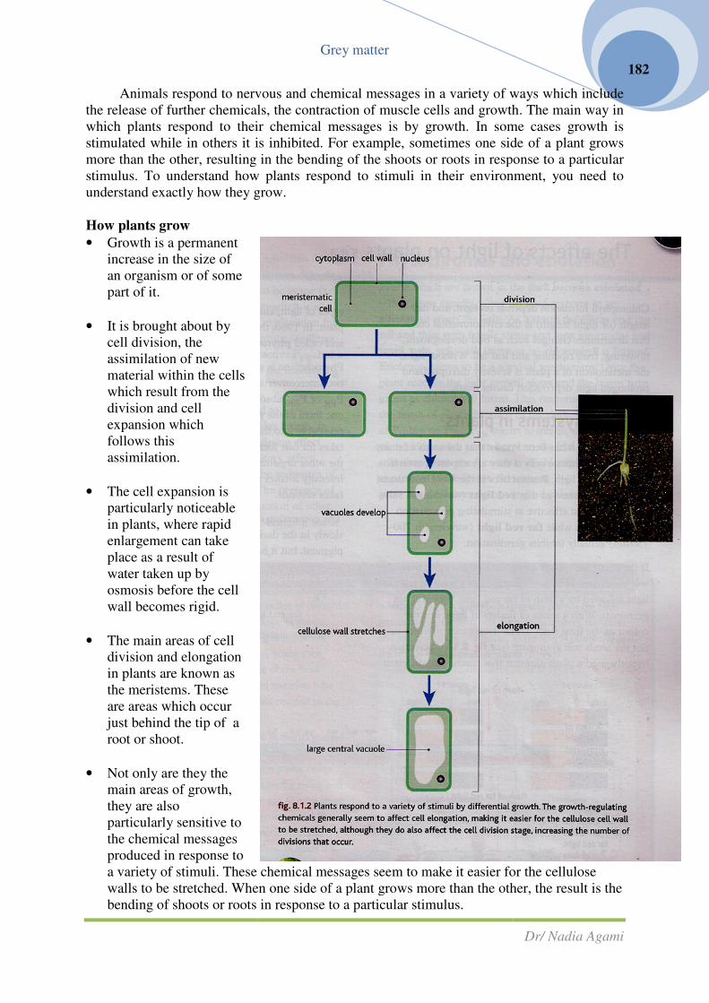
How plants grow
• Growth is a permanent
increase in the size of
an organism or of some
part of it.
• It is brought about by
cell division, the
assimilation of new
material within the cells
which result from the
division and cell
expansion which
follows this
assimilation.
• The cell expansion is
particularly noticeable
in plants, where rapid
enlargement can take
place as a result of
water taken up by
osmosis before the cell
wall becomes rigid.
• The main areas of cell
division and elongation
in plants are known as
the meristems. These
are areas which occur
just behind the tip of a
root or shoot.
• Not only are they the
main areas of growth,
they are also
particularly sensitive to
the chemical messages
produced in response to
a variety of stimuli. These chemical messages seem
walls to be stretched. When one side of a plant grows more than the other, the result is the
bending of shoots or roots in response to a particular stimulus.
Grey matter
Animals respond to nervous and chemical messages in a variety of ways which include
the release of further chemicals, the contraction of muscle cells and growth. The main way in
which plants respond to their chemical messages is by growth. In some cases growth is
stimulated while in others it is inhibited. For example, sometimes one side of a plant grows
more than the other, resulting in the bending of the shoots or roots in response to a particular
stimulus. To understand how plants respond to stimuli in their environment, you need to
understand exactly how they grow.
a variety of stimuli. These chemical messages seem to make it easier for the cellulose
walls to be stretched. When one side of a plant grows more than the other, the result is the
bending of shoots or roots in response to a particular stimulus.
Dr/ Nadia Agami
182
Animals respond to nervous and chemical messages in a variety of ways which include
cle cells and growth. The main way in
which plants respond to their chemical messages is by growth. In some cases growth is
stimulated while in others it is inhibited. For example, sometimes one side of a plant grows
ending of the shoots or roots in response to a particular
stimulus. To understand how plants respond to stimuli in their environment, you need to
to make it easier for the cellulose
walls to be stretched. When one side of a plant grows more than the other, the result is the

Grey matter
Dr/ Nadia Agami
183
Questions:
1- List as many of the different environmental cues that elicit a response in plants as you
can. For each, try to explain why it is important for the plant to respond to that stimulus.
2- Growth in animals usually stops at a certain stage. The meristems of plants, where
growth occurs, remain active throughout the life of the plant. Explain why this difference
is important in the way organisms respond to stimuli.
The effects of light on plants
Chlorophyll formation depends on light, and day length (or night length) is the
environment cue that determines changes such as bud development, flowering, fruit ripening
and leaf fall. Without light, the metabolism of a plant is severely disrupted and prolonged
light deprivation causes death.
Sensory systems in plants
For some times it has been known that the seeds of many plants will germinate only if
they are exposed, even if very briefly, to light. Researchers in the US Department of
Agriculture showed that red light (wavelength 580-660 nm) is most effective at stimulating
germination in lettuce seeds while far red light (wavelength 700-730 nm) actually inhibits
germination.
If the seeds are exposed to a flash of red light, they will germinate. If they are exposed
to a flash of red light followed by a flash of far red light, they will not germinate. With a
series of flashes of light, it is the colour of the final flash which determines whether or not the
seeds will germinate. Scientists hypothesised a plant pigment that reacts with different types
of light, and in turn affects the responses of the plant. In 1960, this pigment was isolated from
plants and called phytochrome.
Flash of red light

Grey matter
Dr/ Nadia Agami
184
Phytochrome is a blue-green pigment which exists in two interconvertible forms: PR or
P660 absorbs red light; PFR or P730 absorbs far red light. When one form of the pigment absorbs
light, it is converter reversibly into the other form. The length of time it takes for one form of
the pigment to be converted into the other depends on the light intensity. In low light
intensity it takes minutes, but in high light intensity it takes seconds.
The conversion of PFR to PR also takes place very slowly in the dark. PR is the more
stable form of the pigment, but it is the PFR which is biologically active.
Or slow conversion in the dark
As normal sunlight contains more red light than far red light, the usual situation in a
plant during daylight hours is for most of the phytochrome to be in the far red form. If the
night period is long enough, it is all converted back into the red form. The explanation for the
control of the germination of the lettuce seedlings is that it is initiated by the biologically
active PFR, but a flash of far red light converts it back to the inactive PR before it has an effect.
Phytochromes enable plants to respond to environmental cues such as changes in day
length. In some cases phytochromes have a stimulating effect on growth in plants, in others
an inhibitory effect. Exactly how phytochromes influence the responses of the plant is still
not fully understood. The presence of phytochromes may stimulate the production of other
growth regulator and plant hormones, bringing about the response to light.
Phytochromes and etiolation
Plants which are grown in the dark become etiolated –they grow rapidly, using up food
reserves in an attempt to reach the light. As a result the plants end up tall and thin, with
fragile stems and long internodes and small, pale, yellowish leaves as no chlorophyll is
formed. Etiolation seems to be a survival mechanism. Once the plant reaches the light growth
slows and the leaves turn green as chlorophyll forms. This response appears to be the result
of phytochromes. In the dark, there is plenty of PR but no PFR.PFR seems to inhibit the
lengthening of the internodes (stem between the leaf nodes), and stimulates both the
formation of chlorophyll and the expansion of the leaves, so without PFR, the internodes grow
but the leaves do not and no chlorophyll forms.
HSW Developing ideas of photoperiodism
In temperate regions such as the UK the period of daylight can vary from around 9 to
15 hours throughout the year. The length of the day and nights give important environmental
cues to living organisms, directing their growth, development and behaviour. One of the most
clearly affected activities is flowering, and scientists have developed models of how plants
sense and respond to day length cues.
Red light (rapid)
Far Red light (rapid)
PR PFR �

In the 1920s W.W. Garner and H.A
Allard in the US studied a particularly tall,
large-leaved form of tobacco plant known as
the “Maryland Mammoth”. Most tobacco
plants flower in the summer but Maryland
Mammoths kept growing and eventually
flowered in December. Garner and Allard
realised the plants were res
environmental cue. The key variable appeared
to be day length. With more than 14 hours of
daylight, no flowers-but below 14 hours a day,
the plants flowered. The critical day length was
thus shown to be 14 hours.
Scientists found that day len
to be the environmental cue affecting flowering
in many plants. Plants flowering when days are
short and nights are long became known as a
short-day plants (SDPs), e.g. strawberries,
chrysanthemums and the tobacco plant.
Plants flowering with relatively long
days and short nights are known as
day plants (LDPs), e.g. snapdragons,
cabbages and henbane. It can be very
difficult to decide whether a plant is a
short- or long-day plant- the two groups
merge. Other plants, such as cucumbers,
tomatoes and pea plants, are unaffected by
the length of the day and are known as
neutral plants (DNPs).
Different flowering pattern allow
plants to take advantage of different
circumstances. In temperate regions short
day plants tend to flower in s
autumn, when the light shading canopy of
leaves either has not developed or has fallen
off. They also flourish near the equator,
where the days are never longer than about
12 hours. Long – day plants flower in the
summer in temperate regions, and
further from the equator where in some seasons there are very long days.
Scientists subsequently discovered that the length of the period of darkness is actually
the environmental cue affection flowering, not day length! It was demonstrated tha
– day plant such as a cocklebur had the long night (period of darkness) interrupted by flash
of light, they do not flower.
Grey matter
In the 1920s W.W. Garner and H.A
studied a particularly tall,
leaved form of tobacco plant known as
the “Maryland Mammoth”. Most tobacco
plants flower in the summer but Maryland
Mammoths kept growing and eventually
flowered in December. Garner and Allard
realised the plants were responding to an
environmental cue. The key variable appeared
to be day length. With more than 14 hours of
but below 14 hours a day,
the plants flowered. The critical day length was
Scientists found that day length appeared
to be the environmental cue affecting flowering
in many plants. Plants flowering when days are
short and nights are long became known as a
(SDPs), e.g. strawberries,
chrysanthemums and the tobacco plant.
th relatively long
days and short nights are known as long-
(LDPs), e.g. snapdragons,
cabbages and henbane. It can be very
difficult to decide whether a plant is a
the two groups
merge. Other plants, such as cucumbers,
omatoes and pea plants, are unaffected by
the length of the day and are known as day-
Different flowering pattern allow
plants to take advantage of different
circumstances. In temperate regions short –
day plants tend to flower in spring and
autumn, when the light shading canopy of
leaves either has not developed or has fallen
off. They also flourish near the equator,
where the days are never longer than about
day plants flower in the
summer in temperate regions, and are found
further from the equator where in some seasons there are very long days.
Scientists subsequently discovered that the length of the period of darkness is actually
the environmental cue affection flowering, not day length! It was demonstrated tha
day plant such as a cocklebur had the long night (period of darkness) interrupted by flash
.
Dr/ Nadia Agami
185
Scientists subsequently discovered that the length of the period of darkness is actually
the environmental cue affection flowering, not day length! It was demonstrated that if a short
day plant such as a cocklebur had the long night (period of darkness) interrupted by flashes

How is the length of the dark signal received?
All the research on photoperiodism points to the involvement of the phytochromes in
the sensitivity of plants to the photoperiod. The changes in flowering patterns which can
brought about by disturbing the dark periods can also be affected by red
Red light inhibits the flowering of short
light, the inhibition is lifted.
Red light leads to the formation of
PFR Far red light or a long period of dark
converts this back to PR. T
hypothesis is that PFR inhibits flowering,
and the lack of PFR allows flowering to
occur. It is thought that it is the lack of
PFR rather than the build up of P
leads to flowering. As the two forms of
phytochrome are almost always present
to some degree in a plant, it is the
balance between them which is affected
by varying periods of light and dark, and
which in turn affect flowering. In long
day plants the situation is reversed, and
it appears that a build – up of P
the daylight hours stimulates flowering.
The detection of the photoperiod seems to take place in the
presence of a plant hormone known as
physiologist Mikhail Charilakhyan in the 1930s. it was
response to the changing levels of phytochromes and carried in the plant transport system to
the flower buds. The evidence included the following findings:
Grey matter
How is the length of the dark signal received?
All the research on photoperiodism points to the involvement of the phytochromes in
the sensitivity of plants to the photoperiod. The changes in flowering patterns which can
brought about by disturbing the dark periods can also be affected by red or
Red light inhibits the flowering of short – day plants, but if the red light is followed by far red
Red light leads to the formation of
Far red light or a long period of dark
. The current
inhibits flowering,
allows flowering to
occur. It is thought that it is the lack of
rather than the build up of PR which
leads to flowering. As the two forms of
phytochrome are almost always present
to some degree in a plant, it is the
balance between them which is affected
by varying periods of light and dark, and
which in turn affect flowering. In long –
day plants the situation is reversed, and
up of PFR during
ht hours stimulates flowering.
The detection of the photoperiod seems to take place in the leaves
presence of a plant hormone known as florigen was hypothesised by the Russian plant
physiologist Mikhail Charilakhyan in the 1930s. it was thought that florigen was made in
response to the changing levels of phytochromes and carried in the plant transport system to
the flower buds. The evidence included the following findings:
Dr/ Nadia Agami
186
All the research on photoperiodism points to the involvement of the phytochromes in
the sensitivity of plants to the photoperiod. The changes in flowering patterns which can
or far red light alone.
day plants, but if the red light is followed by far red
leaves of the plant. The
was hypothesised by the Russian plant
thought that florigen was made in
response to the changing levels of phytochromes and carried in the plant transport system to

• If whole plant is kept in the dark apart f
periods of light and dark, flowering occurs as normal. A plant kept in total darkness does
not flower.
• Using the same experimental set
immediately after the stimulus, th
few hours, it does flower.
• In some species, if a light
new plant will flower.
For years no one could isolate the theoretical
disrepute. However, recently scientists have shown that when a leaf is exposed to a given
amount of light and dark, a particular form of mRNA is produced in the leaf, linked with a
gene associated with flowering (t
a large molecule like this FTmRNA could not be florigen as it would not be able to leave the
cell. Now it has been demonstrated that such molecules as this mRNA can move form cell to
cell to the transport tissues through the plasmodesmata, and that the FTmRNA travels to the
apex of the shoot, where other genes associated with flowering are activated. So FTmRNA
looks as if it may well be florigen!
Questions
1- Draw up a table to summarise the ef
build – up) on different parts of a plant.
2- How does the evidence presented support the idea of the plant hormone florigen? Take
each bullet point in turn and explain its relevance.
3- Produce a flowchart to show how phytochromes and florigen interact to bring about
flowering in a plant
Tropic responses
in plants
The way in which plants respond
to environmental cues is not confined to
the control of their overall growth and
flowering. The growth responses known
as tropisms are made to a number of
different stimuli. The movements of
plants in response to light which comes
from one direction only (phototropism),
and to gravity (geotropism), chemicals
(chemotropism) and touch
(thigmotropism) also involve chemical
messages.
Once a seed begins to germinate
in the soil the shoot and root must keep growing if the developing plant is to survive. But
growth must take place in the right direction. The shoot must grow up towards the light
source for photosynthesis. The roots must grow downwards into the soil which will provide
support, minerals and water for the plant. The movements of these parts of the plant take
Grey matter
ant is kept in the dark apart from one leaf which is exposed to the appropriate
periods of light and dark, flowering occurs as normal. A plant kept in total darkness does
Using the same experimental set – up, if the photoperiodically exposed leaf
e stimulus, the plant does not flower. If the leaf is left in place for a
In some species, if a light – induced leaf from one plant is grafted onto another plant the
For years no one could isolate the theoretical hormone and the florigen theory fell into
disrepute. However, recently scientists have shown that when a leaf is exposed to a given
amount of light and dark, a particular form of mRNA is produced in the leaf, linked with a
gene associated with flowering (the FT gene or flowering Locus. T). It had been believed that
a large molecule like this FTmRNA could not be florigen as it would not be able to leave the
cell. Now it has been demonstrated that such molecules as this mRNA can move form cell to
transport tissues through the plasmodesmata, and that the FTmRNA travels to the
apex of the shoot, where other genes associated with flowering are activated. So FTmRNA
looks as if it may well be florigen!
Draw up a table to summarise the effects red light (PFR build – up) and far red light (P
up) on different parts of a plant.
How does the evidence presented support the idea of the plant hormone florigen? Take
each bullet point in turn and explain its relevance.
lowchart to show how phytochromes and florigen interact to bring about
Tropic responses
The way in which plants respond
to environmental cues is not confined to
the control of their overall growth and
h responses known
as tropisms are made to a number of
different stimuli. The movements of
esponse to light which comes
m one direction only (phototropism),
and to gravity (geotropism), chemicals
(chemotropism) and touch
involve chemical
Once a seed begins to germinate
in the soil the shoot and root must keep growing if the developing plant is to survive. But
growth must take place in the right direction. The shoot must grow up towards the light
osynthesis. The roots must grow downwards into the soil which will provide
support, minerals and water for the plant. The movements of these parts of the plant take
Dr/ Nadia Agami
187
is exposed to the appropriate
periods of light and dark, flowering occurs as normal. A plant kept in total darkness does
up, if the photoperiodically exposed leaf is removed
e plant does not flower. If the leaf is left in place for a
induced leaf from one plant is grafted onto another plant the
hormone and the florigen theory fell into
disrepute. However, recently scientists have shown that when a leaf is exposed to a given
amount of light and dark, a particular form of mRNA is produced in the leaf, linked with a
he FT gene or flowering Locus. T). It had been believed that
a large molecule like this FTmRNA could not be florigen as it would not be able to leave the
cell. Now it has been demonstrated that such molecules as this mRNA can move form cell to
transport tissues through the plasmodesmata, and that the FTmRNA travels to the
apex of the shoot, where other genes associated with flowering are activated. So FTmRNA
up) and far red light (PR
How does the evidence presented support the idea of the plant hormone florigen? Take
lowchart to show how phytochromes and florigen interact to bring about
in the soil the shoot and root must keep growing if the developing plant is to survive. But
growth must take place in the right direction. The shoot must grow up towards the light
osynthesis. The roots must grow downwards into the soil which will provide
support, minerals and water for the plant. The movements of these parts of the plant take

Grey matter
Dr/ Nadia Agami
188
place in direct response to environmental stimuli. The direction of the response is related to
the direction from which the stimulus come.
Phototropism
If plants are grown in bright, all round light, they grow more – or less straight upwards.
In even but low light they will also grow straight upwards, and in fact will grow faster and
taller than in bright light. But if the light to which plants are exposed is brighter on one side
than another or only shines from one side (unilateral light) then the shoots of the plant will
bend towards that light and the roots, if they are at all exposed, will grow away. Shoots are
said to be positively phototropic and roots are negatively phototropic. This response has an
obvious survival value for a plant. It helps to ensure that the shoots receive as much all –
round light as possible, allowing the maximum amount of photosynthesis to take place. Also,
if the roots should emerge from the soil – as they might do after particularly, heavy rain, for
example – they will rapidly turn back to the to the soil. But how are phototropisms brought
about?
To answer this satisfactorily we need to start with a model of how growth is controlled
in a shoot under conditions of all – round light.
HSW What’s the evidence?
Much of the evidence for and work on tropisms has been carried out using germinating
seeds and very young seedlings. This is because they are easy to work with and manipulate.
As they are growing rapidly any changes show up quickly and tend to affect the whole
organism rather than a small part as might be the case with a mature plant. The most widely
used seedlings are those of monocotyledonous plants usually cereals such as oats and wheat.
This is because the shoot when it emerges, is a single, spike with no leaves apparent. This
maker manipulation and observation even easier than in dicotyledonous shoots. The newly
emerged oat shoot is known as a coleoptile, although the more general term shoot will be
used in this book. These early shoots are relatively simple plant systems, so the control of the
response to light in an intact adult plant may well be more complex than our basic model
allows.

HSW Building a model
A succession of research over many years has helped explain phototropism. In the
nineteenth century Charles Darwin and his son France carried out some experiments on oat
coleoloptiles and showed that the phototropic response of plants to light was due to s
of message being passed from the root tip to the growing region.
From the late 1920s onwards the Dutch plant physiologist frits went showed that the
control of growth in plants relied on chemical messages. He had the simple idea of attempting
to block or collect any message that passed from the tip to the growing region behind it, in
order to show the nature of the message.
Questions
1- Describe how phototropism’s differ form photoperiodism.
2- If a block of cocoa butter is used experiment C ins
decapitated shoot. Explain how this informs scientists that the message is water soluble.
3- Evaluate Went’s experimental procedures, listing the dependent and independent
variables involved. How valid and
investigation to be?
Grey matter
A succession of research over many years has helped explain phototropism. In the
nineteenth century Charles Darwin and his son France carried out some experiments on oat
coleoloptiles and showed that the phototropic response of plants to light was due to s
of message being passed from the root tip to the growing region.
From the late 1920s onwards the Dutch plant physiologist frits went showed that the
control of growth in plants relied on chemical messages. He had the simple idea of attempting
block or collect any message that passed from the tip to the growing region behind it, in
order to show the nature of the message.
Describe how phototropism’s differ form photoperiodism.
If a block of cocoa butter is used experiment C instead of agar, there is no response in the
decapitated shoot. Explain how this informs scientists that the message is water soluble.
Evaluate Went’s experimental procedures, listing the dependent and independent
variables involved. How valid and reliable would you assess the results of this type of
Dr/ Nadia Agami
189
A succession of research over many years has helped explain phototropism. In the
nineteenth century Charles Darwin and his son France carried out some experiments on oat
coleoloptiles and showed that the phototropic response of plants to light was due to some sort
From the late 1920s onwards the Dutch plant physiologist frits went showed that the
control of growth in plants relied on chemical messages. He had the simple idea of attempting
block or collect any message that passed from the tip to the growing region behind it, in
tead of agar, there is no response in the
decapitated shoot. Explain how this informs scientists that the message is water soluble.
Evaluate Went’s experimental procedures, listing the dependent and independent
reliable would you assess the results of this type of

Unilateral light and phototropisms
The basic model of the way plants respond to light as they grow was based on shoots
kept entirely in the dark or in full illu
situation where the light form one side is stronger than the other. Experiments done on shoots
illuminated form one side only (unilateral light)
and add more detailed information of their own.
Phototropisms are the result of a
chemical message made in the tip of the
shoot which is transported to the growing
region where it has an effect. The message
is known as a plant hormone or plant
growth substance. The growth substan
involved in phototropisms is celled an
auxin.
Auxins are powerful growth
stimulants and are effective in extremely
low concentrations. The first auxin
discovered was IAA (indoleacetic acid).
The term auxins is now used to cover
substances including IAA which do
identical jobs, referring to the whole group
of chemicals.
Auxins are produced commercially. Gardeners use them to help cuttings root quickly.
They are also proving important in agriculture for improving the yield of crops.
Grey matter
Unilateral light and phototropisms
The basic model of the way plants respond to light as they grow was based on shoots
kept entirely in the dark or in full illumination. However, in real life plants are usually in a
situation where the light form one side is stronger than the other. Experiments done on shoots
illuminated form one side only (unilateral light) confirm the results from earlier experiments
mation of their own.
Phototropisms are the result of a
chemical message made in the tip of the
shoot which is transported to the growing
region where it has an effect. The message
is known as a plant hormone or plant
growth substance. The growth substance
involved in phototropisms is celled an
Auxins are powerful growth
stimulants and are effective in extremely
low concentrations. The first auxin
discovered was IAA (indoleacetic acid).
The term auxins is now used to cover
A which do
identical jobs, referring to the whole group
Auxins are produced commercially. Gardeners use them to help cuttings root quickly.
They are also proving important in agriculture for improving the yield of crops.
Dr/ Nadia Agami
190
Unilateral light and phototropisms
The basic model of the way plants respond to light as they grow was based on shoots
plants are usually in a
situation where the light form one side is stronger than the other. Experiments done on shoots
m earlier experiments
Auxins are produced commercially. Gardeners use them to help cuttings root quickly.
They are also proving important in agriculture for improving the yield of crops.

How do auxins work in a plant?
Auxins seem to affect the ability of the plant cell walls to stretch for example,
Grey matter
auxins work in a plant?
Auxins seem to affect the ability of the plant cell walls to stretch for example,
Dr/ Nadia Agami
191
Auxins seem to affect the ability of the plant cell walls to stretch for example,

Grey matter
Dr/ Nadia Agami
192
• IAA is made in the tip of the shoot and diffuses back towards the zone of
elongation.
• The molecules of IAA bind to specific receptor sites on the cell surface membranes,
• Activating the active pumping of hydrogen ions into the cytoplasm.
• This changes the hydrogen ion concentration, providing the optimum pH for the
enzymes which break bonds between adjacent cellulose microfibrils and keep the
wall flexible.
• The cells absorb water by osmosis and the very flexible cell walls stretch and allow
the cells to expand. Eventually, as the cells mature, the IAA is destroyed by
enzymes, the pH of the cell walls rises and bonds form between the cellulose
microfibrils. As a result the cell was becomes more rigid – and the cell can no
longer expand.
Research shows that the side of a shoot exposed to light contains less auxin than the
side which is not illuminated, Light seems to cause the auxin to move laterally across the
shoot, producing a greater concentration on the unilluminated side.
This movement means the shoot tip acts as a photoreceptor. More hormone diffuses
down to the region of cell elongation on the dark side. This stimulates cell elongation and so
growth on the dark side, resulting in the shoot bending towards the light.
Once the shoot is growing directly towards the light, the unilateral stimulus is removed.
The asymmetric transport of auxin ends and the shoot grows straight toward the light.
The original theory was that light destroyed the auxin. However, experiments such as
those in fig 8.1.13 show that the levels of auxin in shoots are much the same regardless of
whether have been kept in the dark or under unilateral illumination.
Although the basic principles of tropisms have been understood for over a hundred
years, this is still an area of active research. At the moment, there is no clear relationship
between the phytochromes, which are such important photoreceptors in photoperiodism, and
tropism, where the movement of hormones such as IAA are involved in the responses to
light. It was assumed they were quite separate processes. However, research is increasingly
showing a link between the phytochromes and both phototropisms and geotropisms.
For example it appears that phototropism in a very young shoot cannot take place until
the phytochromes have been activated, and that geotropisms are similarly dependent on
phytochromes.
Questions
1- Explain in detail how IAA brings about a phototropic response in an oat coleoptile
exposed to unilateral light.
2- a Explain the importance of the Went bioassay in interpreting the results of the
experiment shown?
3- How does this experiment confirm the current model of phototropisms?

Grey matter
Dr/ Nadia Agami
193
How the nervous system works
The nervous system
You looked at the need for living organisms to have a system of control, and at the way
coordination is achieved in plants using chemical communication systems. However, many
animals, including humans, need a more rapid and more specifically targeted system of
communication, and this is where a nervous system of communication and this is where a
nervous system comes into its own.
A nervous system is made up of interconnected neurons (nerve cells) specialized for the
rapid transmission of impulses throughout the organism. They carry impulses from special
receptor cells, giving information about both the internal and the external environment.
Neurons also carry impulses to specialised effector cells, often muscles, which then bring
about the appropriate response.
The simplest nervous systems are made up of receptor cells, neurons and the nerve
endings associated with the effectors. However, many nervous systems are much more
complex. As well as single receptor cells, groups of receptors have evolved to work together
in sensory organs such as the eye and the ear. Simple nerve nets are replaced by complex
nerve pathways. Some neurons only carry information from the internal or external
environment into the central processing areas of the nervous system. They are known as
sensory neurons.
As animals increase in size and complexity they develop more specialised
concentrations of nerve cells which form a central nervous system (CNS). This is an area
where incoming information is processed, and from where impulses are sent out through
motor neurons which carry impulses to the effector organs. In vertebrates the central nervous
system consists of the brain and spinal cord.
Neurones are individual cells and each one has a long nerve fibre which carries the
nerve impulse. Nerves are bundles of nerve fibres (axons or dendrons) Some nerves carry
only motor fibres and are known as motor nerves, some carry only sensory fibres are known
as sensory nerves, while others carry a mixture of motor and sensory fibres and are called
mixed nerves. It is important to be very clear whether you are talking about neurons or
nerves.

Grey matter
Dr/ Nadia Agami
194
The structure and function of neurons
Neurones are the basic unit of a nervous system millions of neurones that work together
as an integrated while in mammals such as ourselves.
Neurones are cells specialised for the transmission of electrical signals (impulses).
They have a cell body which
cell nucleus, mitochondria and other organelles, along with Nissl’s granules which are
prominent groups of rough endoplasmic reticulum (ER) and ribosomes needed for the
synthesis of the neurotransmitter molecules. The cell
called dendrites which connect to neighbouring nerve cells.
The most distinctive feature of all nerve cells is the nerve fibre, which is extremely long
and thin and carries the nerve
body are called axons. Fibres which transmit impulses towards the cell body (in sensory
neurones) are known as dendrons.
Short relay or connector neurones are found in the CNS and they connect
sensory neurones. They are also the same cell body.
Myelinated nerve fibres
Most vertebrate neurones are associated with another very specialised type of cell, the
Schwann cell. The Schwann cell membrane wraps itself repeatedly around the nerve
forming a fatty layer known as the myelin sheath. There are gaps between the Schwann cells
known as the nodes of Ranvier. The myelin sheath is important for two reasons it protects the
nerves from damage and speeds up the transmission of the nerve i
Speedy nerve impulses
The role of the nerve cells is to carry electrical impulses from one area of the body to
another quickly. The speed at which the
impulses can be carried depends largely on t
things.
1-The first is the diameter of the ne
fibre generally the thicker the fibre, the more
rapidly impulses travel along it.
2- The second is presence or absence of a
myelin sheath. Myelinated nerve fibres can
carry impulses much faster than unmyelinated
ones.
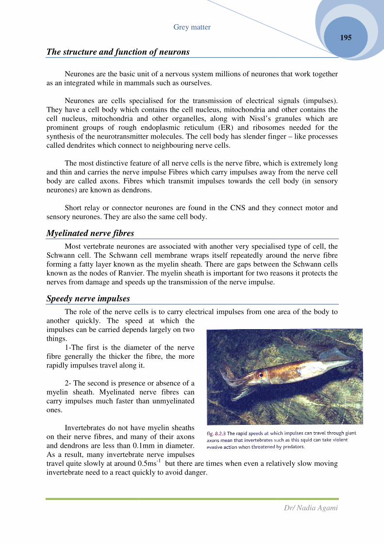
Invertebrates do not have myel
on their nerve fibres, and many of their axons
and dendrons are less than 0.1mm in diameter.
As a result, many invertebrate nerve impulses
travel quite slowly at around 0.5ms
invertebrate need to a react quickly to avoid danger.
Grey matter
The structure and function of neurons
Neurones are the basic unit of a nervous system millions of neurones that work together
as an integrated while in mammals such as ourselves.
Neurones are cells specialised for the transmission of electrical signals (impulses).
contains the cell nucleus, mitochondria and other contains the
cell nucleus, mitochondria and other organelles, along with Nissl’s granules which are
prominent groups of rough endoplasmic reticulum (ER) and ribosomes needed for the
ransmitter molecules. The cell body has slender finger
called dendrites which connect to neighbouring nerve cells.
The most distinctive feature of all nerve cells is the nerve fibre, which is extremely long
and thin and carries the nerve impulse Fibres which carry impulses away f
body are called axons. Fibres which transmit impulses towards the cell body (in sensory
neurones) are known as dendrons.
Short relay or connector neurones are found in the CNS and they connect
sensory neurones. They are also the same cell body.
Most vertebrate neurones are associated with another very specialised type of cell, the
Schwann cell. The Schwann cell membrane wraps itself repeatedly around the nerve
y layer known as the myelin sheath. There are gaps between the Schwann cells
known as the nodes of Ranvier. The myelin sheath is important for two reasons it protects the
nerves from damage and speeds up the transmission of the nerve impulse.
The role of the nerve cells is to carry electrical impulses from one area of the body to
another quickly. The speed at which the
impulses can be carried depends largely on two
is the diameter of the nerve
fibre generally the thicker the fibre, the more
rapidly impulses travel along it.
The second is presence or absence of a
myelin sheath. Myelinated nerve fibres can
carry impulses much faster than unmyelinated
Invertebrates do not have myelin sheaths
on their nerve fibres, and many of their axons
and dendrons are less than 0.1mm in diameter.
As a result, many invertebrate nerve impulses
travel quite slowly at around 0.5ms-1
but there are times when even a relatively slow moving
need to a react quickly to avoid danger.
Dr/ Nadia Agami
195
Neurones are the basic unit of a nervous system millions of neurones that work together
Neurones are cells specialised for the transmission of electrical signals (impulses).
contains the cell nucleus, mitochondria and other contains the
cell nucleus, mitochondria and other organelles, along with Nissl’s granules which are
prominent groups of rough endoplasmic reticulum (ER) and ribosomes needed for the
body has slender finger – like processes
The most distinctive feature of all nerve cells is the nerve fibre, which is extremely long
impulse Fibres which carry impulses away from the nerve cell
body are called axons. Fibres which transmit impulses towards the cell body (in sensory
Short relay or connector neurones are found in the CNS and they connect motor and
Most vertebrate neurones are associated with another very specialised type of cell, the
Schwann cell. The Schwann cell membrane wraps itself repeatedly around the nerve fibre
y layer known as the myelin sheath. There are gaps between the Schwann cells
known as the nodes of Ranvier. The myelin sheath is important for two reasons it protects the
mpulse.
The role of the nerve cells is to carry electrical impulses from one area of the body to
but there are times when even a relatively slow moving

Grey matter
Dr/ Nadia Agami
196
Many invertebrate groups have evolved a number of giant axons, which are nerve fibres
with diameters of around 1mm. These allow impulses to travel at approximately 100ms-1
, fast
enough for most escape strategies to have a chance of success.
Vertebrates have both unmyelinated and myelinated nerves. The voluntary motor
neurones that transmit impulses to voluntary muscles for example to control movement are
all myelinated.
However, the autonomic neurones that control involuntary muscles, like those of the
digestive system, have some unmyelinated fibre. The effect of the myelin sheath is to speed
up the transmission of a nerve impulse without the need for giant axons which take up quite a
lot of space. A more versatile network of relatively small nerve fibres can carry impulses
extremely rapidly, at speeds of up to 120ms-1
HSW the cause of nerve impulses
It has taken many years for our current model of nerve impulses to emerge. For
example, over two centuries ago two famous scientists had a dispute over the electrical
activity of the body. In 1791 Luigi Galvbani discovered that the muscles in severed forgs’
legs twitched when touched by brass and iron simultaneously.
He thought this was the result of what he called animal electricity produced by the
nerves and muscles of his subjects. A few years later Alessandro Volta insisted that the effect
was due to the difference in electrical potential between the two metals and nothing to do
with animal electricity in the muscles at all. This dispute at the end of the eighteenth century
was the starting point for a huge range of experiments in physiology, physics and physical
chemistry which has led to our present day understanding of the nature of the nerve impulse.
The current model of the nerve impulse is of a minute electrical event which is the
result of charge differences between the outside and inside of the nerve fibre membrane. It is
based on ion movements through specialised protein pores and by an active pumping
mechanism. To look at the events of a verve impulse it is easiest to consider a typical nerve
fibre ignoring for the moment size myelination or type.
Investigating nerve impulses
Since nerve impulses are electrical events, albeit very small one3s, one of the most
effective ways of investigating them has been to record and measure the electrical changes.
This is done using apparatus sensitive to small electrical changes. Early work was done using
a pair of recording electrodes place on a verve which was then given a controlled stimulus.
The impulses which resulted were recorded by the electrodes and displayed on a screen.

External electrodes record the responses of entire nerves, made up of large number of
different nerve fibres. The fibre
the results of the recordings can be difficult to interpret correctly and the technique has been
of limited value.
To make real progress, recordings had to be made from within individual ner
Much of the work on nerve fibres has been done using axons, because they are relatively easy
to use.
Sensory nerve fibres often run from a sense organ in the head directly to the brain or
from individual sensory receptors in the skin to the sp
at.
Motor axons, on the other hand, run directly to muscles, often in large motor nerves.
This makes them relatively easy to access, and the effect of stimulating them can be seen
immediately with the twitch of a mu
axons but the same events are seen in all nerve fibres.
Most axons are extremely small around 20
inside is not an easy procedure. About 40 years ago Alan Hod
work on the giant unmyelinated axons of the squid which are around 1mm in diameter. These
axons allow for very rapid nerve transmission to particular muscles in situations when the
squid needs to move in a hurry.
Hodgkin and Huxley used very fine glass microelectrodes inserted into the giant axon.
Another electrode recorded the electrical potential from the outside. This allowed the changes
which occur during the passage of an individual nerve impulse to be recorded accurately
Grey matter
External electrodes record the responses of entire nerves, made up of large number of
different nerve fibres. The fibres are of varying diameter and sensitivity, and
the results of the recordings can be difficult to interpret correctly and the technique has been
To make real progress, recordings had to be made from within individual ner
Much of the work on nerve fibres has been done using axons, because they are relatively easy
Sensory nerve fibres often run from a sense organ in the head directly to the brain or
from individual sensory receptors in the skin to the spinal cord, making them difficult to get
Motor axons, on the other hand, run directly to muscles, often in large motor nerves.
This makes them relatively easy to access, and the effect of stimulating them can be seen
immediately with the twitch of a muscle. So, much of your work on nerve impulses refers to
axons but the same events are seen in all nerve fibres.
Most axons are extremely small around 20µm in diameter so making a recording from
inside is not an easy procedure. About 40 years ago Alan Hodgkin and Andrew Huxley began
work on the giant unmyelinated axons of the squid which are around 1mm in diameter. These
axons allow for very rapid nerve transmission to particular muscles in situations when the
squid needs to move in a hurry.
Huxley used very fine glass microelectrodes inserted into the giant axon.
Another electrode recorded the electrical potential from the outside. This allowed the changes
which occur during the passage of an individual nerve impulse to be recorded accurately
Dr/ Nadia Agami
197
External electrodes record the responses of entire nerves, made up of large number of
s are of varying diameter and sensitivity, and so the results of
the results of the recordings can be difficult to interpret correctly and the technique has been
To make real progress, recordings had to be made from within individual nerve fibres.
Much of the work on nerve fibres has been done using axons, because they are relatively easy
Sensory nerve fibres often run from a sense organ in the head directly to the brain or
inal cord, making them difficult to get
Motor axons, on the other hand, run directly to muscles, often in large motor nerves.
This makes them relatively easy to access, and the effect of stimulating them can be seen
scle. So, much of your work on nerve impulses refers to
m in diameter so making a recording from
gkin and Andrew Huxley began
work on the giant unmyelinated axons of the squid which are around 1mm in diameter. These
axons allow for very rapid nerve transmission to particular muscles in situations when the
Huxley used very fine glass microelectrodes inserted into the giant axon.
Another electrode recorded the electrical potential from the outside. This allowed the changes
which occur during the passage of an individual nerve impulse to be recorded accurately for

the first time. This technique has been refined so that now internal electrodes can be used
with almost any nerve fibre.
Questions
1- Outline the differences between a nerve and a nerve fibre.
2- Summarise how the structure of a motor neurone is rel
3- The speed of transmission of a nerve impulse in part related to the diameter of the nerve
fibre. Explain why vertebrate nerve fibres always have relatively small diameters, yet the
speed of the transmission of the nerve impulses is m
4- Squid giant axons are widely used in neurophysiology. Explain why.
Nerve impulses
The concentration of sodium ions (Na
particles is different outside the axon from that inside: this is the basis of the nerve impulse.
• The membrane of an axon, like any other cell surface membrane, is partially permeable. It
is the difference in permeability of this membr
potassium ions which gives it its special conducting properties. An axon is said to be at
rest when it is not conducting a nerve impulse.
• At rest, the axon membrane is relatively impermeable to sodium ions, but quite
permeable to potassium ions (see AS Biology). It also contains a very active
sodium/potassium ions pump (often referred to as the sodium pump) which uses ATP to
move sodium ions out of the axon and potassium ions in (See A
• The effect of this is to lower the concentration of sodium ions inside the axon they are
pumped out and cannot diffuse back in again. At the same time, potassium ions are
moved in, but they then diffuse out again along a concentration gradient through open
potassium ion channels. Eventually the movement of positively charged potassium ions
out of the cell along the concentration gradient is opposed by the electrochemical
gradient. As a result, the inside of the cell is left slightly negative relative to the
is polarised.
Grey matter
time. This technique has been refined so that now internal electrodes can be used
Outline the differences between a nerve and a nerve fibre.
Summarise how the structure of a motor neurone is related to function.
The speed of transmission of a nerve impulse in part related to the diameter of the nerve
fibre. Explain why vertebrate nerve fibres always have relatively small diameters, yet the
speed of the transmission of the nerve impulses is much faster than that in invertebrates.
Squid giant axons are widely used in neurophysiology. Explain why.
impulses
The concentration of sodium ions (Na+), potassium ions (K
+) and other charged
particles is different outside the axon from that inside: this is the basis of the nerve impulse.
The membrane of an axon, like any other cell surface membrane, is partially permeable. It
is the difference in permeability of this membrane to positively charged sodium and
potassium ions which gives it its special conducting properties. An axon is said to be at
rest when it is not conducting a nerve impulse.
At rest, the axon membrane is relatively impermeable to sodium ions, but quite
permeable to potassium ions (see AS Biology). It also contains a very active
sodium/potassium ions pump (often referred to as the sodium pump) which uses ATP to
move sodium ions out of the axon and potassium ions in (See As Biology 9).
The effect of this is to lower the concentration of sodium ions inside the axon they are
pumped out and cannot diffuse back in again. At the same time, potassium ions are
moved in, but they then diffuse out again along a concentration gradient through open
potassium ion channels. Eventually the movement of positively charged potassium ions
out of the cell along the concentration gradient is opposed by the electrochemical
gradient. As a result, the inside of the cell is left slightly negative relative to the
Dr/ Nadia Agami
198
time. This technique has been refined so that now internal electrodes can be used
ated to function.
The speed of transmission of a nerve impulse in part related to the diameter of the nerve
fibre. Explain why vertebrate nerve fibres always have relatively small diameters, yet the
uch faster than that in invertebrates.
) and other charged
particles is different outside the axon from that inside: this is the basis of the nerve impulse.
The membrane of an axon, like any other cell surface membrane, is partially permeable. It
ane to positively charged sodium and
potassium ions which gives it its special conducting properties. An axon is said to be at
At rest, the axon membrane is relatively impermeable to sodium ions, but quite freely
permeable to potassium ions (see AS Biology). It also contains a very active
sodium/potassium ions pump (often referred to as the sodium pump) which uses ATP to
Biology 9).
The effect of this is to lower the concentration of sodium ions inside the axon they are
pumped out and cannot diffuse back in again. At the same time, potassium ions are
moved in, but they then diffuse out again along a concentration gradient through open
potassium ion channels. Eventually the movement of positively charged potassium ions
out of the cell along the concentration gradient is opposed by the electrochemical
gradient. As a result, the inside of the cell is left slightly negative relative to the outside it

Grey matter
Dr/ Nadia Agami
199
When you look at the events in neurones, the figures are always relative, comparing the
inside of the nerve fibre to the outside solution. There is a potential difference across the
membrane of around 70mV, which is known as the resting potential.
The action potential
When an impulse travels along an axon, the key event is a change in the permeability of
the cell membrane to sodium ions. This change occurs in response to a stimulus for example
light, sound, touch, taste or smell, in a sensory neurone, or the arrival of a neurotransmitter
chemical in a motor neurone. In the experimental situation, the stimulus is usually a minute
and precisely controlled electrical impulse.
1- When a neurone is stimulated, the axon membrane shows a sudden and dramatic
increase in its permeability to sodium ions.
2- Specific sodium ion channels or sodium gates open up, allowing sodium ions to
diffuse rapidly down their concentration and electrochemical gradients.
3- As a result the potential difference across the membrane is briefly reversed, with the
cell becoming positive on the inside with respect to the outside. This
depolarisation lasts about 1millisecond.
4- The potential difference across the membrane at this point is about 40mV. This is
known as the action potential. Remember that these events happen in any nerve
fibre, not just axons.
5- At the end of this brief depolarisation, the sodium ion channels close again and the
excess sodium ions are rapidly pumped out by the sodium pump. This active
transport system uses ATP.
6- Also, the permeability of the membrane to potassium ions is temporarily increased
as voltage dependent potassium ion channels open as a result of the depolarisation.
As a result potassium ion diffuse out of the axon down their concentration gradient
and an electrochemical gradient, attracted by the negative charge on the outside of
the membrane.
7- As the positive sodium and potassium ions leave the cell, the inside becomes
negative relative to the outside once again. It takes a few milliseconds before the
resting potential is restored and the axon is ready to carry another impulse.
The events of the action potential can be recorded clearly using the internal/external
electrode combination you have already seen. The oscilloscope trace is often referred to as
the “spike” because of its shape. It clearly shows the change in the potential difference across
the membrane with the inrush of sodium ions followed by a return to the resting potential as
the permeability of the membrane changes again. The threshold for any nerve fibre is the
point at which sufficient sodium ion channels open for the rush of sodium ions into the axon
to be greater than the outflow of potassium ions. Once the threshold has been reached, the
action potential occurs. The size of this action potential is always the same it is an all-or-
nothing response.

Grey matter
Dr/ Nadia Agami
200
8- The recovery time of an axon (known as the refractory period) is the time it takes
for an area of the axon membrane to recover after an action potential, that is the
time it takes for ionic movements to repolarise the membrane and restore the
resting potential. This depends both on the sodium/potassium pump and on the
membrane permeability to potassium ions.
• For the first millisecond or so after the action potential it is impossible to
restimulate the fibre the sodium ion channels are completely blocked and the
resting potential has not been restored. This is known as the absolute
refractory period.

• After this there is a period of several
milliseconds when th
restimulated, but it will only respond to a
much stronger stimulus than before the
threshold has effectively been raised. This
is known as the
period. During this time they voltage
dependent potassium ion channels are stil
open. It is not until they are closed that the
normal resting potential can be fully
restored.
The refractory period is important in the functioning
of the nervous system as a whole.
� It limits the rate at which
impulses may flow along a fibre
to 500
� It also ensures that impulses flow
in only one direction along
nerves. Until the resting potential
is restored, the part of the nerve
fibre that the impulse has just left
cannot conduct another impulse.
This means the impulse can only
continue traveling in the same
direction it cannot spread
backwards as well.
HSW building up the evidence
Some of the most convincing evidence for this model
of the nerve impulse comes from work done using poisons.
A metabolic poison such as dinitrophenol
production of ATP. It also prevents the axon from
functioning properly. But how does this confirm our model
of a nerve impulse based around the active transport of
sodium ions out of the axon and diffusion of potassium
ions due to differential permeability?
• When an axon is treated with a metabolic poison the
sodium pump stops working as ATP is used up, and the
resting potential is lost at the same rate as the
concentration of ATP decreases. This suggests that the
ATP is being used to power
runs out, the pump no longer works.
• If the poison is washed away, the metabolism returns to
normal and ATP production begins again. The resting
potential is restored, suggesting that the sodium pump
has started up again with the r
Grey matter
After this there is a period of several
milliseconds when the axon may be
restimulated, but it will only respond to a
much stronger stimulus than before the
threshold has effectively been raised. This
is known as the relative refractory
During this time they voltage
dependent potassium ion channels are still
open. It is not until they are closed that the
normal resting potential can be fully
The refractory period is important in the functioning
of the nervous system as a whole.
It limits the rate at which
impulses may flow along a fibre
to 500-1000 each second.
It also ensures that impulses flow
in only one direction along
nerves. Until the resting potential
is restored, the part of the nerve
fibre that the impulse has just left
cannot conduct another impulse.
This means the impulse can only
ontinue traveling in the same
direction it cannot spread
backwards as well.
HSW building up the evidence
Some of the most convincing evidence for this model
of the nerve impulse comes from work done using poisons.
A metabolic poison such as dinitrophenol prevents the
It also prevents the axon from
functioning properly. But how does this confirm our model
of a nerve impulse based around the active transport of
sodium ions out of the axon and diffusion of potassium
l permeability?
When an axon is treated with a metabolic poison the
sodium pump stops working as ATP is used up, and the
resting potential is lost at the same rate as the
concentration of ATP decreases. This suggests that the
ATP is being used to power the sodium pump when it
runs out, the pump no longer works.
If the poison is washed away, the metabolism returns to
normal and ATP production begins again. The resting
potential is restored, suggesting that the sodium pump
has started up again with the return of ATP.
Dr/ Nadia Agami
201

Grey matter
Dr/ Nadia Agami
202
• If a poisoned axon is supplied with ATP by experimenters, the resting potential will be at
least partly restored. This again
confirms our model, suggesting that
the poison is acting by depriving the
sodium pump of energy rather than
by interfering with the membrane
structure and its permeability. If the
latter were the case, even supplying
ATP directly to the pump would have
no effect because ions would move
freely across the membrane and a
potential difference could not be
maintained.
HSW the importance of internal
electrodes
Our understanding of the nervous
system has grows through use of both
external and internal electrodes. The
way the different techniques have contributed to our understanding can best be explained
using a specific example.
whole nerve gets no response until a threshold is reached. However, once over this
threshold, as the strength of the stimulus increases, so does the response of the nerve until a
maximum is reached. Without further knowledge, it looks as if the strength of the nerve
impulse increases in response to the strength of the stimulus.
A stimulus applied to a single axon, using an internal electrode, also elicits no response
until it reaches a certain threshold level. But beyond that threshold, the size of the response is
always the same. However much the stimulus increases in size, the impulse in the axon is
identical. This is the “all-or-nothing law” in action a nerve fibre either carries an impulse or it
does not.

Grey matter
Dr/ Nadia Agami
203
So as stimulus strength
is increased, although an
individual neurone either
responds or it doesn’t, in a
whole nerve the threshold of
more and more nerve fibres is
reached until all of the fibres
in the nerve are responding.
Questions
1- Describe the resting
potential of a neurone and
explain how it is
maintained.
2- a- Describe an action
potential and explain the
importance of the refractory
period.
b- Explain how an action
potential can be most
accurately measured.
3- Explain how the graph in
Fig. ( 8.2.8) provides
evidence for the role of the
sodium pump in
maintaining the resting
potential.
4- Identify why traces A and B
in Fig. (8.2.9 ) are so
different, and explain what
this tells us about the
difference between nerves
and neurones.

Grey matter
Dr/ Nadia Agami
204
The neurons in action
Once an action potential is set up as a response to a stimulus, it will travel the entire
length of that nerve fibre, which may be many centimetres or even meters long. The
movement to the nerve impulse along the fibre is the result of local currents set up by the ion
movements at the action potential itself. They occur both in front of and behind the action
potential.
They depolarize the membrane in
front of the action potential sufficiently to
cause the sodium ion channels to open.
The sodium ion channels behind the
action potential cannot open due to the
refractory period of the membrane behind
the spike. In this way the impulse is
continually conducted in the required
direction.
In myelinated neurons, the situation
is more complex. Ions can only pass in
and out of the axon freely at the nodes of
Ranvier, which are about 1 mm apart.
This means that action potentials can only
occur at the nodes and so they appear to
jump from one to the next.
The effect of this is to speed up
transmission as the ionic movements
associated with the action potential
occur much less frequently, taking less
time. It is known as salutatory conduction
from the Latin verb saltare, which means
“to jump”.
Synapse Neurones must be able to
intercommunicate. Receptors must pass
their information into the sensory nerves,
which in turn must relay the information
to the central nervous system.
Information needs to pass freely around
the central nervous system and the
impulses sent along the motor nerves
must be communicated to the effector
oranges so that action can be taken.
Wherever two neurons meet they
are linked by a synapse.
Every cell in the central nervous

Grey matter
Dr/ Nadia Agami
205
system is covered with synaptic knobs from other cells – several hundred in some cases.
Neurones never actually touch their target cell, so a synapse involves a gap between
two cells, a gap which the nerve impulses must somehow cross.
1- The functioning of synapses,
rather like the contraction of
muscle fibres, depends on the
movement of calcium ions. The
arrival of an impulse at the
synaptic knob increases the
permeability of the presynaptic
membrane to calcium ions as
calcium ion channels open up.
2- Calcium ions then move into
the synaptic knob down their
concentration gradient. The
effect of the influx of calcium
ions is to cause the synaptic
vesicles which contain a
transmitter substance or
neurotransmitter to move to the
presynaptic membrane.
3- Each vesicle contains about
3000 molecules of transmitter.
Some of the vesicles fuse with
the presynaptic membrane and
release the transmitter
substance into the synaptic cleft.
4- These molecules diffuse across the gap and become attached to specific protein
receptor sites on the post – synaptic membrane.
5- This opens sodium ion channels in the membrane and there is an influx of
sodium ions into the fiber, causing a change in the potential difference across
the membrane and an excitatory post – synaptic potential (EPSP) to be set up.
6- If there are sufficient of these EPSPs, the positive charge in the post- synaptic
cell exceeds the threshold level, and an action potential is set up which then
travels on along the post-synaptic neurone.
7- In some cases the neurotransmitter has the opposite effect. Different ion
channels open in the membrane, allowing the inwards movement of negative
ions, which makes the inside more negative than the normal resting potential.
An inhibitory post – synaptic potential results, which makes it less likely that an
action potential will occur in the post – synaptic fibre. These IPSPs play a part
in the way we hear patterns of sound, for example, and they are thought to be
important in the way birds learn songs.

Grey matter
Dr/ Nadia Agami
206
8- Once the transmitter has had an effect, it is destroyed by enzymes in the
synaptic cleft so that the receptors on the post – synaptic membrane are emptied
and can react to a subsequent impulse.
9- Something similar to a synapse occurs where a motor neurone meets an effector.
For example, neuromuscular junctions are found between motor neurons and
muscle cells. When the transmitter is released, it stimulates a contraction in the
muscle cell.
What are the transmitter substances?
One common neurotransmitter, found at the majority of synapses in humans, is
acetylcholine (Ach). It is synthesized in the synaptic knob using ATP produced in the many
mitochondria present. Nerves using acetylcholine as their transmitter are known as
cholinergic nerves.
Once the acetylcholine has done its job it is very rapidly hydrolysed by the enzyme
cholinesterase. This makes sure that it no longer affects the post-synaptic membrane and also
releases the components to be recycled. They pass back into the synaptic knob and are
resynthesised into more acetylcholine.
Not all vertebrate nerves use acetylcholine as their synaptic transmitter substance.
Some, particularly those of the sympathetic nervous system, produce noradrenaline in their
synaptic vesicles and are known as adrenergic nerves. Dopamine is another important
neurotransmitter found only in the CNS.
HSW investigating the synapse
For many years it was suspected that transmission at the synapses was not electrical but
chemical, but it was impossible to prove until the electron microscope was developed. There
are several convincing strands of evidence which support the current model of the working of
the synapse.
• Once the structure of the synapse had been seen using the electron microscope, the
synaptic gap could be measured. This settled the argument as to whether synaptic
transmission was electrical (the nerve impulse “jumping” across the synapse) or chemical.
The gap was about 20nm simply too wide for an impulse the size of an action potential to
jump across it.
• Electron micrographs taken after a nerve has been strongly stimulated for some time show
a lack of synaptic vesicles. This reflects the observed fact that after a period of stimulation
a neurone can no longer respond (it becomes accommodated, see later). It suggests that the
reason for this is that all of the transmitter substance has been used up. This supports the
deduction that normal transmission across the synapse results from the release of
transmitter from a small number of vesicles.
• A variety of drugs and poisons interfere with the working of synapses or neuromuscular
junctions. A look at the effect of some of these substances throws light on the normal
working of the system. Any substance which interferes with the formation of
acetylcholine, stops it being released, prevents it interacting with the post synaptic
receptors or reduces the rate at which it is broken down will in turn have a major effect on
the nervous communication system of the body.
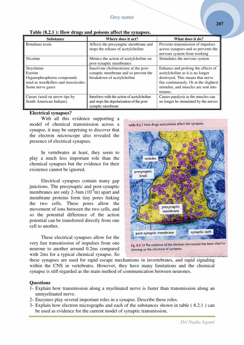
Grey matter
Dr/ Nadia Agami
207
Table (8.2.1 ): How drugs and poisons affect the synapses.
Substance Where does it act? What does it do?
Botulinus toxin Affects the presynaptic membrane and
stops the release of acetylcholine
Prevents transmission of impulses
across synapses and so prevents the
nervous system from working
Nicotine Mimics the action of acetylcholine on
post-synaptic membranes
Stimulates the nervous system
Strychnine
Eserine
Organophosphorus compounds
used as weedkillers and insecticides
Some nerve gases
Inactivate cholinesterase at the post-
synaptic membrane and so prevent the
breakdown of acetylcholine
Enhance and prolong the effects of
acetylcholine as it is no longer
destroyed. This means that nerve
fire continuously. Or at the slightest
stimulus, and muscles are sent into
tetanus.
Curare (used on arrow tips by
South American Indians)
Interferes with the action of acetylcholine
and stops the depolarisation of the post-
synaptic membrane
Causes paralysis as the muscles can
no longer be stimulated by the nerves
Electrical synapses?
With all this evidence supporting a
model of chemical transmission across a
synapse, it may be surprising to discover that
the electron microscope also revealed the
presence of electrical synapses.
In vertebrates at least, they seem to
play a much less important role than the
chemical synapses but the evidence for their
existence cannot be ignored.
Electrical synapses contain many gap
junctions. The presynaptic and post-synaptic
membranes are only 2-3nm (10-9
m) apart and
membrane proteins form tiny pores linking
the two cells. These pores allow the
movement of ions between the two cells, and
so the potential difference of the action
potential can be transferred directly from one
cell to another.
These electrical synapses allow for the
very fast transmission of impulses from one
neurone to another around 0.2ms compared
with 2ms for a typical chemical synapse. So
these synapses are used for rapid escape mechanisms in invertebrates, and rapid signaling
within the CNS in vertebrates. However, they have many limitations and the chemical
synapse is still regarded as the main method of communication between neurones.
Questions
1- Explain how transmission along a myelinated nerve is faster than transmission along an
unmyelinated nerve.
2- Enzymes play several important roles in a synapse. Describe these roles.
3- Explain how electron micrographs and each of the substances shown in table ( 8.2.1 ) can
be used as evidence for the current model of synaptic transmission.

Interactions between neurones
Neurones interact in a variety of complex ways. In many cases a single nerve fibre will
carry an action potential to a synapse with another cell, and transmission across that synapse
will set up the next action potential. However, the situation can be much more complex than
this.
Summation and facilitation
Often a single synaptic knob does not release enough transmitter substance to set up an
action potential in the post-synaptic fibre. However, if two or more sy
stimulated and release transmitter at the same time onto the same post
the effect add together and a post
summation.
In other cases, a single knob does not
impulse to stimulate the post-synaptic nerve fibre but if a second impulse is received from the
same knob in quick succession an action potential results. This effect is known as temporal
summation (adding over time), and it involves facilitation. In other words, the first impulse
does not trigger a response but it makes easier (or facilitates) the passage of the next impulse.
Accommodation
When you first put on perfume or aftershave you are very aware of the sc
short time you lose that awareness. If you reapply your scent another day, you can smell it
Grey matter
Interactions between neurones
in a variety of complex ways. In many cases a single nerve fibre will
carry an action potential to a synapse with another cell, and transmission across that synapse
otential. However, the situation can be much more complex than
Summation and facilitation
Often a single synaptic knob does not release enough transmitter substance to set up an
synaptic fibre. However, if two or more sy
stimulated and release transmitter at the same time onto the same post-synaptic membrane,
the effect add together and a post-synaptic action potential results. This is known as spatial
In other cases, a single knob does not release enough transmitter substance from one
synaptic nerve fibre but if a second impulse is received from the
same knob in quick succession an action potential results. This effect is known as temporal
time), and it involves facilitation. In other words, the first impulse
does not trigger a response but it makes easier (or facilitates) the passage of the next impulse.
When you first put on perfume or aftershave you are very aware of the sc
short time you lose that awareness. If you reapply your scent another day, you can smell it
Dr/ Nadia Agami
208
in a variety of complex ways. In many cases a single nerve fibre will
carry an action potential to a synapse with another cell, and transmission across that synapse
otential. However, the situation can be much more complex than
Often a single synaptic knob does not release enough transmitter substance to set up an
synaptic fibre. However, if two or more synaptic knobs are
synaptic membrane,
synaptic action potential results. This is known as spatial
release enough transmitter substance from one
synaptic nerve fibre but if a second impulse is received from the
same knob in quick succession an action potential results. This effect is known as temporal
time), and it involves facilitation. In other words, the first impulse
does not trigger a response but it makes easier (or facilitates) the passage of the next impulse.
When you first put on perfume or aftershave you are very aware of the scent, but after a
short time you lose that awareness. If you reapply your scent another day, you can smell it

Grey matter
Dr/ Nadia Agami
209
again. This reaction is the same as that of a sea anemone which, when poked with a pointer,
will withdraw its tentacles.
If the sea anemone is poked repeatedly, the response is lost. If left alone for a while, the
sea anemone reacts when poked again. These are examples of a process known as
accommodation.
Each time an impulse arrives at a synapse, vesicles full of neurotransmitter discharge
their contents into the synaptic cleft. These vesicles can be synthesised only at a certain rate.
If the synapse is used too often, all of the vesicles are discharged into the synaptic cleft and
the rate of synthesis simply cannot keep up. At this point the neurones can no longer respond
to the stimulus they are said to have accommodated or fatigued.
A short rest restores the response as new vesicles and transmitter molecules are made.
Some synapses never fatigue, as they have an extremely rapid resynthesis rate, while others
accommodate very quickly. The impact of this is seen particularly clearly in the neurones
linked with the sensory organs.
What does all this mean for living animals?
The nerve fibres and synapses which we have been considering in isolation make
enormously complicated systems in living animals. Individual neurones carry impulses in one
direction only. The bundles of nerve fibres that make up nerves are capable of carrying vast
numbers of impulses in different direction all at the same time. The nervous system as a
whole gathers the available information and controls all the actions of the body, with the
central nervous system collating information and sending out instructions. Synapses allow for
great flexibility and intercommunications between cells and play a vital but incompletely
understood role in the brain, closely linked with both learning and memory.
For simpler animals most of their nervous activity and behaviour involves reflex
actions which have a minimum of input from the central nervous system. Humans also have
many reflex actions, including the pupil reflex that you will study on page 218. However, we
also have an extensive range of nerves which are under conscious, voluntary control. The two
aspects which are crucial to an effective nervous system are a sensory system which detects
stimuli, and a motor system by which effectors are made to respond.
Sensory systems
The coordinated activity of any organism does not depend purely on the passage of
action potentials along nerve fibres or successful reflex arcs. There must be a continuous
input of information from both the outside world and the internal environment if the animal is
to be able to modify its behaviour and survive as situations change. Sensory receptors play a
vital role in providing an animal with information about both its internal and external
environment.
Simple sensory receptors are just neurones which have a dendrite that is sensitive to
one particular stimulus. When the dendrite receives a stimulus, chemical events occur that
result in an action potential in the nerve fibre of the neurone. This type of cell is known as a
primary receptor.

A secondary receptor
completely specialised cells (not neurones) which are sensitive to a particular type of
stimulus. These cells synapse with a normal sensory
central nervous system. The retinal cells in the retina of the eye are an example of these.
As animals become increasingly complex, so do their sensory systems. While many
sensory receptors are always found as iso
are increasingly found associated together in systems known as sense organs.
Questions
1- Define what is meant by accommodation in a neurone, and explain why it is important in
the nervous system.
2- Explain how temporal and spatial summation help to make an organism more responsive
to environmental cues.
How sensory receptors work
Sensory receptors transfer the energy of a particular stimulus
movement into the electrochemical
energy of an action potential in a sensory
nerve fibre. There are various different
types of receptors and a variety of ways
of categorizing them these are the main
ones:
• Exteroceptors respond to stimuli
outside the body.
• Interoceptors respond to stimul
the body.
• Proprioceptors are sensitive to the
relative positions of the skeleton and
degree of muscle contraction, and are
very important for maintaining
posture.
• Chemoreceptors are sensitive to
chemical stimuli such as smell, taste and the pH
• Mechanoreceptors are sensitive to mechanical stimuli such as pressure, tension, movement
and gravity.
• Photoreceptors are sensitive to electromagnetic stimuli for humans this is particularly
visible light, while many insects are s
• Thermoreceptors respond to heat stimuli, and are particularly effective at detecting
changes and differences in temperature.
Grey matter
A secondary receptor is slightly more complicated. It consists of one or more
completely specialised cells (not neurones) which are sensitive to a particular type of
stimulus. These cells synapse with a normal sensory neurone which carries the impulse to the
central nervous system. The retinal cells in the retina of the eye are an example of these.
As animals become increasingly complex, so do their sensory systems. While many
sensory receptors are always found as isolated entities, in higher animals sensory receptors
are increasingly found associated together in systems known as sense organs.
Define what is meant by accommodation in a neurone, and explain why it is important in
Explain how temporal and spatial summation help to make an organism more responsive
How sensory receptors work
Sensory receptors transfer the energy of a particular stimulus-light, heat, sound,
movement into the electrochemical
rgy of an action potential in a sensory
nerve fibre. There are various different
types of receptors and a variety of ways
of categorizing them these are the main
Exteroceptors respond to stimuli
Interoceptors respond to stimuli inside
Proprioceptors are sensitive to the
relative positions of the skeleton and
degree of muscle contraction, and are
very important for maintaining
Chemoreceptors are sensitive to
chemical stimuli such as smell, taste and the pH levels of the blood.
Mechanoreceptors are sensitive to mechanical stimuli such as pressure, tension, movement
Photoreceptors are sensitive to electromagnetic stimuli for humans this is particularly
visible light, while many insects are sensitive to UV light.
Thermoreceptors respond to heat stimuli, and are particularly effective at detecting
changes and differences in temperature.
Dr/ Nadia Agami
210
is slightly more complicated. It consists of one or more
completely specialised cells (not neurones) which are sensitive to a particular type of
neurone which carries the impulse to the
central nervous system. The retinal cells in the retina of the eye are an example of these.
As animals become increasingly complex, so do their sensory systems. While many
lated entities, in higher animals sensory receptors
are increasingly found associated together in systems known as sense organs.
Define what is meant by accommodation in a neurone, and explain why it is important in
Explain how temporal and spatial summation help to make an organism more responsive
light, heat, sound,
Mechanoreceptors are sensitive to mechanical stimuli such as pressure, tension, movement
Photoreceptors are sensitive to electromagnetic stimuli for humans this is particularly
Thermoreceptors respond to heat stimuli, and are particularly effective at detecting

How do receptors work?
Just like nerve fibres, receptor cells have a resting potential which is
maintenance of an interior that is negative in relation to the outside by membrane sodium
pumps. When a receptor cells receives a stimulus, sodium ions move rapidly across the cell
membrane along concentration and electrochemical gradients
up a generator potential. A small stimulus results in a small generator potential and a large
stimulus results in a large generator potential generator potentials do not obey the all
nothing law. If the generator potent
sensory neurone, an action potential will result in that neurone. If is not, there will be no
action potential the action potential does obey the all
This process: Stimulus → local change in permeability
action potential.
Is common in one form or another to most sensory receptors.
In sensory organs such as the eye, several receptor cells will often synapse with a
single sensory neurone. This means that even if the generator potential from an individual
receptor cell is insufficient to set up a synapse, the potentials from several may add together
or summate and trigger an action potential. This is known as
adaptation for increasing the sensitivity of a sensory system to low
see this is an important feature of the retina cells, the light
When the received stimulus is weak, only a low frequency of acti
the sensory neurone results. But if the stimulus received by the receptors is strong, then a
rapid stream of action potentials is fired along the sensory neurone. The importance of this is
that, although the axon obeys the all
potential, a graded response is still possible,
stimulus. What this means for the eye, for example, is that we are not only aware of the
difference between light and dark
Grey matter
Just like nerve fibres, receptor cells have a resting potential which is
maintenance of an interior that is negative in relation to the outside by membrane sodium
pumps. When a receptor cells receives a stimulus, sodium ions move rapidly across the cell
membrane along concentration and electrochemical gradients, and this generator current sets
up a generator potential. A small stimulus results in a small generator potential and a large
stimulus results in a large generator potential generator potentials do not obey the all
nothing law. If the generator potential produced is large enough to reach the threshold of the
sensory neurone, an action potential will result in that neurone. If is not, there will be no
action potential the action potential does obey the all-or-nothing law.
local change in permeability → generator current → generator potential
Is common in one form or another to most sensory receptors.
In sensory organs such as the eye, several receptor cells will often synapse with a
one. This means that even if the generator potential from an individual
receptor cell is insufficient to set up a synapse, the potentials from several may add together
or summate and trigger an action potential. This is known as convergence
adaptation for increasing the sensitivity of a sensory system to low-level stimuli. As you will
see this is an important feature of the retina cells, the light-sensitive cells of the eye.
When the received stimulus is weak, only a low frequency of acti
the sensory neurone results. But if the stimulus received by the receptors is strong, then a
rapid stream of action potentials is fired along the sensory neurone. The importance of this is
that, although the axon obeys the all-or nothing law in terms of each individual action
graded response is still possible, giving information about the strength of the
stimulus. What this means for the eye, for example, is that we are not only aware of the
difference between light and dark, but of all the varying degrees of light and shade.
Dr/ Nadia Agami
211
Just like nerve fibres, receptor cells have a resting potential which is dependent on the
maintenance of an interior that is negative in relation to the outside by membrane sodium
pumps. When a receptor cells receives a stimulus, sodium ions move rapidly across the cell
, and this generator current sets
up a generator potential. A small stimulus results in a small generator potential and a large
stimulus results in a large generator potential generator potentials do not obey the all-or-
ial produced is large enough to reach the threshold of the
sensory neurone, an action potential will result in that neurone. If is not, there will be no
generator potential →
In sensory organs such as the eye, several receptor cells will often synapse with a
one. This means that even if the generator potential from an individual
receptor cell is insufficient to set up a synapse, the potentials from several may add together
convergence and it is a useful
level stimuli. As you will
sensitive cells of the eye.
When the received stimulus is weak, only a low frequency of action potentials along
the sensory neurone results. But if the stimulus received by the receptors is strong, then a
rapid stream of action potentials is fired along the sensory neurone. The importance of this is
g law in terms of each individual action
giving information about the strength of the
stimulus. What this means for the eye, for example, is that we are not only aware of the
, but of all the varying degrees of light and shade.

Grey matter
Dr/ Nadia Agami
212
Adaptation of the receptors
The most important information carried by the sensory system is about changes in the
environment that are potential problems to be dealt with by an organism.
So if a receptor is exposed to a steady stimulus that continues unchanged for a period
of time, the information become of decreasing importance to the animal unless for some
reason it changes.
The nervous system is adapted to cope with this situation. Instead of simply
responding constantly to a steady stimulus, most receptors show a gradual decline in the
generator potentials produced and so the action potentials in the sensory neurone become less
frequent and may eventually stop. This is known as adaptation.
The mechanism by which adaptation is brought about is not fully understood, but
seems to involve changes at the membrane. It may well be linked to the accommodation of
the synapses in the pathway too. Some receptors adapt very rapidly, others only slowly or not
at all. But in general, adaptation serves to prevent the system from carrying too many
unnecessary impulses and frees the CNS from irrelevant information. Thus an unpleasant
smell rapidly becomes tolerable, a hot bath feels comfortable and an irritating label in a
jumper is forgotten-unless in some way the stimulus changes, when we become sensitised
again.
Question
1- a- Describe how a sensory receptor works.
b- Explain how convergence increases the sensitivity of a system to low-level stimuli.
2- Accommodation and adaptation have similar functions. Explain how the two processes
differ and why they are important in the nervous system and for the living animal.

Grey matter
Dr/ Nadia Agami
213
The human eye
Single sensory receptor cells are very useful and can carry vital information, even for
very complex organisms. But groups of receptor cells all speicalised for responding to a
particular stimulus can be even more useful as they can respond in a more varied way.
Relatively early in animal evolution, collections of receptors developed together to
form specialised regions known as sense organs. Throughout the animal kingdom, the most
common sense organs are those that respond to light, and to sound or vibration.
Sensitivity to light is found in some of the simplest living organisms. In humans it has
developed far beyond a simple sensitivity to the absence or presence of light into a
remarkably sophisticated optical system, allowing clear and focused vision in a wide variety
of circumstances. Our eyes are sensitive to electromagnetic radiation with a wavelength
between 400 and 700nm. The ranges of other species often vary from this.
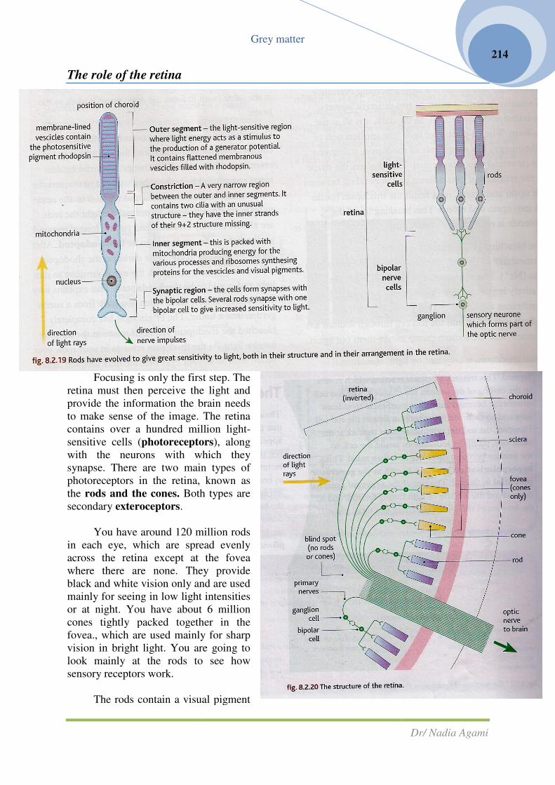
The role of the retina
Focusing is only the first step. The
retina must then perceive the light and
provide the information the brain needs
to make sense of the image. The retina
contains over a hundred million light
sensitive cells (photoreceptors
with the neurons with which they
synapse. There are two main types of
photoreceptors in the retina, known as
the rods and the cones. Both types are
secondary exteroceptors.
You have around 120 million rods
in each eye, which are spread evenly
across the retina except at the fovea
where there are none. They provide
black and white vision only and are used
mainly for seeing in low light intensities
or at night. You have about 6 million
cones tightly packed together in the
fovea., which are used mainly for sharp
vision in bright light. You are going to
look mainly at the rods to see how
sensory receptors work.
The rods contain a visual pigment
Grey matter
Focusing is only the first step. The
then perceive the light and
provide the information the brain needs
to make sense of the image. The retina
contains over a hundred million light-
photoreceptors), along
with the neurons with which they
synapse. There are two main types of
hotoreceptors in the retina, known as
Both types are
You have around 120 million rods
in each eye, which are spread evenly
across the retina except at the fovea
where there are none. They provide
white vision only and are used
mainly for seeing in low light intensities
or at night. You have about 6 million
cones tightly packed together in the
fovea., which are used mainly for sharp
vision in bright light. You are going to
o see how
The rods contain a visual pigment
Dr/ Nadia Agami
214

called rhodopsin (visual purple).
of them synapse with the same sensory neurone. Although this means they do not give a
particularly clear picture, it does make them extremely sensitive both to low light levels and
to movements in the visual field, because several small generator potentials can trigger an
action potential to the CNS through the summation effect.
Note that the arrangement of the retina is
actually next to the choroid, and the neurons are at the interior edge of the eyeball. The light
has to pass through the synapses and the inner segments before reaching the outer segment
containing the visual pigments. The reason for this somewhat unexpected arrangement is the
origin of the retinal cells in the embryo and the way in which the eye is formed during
embryonic development. This is why there is a blind spot where all the neur
the layers of the eye to go into the brain.
How do the rods work?
• The sensitivity of the rods is based on the reactions of the visual pigment rhodopsin with
light.
• Rhodopsin is formed from two components, opsin and retinal.
• Opsin is a combination of lipid and protein (a lipoprotein) and retinal is a light
derivative of vitamin A.
• retinal exists as two different isomers, cis
the cis-form. When a photon of light (the small
rhodopsin, it converts the cis
retinal, and puts a strain on the bonding between the opsin and retinal.
Grey matter
rhodopsin (visual purple). The rods are not very tightly packed together and several
of them synapse with the same sensory neurone. Although this means they do not give a
larly clear picture, it does make them extremely sensitive both to low light levels and
to movements in the visual field, because several small generator potentials can trigger an
action potential to the CNS through the summation effect.
rrangement of the retina is “back-to-front”. The “outer segments” are
actually next to the choroid, and the neurons are at the interior edge of the eyeball. The light
has to pass through the synapses and the inner segments before reaching the outer segment
containing the visual pigments. The reason for this somewhat unexpected arrangement is the
origin of the retinal cells in the embryo and the way in which the eye is formed during
embryonic development. This is why there is a blind spot where all the neur
the layers of the eye to go into the brain.
How do the rods work?
The sensitivity of the rods is based on the reactions of the visual pigment rhodopsin with
Rhodopsin is formed from two components, opsin and retinal.
a combination of lipid and protein (a lipoprotein) and retinal is a light
retinal exists as two different isomers, cis-retinal and trans-retinal. In the dark it is all in
form. When a photon of light (the smallest unit of light energy) hits a molecule of
rhodopsin, it converts the cis-retinal into trans-retinal. This changes the shape of the
retinal, and puts a strain on the bonding between the opsin and retinal.
Dr/ Nadia Agami
215
The rods are not very tightly packed together and several
of them synapse with the same sensory neurone. Although this means they do not give a
larly clear picture, it does make them extremely sensitive both to low light levels and
to movements in the visual field, because several small generator potentials can trigger an
The “outer segments” are
actually next to the choroid, and the neurons are at the interior edge of the eyeball. The light
has to pass through the synapses and the inner segments before reaching the outer segments
containing the visual pigments. The reason for this somewhat unexpected arrangement is the
origin of the retinal cells in the embryo and the way in which the eye is formed during
embryonic development. This is why there is a blind spot where all the neurons pass through
The sensitivity of the rods is based on the reactions of the visual pigment rhodopsin with
a combination of lipid and protein (a lipoprotein) and retinal is a light-absorbing
retinal. In the dark it is all in
est unit of light energy) hits a molecule of
retinal. This changes the shape of the

Grey matter
Dr/ Nadia Agami
216
• As a result the rhodopsin breaks up into opsin and retinal. This breaking up of the
molecule is referred to as bleaching.
• The bleaching of the rhodopsin changes the permeability of the cell membrane of the rod
to sodium ions (Na+). The membranes of most neurons are relatively impermeable to
sodium ions but rod cell membranes are normally very permeable to them. Sodium ions
move into the rod cell through sodium ion channels, and the sodium pump moves them
out again.
• When rhodopsin is bleached by light, trans-retinal is formed. This triggers a cascades
reaction which results in the closing of the sodium ion channels. The rod cell membrane
becomes much less permeable to sodium ions and so there are fewer sodium ions in the
rod cell.
• However, the sodium pump continues to work at the same rate, pumping sodium ions out
of the cell, so the interior becomes more negative than usual. This hyperpolarisation is
what is known as the generator potential in the rod. The size of the generator potential
depends on the amount of light hitting the rod, and so the amount of rhodopsin bleaching
that takes place. If it is large enough to reach the threshold, or if several rods are
stimulated at once, neurotransmitter substances are released into the synapse with the
bipolar cell.
• An action potential is then set up in the bipolar cell which passes across the synapse to
cause an action potential in the sensory neurone. All the sensory neurons leave the eye at
the same point to form the optic nerve leading to the brain.
• Once the visual pigment has been bleached, the rod cannot be stimulated again until the
rhodopsin is resynthesised. It takes ATP energy produced by the many mitochondria in
the inner segment to convert the trans-retinal back to cis-retinal and rejoin it to the opsin
to form rhodopsin again.
• In normal daylight the rods are almost entirely bleached and can no longer respond to dim
light-the eye is said to be light-adapted. After about 30 minutes in complete darkness the
rhodopsin will be almost fully reformed, the eye is sensitive to dim light and is said to be
dark-adapted. This explains why you become almost blind when you walk from a sunny
garden into a house. The bright light has completely bleached the rhodopsin you need to
see in the dimmer interior light. As the rhodopsin reforms in your rods, your vision
returns as your eyes become dark adapted again.

Grey matter
Dr/ Nadia Agami
217
The cones and colour vision
• The cones work in a very similar way to rods, except that their visual pigment is known as
iodopsin.
• There appear to be three types of iodopsin, each sensitive to one of the primary colours of
light. Iodopsin needs to be hit with more light energy than rhodopsin in order to break
down, and so it is not sensitive to low light intensities. But the cones provide colour
vision, because the brain interprets the numbers of different types of cones stimulate as
different colours. This complements the low light vision and sensitivity to movmenet
provided by the rods.
HSW investigating the graded response of the rods
The rods of your eye do not give you an all-or-nothing response. You are aware of
varying levels of light intensity. Yet you know that the axons which fire off potentials
through the optic nerve operate on an all-or-nothing response
Scientists have developed a model based on a graded response in the receptor cells
which is amplified through a cascade system that causes the hyperpolarisation in the receptor
cells. A single photon of light bleaches a single molecule of rhodopsin, but through the
cascade that is set in place, several thousand sodium ion channels can be closed.
The brighter the light, the more rhodopsin is bleached and so the more sodium ion
channels are closed. This gives a bigger depolarization of the cell which lasts longer, so the
cell can respond in a measurably different way to very small differences in light intensity.
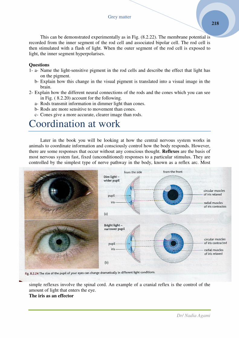
Grey matter
Dr/ Nadia Agami
218
This can be demonstrated experimentally as in Fig. (8.2.22). The membrane potential is
recorded from the inner segment of the rod cell and associated bipolar cell. The rod cell is
then stimulated with a flash of light. When the outer segment of the rod cell is exposed to
light, the inner segment hyperpolarises.
Questions
1- a- Name the light-sensitive pigment in the rod cells and describe the effect that light has
on the pigment.
b- Explain how this change in the visual pigment is translated into a visual image in the
brain.
2- Explain how the different neural connections of the rods and the cones which you can see
in Fig. ( 8.2.20) account for the following.
a- Rods transmit information in dimmer light than cones.
b- Rods are more sensitive to movement than cones.
c- Cones give a more accurate, clearer image than rods.
Coordination at work
Later in the book you will be looking at how the central nervous system works in
animals to coordinate information and consciously control how the body responds. However,
there are some responses that occur without any conscious thought. Reflexes are the basis of
most nervous system fast, fixed (unconditioned) responses to a particular stimulus. They are
controlled by the simplest type of nerve pathway in the body, known as a reflex arc. Most
simple reflexes involve the spinal cord. An example of a cranial reflex is the control of the
amount of light that enters the eye.
The iris as an effector
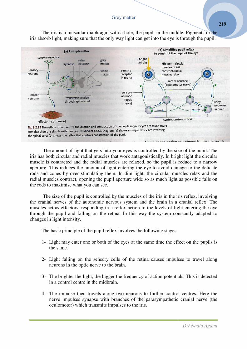
The iris is a muscular diaphragm with a hole, the pupil, in the middle. Pigments in the
iris absorb light, making sure that the only way light
The amount of light that gets into your eyes is controlled by the size of the pupil. The
iris has both circular and radial muscles that work antagonistically. In bright light the circular
muscle is contracted and the radial muscles are relaxed, so the pupil is reduce to a narrow
aperture. This reduces the amount of light entering the eye to avoid damage to the delicate
rods and cones by over stimulating them. In dim light, the circular muscles relax and the
radial muscles contract, opening the pupil aperture wide so as much light as possible falls on
the rods to maximise what you can see.
The size of the pupil is controlled by the muscles of the iris in the iris reflex, involving
the cranial nerves of the autonomic nervous system an
muscles act as effectors, responding in a reflex action to the levels of light entering the eye
through the pupil and falling on the retina. In this way the system constantly adapted to
changes in light intensity.
The basic principle of the pupil reflex involves the following stages.
1- Light may enter one or both of the eyes at the same time the effect on the pupils is
the same.
2- Light falling on the sensory cells of the retina causes impulses to travel along
neurons in the optic nerve to the brain.
3- The brighter the light, the bigger the frequency of action potentials. This is detected
in a control centre in the midbrain.
4- The impulse then travels along two neurons to further control centres. Here the
nerve impulses synapse with branches of the parasympathetic cranial nerve (the
oculomotor) which transmits impulses to the iris.
Grey matter
The iris is a muscular diaphragm with a hole, the pupil, in the middle. Pigments in the
iris absorb light, making sure that the only way light can get into the eye is through the pupil.
The amount of light that gets into your eyes is controlled by the size of the pupil. The
iris has both circular and radial muscles that work antagonistically. In bright light the circular
and the radial muscles are relaxed, so the pupil is reduce to a narrow
aperture. This reduces the amount of light entering the eye to avoid damage to the delicate
rods and cones by over stimulating them. In dim light, the circular muscles relax and the
dial muscles contract, opening the pupil aperture wide so as much light as possible falls on
the rods to maximise what you can see.
The size of the pupil is controlled by the muscles of the iris in the iris reflex, involving
tonomic nervous system and the brain in a cranial reflex. The
muscles act as effectors, responding in a reflex action to the levels of light entering the eye
through the pupil and falling on the retina. In this way the system constantly adapted to
The basic principle of the pupil reflex involves the following stages.
Light may enter one or both of the eyes at the same time the effect on the pupils is
Light falling on the sensory cells of the retina causes impulses to travel along
neurons in the optic nerve to the brain.
The brighter the light, the bigger the frequency of action potentials. This is detected
in a control centre in the midbrain.
mpulse then travels along two neurons to further control centres. Here the
nerve impulses synapse with branches of the parasympathetic cranial nerve (the
oculomotor) which transmits impulses to the iris.
Dr/ Nadia Agami
219
The iris is a muscular diaphragm with a hole, the pupil, in the middle. Pigments in the
can get into the eye is through the pupil.
The amount of light that gets into your eyes is controlled by the size of the pupil. The
iris has both circular and radial muscles that work antagonistically. In bright light the circular
and the radial muscles are relaxed, so the pupil is reduce to a narrow
aperture. This reduces the amount of light entering the eye to avoid damage to the delicate
rods and cones by over stimulating them. In dim light, the circular muscles relax and the
dial muscles contract, opening the pupil aperture wide so as much light as possible falls on
The size of the pupil is controlled by the muscles of the iris in the iris reflex, involving
the brain in a cranial reflex. The
muscles act as effectors, responding in a reflex action to the levels of light entering the eye
through the pupil and falling on the retina. In this way the system constantly adapted to
The basic principle of the pupil reflex involves the following stages.
Light may enter one or both of the eyes at the same time the effect on the pupils is
Light falling on the sensory cells of the retina causes impulses to travel along
The brighter the light, the bigger the frequency of action potentials. This is detected
mpulse then travels along two neurons to further control centres. Here the
nerve impulses synapse with branches of the parasympathetic cranial nerve (the

Grey matter
Dr/ Nadia Agami
220
5- These stimulate the effectors (the muscles of the iris). The circular muscles contract
and the radial muscles relax so the pupil constricts.
If the frequency of action potentials from the retina falls (when light levels drop),
impulses travel from the control centres along sympathetic nerves to the iris, causing the
circular muscles to relax and the radial muscles to contract and widen the pupil. This negative
feedback system controls the amount of light entering the eye. The reflex response of either
eye controls the dilation or constriction of the pupils of both eyes so a bright light shone into
one eye only will cause the pupils of both eyes to constrict.
The pupils respond to emotional cues as well as light. So, for example, the release of
the stress hormone adrenaline causes the pupils to widen, making sure you can use all the
available light to see as well as possible. Pupils also dilate when you see someone you like
(or are physically attracted too) and constrict when you see someone you don’t like or don’t
find attractive.
Comparing mechanisms of coordination
You looked at coordination in plants, which is brought about by plant hormones of
different types. Some coordination in animals is also the result of hormones, such as aspects
of growth and sexual maturity as well as responses to stress and the control of blood sugar
levels in mammals. However, animals also have nervous systems. How do they compare?
Chemical control is relatively slow but it can be very long lasting. Hormones are
carried around the body of an animal in the plasma as it moves around the circulatory system.
They move into the target cells by diffusion and are picked up by receptors on cell
membranes. Plant hormones also move in the transport systems of plants (the phloem) and by
diffusion between cells. Chemical controls is often linked to changes involving growth of an
organisms. It allows for long-term responses to environmental changes. However, it can also
be used for day-do-day responses and is well suited to delicate control mechanisms such as
negative feedback systems.
Nervous control is usually very rapid, making it an ideal form of internal
communication and control for organisms that move their whole bodies about. If you need to
respond quickly to environmental cues, nerve impulses give you the speed you need.
Questions
1- Suggest why chemical coordination alone works for plants, while most animals use both
chemical and electrical means of coordination.
2- Draw up a table to compare the mechanisms of coordination in plants and animals, and
state appropriate examples from the pages.
3- Describe how the pupil reflex works and explain why it is important as a reflex response.
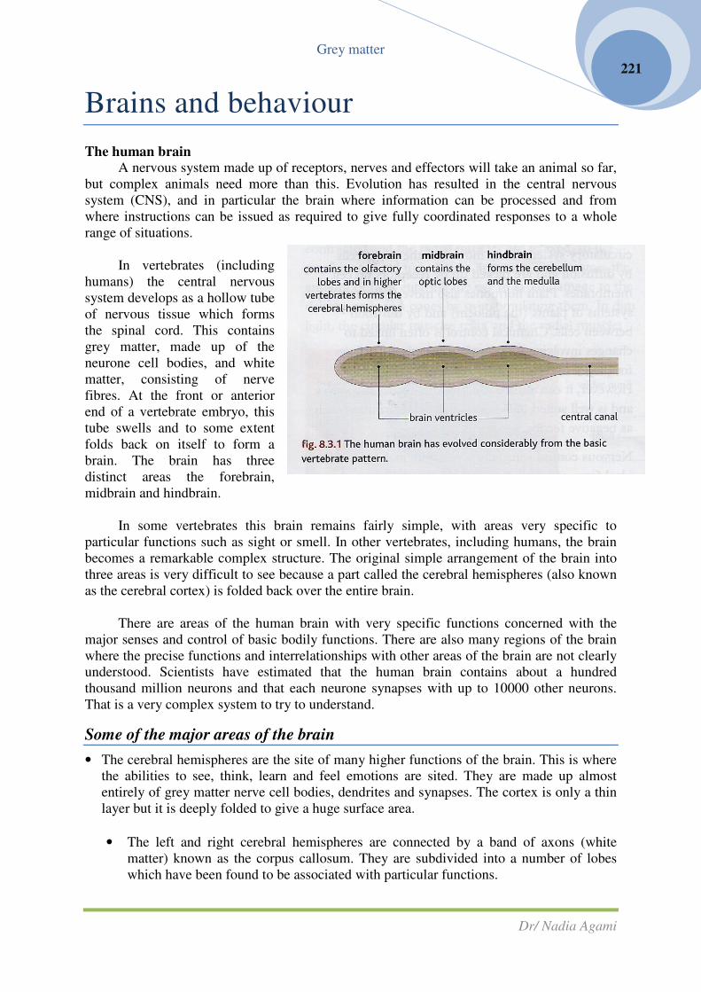
Grey matter
Dr/ Nadia Agami
221
Brains and behaviour
The human brain
A nervous system made up of receptors, nerves and effectors will take an animal so far,
but complex animals need more than this. Evolution has resulted in the central nervous
system (CNS), and in particular the brain where information can be processed and from
where instructions can be issued as required to give fully coordinated responses to a whole
range of situations.
In vertebrates (including
humans) the central nervous
system develops as a hollow tube
of nervous tissue which forms
the spinal cord. This contains
grey matter, made up of the
neurone cell bodies, and white
matter, consisting of nerve
fibres. At the front or anterior
end of a vertebrate embryo, this
tube swells and to some extent
folds back on itself to form a
brain. The brain has three
distinct areas the forebrain,
midbrain and hindbrain.
In some vertebrates this brain remains fairly simple, with areas very specific to
particular functions such as sight or smell. In other vertebrates, including humans, the brain
becomes a remarkable complex structure. The original simple arrangement of the brain into
three areas is very difficult to see because a part called the cerebral hemispheres (also known
as the cerebral cortex) is folded back over the entire brain.
There are areas of the human brain with very specific functions concerned with the
major senses and control of basic bodily functions. There are also many regions of the brain
where the precise functions and interrelationships with other areas of the brain are not clearly
understood. Scientists have estimated that the human brain contains about a hundred
thousand million neurons and that each neurone synapses with up to 10000 other neurons.
That is a very complex system to try to understand.
Some of the major areas of the brain
• The cerebral hemispheres are the site of many higher functions of the brain. This is where
the abilities to see, think, learn and feel emotions are sited. They are made up almost
entirely of grey matter nerve cell bodies, dendrites and synapses. The cortex is only a thin
layer but it is deeply folded to give a huge surface area.
• The left and right cerebral hemispheres are connected by a band of axons (white
matter) known as the corpus callosum. They are subdivided into a number of lobes
which have been found to be associated with particular functions.

Grey matter
Dr/ Nadia Agami
222
• The frontal lobe is associated with the higher brain functions such as emotional responses,
planning ahead, reasoning and decision making. It is very much the “conscious” area of
the brain, involved with combining information from several area of the cortex to form
associations and develop ideas. It is linked with your “personality”. The frontal lobe also
contains the primary motor cortex of the brain which is involved in the control of many of
the body movements. The control comes via motor neurones which pass through the
hindbrain and the spinal cord carrying impulses to the muscles.
• The temporal lobe is involved with processing auditory information in other words, sound
recognition, hearing and speech. It also seems to be involved in the memory functions of
the brain.
• The occipital lobe is also known as the visual cortex. It is concerned with processing
visual information- in other words it deals with the input from the eyes and is involved in
vision, shape recognition, colour vision and the sense of perspective.
• The parietal lob has a number of varied functions. It is associated with some aspects of
memory and recognition. It also concerned with the ability to calculate, with movement
and sensation, and with your ability to orientate yourself in space.
• There are a number of another areas of the brain which are known to be linked to specific
aspects of the way your body works. Many of these are involved in the unconscious
responses of your body which maintain the processes of life.
• The hypothalamus coordinates the autonomic (unconscious) nervous system. It plays a
major part in the thermoregulation of the body. It monitors the chemistry of the blood and
controls the hormone secretions of the pituitary gland. It also controls many basic drivers-
thirst, hunger, aggression and reproductive behaviour.
• The cerebellum coordinates smooth movements and uses information from the muscles
and the ears to maintain posture and balance.
• The medulla oblongata (often referred to as the medulla) is the most primitive part of the
brain. Contains reflex centers which control functions such as the heart rate, blood
pressure, breathing rate, coughing, sneezing, swallowing, saliva production and peristalsis.
It is this region which may maintain the basic life responses even when the higher areas of

the brain have been effectively destroyed. Figure 8.3.2 gives you a more detailed
illustration of the various areas of the brain.
Question
1- Degree of folding in the cerebral hemispheres varies among species, w
humans than other primates of example. Dolphins have much folding but the layer of
tissue is thinner. What is explanation for these differences?
2- Make a table to summaries the functions of the main areas of the human brain.
How the human brain works
There are several hundred million nerve cells working together in the human brain.
The great nerve tracts from the spinal cord cross
that the left-had side of the brain receives informatio
the body; and vice versa.
The cerebral cortex is only about 3 mm thick, and yet it controls most of the functions
that make us what we are. The brain contains centers of nuclei made up of cell bodies which
may have hundreds of synapses, making intercommunication between thousands and indeed
millions of cells possible. You are going to look at some of the ways in which scientists
investigate the brain, and then look at the models of learning and behaviour which ha
developed as a result.
For many years our understanding of the human brain has been developed using
evidence from two different sources: animal studies and studying human brains.
Animal studies
Many investigations have been carried out on the
has been related directly to the working of the human brain, although this is not always
possible. Animal studies can approach the problem of understanding the brain in a number of
different ways. Some experiments look
happens when obstacles are placed in the way
of that development.
Another approach is to damage or
remove areas of the brain of an animal and
observe the effect on its behaviour. For
example, the effect of removing the cerebral
hemispheres from animals such as dogs and
monkeys has been studies, as well as
implanting electrodes to see the effect on
behaviour of artificially stimulating a region
of the brain.
Observations of normal behaviour and
learning can also be made and (sometimes)
compared with post-mortem changes in the
brain. One problem with this is the human
tendency to interpret animal behaviour in the
light of human responses
(anthropomorphism). So, for example, cats
used to a caged existence, or ra
Grey matter
the brain have been effectively destroyed. Figure 8.3.2 gives you a more detailed
illustration of the various areas of the brain.
Degree of folding in the cerebral hemispheres varies among species, w
humans than other primates of example. Dolphins have much folding but the layer of
tissue is thinner. What is explanation for these differences?
Make a table to summaries the functions of the main areas of the human brain.
human brain works
There are several hundred million nerve cells working together in the human brain.
The great nerve tracts from the spinal cord cross-over as they enter and leave the brain, so
had side of the brain receives information from the controls the right
The cerebral cortex is only about 3 mm thick, and yet it controls most of the functions
that make us what we are. The brain contains centers of nuclei made up of cell bodies which
ave hundreds of synapses, making intercommunication between thousands and indeed
millions of cells possible. You are going to look at some of the ways in which scientists
investigate the brain, and then look at the models of learning and behaviour which ha
For many years our understanding of the human brain has been developed using
evidence from two different sources: animal studies and studying human brains.
Many investigations have been carried out on the brains of animals. Some of the work
has been related directly to the working of the human brain, although this is not always
possible. Animal studies can approach the problem of understanding the brain in a number of
different ways. Some experiments look at the way the brain develops normally, and what
happens when obstacles are placed in the way
Another approach is to damage or
remove areas of the brain of an animal and
observe the effect on its behaviour. For
emoving the cerebral
hemispheres from animals such as dogs and
monkeys has been studies, as well as
implanting electrodes to see the effect on
behaviour of artificially stimulating a region
Observations of normal behaviour and
lso be made and (sometimes)
mortem changes in the
brain. One problem with this is the human
tendency to interpret animal behaviour in the
light of human responses
(anthropomorphism). So, for example, cats
used to a caged existence, or rats which
Dr/ Nadia Agami
223
the brain have been effectively destroyed. Figure 8.3.2 gives you a more detailed
Degree of folding in the cerebral hemispheres varies among species, with more folding in
humans than other primates of example. Dolphins have much folding but the layer of
Make a table to summaries the functions of the main areas of the human brain.
There are several hundred million nerve cells working together in the human brain.
over as they enter and leave the brain, so
n from the controls the right-hand side of
The cerebral cortex is only about 3 mm thick, and yet it controls most of the functions
that make us what we are. The brain contains centers of nuclei made up of cell bodies which
ave hundreds of synapses, making intercommunication between thousands and indeed
millions of cells possible. You are going to look at some of the ways in which scientists
investigate the brain, and then look at the models of learning and behaviour which have been
For many years our understanding of the human brain has been developed using
evidence from two different sources: animal studies and studying human brains.
brains of animals. Some of the work
has been related directly to the working of the human brain, although this is not always
possible. Animal studies can approach the problem of understanding the brain in a number of
at the way the brain develops normally, and what

Grey matter
Dr/ Nadia Agami
224
frequently explore mazes in the interests of science, are likely to be well-fed, healthy
individuals display normal behavior for their species. But our natural response may be to feel
sorry for them, even deciding that they look unhappy, because we are endowing them with
our own human reactions to a situation.
HSW the moral and ethical dilemmas of using animals
Many people find the use of animals in brain research distasteful. Some feel that it
should never be done (absolutists) and others think that there may be times when it is justified
(relativists). Individual viewpoints will be affected by the moral code, or norm, of the society
in which we live, as well as personal ethics, what we consider right and wrong or the best
approach in that situation. For example, the strength of feeling may depend on the type of
animals used. There is relatively little objection to the using sea slugs or squids, but suing
dogs and cats and most particularly primates is more likely to raise ethical objections.
Ethics committees decide what research can and can’t be done. Their decision are
made in a relativist way, comparing the effect on the animals with the potential improvement
for human life particularly for people affected by brain diseases and damage.
Studying human brains
Obviously experiments interfering with or damaging the brain are not carried out on
humans. The nearest kind of research to this is when brain surgery patients have allowed their
brains to be artificially stimulated. Brain surgery is often completed under local anesthetic
because there are no sensory nerve
endings in the brain. The conscious
patients describe the sensations
relating to the stimulation, and this has
shown that certain areas of the brain
are associated with very particular
functions.
Most of our information about
the functions of the human brain
comes from situation where parts of
the brain are damaged or missing at
birth or as the results of illness or
injury.
For example, in 1848 Phineas
Gage was foreman on US railway
construction company. He was likable,
reliable, hardworking and very
responsible. In an explosives accident
an iron bar passed through his head. It
didn’t kill him, but it destroyed much of the front part of the left-had side of his brain. Gage
could still walk, talk and carry on a semblance of normal life, but this personality changes
dramatically. He became impatient, irresponsible, foulmouthed and unpleasant. He lost his
job, and died 12 years later.

Grey matter
Dr/ Nadia Agami
225
Researcher at Harvard have used computer graphics along with images of the injury
and the skull to show that the bar destroyed much of the connection between the left-side
frontal lobes and the midbrain. Because of this Phineas Gage lost the ability to control his
emotional behaviour.
Diseases that affect the brain can cause terrible problems for the people affected, but they can
also help scientists understand how the healthy brain works.
In some cases they can also point the way to possible treatments. Dr Oliver Sachs, a clinical
neurologist, has described a number of unusual cases, such as a man who mistook his wife for
a hat! This man had a disease affecting the visual areas of the brain. He could see and
describe things but he had lost the ability to make the normal connections between what he
saw and what the object was.
Another patients had a massive stroke which affected the deeper and back portions of her
cerebral hemispheres. Her personality and ability to talk were unimpaired-but she completely
lost the concept of the left-hand side, both of her own body and the world around her. She
would eat only the right-hand portion of her food and applied make-up to only to the right-
hand side of her face. These patients show clearly the location of function and awareness in
the different sides of the brain.
Medical imaging technology
The development of medical imaging
technology has had a profound effect on our ability
to investigate the development of the brain and to
link structures to functions. It has also made it
possible to understand the progress of many brain
diseases and how they affect the people who have
them.
• X-rays are great for taking images of hard
tissue such as bones. They are much less
useful for producing images of soft tissues
such as the brain.
• Computerised tomography (also known as a
CT scan) was developed to allow scientists
and doctors to see inside the brain. A CT scan
involves thousands of tiny beams of X-rays
which are passed through an area of the body
such as the head. Each beam is reduced is
strength (attenuated) by the density of the
tissue it passes through. The X-rays which
make it through are detected and measured.
All of the data are put together in a computer to produce a cross-sectional image of a
thin slice through the body. Sometimes special dyes are injected into the blood or
tissues to make particular areas X-ray opaque so they show up more clearly in the
scan.
A CT scan can identify major structures in the brain and detect problems such as brain
tumours, bleeding in the brain or swellings of the arteries in the brain (aneurisms). It does not

Grey matter
Dr/ Nadia Agami
226
pick up very fine structural details. The images arte like photograph-images frozen in time.
So CT scans cannot be used to show how areas of the brain are used or change during
different activities. However, evidence from CT scan images linked to observed changes in
behaviour can indicate the importance of certain areas of the brain in particular functions.
• Magnetic resonance imaging or MRI scans produce images that show much finer
detail than CT scans. They are produced using magnetic fields and radio waves to
image the soft tissues, which removes even the small risk of damage from the X-
rays used in CT scans. Hydrogen atoms are the most commonly imaged element,
partly because so much of the body is made of water (containing lots of hydrogen
atoms), and partly because hydrogen atoms produce a particularly strong MRI
signals. The signals produced are analysed by a computer and used to produce an
image.

� Different tissue respond differently to the magnetic field from the radio waves,
depending on many factors including the amount of water in the structure. As
a result, distinct regions of the brain can be recognised in the image. Thin
sections of the body are usually examined and these produce 2D MRI scans.
However, the computer can put these slices together to produce a 3 D image.
� MRI produces very detailed image which giv
information about the living brain. It is widely used to diagnose brain injuries,
strokes, tumours and infections of the brain or the spine. By showing up areas
of damage in the brain very clearly, it has enabled doctors and scien
make links between the structures in the brain and patterns of behaviour seen
in their patients. However, MRI scans give an historical image of the brain
like CT scans, they don’t show it as it works.
� An even more recent development in technolog
resonance imaging, or fMRI, makes it possible to watch the different areas of
the brain in action while people carry out different takes, fMRI monitors the
uptake of oxygen in different brain areas. Deoxyhaemoglobin absorbs the
radiowave signal and later re
an area of the brain is active, the blood flow to that area increase and more
oxyhaemoglobin is delivered to supply the active cells with the oxygen they
need for aerobic respiration.
active area of the brain absorbs (and so emits) less energy than a less active
area. This can be observed in real time, as different areas of the brain “light
up” on the image as they become active.
� The number of fMRI images which can be recorded each second is constantly
increasing, so the ability of scientists of observe changes in the brain activity
Grey matter
Different tissue respond differently to the magnetic field from the radio waves,
depending on many factors including the amount of water in the structure. As
nct regions of the brain can be recognised in the image. Thin
sections of the body are usually examined and these produce 2D MRI scans.
However, the computer can put these slices together to produce a 3 D image.
MRI produces very detailed image which give doctors a great deal of
information about the living brain. It is widely used to diagnose brain injuries,
strokes, tumours and infections of the brain or the spine. By showing up areas
of damage in the brain very clearly, it has enabled doctors and scien
make links between the structures in the brain and patterns of behaviour seen
in their patients. However, MRI scans give an historical image of the brain
like CT scans, they don’t show it as it works.
An even more recent development in technology, functional magnetic
resonance imaging, or fMRI, makes it possible to watch the different areas of
the brain in action while people carry out different takes, fMRI monitors the
uptake of oxygen in different brain areas. Deoxyhaemoglobin absorbs the
wave signal and later re-emits it,, while oxygaemoglobin does not. When
an area of the brain is active, the blood flow to that area increase and more
oxyhaemoglobin is delivered to supply the active cells with the oxygen they
need for aerobic respiration. Less of the signals is absorbed as a result. So an
active area of the brain absorbs (and so emits) less energy than a less active
area. This can be observed in real time, as different areas of the brain “light
up” on the image as they become active.
umber of fMRI images which can be recorded each second is constantly
increasing, so the ability of scientists of observe changes in the brain activity
Dr/ Nadia Agami
227
Different tissue respond differently to the magnetic field from the radio waves,
depending on many factors including the amount of water in the structure. As
nct regions of the brain can be recognised in the image. Thin
sections of the body are usually examined and these produce 2D MRI scans.
However, the computer can put these slices together to produce a 3 D image.
e doctors a great deal of
information about the living brain. It is widely used to diagnose brain injuries,
strokes, tumours and infections of the brain or the spine. By showing up areas
of damage in the brain very clearly, it has enabled doctors and scientist to
make links between the structures in the brain and patterns of behaviour seen
in their patients. However, MRI scans give an historical image of the brain-
y, functional magnetic
resonance imaging, or fMRI, makes it possible to watch the different areas of
the brain in action while people carry out different takes, fMRI monitors the
uptake of oxygen in different brain areas. Deoxyhaemoglobin absorbs the
emits it,, while oxygaemoglobin does not. When
an area of the brain is active, the blood flow to that area increase and more
oxyhaemoglobin is delivered to supply the active cells with the oxygen they
Less of the signals is absorbed as a result. So an
active area of the brain absorbs (and so emits) less energy than a less active
area. This can be observed in real time, as different areas of the brain “light
umber of fMRI images which can be recorded each second is constantly
increasing, so the ability of scientists of observe changes in the brain activity

Grey matter
Dr/ Nadia Agami
228
as different tasks are performed is improving all the time. fMRI gives an
extremely spatially accurate image of the brain. At the moment it is used
mainly to investigate normal brain structure and function. For example, clear
differences are visible in the activity of the brains of people with dyslexia
when reading compared with the brains of normal readers. However,
increasingly researchers are looking at ways of using fMRI to diagnose
disease such as the early signs of stroke damage and the onset of Alzheimer’s.
There are some drawbacks with fMRA, however. :
1- Like MRI, it is a noisy procedure which some people find very stressful.
2- It has to be carried out with the patient’s head remaining completely still- any
movement reduces the accuracy of the image. This limits what can be tested
usually to a response to images, although increasingly scientists are finding ways
to allow movement in other body areas. For example, in one investigation scientist
studies the brains of dancers as they moved their feet as it dancing to music. The
areas which lit up were strongly linked to the speech areas of the brain.
Some neurophysiologists have queried the validity of fMRI scans. They argue that the
blood flow to different areas of the brain when a subject is looking at different stimuli is a
case of correlation, not causation. Further Research will be need to confirm whether or not it
is a causal link. For the moment, the majority of scientists in this field are convinced that the
areas shown up by this imaging technique really are the active regions of the brain involved
in an activity.
One major drawback current brain imaging techniques is that the individuals need to
keep still. However, one relatively new brain mapping technique can be used in real time as
people actively engage in real-life situations, opening up whole new areas of research.
Quantified electroencephalography (qEEG) uses sensors attached to the outside of the
skull to measure the activity of the brain as patients carry out different activities. It allows
scientists to build up brain maps indicating which areas are used in different activities and
skills. So far the main use is research into the functioning of the brain rather than diagnosis of
diseases and so the fact that there are no known risks for the subjects, who are volunteers, is a
big advantage. The small risk associated with, for example, the X-rays used in CT scans for
diagnosis, is justified by clinical need. However, research done on healthy patients should
have no element of risk for them in the procedure. The major disadvantage of qEEGs is that
they are not as spatially accurate in the brain as fMRI can be.
Dr David Lewis, the Director of Neuroscience in a company called MindLab
international, has investigated a wide range of activities. In one case, he was asked to
compare the impact of kissing with eating chocolate on brain activity in a number of young
couples. Perhaps surprisingly, the chocolate stimulated many more areas of the brain than
kissing did-although as Dr Lewis commented, being wired up to recording equipment
possibly doesn’t put you in the most romantic mood but the impact of chocolate is all down
to the chemicals it contains!
In further exciting development neuroscientists are attempting to combine fMRI with
qEEG to give greater accuracy to the brain maps they are producing.

Questions:
1- Summaries the main imaging techniques used to investigate brain structure and function.
Explain how they work, advantage and limitation of each and give examples of their use
in both understanding brain structure and function and in diagnosing disease.
2- Summaries both absolutists and relativist viewpoints on the use of animal models in
understanding how brains work. Explain how each group feels they have the moral high
ground.
Nature and nur
The human brain takes many yea
conception a basic neural tube has formed.
By the time a baby is born this simple
starting point has developed into a
complex brain made up of about
100000000000 (1010
) neurons!
Once a baby is born the brain
continues to grow, with the formation of
new synapses and the myelination of
axons rather than the formation of new
neurones.
A newborn baby is exposed to
many new stimuli and the immature neurones move around and make the connections which
will result in the mature anatomy of the brain.
By 2 years old the brain is about 80% of the adult size. Scientists used to think that
brain growth and development were complete the around 7 years of age. Evidence now
suggests that myelination and the main structura
but that changes such as those involved in memory formation can continue throughout life.
The way your brain develops is in part controlled by the genes you have inherited.
However, there is strong evidence fr
development of the brain also depends on your getting the right environmental stimuli. Both
nature and nuture play their parts!
Critical windows of development
One current model used to explain certain asp
and in other animals is that there are critical “windows” of development. These are periods of
time during which vital neural connections are made in the brain in response to specific
stimuli. In theory, if the brain do
will not develop normally. This model has been extensively investigated using the
development of the visual cortex in animals as varied as monkeys and ferrets.
Studies of the growth of the bra
light-sensitive cells in the retina grow through to the area know
synapses are developed in a very regular way. This results in an ordered arrangement of
Grey matter
Summaries the main imaging techniques used to investigate brain structure and function.
Explain how they work, advantage and limitation of each and give examples of their use
in both understanding brain structure and function and in diagnosing disease.
Summaries both absolutists and relativist viewpoints on the use of animal models in
understanding how brains work. Explain how each group feels they have the moral high
urture in brain development
The human brain takes many years to develop fully. As early as 21 days after
conception a basic neural tube has formed.
orn this simple
has developed into a
complex brain made up of about
) neurons!
Once a baby is born the brain
continues to grow, with the formation of
new synapses and the myelination of
axons rather than the formation of new
A newborn baby is exposed to
many new stimuli and the immature neurones move around and make the connections which
in the mature anatomy of the brain.
By 2 years old the brain is about 80% of the adult size. Scientists used to think that
brain growth and development were complete the around 7 years of age. Evidence now
suggests that myelination and the main structural arrangement of the brain is complete by 18,
as those involved in memory formation can continue throughout life.
The way your brain develops is in part controlled by the genes you have inherited.
However, there is strong evidence from both human and animal studies that the normal
development of the brain also depends on your getting the right environmental stimuli. Both
ture play their parts!
Critical windows of development
One current model used to explain certain aspects of brain development in humans
and in other animals is that there are critical “windows” of development. These are periods of
time during which vital neural connections are made in the brain in response to specific
stimuli. In theory, if the brain does not receive these stimuli at the crucial time the pathways
will not develop normally. This model has been extensively investigated using the
development of the visual cortex in animals as varied as monkeys and ferrets.
ies of the growth of the brain in animal fetuses show that the axons from the
sensitive cells in the retina grow through to the area known as the thalamus where the
synapses are developed in a very regular way. This results in an ordered arrangement of
Dr/ Nadia Agami
229
Summaries the main imaging techniques used to investigate brain structure and function.
Explain how they work, advantage and limitation of each and give examples of their use
in both understanding brain structure and function and in diagnosing disease.
Summaries both absolutists and relativist viewpoints on the use of animal models in
understanding how brains work. Explain how each group feels they have the moral high
velopment
rs to develop fully. As early as 21 days after
many new stimuli and the immature neurones move around and make the connections which
By 2 years old the brain is about 80% of the adult size. Scientists used to think that
brain growth and development were complete the around 7 years of age. Evidence now
l arrangement of the brain is complete by 18,
as those involved in memory formation can continue throughout life.
The way your brain develops is in part controlled by the genes you have inherited.
m both human and animal studies that the normal
development of the brain also depends on your getting the right environmental stimuli. Both
ects of brain development in humans
and in other animals is that there are critical “windows” of development. These are periods of
time during which vital neural connections are made in the brain in response to specific
es not receive these stimuli at the crucial time the pathways
will not develop normally. This model has been extensively investigated using the
development of the visual cortex in animals as varied as monkeys and ferrets.
in animal fetuses show that the axons from the
as the thalamus where the
synapses are developed in a very regular way. This results in an ordered arrangement of

Grey matter
Dr/ Nadia Agami
230
neurones in the visual cortex. Stained sections show that it is organised into columns of cells,
known as ocular dominance columns.
Neighboring columns of cells receive input from the same area of the retina in the left
eye and the right eye. For some time scientists thought this arrangement was the result of the
stimulation of the eye during a critical period after birth. However, later works has shown
that this isn’t the case- the columns of cells are already present in the visual cortex at birth.
The existence of the columns is the result of nature-the genetics of the developing animal.
They are modified during a later critical periods.
HSW confirming a model
J. C. Horton and D. R. Hocking worked on macaque monkeys to confirm the “nature”
hypothesis that the ocular dominance columns are in place before birth rather than developing
in response to environmental cues (nurture). They delivered three foetal monkeys by
Caesarean section 8 days before the end of normal gestation. This meant their eyes were at an
earlier stage of development than a normal newborn monkey.
The infant monkeys were kept completely in the dark throughout their lives. A
radioactive tracer was injected into the right eye of each monkey 1 day after they were
delivered. A week later, when they would naturally have been at full term, sections of the
visual cortex were prepared and examined. They showed well- developed ocular dominance
columns. This work has been confirmed in a number of other species. It has been very useful
in developing our model of how the brain develops. However, there are clear ethical issue in
this type of work which some people find unacceptable.
The work of Horton, Hocking and others demonstrated that the basic organization of
the visual cortex is present at birth and is therefore the result of genetic inheritance (nature).
More work was needed to see if there really are any critical windows (also referred to as
sensitive periods) of development in the brain when nurture (the environment) contributes to
the development and can override the genetic influence.

Grey matter
Dr/ Nadia Agami
231
Evidence for the effect of nurture
In the 1960s a great deal of
work was done on the
development of the visual
cortex using kittens and
monkeys. These animals
were used because, unlike
the files, rats and mice used
in much animal research,
kittens and monkeys have
visual systems which are
very similar to our own.
Newborn kittens and
monkeys have very different
visual abilities-the monkey
appears visually alert and
can look at specific object
while a newborn kitten is
blind, with its eyes still
closed permanently.

Grey matter
Dr/ Nadia Agami
232
Experiments suggested that in newborns the basic ability to see, and even binocular vision, is
already in place.
In a series of classic experiments, David Hubel and Torsten Weisel showed that there
appears to be a critical window for the development of the mature visual cortex in cats and
monkeys. In the best known of these experiments they stitched shut (sutured) on or both of
the eyes of infant kittens and monkeys. The age at which the animals were sutured and
deprived of vision, and the length of time the eye remained closed, was varied. The effects of
this early sensory deprivation on the firing of neurons in the visual cortex were then
observed, sometimes many years later. Hubel and Weisel reared anima ls in the dark, and in
the light environments without any patterns such as vertical lines and measured the effects of
these treatments on the development of the visual cortex. Some of the findings are
summarized below.
• When kittens had the lid of one eye sutured shut at 1 week old (i.e. before their eyes had
opened) they developed apparently normally. They could seen perfectly well with their
open eye. After times which varied from 1 to 3 months the closed eye was allowed to open
and the other eye was stitched shut. It immediately became clear that the kittens were
effectively blind in the eye that had previously been closed. They bumped into things and
fell off steps and tables. Their eyes shoed no physical signs of blindness, but when
recordings were made in the visual cortex of these animals it was clear that very few of the
cells that corresponded to the original closed eye were firing. Similar results were
achieved using newborn monkeys, where the eyes were kept stitched shut for 6 months.
• When the lid of one eye is closed in a cat over 4 months age which could see normally
until then, it has no effect on the vision in the eye or the recording made in the visual
cortex when the eye is eventually opened again.
• The period of susceptibility in kittens is highest between the fourth and fifth weeks after
birth. Closing eye for a little as 3-4 days during this critical period has a profound effect
on the number of neurons which then fire in the linked areas of the visual cortex. A short
closure at the critical time has as much effect as closing the eye from birth.
• The critical period for the development of the visual cortex in monkeys appears to be
between 6 and 8 weeks after birth.
How the visual cortex develops
The evidence shows that the columns of the cortex are there by the time an animal is
born. So how do scientist explain the importance of the environmental stimulus on these
columns during the critical period? All the research suggests that there is further visual
development after birth. The model which best explains the observed events is as follows.
In a newborn kitten, monkey or human there is considerable overlap between the
dendrites and synapses of the axons in the different columns. In the adult there is
considerable less overlap. Experimental evidence from animal with one eye artificially closed
suggests that the change is brought about as a result of visual experience.

Axons compete for target
cells in the visual cortex.
Every time an action
potential passes along an
axon and arrives at a target
cell it releases
neurotransmitter from the
synapse which establishes it
more firmly. If another
neurone also synapses with
the same target cell, but no
impulses pass along it, it
does not release
neurotransmitter and
eventually the synapses
which are not firing will be
weakened and destroyed.
This happens throughout the
nervous system to reduce the
numbers of unwanted axons
and synapses and to make
sure that messages are
carried efficiently to exactly
where they are needed.
one eye is deprived of light, none of the axons from that eye will be carrying impulses
to the target cells in the visual cortex. in any area where the neurons from both eyes overlap,
the synapse on the axons carrying impulses
synapses from the closed eye will be inactive and so they will be weakened and eventually
lost, a result, the axons and synapses from the active eye will come to dominate the cortex.
Questions
1- Describe two ethical positions on the work carried out by:
a- Crowley and Katz on infant monkeys
b- Weisel and Hubel on kittens.
2- Summarise the evidence, described here from animal experiments, for the existence of
critical windows of development of sight in mammals.
3- Suggest under what circumstances scientists might collect evidence of similar critical
windows of development in humans.
4- Hubel and wiesel were awarded a Nobel prize for their work. Why are their findings so
important?
Grey matter
Axons compete for target
cells in the visual cortex.
ction
potential passes along an
axon and arrives at a target
cell it releases
neurotransmitter from the
synapse which establishes it
more firmly. If another
neurone also synapses with
the same target cell, but no
impulses pass along it, it
neurotransmitter and
eventually the synapses
which are not firing will be
weakened and destroyed.
This happens throughout the
nervous system to reduce the
numbers of unwanted axons
and synapses and to make
sure that messages are
ctly
one eye is deprived of light, none of the axons from that eye will be carrying impulses
to the target cells in the visual cortex. in any area where the neurons from both eyes overlap,
the synapse on the axons carrying impulses form the open eye will be very active. The
synapses from the closed eye will be inactive and so they will be weakened and eventually
lost, a result, the axons and synapses from the active eye will come to dominate the cortex.
ical positions on the work carried out by:
Crowley and Katz on infant monkeys
Weisel and Hubel on kittens.
Summarise the evidence, described here from animal experiments, for the existence of
critical windows of development of sight in mammals.
Suggest under what circumstances scientists might collect evidence of similar critical
windows of development in humans.
Hubel and wiesel were awarded a Nobel prize for their work. Why are their findings so
Dr/ Nadia Agami
233
one eye is deprived of light, none of the axons from that eye will be carrying impulses
to the target cells in the visual cortex. in any area where the neurons from both eyes overlap,
form the open eye will be very active. The
synapses from the closed eye will be inactive and so they will be weakened and eventually
lost, a result, the axons and synapses from the active eye will come to dominate the cortex.
Summarise the evidence, described here from animal experiments, for the existence of
Suggest under what circumstances scientists might collect evidence of similar critical
Hubel and wiesel were awarded a Nobel prize for their work. Why are their findings so

Grey matter
Dr/ Nadia Agami
234
Development of
the human brain
The development of the human brain is
brought about by a combination of genetics
(nature) and the environment in which it
grows (nature). To compare the contributions
of nature is not easy, and to do it scientists use
a combination of many different methods.
These include the animal experiments you
have looked at showing that in the
development of the visual cortex of certain
mammals (which development of the
influence of the genes) there is a critical
window where environmental influences
have a m ajor effect.
But we cannot just assume the same is
true for human brains. Some of the research
has been carried out on primates, our closest
mammalian relatives, which increases the likelihood that humans will have a similar critical
window when external stimuli are crucial. Experiments of the type done kittens and monkeys
would be completely unethical on human babies. So more evidence enabling us to compare
the relative influence of nature is needed. This is often gathered in indirect, less invasive
ways.
Individuals with damaged brains
As you saw observations on people with damaged brain areas have been vital in
helping to build up a picture of the structure of the different brain areas that develop and their
function. This knowledge is fundamental in understanding how the brain develops, but has
not been considered of major importance in the comparison of the contributions of nature and
nurture.
However, studies looking at how the brain changes and develops after areas are
damaged after strokes or accidents are bringing in new evidence, it was thought that the
neural connections of the cortex that control voluntary movement, speech and reasoning
become fixed after a certain stage. Evidence now suggests that new connections may be made
which bypass the damaged area, with a different part of the brain taking over the job.
Using newborn babies
The observations of doctors treating newborn babies are very useful tools for
unraveling the interaction of nature and nurture in brain development. for example, babies
may be born with limit light reaching the retina) in one or both eyes. If the cataract is
removed when the child is very young, sight develops normally. But if surgery is left until the
child is older, sight cannot be restored this suggests that a critical period of development is
involved, just like the young animals. Researchers looked at the outcomes of surgery carried
out on babies who had a dense cataract in one eye diagnosed at between 1 and 10 days after
birth. They followed up a group of 48 of these children when they were aged between 5 and 8

Grey matter
Dr/ Nadia Agami
235
years old, looking at the timing of their
surgery. They investigated whether the
pattern of sight restored suggested that the
environment (nature) inflicted a gradual
change on brain development or whether
it pointed to a critical early window,
pinpointed by when treatment was
maximally effective.
The evidence strongly suggested
that operating to remove the cataract
before 6 weeks of age minimized any
future sight problems.
The research results of the effect of
the timing of catartct removal operations
on subsequent levels of vision, shown as
dots.
They match the pattern of the critical window method better than the gradual change
model. 20/20 vision is the best you can have!
This evidence points to the idea that there is a critical window of development of the
visual cortex in humans, when nurture the an overriding influence on the patterns laid down
by nature. This pattern reflects the development patterns seen in young monkeys and kittens,
confirming the usefulness of that research.
Another study published in 2008 by Francesca Simion, Lucia Regolin and Hermann
Bulf showed that babies as young as 2 days old recognise the difference between movement
by a living, biological organism and non-biological movement. Using light patterns recorded
from the joints and random light patterns, newborn infants showed that they could
discriminate between the two different patterns of motion. They clearly preferred to look at

the biological movement (it held their attention much longer) and they also looked much
longer at biological displays
biological movement presented to the infants was actually from chickens, and this type of
biological movement discrimination in the brain has been seen in a number of animals
including chicks, cats, pigeons, monkeys, apes and dolphins. It appears to be genetically
wired into the brain- and again this makes evolutionary sense. So in this area of brai
development, nature appears to be all important.
Other work using the abilities of young babies suggests a genetic tendency towards face
recognition (see also the twin studies below). It also sugg
of brain development when we learn to distinguish human faces from others. Charles nelson
in the US suggested that as babies gain experience of
cortex becomes particularly sensitive to the type of faces they see most often
. At the same time they lose the ability to distinguish between less familiar types of face such
as monkeys. Two UK researches showed experimentally that younger babies are much
better than older babies at recognizing the faces of different monkey
distinguish between different human faces. There seemed to be some level of critical window
around 6 months of age when the ability to recognise faces narrows down. However,
familiarity can override these changes in the brain. Adult
for example, soon learn to distinguish them easily by their faces alone. This work showed a
clear interaction between nature and nurture in the development of the relevant brain areas.
Evidence from other research method
environmental influences can impact on the development of the human of human brain and
some situations where the genetic ‘hard
effect.
Research methods include the use of imaging techniques, animal experiments and
human observations you have met already. Other methods include twin studies wh
met in AS Biology . Twins offer researchers an almost unique tool for observing the impact
of nature and nurture on development.
HSW studying human deprivation
Although it is impossible to carry out sensory deprivation experiments on human
infants, there are times when this happens
as a result of deliberate cruelty form the
parents or carers. Studies carried out
severely neglected children in the US
during the 199os showed that when
children are deprived of normal sensory
input, their brains fail to develop properly.
These children had suffered appalling
neglect, with some kept in dark cupboards
with virtually no human contact. The
deprivation was not just for sight
involved touch, warmth affection language
and social interactions.
This type of evidence, while
shocking, is extremely useful in comparing
the contributions of nature and nurture to
brain development in human beings.
Grey matter
movement (it held their attention much longer) and they also looked much
longer at biological displays which where the right way up rather than upside down. The
biological movement presented to the infants was actually from chickens, and this type of
gical movement discrimination in the brain has been seen in a number of animals
including chicks, cats, pigeons, monkeys, apes and dolphins. It appears to be genetically
and again this makes evolutionary sense. So in this area of brai
development, nature appears to be all important.
Other work using the abilities of young babies suggests a genetic tendency towards face
in studies below). It also suggests that there is a critical window
when we learn to distinguish human faces from others. Charles nelson
in the US suggested that as babies gain experience of looking at facts one area of the cerebral
cortex becomes particularly sensitive to the type of faces they see most often
. At the same time they lose the ability to distinguish between less familiar types of face such
as monkeys. Two UK researches showed experimentally that younger babies are much
han older babies at recognizing the faces of different monkeys but all babies could
distinguish between different human faces. There seemed to be some level of critical window
around 6 months of age when the ability to recognise faces narrows down. However,
miliarity can override these changes in the brain. Adults who work with monkeys and apes
for example, soon learn to distinguish them easily by their faces alone. This work showed a
clear interaction between nature and nurture in the development of the relevant brain areas.
Evidence from other research methods confirms that there are many ways in which
environmental influences can impact on the development of the human of human brain and
some situations where the genetic ‘hard-wiring’ is there from the start and nurture has little
ude the use of imaging techniques, animal experiments and
human observations you have met already. Other methods include twin studies wh
. Twins offer researchers an almost unique tool for observing the impact
on development.
HSW studying human deprivation
Although it is impossible to carry out sensory deprivation experiments on human
infants, there are times when this happens
as a result of deliberate cruelty form the
parents or carers. Studies carried out on
severely neglected children in the US
during the 199os showed that when
children are deprived of normal sensory
input, their brains fail to develop properly.
These children had suffered appalling
neglect, with some kept in dark cupboards
no human contact. The
deprivation was not just for sight-it
involved touch, warmth affection language
This type of evidence, while
shocking, is extremely useful in comparing
the contributions of nature and nurture to
ment in human beings.
Dr/ Nadia Agami
236
movement (it held their attention much longer) and they also looked much
which where the right way up rather than upside down. The
biological movement presented to the infants was actually from chickens, and this type of
gical movement discrimination in the brain has been seen in a number of animals
including chicks, cats, pigeons, monkeys, apes and dolphins. It appears to be genetically
and again this makes evolutionary sense. So in this area of brain
Other work using the abilities of young babies suggests a genetic tendency towards face
ests that there is a critical window
when we learn to distinguish human faces from others. Charles nelson
looking at facts one area of the cerebral
cortex becomes particularly sensitive to the type of faces they see most often-i.e. human faces
. At the same time they lose the ability to distinguish between less familiar types of face such
as monkeys. Two UK researches showed experimentally that younger babies are much
s but all babies could
distinguish between different human faces. There seemed to be some level of critical window
around 6 months of age when the ability to recognise faces narrows down. However,
s who work with monkeys and apes
for example, soon learn to distinguish them easily by their faces alone. This work showed a
clear interaction between nature and nurture in the development of the relevant brain areas.
s confirms that there are many ways in which
environmental influences can impact on the development of the human of human brain and
wiring’ is there from the start and nurture has little
ude the use of imaging techniques, animal experiments and
human observations you have met already. Other methods include twin studies which you
. Twins offer researchers an almost unique tool for observing the impact
Although it is impossible to carry out sensory deprivation experiments on human

Grey matter
Dr/ Nadia Agami
237
Twin studies and face recognition
people are social animals. Through life we need to recognise other people to make
friends, support our family, find and recognise a mate, etc. But as we all know, some people
are better at remembering faces the others. A great deal of research has gone into how
recognition develops in the brain, and some very recent twin studies suggest that there are
strong genetic factors.
In 2007 Dr Thad Polk and his team at the University of Illinois measured activity in the
visual cortex of 24 sets of fraternal and identical twins using FMRI. The twins using FMRL.
The twins were presented with several sets of images including houses, letters strung together
and people’s faces.
The team used scrambled images which couldn’t be recognized as a baseline
measurement.
Analysis of brain activity patterns from the twins showed considerably more similarity
in the brain activity of identical twins than fraternal twins when we are looking at faces or
places. However, the patterns in response to words and letters showed little difference
between the two types of twins.
These results suggest that there may well be a strong genetic basis for our response to
faces and familiar places, but a less strong genetic link-and therefore probably a stronger
environmental impact-for our word recognition.
This would make evolutionary sense as we have had hundreds of thousands of years in
which it has been very important to recoginse other individuals as friends or enemies-but
only a very short time when letters and words have been important to most people.

Grey matter
Dr/ Nadia Agami
238
Cross-cultural studies
Another approach to understanding the impact of nature and nurture on the
development of the brain is to look at the way the brain develops in different cultures. One
clear example of this is in depth perception. When you look at close objects, less than 30 cm
away, information from both eyes falls on the visual cortex. You are effectively seeing the
same object from two different angles. Within the visual cortex of your brain, the two images
are effectively superimposed to give you a 3 dimensional image.
However, when you look at objects which are further away, both eyes see much the
same thing so you no longer have detailed stereoscopic vision. You brain uses other visual
clues from the surroundings, as well as past experience, to give you depth perception. As a
result you can tell the difference between a cow standing in a filed a long distance and a toy
cow on a windowsill relatively close to you. The images of the cows may be much the same
size-but other clues and your life experience mean that you ‘know’ that one animal is big and
far away’ while the other is small and close to you!
The clues and cues we use to give us depth perception are largely unconscious. We
only become only become aware of how much we depend on them when we are faced with
an optical illusion which challenges us. And this is when differences between people reaised
in different cultures really emerge, suggesting that the brain development that gives us
distance perception is the result of our environment rather than innate.
A common definition of culture is that it consist of a system of beliefs shared among a
group of people these beliefs shape the behaviour that is acceptable within the group. The
experience of and individual growing up from babyhood to adulthood will depend heavily on
the culture of their group-and so scientists can see if this affects the development of the brain.
For example, you may well have come across the Muller-Lyer illusion in puzzle books
when you were young. Look at it again-which of the two vertical lines, X or Y, is longer?
Many people in the UK see line Y as longer-although in fact both lines are the same length.
People from the Zulu culture of African-and other African cultural groups-rarely misjudge
the length of the lines. Many scientists believe this is due to cultural differences.
In the UK we live what is sometimes known as a carpentered world, full of straight
lines and right angles, corners and rectangular buildings . As a result we interpret lines and
angles as if they belong in a rectangular room. This is that context-which makes y look longer
than X . Hover, the Zulu people have a circular rather than a carpentered culture. Their homes
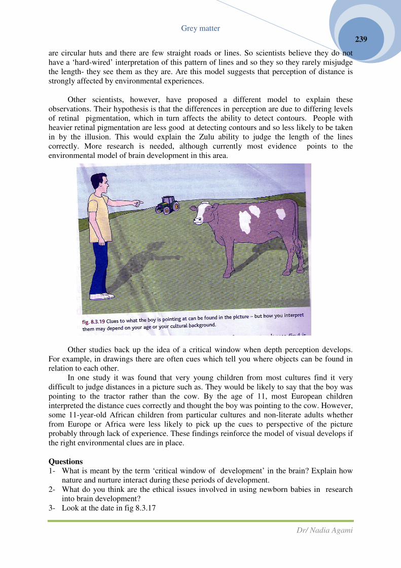
Grey matter
Dr/ Nadia Agami
239
are circular huts and there are few straight roads or lines. So scientists believe they do not
have a ‘hard-wired’ interpretation of this pattern of lines and so they so they rarely misjudge
the length- they see them as they are. Are this model suggests that perception of distance is
strongly affected by environmental experiences.
Other scientists, however, have proposed a different model to explain these
observations. Their hypothesis is that the differences in perception are due to differing levels
of retinal pigmentation, which in turn affects the ability to detect contours. People with
heavier retinal pigmentation are less good at detecting contours and so less likely to be taken
in by the illusion. This would explain the Zulu ability to judge the length of the lines
correctly. More research is needed, although currently most evidence points to the
environmental model of brain development in this area.
Other studies back up the idea of a critical window when depth perception develops.
For example, in drawings there are often cues which tell you where objects can be found in
relation to each other.
In one study it was found that very young children from most cultures find it very
difficult to judge distances in a picture such as. They would be likely to say that the boy was
pointing to the tractor rather than the cow. By the age of 11, most European children
interpreted the distance cues correctly and thought the boy was pointing to the cow. However,
some 11-year-old African children from particular cultures and non-literate adults whether
from Europe or Africa were less likely to pick up the cues to perspective of the picture
probably through lack of experience. These findings reinforce the model of visual develops if
the right environmental clues are in place.
Questions
1- What is meant by the term ‘critical window of development’ in the brain? Explain how
nature and nurture interact during these periods of development.
2- What do you think are the ethical issues involved in using newborn babies in research
into brain development?
3- Look at the date in fig 8.3.17

Grey matter
Dr/ Nadia Agami
240
a- How many severely neglected children were studied?
b- How many of the neglected children studied had brains that fell on or below the
bottom 10 percentile in size?
c- How would you explain this effect of sever neglect in terms of the development of
the brain?
4- Summarise the main methods by which the relative contributions of nature and nurture to
brain development may be studied. For each one discuses the strengths and weaknesses
of the method.
Learning and memory
Another way of considering the impact of nature and nurture on the development of the
brain is to look at the changes in the brain during learning, as memories are laid down.
Learning is usually observed through changes in behaviour. There are two main types of
behaviour, innate or species-characteristic behaviour and learned or individual-characteristic
behaviour. Both can tell us a considerable amount about the brain and how it works, but here
is only space for a very brief overview in this book.
Species-characteristic (innate) behaviour
Innate behaviour is a large collection of responses which are usually seen in every
member of a particular species hence species-characteristic. This type of behaviour is not
learned. It is a genetically determined response to a particular stimulus, and it occurs as
specific nerve pathways are laid down in the embryo from the instruction of the DNA.
Innate behaviour covers an enormous range of types of response, from the simplest
avoidance reflexes to highly complex courtship and territorial displays. The stimulus for a
piece of innate behaviour will always elicit the same response, which has been selected over
generations for its survival value. Nature is completely dominant in the development of these
neural pathways in the brain.
This type of behaviour is important in non-vertebrates which are frequently very short-
lived. They do not have time to learn by trial-and-error if they are to successfully complete
their life cycles. But even in vertebrates, instinctive behaviour is important both in the young,
which have not had time to learn by trial-and-error, and in adults where a ready-made set of
responses to a given situation leaves the higher areas of the brain free for more complex
learned behaviour.
Individual-characteristic (learned) behaviour
Learned behaviour is an adaptive change in the behaviour of an individual which occurs
as the result of experience. An individual learns from an experience, and modifies their
behaviour accordingly. An example of learned behaviour is the reaction of a child to
touching a hot oven door. The hand will be withdrawn rapidly as a result of a piece of reflex,
innate behaviour. But he child will learn from the experience, and is unlikely to touch the
door again deliberately. This learning will vary from child to child. Some will only need one
experience of the heat to modify their behaviour. Others will try the experiment several times
before the change in behaviour is made. Some individuals may not even need to touch the hot
door themselves seeing another child’s reaction or being warned by an adult may be enough.

Grey matter
Dr/ Nadia Agami
241
So nurture is important in the way the brain develops and learning takes place in
response to the environment but the ease with which learning takes place is at least in part
purely down to genetics (nature).
• Habituation occurs when a stimulus is repeated many times and nothing happens there is
neither “punishment” nor “reward”. The animal learns to simply ignore the stimulus and
make no response. It is not a simple adaptation of the sensory system because once a
response is habituated or lost it does not return unless the animal is specifically sensitized.
Examples include the way birds learn to ignore a scarecrow and babies learn not to
“startle” at every sudden noise. You will be looking at habituation in more detail.
• Conditioned reflexes are the result of animals learning to associate new stimuli with an
existing unconditioned reflex, as in the case of Pavlov’s dogs.
• Trial-and-error (operant) learning occurs when a piece of trial behaviour is either
rewarded (something good happens such as food is found) or punished (something bad
happens such as the animal is hurt). If the animal associates the outcome of a piece of
behaviour with a reward, that behaviour is likely to be repeated. If the behaviour is
associated with punishment, it is less likely to be tried again. The American psychologist
B.F. Skinner did a great deal of work on this type of learning using pigeons.
• Imprinting is a simple and specialised sort of learning which occurs only in very young
animals. At one receptive stage the young animal identifies with another organism, which
is usually the parent or, if no parent is available, another large object. It will then follow
this object and relate to other similar objects throughout its life.
• Exploratory (latent) learning takes place when an animal explores new surroundings and
learns them without any immediate reward or punishment. The information may then be
useful at another time.
• Insight learning is based on thought and reasoning. It is mainly seen in mammals,
particularly primates, and is regarded as the highest sort of learning. Once a problem has
been solved, the solution is then remembered.
Questions
1- What does innate behaviour tell us about the development of the brain?
2- How can animal behaviour be used to help compare the contributions of nature and
nurture to brain development?
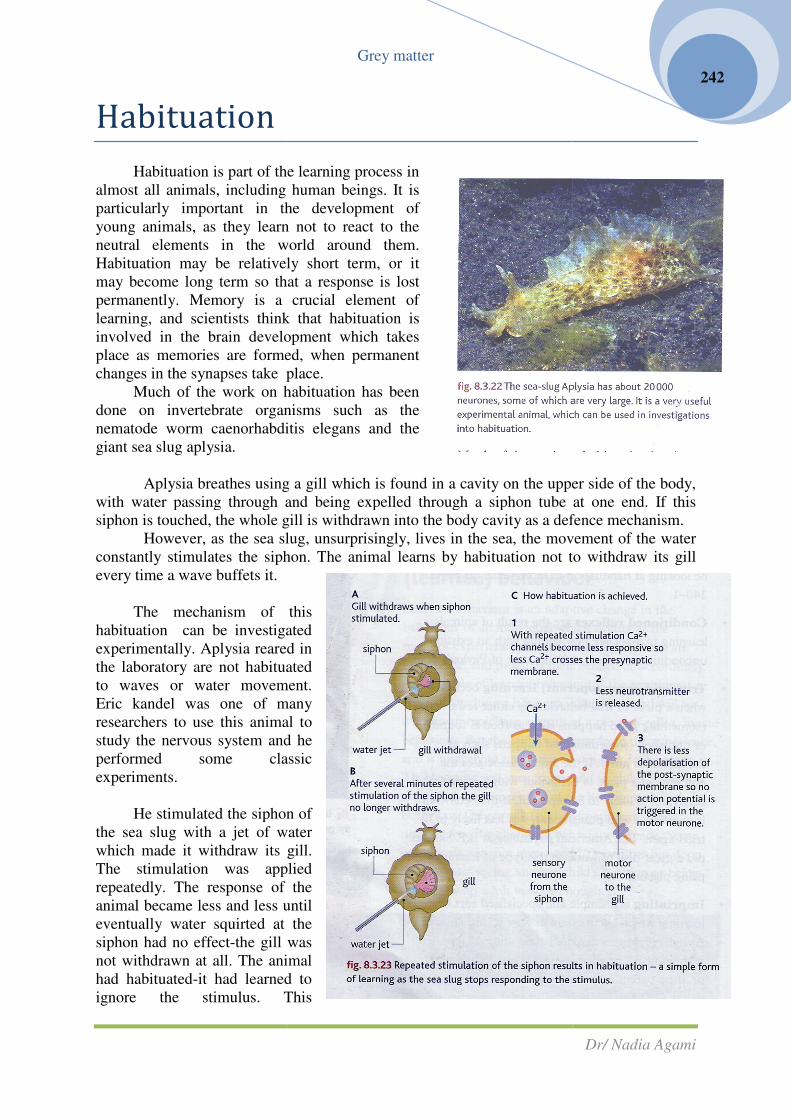
Habituation
Habituation is part of the learning proce
almost all animals, including
particularly important in the development of
young animals, as they learn not to react to the
neutral elements in the world around them
Habituation may be relatively short term, or it
may become long term so that a resp
permanently. Memory is a crucial element of
learning, and scientists think that habituation is
involved in the brain development which takes
place as memories are formed, when permanent
changes in the synapses take place.
Much of the work on habituation
done on invertebrate organisms such a
nematode worm caenorhabdi
giant sea slug aplysia.
Aplysia breathes using a gill which is found in a cavity on the upper side of the body,
with water passing through and being expelled
siphon is touched, the whole gill is withdrawn into the body cavity as a defence mechanism.
However, as the sea slug, unsurprisingly, lives in the sea, the movement of the water
constantly stimulates the siphon. T
every time a wave buffets it.
The mechanism of this
habituation can be investigated
experimentally. Aplysia reared in
the laboratory are not habituated
to waves or water movement.
Eric kandel was one of many
researchers to use this animal to
study the nervous system and he
performed some classic
experiments.
He stimulated the siphon of
the sea slug with a jet of water
which made it withdraw its gill.
The stimulation was applied
repeatedly. The response of the
animal became less and less until
eventually water squirted at the
siphon had no effect-the gill was
not withdrawn at all. The animal
had habituated-it had learned to
ignore the stimulus. This
Grey matter
is part of the learning process in
human beings. It is
particularly important in the development of
young animals, as they learn not to react to the
ements in the world around them.
Habituation may be relatively short term, or it
become long term so that a response is lost
rmanently. Memory is a crucial element of
learning, and scientists think that habituation is
involved in the brain development which takes
place as memories are formed, when permanent
changes in the synapses take place.
Much of the work on habituation has been
done on invertebrate organisms such as the
nematode worm caenorhabditis elegans and the
Aplysia breathes using a gill which is found in a cavity on the upper side of the body,
with water passing through and being expelled through a siphon tube at one end. If this
siphon is touched, the whole gill is withdrawn into the body cavity as a defence mechanism.
However, as the sea slug, unsurprisingly, lives in the sea, the movement of the water
constantly stimulates the siphon. The animal learns by habituation not to withdraw its gill
The mechanism of this
habituation can be investigated
experimentally. Aplysia reared in
the laboratory are not habituated
to waves or water movement.
ne of many
researchers to use this animal to
study the nervous system and he
performed some classic
He stimulated the siphon of
the sea slug with a jet of water
s gill.
The stimulation was applied
ponse of the
animal became less and less until
eventually water squirted at the
the gill was
not withdrawn at all. The animal
it had learned to
ignore the stimulus. This
Dr/ Nadia Agami
242
Aplysia breathes using a gill which is found in a cavity on the upper side of the body,
through a siphon tube at one end. If this
siphon is touched, the whole gill is withdrawn into the body cavity as a defence mechanism.
However, as the sea slug, unsurprisingly, lives in the sea, the movement of the water
he animal learns by habituation not to withdraw its gill

Grey matter
Dr/ Nadia Agami
243
habituation was retained over time, showing that it was not a simple case of accommodation .
Kandel then identified the neurones involved in the gill reflex and investigated what
happened when the siphon was synapses the calcium channels in the presynaptic membrane
become less responsive with repeated stimulations. With fewer calcium channels open, fewer
calcium ions cross into the presynaptic knob. As a result, fewer vesicles move to the
presynaptic membrane, fuse and discharge their neurotransmitter. With less neurotransmitter
available to bind to the post-synaptic membrane, the post-synaptic excitatory potential is not
high enough to trigger an action an action potential-
and so there is no response!
Investigating habituation
It is possible to investigate habituation in the
school laboratory. Various different organisms can be
used to demonstrate t his simple form of learning,
including worms, snails and even people!
Habituation in humans
Habituation is an important form of learning in
humans, in both the short term and the long term. For
example, if you visit someone who lives near a
railway line, the first few times a train passes you will
be startled or at least aware of the noise.
After a few hours, you will no longer
notice the trains-you have become
habituated. If you don’t visit again for some
time, your awareness of the trains passing
will be reinstated-but you will habituate
more rapidly on your second visit.
Habituation is very important in the
development of newborn babies who have a
number of startle reflexes. These gradually
become habituated as the baby grows and
matures until they are lost completely. For
example, the Moro reflex where a baby
arched its back, and throws its arms up and
out in a gasping movement if it is startled or
its head is lowered suddenly, is fading fast
in a normal baby within about 8 weeks of
birth, allowing the baby to interact more
calmly with the world around it. Modern
research suggests that one of the problems
for people affected by schizophrenia may be that they do not habituate as readily as other
people .
The study of the brain and behaviour continues-we still have enormous amounts to
discover about the workings of our own brains!

Grey matter
Dr/ Nadia Agami
244
Questions
1- Sea slugs and other similar non-vertebrates can transmit neurons. Why does this make
them such useful experimental animals?
2- How does habituation differ from accommodation and why is this important in the
development of the brain?
Brains, the genome
and medicine
The chemical balance of brains
Communication between the neurons
around your body and in your brain uses a
number of different neurotransmitters, some of
which are found only in brain synapses (e.g.
dopamine, serotonin).
An imbalance in these transmitters can
result in both mental and physical symptoms.
Treating diseases caused by such
imbalances means getting drugs across the
blood-brain barrier. This barrier is formed by
the endothelial cells that line the capillaries of
the brain which are very tightly joined together.
This makes it difficult for bacteria to cross into
the brain and cause infections, which is good,
but it also makes it difficult to get therapeutic
drugs into the brain. Drugs that affect the brain
are usually active at the synapses-and there are
a number of stages in synaptic transmission
where they can be targeted.
Parkinson’s disease
Parkinson’s disease involves the loss of nerve cells in an area of the midbrain known as
the supstantia nigra. In a healthy brain these cells produce the neurotransmitter dopamine,
and the axons from them spread thought the frontal cortex, the brain stem and the spinal cord,
so they are closely involved in the control and coordination of movement.
In Parkinson’s disease, these dopamine-producing cells die and motor control is
gradually lost. For a long time the brain compensates for the loss-most people don’t show
symptoms of the disease until around 8% of their dopamine- producing cells have gone
Parkinson’s disease usually develops in people over 50 years old, althought it can
appear in younger people. The causes are still not clear. In young-onset Parkinson’s disease,
the genetic link is much weaker, and other factors are also involved environmental triggers
may play a part and there may be a link with a number of toxins, herbicides and pesticides.
Around 120 000 people in the UK are affected by the condition.

Grey matter
Dr/ Nadia Agami
245
The ways individuals react to the falling levels of dopamine in Parkinson’s disease
vary. common symptoms include the following:
• Tremor (shaking) which usually begins in just one hand. This is the first symptom for
around 70% of affected people.
• Slowness of movements-it is hard to get started when you want to make a movement, and
movements take longer to perform.
• stiffness (rigidity) of the muscles-this can make it difficult to turn over in bed or stand up
after sitting in a chair.
Other problems that arise as the disease progresses may include poor balance, difficulty
in walking, problems with sleeping, depression and even difficulties with speech and
breathing.
Treating the symptoms
At the moment there is no cure for
Parkinson’s disease. However, there are a number
of drugs which can be very effective at easing or
delaying the symptoms. Most of the drugs aim
either to replace the natural dopamine in the brain
or to allow the body to make the best use of the
dopamine it still produces. Treatment can include
the following:
♦ Levodopa (L-dopa): dopamine cannot cross the
blood-brain barrier, so it cannot be used to treat
Parkinson’s. however L-dopa is a precursor of
dopamine which can cross the barrier and has
been in use since the 1960s. supplying the brain
with L-dopa allows the remaining cells to make
as much dopamine as possible. This can greatly
relieve stiffness and slowness of movements.
Eventually it becomes less effective as brain
cells continue to die off.
♦ Dopamine agonists: these chemicals bind to
dopamine receptors in brain synapses and mimic
the effect of dopamine. They are often used at
the beginning of the disease when they are most
effective, saving L-dopa for later.
♦ MAOB inhibitors: in AS biology you studied the
enzyme monoaminoxidase A (MAOA) in the context of genetics and behaviour.
Monoaminoxidase B (MAOB) is a similar enzyme which breaks down dopamine in the
brain synapses. MAOB inhibitors inhibit this enzyme, reducing the destruction of what
little dopamine gets made.

Grey matter
Dr/ Nadia Agami
246
HSW Developing new treatments for Parkinson’s disease
Research into possible new drug treatments for Parkinson’s disease continues. However
there are two more radical areas of research.
Gene therapy
Scientists are using knowledge gained from the results of the human Genome project
(see As biology) to help them develop gene therapies. Although Parkinson’s disease is not
a genetic disease, scientists are investigating the possibility of inserting healthy genes into
the affected cells.
There are two main approaches:
1- adding genes to prevent the dopamine-producing cells of the midbrain from dying.
2- and adding genes to enhance the levels of dopamine production in the remaining
cell. Both approaches have produced some encouraging results. However, there
are major problems in delivering the healthy genes to the cells of the midbrain,
and safety must be a prime concern, so using gene therapy like this is still some
years away.
Stem cell therapy
This research aims to provide a cure for Parkinson’s disease rather than a therapy to
relieve the symptoms. Scientists are hoping to use embryonic stem cells to replace the failing
dopamine-producing cells in the brains of Parkinson’s sufferers (as biology). Current research
using mouse models is very promising, but the ethical issue of using embryonic cells remain.
There is also the risk in using stem cells results. As scientists learn how to stimulate
embryonic stem cells to become dopamine-producing cells in the laboratory, it will also
provide them with a valuable tool for developing further and possibly more effective drug
therapies.
Depression
Everyone feels fed up or down from time to time,
and people often say they are feeling a bit depressed.
However, this is not true depression which is a serio us
illness that affects many people during their lives. The
causes of depression are complex and are not fully
understood. However, one cause may be problems with
the neurotransmitter serotonin in the brain.
Serotonin is the synaptic transmitter in a group of
cells in the brain stem which have axons that spread
throughout the brain into the cortex, the cerebellum
and the spinal cord. They have a widespread influence,
and low levels of serotonin result in fewer nerve
impulses travelling around the brain so overall brain
activity is suppressed. It has been shown that the
serotonin pathways are often abnormal in people
suffering from depression.

Grey matter
Dr/ Nadia Agami
247
Sometimes depression is triggered as a result of external factors such as work or
relationship stress or bereavement. At others it seems to be purely the results of chemical
changes in the brain. Serotonin is not the only neurotransmitter implicated in depression.
Dopamine and noradrenalin may also play a role in the condition for some people.
HSW Genetics and depression
As a result to research including the human Genome project (see As biology), scientists
have discovered a gene (5-HTT) which
appears to be linked to the production of
serotonin in the brain.
Evidence suggests that people with
the ‘short’ form of this gene are more likely
to suffer depression after stressful life
events than people with the ‘long’ allele.
The 20% of the population who are
homozygous for the short form seem most
likely to experience depression after
difficult times in their lives.
Heterozygotes appear to have an
intermediate risk and those homozygous for
the long form have a very low risk of
depression.
The gene codes for a reuptake protein
that enables sero tonin to be taken back into
the presynaptic membrane after release into
the synapse. Although this serotonin
reuptake protein is produced whichever
form of the gene is inherited, the long form of the gene means more of it is produced. People
who have homozygous long forms produce more reuptake proteins than heterozygotes who
produce more than homozygous short forms. The data in fig.8.4.4 come from an Australian
study was of a group of students over a 25-year period. The group was relatively small (127
members) but the information collected was detailed and the results support other larger
studies.
Building a hypothesis
There are some problems when it comes to using this evidence to develop a hypothesis
for the causes of depression. The evidence suggests that the more selective serotonin
reuptake proteins there are in the presynaptic membrane, the less likely you are to
suffer from depression after stressful life events. The role of these proteins is to remove
serotonin from the synaptic cleft. This, in turn, limits the stimulation of the post-synaptic
membrane without breaking down and destroying the serotonin. Scientists are not sure why a
larger number of these proteins reduce the risk of stress-related depression.

Grey matter
Dr/ Nadia Agami
248
The most successful treatment for depression is the use of SSRIs (see below) which
block these reuptake proteins and raise the levels of serotonin in the synapse, increasing the
levels of post-synaptic action potentials. So the working of the therapeutic drugs does not
seem to match the apparent role of the reuptake proteins. There is still a lot of research to be
done before we fully understand the biochemical basis of depression and how it may be
treated and avoided.
Treating depression
Treatment for depression includes ‘talking therapies’ which can help a patient come to
terms with adverse life events, and the use of drugs. Many of these drugs are linked to the
serotonin synapse systems and other neurotransmitters.
1- Some of the best-known antidepressant drugs are the SSRIs (selective serotonin
reuptake inhibitors). These drugs inhibit the reuptake proteins in the presynaptic
membrane, so that more serotonin remains in the synaptic cleft, more impulses
travel along the post-synaptic axon and the result is relief of symptoms such as a
lifting of the mood and an improved ability to sleep.
2- Other treatments also involve neurotransmitters. Tricyclic antidepressants (TCAs)
work by increasing the levels of serotonin and noradrenalin in the brain, while
3- monoaminoxidase inhibitors (like the MAOB used in treating Parkinson’s
disease) inhibit the enzymes which usually cause the breakdown of
neurotransmitters in the synapses of the brain.
Illegal drugs and the brain
Drugs such as L-dopa and SSRIs can have a clear and measurable effect on the way
your brain works. These drugs are therapeutic and legal.
Other drugs are enjoyable and legal, such as the caffeine from coffee, tea, colas or
energy drinks. Caffeine crosses the blood-brain barrier and affects the brain in a number of
way which include slowing down the rate of dopamine reabsorption at dopamine
synapses. But there are some drugs which are used specifically because they have an impact

Grey matter
Dr/ Nadia Agami
249
on the way the brain works. These drugs are often illegal. One which is widely used is
ecstasy (MDMA or 3,4- methylenedioxy-N-methylamphetamine).
Ecstasy has a marked effect on the brain. :
� It acts as a stimulant, increasing the heart rate, rather like amphetamine, and
also as a psychotropic drug which alters the way a person ‘sees’ the world. So
the short-term effects of the drug are to change the mood, making people feel
happy, sociable, full of energy, warm and empathetic.
� The drug brings about these changes by affecting the serotonin synapses of the
brain.
� Serotonin has a major effect on the way the brain works. Depression seems to
be the result of too little serotonin in some way. Ecstasy blocks the serotonin
reuptake transport system so that the synapses are completely flooded with
serotonin, which cannot be returned to the presynaptic knob.
� There is also some evidence that the drugs makes the transport system work in
reverse, so that all of the serotonin in the presynaptic knob is moved out into the
synapse which affects the post-synaptic membrane and floods the brain with
impulses.
� Ecstasy may also affect the dopamine systems, with the high levels of serotonin
stimulating the release of more dopamine, adding to the pleasure sensation, but
there is some debate among scientists about whether this is really the case.
Side effects of ecstasy :
� Using ecstasy causes physical changes, such as increased heart rate, and can
cause problems in the body’s thermoregulatory system.
� There may be no desire to drink which can lead to hyperthermia (overheating)
with raised blood pressure and irregular heartbeat. In a small number of cases
this can lead to death.
� Ecstasy can also affect the hypothalamus so that it secretes more antdiuretic
hormone, which is normally secreted when the body needs to conserve water.
This effectively stops the kidneys from producing urine and can lead to
problems if someone keeps drinking water in an attempt to stay hydrated and
cool down. They retain so much water that osmosis destroys their cell. This is
what killed Leah Betts in 1995.
HSW long-term brain damage from ecstasy
There are clear indications that the use of ecstasy can cause long-term damage to the
brain. Scientific evidence appears to show that ecstasy can cause permanent damage to the
serotonin neurones. Classic research using scans of the brain of non-drug users and long-term
ecstasy users showed that the serotonin-linked areas of the ecstasy users have a greatly
reduced ability to bind with the serotonin in normal condition.

Grey matter
Dr/ Nadia Agami
250
A Dutch study published by the British journal of psychiatry in 2008 used MRI scans
and other brain imaging techniques to show that long-term ecstasy use appears to cause both
damage to, and loss of, serotonin axons in the thalamus and lower than normal levels of
serotonin in the cerebral cortex. The study used 71 individuals with a broad variation in drug
use.
Some studies, however, have found no adverse effects from long-term drug use. A team
at the University of East London found ecstasy users outperformed non-drug users at some
tests. Some of the very influential early research was carried out on squirrel monkeys, and
there are arguments that monkey brains are in many ways different from human brains.
Because the drugs are illegal, some scientists have suggested there is a bias towards research
which shows adverse effects. However, ecstasy is now being investigated for possible
therapeutic use.
Questions:
1- Explain how a drug affecting each of the five stages of neurotransmission would have an
effect on how nerve impulses are transmitted and what this might mean in the body.
2- Explain the difference between dopamine and serotonin synapses and their roles in the
body.
3- Compare the action of the drugs L-dopa and ecstasy on the brain and their impact on
human health.
4- Describe the working hypothesis which explains the results of fig.8.4.4. How does the
evidence on the effectiveness of SSSRIs in treating depression undermine this
hypothesis? Suggest further investigations which might help to clarify the situation in the
brain during depression.
The human genome and medicine
The Human Genome project set out to identify all of the genes in the human
chromosomes and to sequence the 3 billion base pairs which make up the human DNA (As
biology). It was an international project and the DNA samples were then chosen
anonymously and at random for analysis.
The project also had specific aims about the storage and analysis of all the data
involved, with the ideal that all of the information about the genome should be freely
available to scientists everywhere. Consideration of the ethical, legal and social issues, which
are inevitably raised when such personal genetic information is unravelled, was also part of
the brief. The human genome project demonstrated that every individual has at least 99.9% of
their DNA in common!
Following on from this initial work another massive international project was set up in
2008. the 1000 genomes project will analyse and compare the DNA of at least a thousand
people from around the word, seeking to provide as much information as possible on
biomedically relevant variations. And as before, the information will be made freely available
to scientists around the globe.

Grey matter
Dr/ Nadia Agami
251
Pharmacogenomics
The ability to find and manufacture a medicine which has the right effect in the right
place is of vital importance in the fight against disease. Think of the impact of antibiotics-
many infectious bacterial disease that were life-threatening 70 years ago are treated easily
with a course of antibiotics today.
New medicines are currently developed from existing chemicals, often those extracted
from plants (As biology). However,
increasingly detailed knowledge of the
human genome has opened up a
number of new and potentially exciting
advances in different areas of drug
development. Each individual -part
from identical twins- is genetically
unique. Until now, drugs have been
produced to suit the majority of people.
In the future it may be possible to tailor
drugs to suit both individuals and
particular ethnic groups with
susceptibilities to specific disease. This
could help the drugs work more
efficiently, in lower doses, with fewer
side effects. The new science of
pharmacogenomics has been developed
to link pharmaceutical expertise with
knowledge of the human genome.
Genetic factors have a marked
effect on the efficiency of some drugs.
For example, when a relatively new
group of pain relievers called kappa-opioids was first tested, the drugs did not seem as
effective as the more commonly used opioids such as morphine and codeine. The initial tests
were done mainly on men. However, later research has shown that they are very effective
painkillers for women! The genetic makeup of the patient makes all the difference to the way
the drug works. Pale-skinned, red-haired women are more responsive to common painkillers
than anyone else and ibuprofen has little or no effect on pain relief in women during the
second half of their menstrual cycle. More detailed knowledge of our individual genetics and
have enormous benefits in terms of drug development.
Scientists expect that pharmacogenomics will also produce much more powerful drugs
because the medicines they design can be exactly targeted to specific diseases or to changes
in the proteins or genetic material of the cells. Knowing the fine details of the human genome
make it easier to produce medicines that affect pathogens or cancer cells but do not damage
healthy body cells.
Using the human genome project to find genome targets for particular diseases could
make it possible t isolate a suitable drug candidate more rapidly. Advances such as these
could be of enormous benefit, making it possible to treat more diseases, and to treat them
more effectively.

Grey matter
Dr/ Nadia Agami
252
Choosing the right drug
The outcomes of the human genome project have the potential to save the NHS a great
deal of money by making it much easier to prescribe the right drug for a patient at the right
dosage. This has important implications. A study carried out in two large hospitals in
Merseyside in 2004 looked at the causes of the admissions of 18820 hospital patients. The
research team found that 1225 admissions were due to adverse drug reactions-6.5% of all the
admission- and the median stay in hospital was 8 days.
More than 20 people in this study died as a result of an adverse drug reaction- around
1000 lose their lives in this way every years in the UK. Based on this research, the average
cost to the NHS each year of adverse drug reactions is around £466 million pounds, using 4%
of the total bed capacity. And that doesn’t take into account the impact of these problems,
which can lead to permanent damage to organs such as the kidneys, on the individuals and
their families in terms of lost earnings and quality of life. Access to a patient’s genome could
help doctors prescribe medication which will work with rather than against their patient’s
cells, reducing the chance of adverse reactions.
Choosing the right dosage
Currently drug dosage usually depends on the age or weight of a person. Genetic
information about individuals could allow doctors to understand just how rapidly a patient’s
body will respond to and excrete a particular medicine. Many people could have much lower
doses of medicines, while those who need it could be given higher doses.
One of the few examples of where pharmacogenomics is being used now is in
screening patients for drugs trials. It has been discovered that the cytochrome P450 gene
codes for certain liver enzymes that break down many different types of drugs so they can be
eliminated from the body, variants of the gene mean some people have less active or even
inactive forms of these enzymes, so they are particularly vulnerable to a drug overdose.
Before anyone is accepted as part of a clinical trial for a new medicine, they are given genetic
tests to see which form of cytochrome P450 genes they have to ensure it is as safe as possible
for them to take part in the trial.

Grey matter
Dr/ Nadia Agami
253
HSW social, moral and ethical aspects of pharmacogenomics
A number of issues will have an impact on how far and how quickly the new science of
pharmacogenomics goes. Many of these issues have social, moral and ethical implications as
well as scientific ones-which makes them all the harder to resolve. Below are some of these
issues for you to consider.
• Identifying genes which affect the response to a particular drug is currently difficult, time
consuming and expensive. There are a lot of drugs –and more to be discovered. There may
be more than one gene involved in the way the human body responds to any particular
drug. The interactions of genotypes and drugs are very complex. Is the potential benefit
worth the financial investment society needs to make? But if we have the ability to do this,
is it moral or ethical not to try?
• There are some tough economics to consider. At the moment the big pharmaceutical
companies develop one or two drugs to treat any particular disease which are effective for
most people. This one size fits all approach has so far worked reasonably well. What if
75% of the population can be treated successfully with one drug, but five different drugs
would be needed to provide the remaining 25% of the population with effective treatment?
An absolutist standpoint would be that if appropriate drugs for an individual can be made
available, they should be, regardless. A relativist position would be that costs and benefits
have to be weighed in the balance, and the money used to develop a drug to treat a small
number of people could be more effectively used elsewhere. Eventually society will
decide- and economics are bound to be part of that decision- making process.
• It may will be possible to tell people a particular drug won’t work for their particular
genetic makeup before alternatives are available. Is it ethical to leave people with certain
genetic variations with no treatment available to them? But is it moral or ethical to give
them drug knowing it will have no effect?
• There are huge financial implications in training doctors and pharmacists to recognise all
the possible drug permutations, and keeping them up to date. This has big financial
implications for society- but deciding not to do it would be an ethical minefield.
• To have medical treatments tailored to the exact genetic profile of an individual means
everyone will need to have their genome analysed and stored. However, there has been
considerable opposition to a DNA database for use in fighting crime or overcoming
immigration issues. For pharmacogenomics to have any future a DNA database becomes
vital- but once it is in place it would surely be used in criminal investigations too. Society
needs to have the ethical debate to decide if this is the route they want to follow before too
great an investment is made in the science of pharmacogenomics. The potential benefits
are enormous- but there are potential hazards as well.
HSW A “heart disease” gene and pharmacogenomics
In early 2000 an international team of scientists published a paper revealing evidence of
a genetic mutation that greatly increases the risk of an individual developing heart disease.
Across the world, around 1 person in 100 carries this mutation, but on the Indian sub-
continent that figure rises to 1 in 25 people. The mutation affects the formation of a specific
heart protein. The mutation doesn’t usually cause problems until people reach middle age the
bodies of younger people deal with the rogue protein and destroy it. As people get older this
mechanism stops working and level of the mutated protein build up. This begins to affect the
way the heart functions until the risk of developing heart disease is seven times higher than
for an unaffected individual. Scientists suggest this means that heart problems are almost
inevitable for people with the mutation. People have no idea that they carry the mutation at

Grey matter
Dr/ Nadia Agami
254
the stage when they are having children, so there is no selection pressure to prevent it being
passed on.
At the moment, nothing can be done to overcome the impact of his mutation. But
pharmacogenomics may well be the route to a solution. By working with the faulty genes it
may be possible to devise a treatment that will neutralise the rogue protein as people get
older. With suitable tests, treatment could begin as the body’s own breakdown mechanism
begins to fail. Alternatively, a drug might be developed that blocks the production of the
protein by interacting with the transcription process of the faulty gene. No one yet knows
how this potential health timebomb is going to be successfully tackled but it certainly seems
likely that pharmacogenomics will play a part. And because so many people are affected, it
will certainly be worth pharmaceutical companies investing the time and money needed to
develop a pharmacogenomcially active drug.
Questions
1- The science of pharmacogenetics depends on the outcome of the Human Genome
Project. Why?
2- What are the main potential benefits of pharmacogenomics?
3- Summarise the main social, moral and ethical issues that are raised by
pharmacogenomics. Describe at least two different positions that can be taken with
respect to each point you raise.
4- Using this resource and others, investigate and describe two new medical treatments that
depend on pharmacogenomics for their development.
Drugs from genetically modified organisms
Pharmacogenomics is an area of drug development and medical treatment that still has
a long way to go before it is in regular use. In AS Biology , you discovered how organisms
can be genetically modified by the insertion of a desired gene. Our growing knowledge of the
human genome is already being used in the development of genetically modified organisms
to produce medicines and vaccines to treat human conditions, but this science too is still in its
infancy.
Microorganisms the genetic modification for a number of reasons:
� They are relatively easy and cheap to culture,
� and because they reproduce so rapidly a transferred gene is copied very rapidly
when the microorganism are allowed to replicate in ideal conditions.
� It is very important to be able to tell which bacteria contain the desired,
recombinant piece of DNA. As one bacterium looks very like another, scientists
transfer special marker genes along with the desired DNA that make it easy to
pick out those microorganisms in which a successful transformation has taken
place. These markers are often characteristics such as antibiotic resistance. The
bacteria identified by the markers can then be cultured on a large scale in
industrial fermenters, and the proteins which they make harvested. One major
perceived risk of these microorganisms is that if they spread into the
environment they carry genes for antibiotic resistance with them.

Grey matter
Dr/ Nadia Agami
255
There is an ever-growing number of chemicals made by genetically engineered
organisms. They include antibiotics such as penicillin, hormone such as growth hormone and
enzymes.
Microorganisms and human insulin
As you saw in AS Biology, people with type 1 diabetes cannot make the insulin needed
for their bodies to work properly. Everyone with type 1 diabetes and some people who have
type 2 diabetes need regular injections of insulin to keep them healthy.
The source of insulin for people with diabetes used to be the pancreases of slaughtered
pigs, sheep and cattle. Although the insulin from these animals is similar to human insulin, it
is not quite the same. This caused problems for some patients because their immune systems
reacted to the foreign antigens on the animal insulin. Also the supply of insulin was not
always reliable, because it depended on how many animals were slaughtered.
So biotechnology was used to develop a way of manufacturing human insulin using
microorganisms. The process was difficult because the insulin molecule is made up of two
polypeptide chains. To tackle this problem, scientists in the 1980s managed to introduce a

Grey matter
Dr/ Nadia Agami
256
synthesized gene for each chain into different bacteria, which were then cultured in huge
numbers. The mixture from the giant fermenters needs downstream processing the
microorganisms and the desired end products have to be separated from the rest of the
mixture.
This produces two pure protein chains which are then oxidized to join them together.
The resulting chemical, often referred to as humulin, appears to function exactly like human
insulin. Its purity is guaranteed, making it easier to calculate doses accurately. It has been a
great advantage for the majority of people with diabetes. Even more recently, a synthetic
gene has been developed that mimics the normal human gene for insulin and allows
proinsulin to be made by just one type of engineered bacteria. At the end of the process,
enzymes convert proinsulin to insulin.
Using microorganisms in this way removes the problems of uncertain supply and
provides a constant, convenient and pure source a human hormone. this is a clear example of
the way genetic engineering and the new biotechnology have had a positive effect in human
medicine.
For most parents, the birth of a baby is followed by years of pleasure and amazement at
the way their offspring grow. But some children grow so slowly that it needs to be
investigated. They may lack growth hormone (GH) or somatotrophinm the hormone secreted
by the pituitary gland which stimulates growth.
For many years the problem was overcome by giving children regular injections of
pituitary extract, made from pituitary glands taken from human cadavers. The extract was
expensive and always in short supply. Genetic engineering has solved the problem of supply.
The hormone can now be produced in commercial amounts by genetically modified
microorganisms.
However, the solution came too late for some unfortunate children. Lack of care in the
extraction of the pituitary glands led to contamination of the drug by brain tissue from
individuals who had died as a result of the very rare Creutzfeldt- Jakob disease, a fatal brain
disease that has been linked to BSE (bovine spongiform encephalopathy, or’ mad cow
disease’) in cattle. Sadly, some people were treated with contaminated pituitary extract before
the new hormone was produced by genetically modified microorganisms, and they died of the
disease. people sometimes express concern about the risks of using genetically modified
organisms in can come from a wide variety of sources, and that genetically modified
organisms can also reduce some of these risks.
Banana genetically modified microorganisms have been fantastically successful at
making certain human proteins such as insulin. However, prokaryotes simply do not possess
the biochemistry to make some of the more complex human proteins, and so work has moved
on the introduce desirable human genes into eukaryotic cells including yeast, and more
recently plants and even mammals.
There are hopes that transgenic plants may become an important weapon in the
worldwide fight against disease. vaccination is a tried and tested way of eliminating serious
diseases. However, the challenge in less economically developed countries is that vaccines
can be relatively expensive and so the countries cannot afford them. They usually require
storing in a fridge and there may be cultural and practical difficulties with both getting to
healthcare workers and allowing them to vaccinate children. Nevertheless, if plants or plant

Grey matter
Dr/ Nadia Agami
257
products such as bananas, potatoes or carrots can be genetically modified to carry vaccines to
human diseases such as infant diarrhea or hepatitis B, then many of these problems are solved
in one go. The plants can be grown by the communities that need them, they are relatively
cheap, there is no need for cool storage and there is no need for trained healthcare workers to
be available for the children to be protected from deadly diseases. Trials are already in place
in the US and china and bananas are emerging as a prime candidate as plant medicines,
particularly for hepatitis B vaccines.
Making transgenic plants
Introducing genes from one type of plant into another, or even from an animal into a
plant, is usually achieved using the bacterium agro bacterium tumefaciens. A tumefaciens
causes tumours in plants which are known as grown galls. It contains a plasmid called the Ti
plasmid which transfers bacterial genetic information directly into the plant DNA. It is this
which normally causes abnormal growth of the plant cells, but modified plasmids can be used
to carry beneficial genes into the plant genome. Then, by the process of plant cloning (AS
Biology ), the modified transgenic cells can be used to produce whole new transgenic plants.
HSW choices for society
There are some concerns that using transgenic plants poses risks for the environment.
Just as in genetically modified bacteria, scientist usually include marker genes in genetically
modified plants to make it easy to identify them. These marker genes may include alleles
which ensure the plant is infertile, so that the plants cannot reproduce and fresh supplies of
transgenic plants have to be bought each time they are needed.
This is more of a concern where food crops are involved than where plants are being
modified to use a medicines, but some of the same issues arise. Also, if some of the
transgenic traits got into the wild population there might be issues of immunity developing to
certain treatments. Some people worry about taking in alien DNA in genetically modified
food plants. They forget that we eat many different types of DNA every day – and when the

Grey matter
Dr/ Nadia Agami
258
genetically modified food contains vital medicines, for many people the risk-benefit balance
shifts dramatically.
At the moment the use of transgenic plants in medicine is relatively new and unknown
– so concerns about risks in the general population are at a low level. Perhaps if the
technology becomes more widely used, or the media coverage becomes negative, worries will
grow!
Genetically modified animals
It is very difficult to transfer new DNA into eukaryotic cells but there have already
been some notable successes. Some of the most exciting and recent research involving
genetic engineering has been the work on inserting human genes into the tissues of sheep and
cattle to produce transgenic animals. In 1991 the first transgenic sheep was born, a ewe called
Tracey who produced the human protein alpha -1-antitrypsin (AAT) in her milk.
This protein is missing in people who suffer from a genetic condition that affects their
livers and lungs, causing emphysema to develop at a very early age.
The production of proteins using transgenic animals involves the introduction of a
copy of a human gene which codes for the desired protein into the genetic material of an egg
of a different animal species. As well as the gene for the specific protein there is also a
promoter sequence which makes sure the gene will be expressed in the mammary gland of
the lactating female. The fertilized and transgenic embryo is then replaced inside a surrogate
mother, is born and grows to maturity. When the animal is mature and produces milk, that
milk is harvested, purified and the human protein extracted.
Making transgenic animals
There are number of techniques for getting new DNA into mammalian cells, which are
being tried with varying degrees of success. Whatever technique is used it must get the new
DNA through the cell membrane and into the cytoplasm –or nucleus – where it can be
accepted and incorporated into the host cell DNA. The methods include the following:
♦ Microinjection (DNA injection) where DNA is injected into a cell through a very fine
micropipette. This s manipulated using a micromanipulator, because the steadiest hand
would tremble enough to destroy the cell. The method is rather hit and miss – many cells
have to be injected before one takes up the DNA successfully, but it is the method that has
resulted in many of the best known transgenic animals – for example, Tracey.
♦ Microprojectiles: - DNA is shot into the cell at high speed carried on minute gold or
tungsten pellets. Some cells survive this treatment and accept the DNA.
♦ A harmless virus can be engineered to carry a desirable gene and then used to infect the
animal’s cells, carrying the DNA with it.
♦ Liposome wrapping – the gene to be inserted is wrapped in liposomes (spheres formed
from a lipid bilayer). These fuse with the cell membrane and can pass through it to deliver
the DNA into the cytoplasm.
So far transgenic sheep, cows, pigs, rabbits and mice have been produced, all capable
of producing human proteins in their milk. Using large animals such as cows and sheep, large

Grey matter
Dr/ Nadia Agami
259
volumes of milk – and therefore human proteins – can be achieved, and the animals can be
increased by breeding or cloning.
Examples of drugs from transgenic animals
More than 20 different human proteins have been produced so far from transgenic
animals, and some of them are already in therapeutic use or are being trialled.
♦ Factor VII and Factor IX are both important components of the human blood clotting
cascade which can be missing in haemophilia and other blood clotting diseases. These
factors are being harvested from transgenic milk.
♦ Alpha-1 antitrypsin is the protein produced by Tracey. The milk of such transgenic sheep
contains up to 35g of human protein in every liter, a very high yield. The milk is very
expensive – it costs several thousand pounds per liter. Trials are underway on treatment
using the transgenic protein and the idea is that as the numbers of transgenic animals
increase, the price will eventually come down. However, there have been major problems
with developing these products commercially. Most of the transgenic sheep were
destroyed when the company that produced them hit financial difficulties.
♦ Activated protein C for treating deep vein thrombosis and modified milk for people with
lactose and other intolerances are also being tested.
As we unravel the mysteries of the human genome in ever more detail, we should come
to know the genetic sequence of every chemical in our body. If many of our faulty molecule
can be made for us by transgenic animals, the impact on future health and medicine could be
enormous.
There are many benefits to using genetically modified organisms to produce drugs both
for humans and for animals. However, there are some aspects of this work which are
controversial and raise a number of ethical issued. As with genetically modified food, there
are concerns about the salary the safety of these products both for the humans that use them
and for the animals that are used to produce them. Environmental concerns about gene
transfer to wild species are also very much present.
Another area of real concern is the fact that this type of technology will be the property
of just a few companies in developed countries. The new advances may be largely based
towards the need of the richer countries – although the work of plant – based vaccines tends
to negate that fear.
Some people have strong objections to the use of animals and other organisms in this
way and feel that genetic modification violates the rights of the organism modified.
These are just some of the areas of concern which have been raised and discussed –
there are others. However, there are many people who feel that the enormous benefits these
developments may bring in medical treatments for outweigh any risks or ethical stumbling
blocks which may arise.
Questions
1- Produce a flow diagram to explain the process by which a drug is made using a
genetically modified bacterium.
2- Identify two ethical positions on the use of genetically modified bacteria in the
production of drugs.

Grey matter
Dr/ Nadia Agami
260
3- a. Compare the production of a drug from a genetically modified plant with that from a
genetically modified microorganism.
b. What are the risks and benefits of using genetically modified plants to produce
vaccines?
4- Investigate the use of genetically modified animals to produce human medicines. Select
one drug and evaluate the success of this process so far.
5- Discuss the risks and benefits associated with the use of genetically modified organisms
(other than the transgenic animals described in your answer to question 4) in the
production of drugs and for other purposes, using example from this text and from your
own research.