aacn pccn webinar session 3 pulmonary
TRANSCRIPT

AACN PCCN Webinar
Session 3 Pulmonary
Presenter: Carol A. Rauen, RN, MS, CCNS, CCRN, PCCN, CEN
Independent Clinical Nurse Specialist & Education Consultant [email protected]

Session 3: Pulmonary
1
Pulmonary
I. INTRODUCTION
AACN-PCCN Blueprint - 14% a. Acute Lung Injury (ALI) b. Exacerbation of COPD c. Obstructive Sleep Apnea d. Pleural Space Abnormalities and Complications (e.g. pneumothorax, Hemothorax, pleural
effusion, empyema) e. Pulmonary Embolism f. Pulmonary Hypertension g. Respiratory Depression (e.g. Mediation-Induced, Decreased-LOC Induced) h. Respiratory Failure
Acute
Chronic i. Respiratory Infections (e.g. Pneumonia) j. Severe Asthma k. Thoracic Surgery
Lobectory
Pneumonectomy
Pulmonary Testable Nursing Actions a. Perform a comprehensive pulmonary assessment b. Monitor normal and abnormal diagnostic test results c. Interpret ABGs and report findings d. Monitor patient for response to pulmonary medications e. Manage patients requiring non-invasive O2 or ventilation delivery systems
nasal cannula, face masks, venti-masks , non-rebreather mask, BiPAP, CPAP f. Manage patients requiring mechanical ventilation – tracheostomy tube g. Manage patients requiring respiratory monitoring devices
continuous SPO2, intermittent SPO2 , end-tidal CO2 (capnography) h. Recognize signs and symptoms of respiratory complications, and seek assistance as needed i. Maintain airway j. Manage patients with chest tubes k. Assist with procedures
thoracentesis, chest tube insertion l. Administer Medications for procedural (conscious) sedation and monitor patient response
Session 3: Pulmonary
2
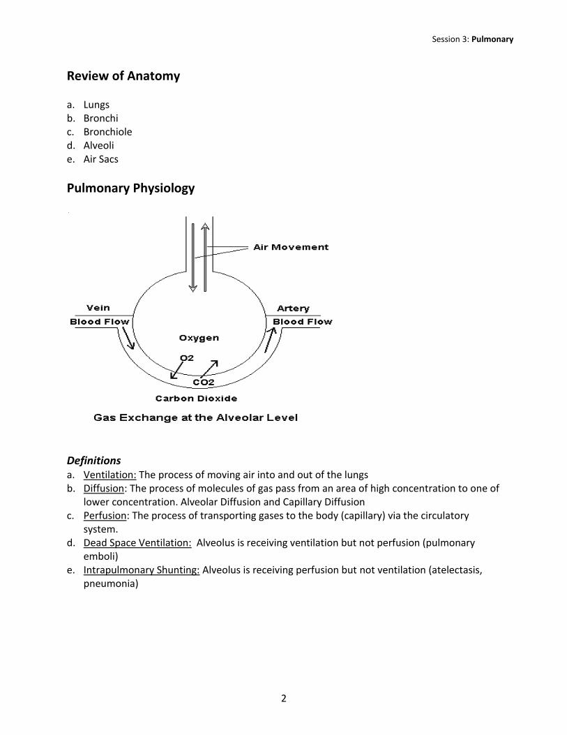
Review of Anatomy a. Lungs b. Bronchi c. Bronchiole d. Alveoli e. Air Sacs
Pulmonary Physiology
Definitions a. Ventilation: The process of moving air into and out of the lungs b. Diffusion: The process of molecules of gas pass from an area of high concentration to one of
lower concentration. Alveolar Diffusion and Capillary Diffusion c. Perfusion: The process of transporting gases to the body (capillary) via the circulatory
system. d. Dead Space Ventilation: Alveolus is receiving ventilation but not perfusion (pulmonary
emboli) e. Intrapulmonary Shunting: Alveolus is receiving perfusion but not ventilation (atelectasis,
pneumonia)

Session 3: Pulmonary
3
II. ASSESSMENT AND GAS EXCHANGE
Work of Breathing a. Respiratory Rate at Rest? b. Pulse Ox c. Color? Central and Peripheral (Clubbing?) d. Dyspnea? e. Breathing Effort Visibly Labored? f. Use of Accessory Muscles? g. Breath Odor? (Sweet or Urine odor) h. Pain Associated with Deep or Rapid Breathing? i. Shallow or Uneven Expansion? j. Cough? Productive? Description of Sputum? k. Physical Position? Can They Breathe in a Supine Position? Orthopnea? l. History of Pulmonary Disorder?
Smoking*
Bronchitis
Emphysema
Asthma
Pneumonia
TB
Environment Pulmonary Stressor
*Smoking cessation is always a teachable function of nursing
Auscultation a. Bronchial (Tracheal) b. Bronchovesicular c. Vesicular
Absent
Diminished
Displaced
Pleural Friction rub
Fine Crackles (rales)
Course Crackles (rales)
Rhonchi
Wheeze
Stridor
Session 3: Pulmonary
4
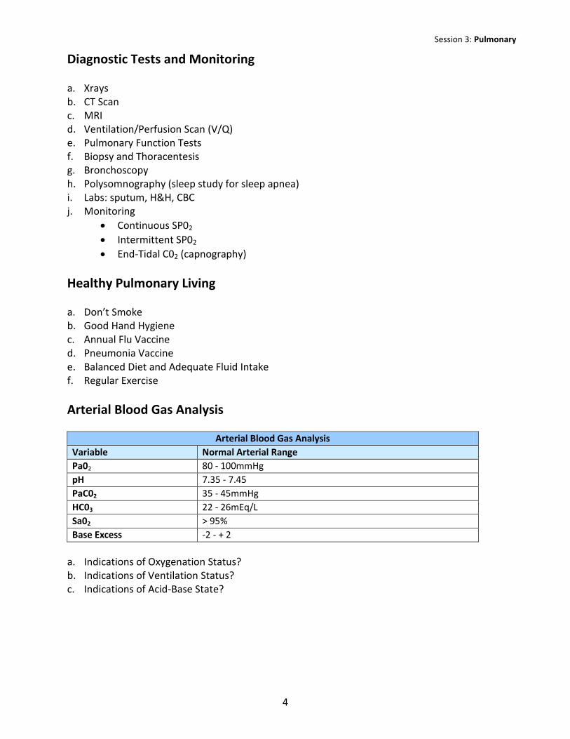
Diagnostic Tests and Monitoring a. Xrays b. CT Scan c. MRI d. Ventilation/Perfusion Scan (V/Q) e. Pulmonary Function Tests f. Biopsy and Thoracentesis g. Bronchoscopy h. Polysomnography (sleep study for sleep apnea) i. Labs: sputum, H&H, CBC j. Monitoring
Continuous SP02
Intermittent SP02
End-Tidal C02 (capnography)
Healthy Pulmonary Living a. Don’t Smoke b. Good Hand Hygiene c. Annual Flu Vaccine d. Pneumonia Vaccine e. Balanced Diet and Adequate Fluid Intake f. Regular Exercise
Arterial Blood Gas Analysis
Arterial Blood Gas Analysis Variable Normal Arterial Range Pa02 80 - 100mmHg pH 7.35 - 7.45 PaC02 35 - 45mmHg HC03 22 - 26mEq/L Sa02 > 95% Base Excess -2 - + 2
a. Indications of Oxygenation Status? b. Indications of Ventilation Status? c. Indications of Acid-Base State?
Session 3: Pulmonary
5
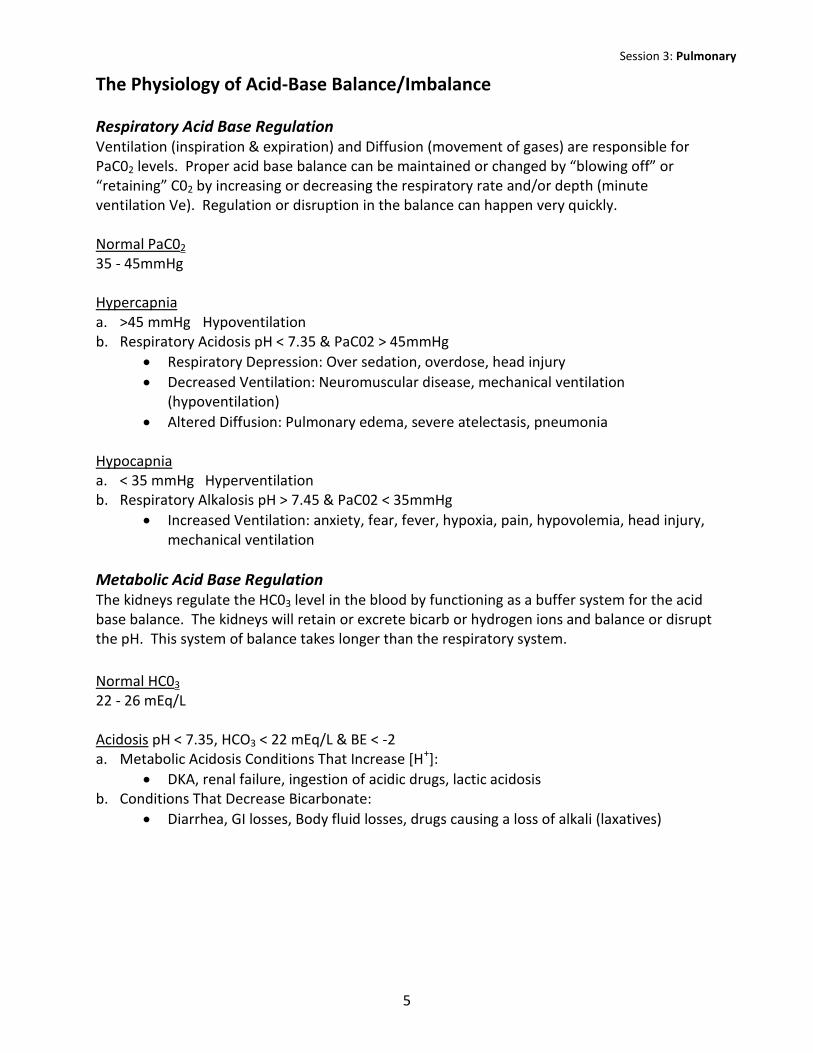
The Physiology of Acid-Base Balance/Imbalance
Respiratory Acid Base Regulation Ventilation (inspiration & expiration) and Diffusion (movement of gases) are responsible for PaC02 levels. Proper acid base balance can be maintained or changed by “blowing off” or “retaining” C02 by increasing or decreasing the respiratory rate and/or depth (minute ventilation Ve). Regulation or disruption in the balance can happen very quickly. Normal PaC02 35 - 45mmHg Hypercapnia a. >45 mmHg Hypoventilation b. Respiratory Acidosis pH < 7.35 & PaC02 > 45mmHg
Respiratory Depression: Over sedation, overdose, head injury
Decreased Ventilation: Neuromuscular disease, mechanical ventilation (hypoventilation)
Altered Diffusion: Pulmonary edema, severe atelectasis, pneumonia Hypocapnia a. < 35 mmHg Hyperventilation b. Respiratory Alkalosis pH > 7.45 & PaC02 < 35mmHg
Increased Ventilation: anxiety, fear, fever, hypoxia, pain, hypovolemia, head injury, mechanical ventilation
Metabolic Acid Base Regulation The kidneys regulate the HC03 level in the blood by functioning as a buffer system for the acid base balance. The kidneys will retain or excrete bicarb or hydrogen ions and balance or disrupt the pH. This system of balance takes longer than the respiratory system.
Normal HC03 22 - 26 mEq/L Acidosis pH < 7.35, HCO3 < 22 mEq/L & BE < -2 a. Metabolic Acidosis Conditions That Increase [H+]:
DKA, renal failure, ingestion of acidic drugs, lactic acidosis b. Conditions That Decrease Bicarbonate:
Diarrhea, GI losses, Body fluid losses, drugs causing a loss of alkali (laxatives)
Session 3: Pulmonary
6
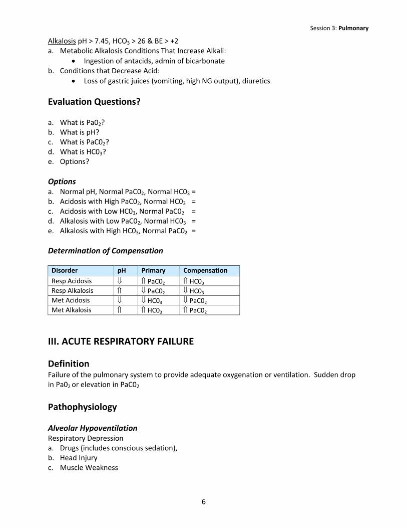
Alkalosis pH > 7.45, HCO3 > 26 & BE > +2 a. Metabolic Alkalosis Conditions That Increase Alkali:
Ingestion of antacids, admin of bicarbonate b. Conditions that Decrease Acid:
Loss of gastric juices (vomiting, high NG output), diuretics
Evaluation Questions? a. What is Pa02? b. What is pH? c. What is PaC02? d. What is HC03? e. Options?
Options a. Normal pH, Normal PaC02, Normal HC03 = b. Acidosis with High PaC02, Normal HC03 = c. Acidosis with Low HC03, Normal PaC02 = d. Alkalosis with Low PaC02, Normal HC03 = e. Alkalosis with High HC03, Normal PaC02 =
Determination of Compensation
Disorder pH Primary Compensation
Resp Acidosis PaC02 HC03 Resp Alkalosis PaC02 HC03 Met Acidosis HC03 PaC02 Met Alkalosis HC03 PaC02
III. ACUTE RESPIRATORY FAILURE
Definition Failure of the pulmonary system to provide adequate oxygenation or ventilation. Sudden drop in Pa02 or elevation in PaC02
Pathophysiology
Alveolar Hypoventilation Respiratory Depression a. Drugs (includes conscious sedation), b. Head Injury c. Muscle Weakness
Session 3: Pulmonary
7
Ventilation-Perfusion Mismatching a. Increased Dead Space b. Intrapulmonary Shunting
Diffusion Impairment a. Hypoventilation b. Low CO States c. Low H/H d. Decreased 02 Consumption: Sepsis, Toxins
Treatment Options a. Ventilate b. Oxygenate c. Treat Underlying Cause d. Treat Acid Base Imbalance e. Supportive Therapy
Airway Management & Oxygen Therapies Many pulmonary disorders and acute respiratory distress/failure are treated with airway devices and oxygen therapies. Review devices: oral/nasal airways, nasal cannula, face masks, CPAP, BiPAP – the use, assessment, potential complications and nursing care concerns.
IV. RESTRICTIVE LUNG DISORDERS Pulmonary Disorders that restrict the lungs from expanding. Lung compliance and volumes are decreased.
Examples include: a. Acute Lung Injury b. Bacterial Infections
Pneumonia
TB
RSV
Lung Abscess
Emphysema
Fungal Infections c. Occupational Lung Diseases d. Sarcoidosis e. Atelectasis

Session 3: Pulmonary
8
Common Signs and Symptoms a. Refractory Hypoxemia b. Dyspnea c. Increased WOB, Shallow Breathing
Acute Resp Distress Syndrome (ARDS) ARDS is a syndrome, not a disease; it is a group of physical manifestations that are primary pulmonary and result from direct or indirect lung injury followed by a significant inflammatory insult. The inflammation and resultant chemical mediator release cause increase capillary permeability, pulmonary edema, and alveolar collapse. These manifestations can, and fre-quently do, cause lung damage, failure and subsequently death. Treatment typically requires intubation and mechanical ventilation (in the ICU setting) and eliminating the causative factor.
Pneumonia An Inflammatory Process of the Lung Parenchyma Caused by Infection that Leads to Alveolar Consolidation.
Etiology Origin a. Bacterial (75% CAP Streptococcus Pneumoniae) b. Viral (RSV primarily in children, highly contagious by droplets, inflammation in
small airways leads to obstruction) c. Fungal d. Aspiration Site a. Bronchial b. Alveolar c. Lobar Source a. Community Acquired Pneumonia (CAP) b. Hospital Acquired Pneumonia (HAP) c. Ventilator Acquired Pneumonia (VAP)
Pathophysiology a. Lower Respiratory Tract Invasion b. Inflammatory Reaction c. Increased Capillary Permeability d. Phagocytotic Cells Migrate to Site e. Alveoli Fill with Exudate

Session 3: Pulmonary
9
f. Impair Gas Exchange From Shunting
Clinical Presentation a. Dyspnea & Tachypnea b. Productive Cough c. Pleuritic Chest Pain d. Fever, Chills, Rigors, Fatigue e. Anorexia f. Night Sweats g. Pleural Effusion h. Crackles, Rhonchi i. Tachycardia
Diagnostic Measures a. Chest X-Ray: Localized Infiltration b. Sputum Culture: Positive for Microbes c. CBC: Positive for Infection Elevated WBC Count d. Bronchoscopy: Visualize Inflammation/Consolidation
Treatment Options a. Antibiotics b. Oxygen c. Mechanical Ventilation d. Positioning: Good Lung Down e. Fluids & Humidification f. Pulmonary Hygiene g. Manage Fever & Pain h. Prevention
Hand Washing
Sterile Suctioning
Mouth Care
HOB > 450
Pneumococcal Vaccine
Stress Ulcer Prophylaxis
Extubate ASAP
V. OBSTRUCTIVE LUNG DISORDERS Pulmonary Disorders where airway obstruction and gas trapping are the primary problem.
Expansion and compliance of the lung tissue is not the problem. Examples include: a. Chronic Obstructive Pulmonary Disease (COPD)
Emphysema
Bronchitis b. Asthma
Session 3: Pulmonary
10
Chronic Obstructive Pulmonary Disease
Etiology Bronchitis a. An inflammatory response to an irritant (infectious or noninfectious) that results in
vasodilation, congestion, mucosal edema, and bronchospasm. Affects the small and large airways rather than the alveoli.
Chronic Bronchitis: Chronic cough w sputum production > 3 months per year for 2 successive years.
Emphysema Smoking is the #1 cause. Other causes include occupational exposure to certain particles (coal dust, asbestos, firefighters) & Alph1-Antitryspsin Disease.
Pathophysiology (Emphysema) a. Irritation and Inflammation of Bronchioles Mucus Production Obstruction Tissue
Injury Decrease Surfactant Bronchiolar Collapse b. Obstruction Air Trapping and Distention of Alveoli Enlargement of Air Sacs and Loss of
Elastic Recoil Multiple Alveoli Actually Fuse to One Large One Decreasing Surface Area for Gas Exchange
c. Increases in FRC d. Hypoxia e. V/Q Mismatch f. Pulmonary Hypertension g. Increased RV Afterload Right Heart Failure (Cor Pulmonale) RV Hypertrophy Drop
in LV Filling/CO
Clinical Presentation a. Dyspnea on Exertion Dyspnea at Rest b. Productive Cough Non Productive Cough c. Tachypnea with Small Tidal Volume d. Dropping FEV1 e. Malnutrition/Muscle Wasting (including diaphragm) f. Increase in AP Diameter g. Diminished Breath Sounds in Bases h. PFTs:
Increased: FRC, RV, TLC
Decreased: FEV1, TV i. ABG: Hypoxia w Respiratory Acidosis. Over Time will Develop a Degree of Metabolic
Compensation j. Example: Pa02 71, PaC02 52, pH 7.29, HC03 34, Sa02 72 k. Chest X-Ray: Flattened Diaphragm, Decreased Vascular Markings and Bullae l. Right Heart Failure m. Chronic Multi-System Dysfunction Related to Chronic Hypoxemia and Hypercapnia

Session 3: Pulmonary
11
Acute Care Concerns COPD is a chronic medical condition. It is relevant to the PCCN exam because these patients are frequently admitted for problems that require acute care as a result of their chronic debilitated state. Assessment and treatment options MUST take into consideration their pulmonary function and dysfunction. Common Reasons for Acute Care Admission a. Pneumonia b. Heart Failure c. Pulmonary Emboli d. Respiratory Failure e. Bronchospasm f. Spontaneous Pneumothorax g. None Compliance with Pulmonary Medical Therapies
Treatment Options a. Treat Primary Cause of Admission b. Oxygen Administration (with caution) c. Hydration & Humidification d. Removal of Secretions e. Pharmacology
Antibiotics
Steroids
Bronchodilators
Mucolytics f. Nutritional Support (high calorie, low carbohydrate)
Severe Asthma
Etiology A hyperactive airway due to an intrinsic or extrinsic factor. Common causes include: a. Respiratory Infection b. Allergic Reaction to Inhaled Antigen c. Emotional Stress d. Exercise e. Idiosyncratic Reaction to NSAID or Beta Blocker f. Environment Toxin g. Mechanical Stimulation (coughing, laughing, cold air) h. Reflux Esophagitis
Pathophysiology a. A disease of inflammation that precipitates bronchospasm (obstruction). b. Affects airways not alveoli and the bronchospasm is reversible c. Inflammation precipitates mucus production (more obstruction)
Session 3: Pulmonary
12
d. Obstruction leads to air trapping and difficulty with expiration (harder, longer and less effective)
e. Decreased Oxygenation and Carbon Dioxide Removal f. Acute Asthma or Status Asthmaticus: the individual’s “typical” asthma therapies don’t work,
the bronchospasm, mucus production and air trapping continue potentially to the point where there is no air movement.
g. Hyperinflation increases intrathoracic pressures which decreases venous return and increases RV afterload
Clinical Presentation a. Stimulation of Asthma Unrelieved by Typical Tx b. Increased Work of Breathing c. Rapid RR with Little Air Movement (Air Trapping) d. Long Expiratory Phase e. Expiratory Wheezes Initially Minimal to No Air Movement on Inspiration or Expiration f. Pulsus Paradoxus g. Restless and Anxious, Calming Down is a Bad Sign
Diagnostic tests a. ABG: Hypoxia and Hypercapnia – Resp Acidosis b. PFT: Drop in FEV1 & Peak Expiratory Flow
Treatment Options a. Psychological Support b. Oxygen Therapy c. Maybe Mechanical Ventilation (big ETT), Low TVs d. Remove Irritate (if known) e. Hydration & Humidification f. Pharmacological
Bronchodilators
ABX
Corticosteroids
Inhaled Anticholinergic Agents
Heliox
Sedatives & Muscle Relaxants
SubQ Epinephrine f. Monitor and Treat Pneumothorax g. Monitor and Treat Heart Failure
VI. OBSTRUCTIVE SLEEP APNEA (OSA) is the most common type. The pt experiences repeated episodes of apnea (stop breathing for > 15 seconds) during sleep secondary to upper airway obstruction. Central Sleep Apnea is not from upper airway obstruction but due to a dysfunction in the brain and sleep
Session 3: Pulmonary
13
center. These apnea episodes can last up to 20 seconds (see box for information specific to central sleep apnea).
Etiology a. Obesity (> 50% of pts) b. Neck Circumference > 17 inches c. Nasal Obstruction d. Men > Women e. Postmenopausal Women f. Age > 50 Years Old g. Smoking h. Hypertension
Pathophysiology In OSA there is an occlusion of the upper airway (nasopharynx and oropharynx) leading to obstruction. This typically happens during REM sleep because the muscle structures become hypotonic. The obstruction typically leads to snoring. The snoring and lack of ventilation awakens the patient, they clear the obstruction and return to sleep. The repeated apnea episodes and subsequent sleep interruptions lead to acute and potential chronic health problems.
Clinical Presentation a. Daytime Sleepiness/Fatigue b. Morning Headaches c. Cognitive, Personality, Behavioral Changes d. Increased Potential for Trauma e. Sexual Dysfunction f. Night Sweats g. Excessive Snoring at Night h. Repeated Sudden Awakenings i. Potential Long Term Complications
Hypertension (diurnal and pulmonary)
Cor Pulmonale
Stroke
MI
Diagnostic Tests a. Sleep Study (Polysomnography) b. Pulmonary Function Tests c. ABG d. Wt and Neck Measurements (not diagnostic)
Session 3: Pulmonary
14
Treatment Options a. Wt Loss b. Mechanical Tx – CPAP or BiPAP c. Dental or Oral Appliances (airways) d. Surgery e. Maintain HOB > 300 f. Avoid ETOH and Sedatives Before Sleep
Central Sleep Apnea Apnea episodes during sleep caused from a problem in the brain not obstruction in upper airway. Common etiologies include: a. Parkinson’s Disease b. Alzheimer’s Disease c. Damage to Brainstem: Encephalitis, Stroke TBI d. Cervical Spine Damage: Injury, Radiation, Severe Arthritis, Degenerative Bone Disorders Treatment centers on treating the primary cause of the apnea
VII. PULMONARY EMBOLI Occlusion in the pulmonary arterial circulation, blocking flow to a region(s) of the lung and creating dead space ventilation.
Etiology a. Fat b. Air c. Amniotic Fluid
d. Thromboembolic – 90% of all PEs from DVT Virchow’s Triad a. Venous Stasis
Immobility
Dehydration
Pregnancy/Oral Contraception/Hormone Replacement Therapy
Paralysis
Obesity
Session 3: Pulmonary
15
b. Hypercoagulability
A-Fib
Tumors/Cancer
Dehydration
Heart Failure (also immobility)
COPD Pt 20 A-Fib, Polycythemia
Previous PE c. Vascular Wall Damage
Trauma
Venous Catheters
Varicose Veins
Elevated LDL
Age
Pathophysiology a. The pathophysiology and presentation must be viewed on a continuum. It will depend on
the size of the blockage and length of time it has been occurring. b. Pulmonary Artery Obstruction c. V/Q Mismatching V > Q = Dead Space Initially d. Non Perfused Alveoli will Collapse Secondary to Decreased Surfactant Production
Intrapulmonary Shunting e. Pulmonary Infarction May Occur f. Increased Pulmonary Vascular Resistance Increases Afterload on Right Ventricule RV
Failure and Potentially Infarction
Clinical Presentation a. Sudden Onset Dyspnea & Pleuritic Chest Pain b. Tachypnea c. Refractory Hypoxemia d. ABG: Hypoxemia with Respiratory Alkalosis
Example: Pa02 71, PaC02 29, pH 7.59, HC03 25, Sa02 72 e. Fat Emboli: Petechiae on Thorax and Upper Extremities f. Cardiac
Tachycardia
Cyanosis
Jugular Venous Distention
RV Failure (Increased Resistance/Afterload & Loud S2)
ECG: RV Hypertrophy, T wave Abnormalities

Session 3: Pulmonary
16
Diagnostic Tests a. Chest X-Ray – Not Diagnostic but Rules Out Other Causes for the Respiratory Distress b. V/Q Scan c. Spiral CT d. + D-Dimer e. Pulmonary Angiogram f. MRI g. Lower Extremity Doppler Studies (not emergent)
Treatment Options a. ABCs b. Oxygen administration c. Intubated if Necessary d. Consider Thrombolytics e. Consider Embolectomy f. IVC Filter Placement g. Pain Management h. Identify Causative Factor and Treat i. Future Prevention
VIII. THORACIC SURGERY Common Thoracic Surgeries Most tissue related lung surgeries are for lung cancer. Segmental or wedge resections are also done for localized inflammatory processes, TB, or abscess. Lung reduction surgeries are primarily done for emphysema. Tracheal Surgery: Placement of temporary to permanent trach for airway management
Pneumonectomy: Removal of entire side of lung
Lobectomy: Removal of one lobe
Segmental Resection: Removal of one segment. Right side has 10 segments and the left has 8.
Wedge Resection: Only a portion, typically a well circumscribed diseased portion
Decortication: A fibrinous peel is removed from the visceral pleura, allowing for re-expansion of the lung. Typically done for empyema.
Session 3: Pulmonary
17
Nursing Care a. Oxygen Therapy b. Hemodynamic Monitoring: CVP c. Positioning d. Initiating Coughing and Deep Breathing e. Promote Abdominal Breathing f. Pain Management g. Nutrition h. Chest Tube Drainage System
Assisting with Chest Tube Insertion
Monitoring Patient with Chest Tube i. Assess for Subcutaneous Emphysema j. Assess for Thoracic Air Leaks k. Special Tx for Pneumonectomy:
Cannot lie on operative side
Palpate trachea for midline position
Pleural Space Abnormalities
Pathophysiology Air Enters the Pleural Space from a tear in the visceral or parietal pleura as the result of Blunt or Penetrating Chest Trauma. Iatrogenic causes include central line placement, invasive chest procedures (biopsy, thoracentesis) and mechanical ventilation (barotrauma). Occasionally spontaneous. Lung collapses because of the change in intrapleural pressure.
Clinical Presentation Classifications a. Tension Pneumothorax b. Simple Pneumothorax c. Hemothorax d. Hemo/Pneumothorax e. Pneumomediastium f. Sucking Chest Wound
Respiratory Distress (degree depends on classification)
Tachycardia and Hypotension
Diminished Breath Sounds Over Affected Area
Tension: Tracheal Deviation, JVD
Visualized on Chest X-Ray
Hypoxia on ABG

Session 3: Pulmonary
18
Treatment Options a. Emergent Needle Decompression (tension) b. Chest Tube Placement (re-expand lungs & evacuate air/blood)
Insert High for Pneumo
Insert Low for Hemo
Suction Utilized
Potential for Air Leak
Milking (not stripping) Recommended for Clot Removal c. Oxygen Delivery & Potentially Intubation d. Sucking Chest Wound: Occlusive dressing on expiration. Monitor status carefully might
need flutter valve e. Air Embolism: Trendelenberg position and Left Side to Trap Air in Heart (RV) f. Surgery May be Required