abbreviations - bib.irb.hrbib.irb.hr/datoteka/704162.final_revision-_hohsteter_cani… · web...
TRANSCRIPT
Canine testicular tumors: two types of seminomas can be differentiated by
immunohistochemistry
Marko Hohšteter1*, Branka Artuković1, Krešimir Severin2, Andrea Gudan Kurilj1, Ana Beck1,
Ivan-Conrado Šoštarić-Zuckermann1 and Željko Grabarević1
1 Department of Veterinary Pathology, Veterinary Faculty, University of Zagreb, Heinzelova
55, 10 000 Zagreb, Croatia
2 Department of Forensic and Judicial Veterinary Medicine, Veterinary Faculty, University of
Zagreb, Heinzelova 55, 10 000 Zagreb, Croatia
Branka Artuković: [email protected]
Krešimir Severin: [email protected]
Andrea Gudan Kurilj: [email protected]
Ana Beck: [email protected]
Ivan-Conrado Šoštarić-Zuckermann: [email protected]
Željko Grabarević: [email protected]
*Corresponding author: Marko Hohšteter, Department of Veterinary Pathology, Veterinary
Faculty, University of Zagreb, Heinzelova 55, 10 000 Zagreb, Croatia
e-mail: [email protected]

Abstract
Background: Testicular tumors are the most common genital neoplasms in male dogs, with
Leydig cell tumors (LCT), seminomas (SEM), and Sertoli cell tumors (SCT) the most
common forms. Human SEM are classified as classical (CSEM) or spermatocytic (SSEM).
Intratubular germ cell neoplasia of undifferentiated origin (IGCNU) is another form of human
testicular tumor. The aim of this study was to verify that CSEM/SSEM classification is valid
in dogs and confirm the existence of canine IGCNU.
Results: Testicular tumors were found in 46% of dogs at necropsy and accounted for 7% of
tumors biopsied. The median age of dogs with tumors at necropsy was 10.16 years; median
age at positive biopsy was 10.24 years. The most common tumors, in decreasing order, were
LCT, mixed tumors, SEM and SCT at necropsy, and SEM, SCT, mixed tumors, LCT,
peripheral nerve sheath tumor, and teratoma in the biopsy group. IGCNU was found in 3% of
testicles at necropsy and in 3% of biopsy samples. Two dogs had testicular tumor metastasis.
Expression of c-KIT was most common in SEM and seminomatous components of mixed
tumors. PLAP was mostly expressed in IGCNU, SEM, teratoma, and some mixed tumors.
Cytokeratin was mainly expressed in SCT. CD30 expression was low in both groups.
Conclusions: The high tumor incidence at necropsy can be attributed to older age. Tumor
incidence in biopsy samples, dog age, and histological classification were consistent with
previous studies. The higher incidence of SEM and SCT in the biopsy group probably resulted
from the obvious clinical expression of these tumor types. The low incidence of metastasis
confirmed the predominance of benign tumors. Low CD30 expression confirmed the low
incidence of testicular embryonal carcinoma. Cytokeratin helps differentiate stromal tumors,
especially SCT, from germ cell tumors. Histology and c-KIT and PLAP expression indicate
that IGCNU exists in dogs. Expression of c-KIT and PLAP confirmed that CSEM and SSEM
classification is valid in dogs.
Keywords: Dog, Testis, Tumors, Seminoma, Immunohistochemistry, Incidence, IGCNU
Background
Testicular tumors are the most common neoplasms of the genital system in male dogs and are
the third most common type of canine tumor after skin and fibrous tissue tumors [1].
Testicular tumors represent more than 90% of all canine male genital tumors and dogs have
the highest incidence of all animal species [2].
Investigations of the incidence of testicular tumors in dogs at necropsy have shown
somewhat discordant results. One dated study found an incidence of 16% [3], while a more
recent paper reported an incidence as high as 27% [4]. According to research conducted by
Gamlem et al., testicular tumors represented 7% of all biopsied tumors in dogs from 1990 to
1998 [5]. In another study, Vescalari et al. [6] found that male genital tumors represented 13%
of all biopsied tumors of male dogs from 2005 to 2008. About 40% of neoplastic testicles
have more than one tumor [7]. Testicular tumors are often classified as mixed tumors,
although they actually result from two different tumor types occurring in the same testis [8].
Primary testicular tumors are histologically classified into germ cell tumors, sex cord-
stromal (gonadostromal) tumors, and mixed germ cell-sex cord stromal tumors [9]. Within
these groups are the three most common canine testicular tumors, which have relatively
similar incidence varying by study. Sertoli cell tumors (SCT) and Leydig cell tumors (LCT)
are sex cord-stromal tumors and seminomas (SEM) are germ cell tumors [1, 4, 10].
Many immunohistochemical markers are used for differentiation of human testicular
tumors and although some of them have been studied in canine testicular tumors, information
about them is still insufficient. c-KIT is used in human patients for differentiation of c-KIT
positive CSEM from c-KIT negative SSEM [11, 12]. c-KIT is also expressed in human
IGCNU contrary to nonseminomatous germ cell tumors and stromal tumors which do not
express c-KIT [11-13]. In dogs some studies have shown that a certain percentage of SEM
express c-KIT [10, 13, 14] which is in disagreement with other reports that describe the

absence or very rare c-KIT expression in canine SEM [15,16]. PLAP is widely used marker
that is expressed in human IGCNU and very often in human CSEM [11, 12]. Investigations
by Grieco et al. [17] and Yu et al. [10] confirmed the expression of PLAP in some canine
SEM, but there are no data on PLAP expression in canine IGCNU. Cytokeratin AE1/AE3 is
used in human diagnostics as a marker for the differentiation of cytokeratine negative
testicular germ cell tumors from cytokeratine positive embryonal carcinoma, yolk sac tumors
and other carcinomas. Cytokeratin is also expressed in SCT and LCT [11, 18]. In veterinary
medicine, there are only few reports of cytokeratin expression in testicular tumors. In all of
them SEM showed no immunoreactivity to cytokeratin which is expressed mainly in SCT and
mixed SCT, and rarely in LCT [14, 19]. According to results of Banco et al. [19] cytokeratin
is not expressed in normal Sertoli cells, so this marker can be useful for differentiation of
SCT, not only from other testicle tumors but also from neoplastically unaltered Sertoli cells.
CD30 shows high expression in human simple and mixed testicular embryonal carcinoma and
is used for differentiation of this tumors from other germ cell tumors [11, 12]. Like in
humans, investigation by Yu et al. [10] confirmed the expression of CD30 in canine
embryonal carcinomas.
Doubts have been raised in recent studies about the classification of SEM in dogs.
Some studies have shown that SEM in dogs, as in humans, can be classified into two types:
classical (CSEM) and spermatocytic (SSEM) [10, 17, 20]. In contrast, Bush et al. [15] and
Thorvaldsen [16], found that canine SEM are predominantly spermatocytic, suggesting that
there are no (or extremely rare) cases of canine CSEM.
In humans, CSEM is the predominant SEM type, with a high incidence among young
men. CSEM originates from transformed gonocytes (prespermatogonia and spermatogonia),
while SSEM are neoplasms of older men and are derived from more differentiated germ cells,
mostly spermatocytes [15, 21-24]. It is probable that this different origin of SSEM determines
its predominantly benign behavior, in contrast to CSEM, which is malignant with a high
metastatic potential [20, 25]. Canine SEM is mostly benign; however, it does metastasize in a
small number of cases [26].
It is also not clear whether intratubular germ cell neoplasia of undifferentiated origin
(IGCNU) or carcinoma in situ is found in canine testicles. These tumors are very common as
precursor lesions of CSEM in men, and recently some authors have suggested that identical
tumors can be observed in canine testicles [17, 27, 28]. In humans, IGCNU is similar to
CSEM, and according to some reports canine CSEM is derived from gonocytes
(prespermatogonia) and spermatogonia. These cells express the germ cell markers c-KIT and
PLAP. SSEM, which is derived from more differentiated cells, namely spermatocytes, does
not or only focally expresses c-KIT and PLAP [10, 11, 13, 17, 20, 27, 29, 30, 31].
The aim of this study was to determine usefulness of immunohistochemical markers
(c-KIT, PLAP, cytokeratin, CD30) in differentiation of canine testicular neoplasia. Further
objectives included verification that differentiation between CSEM and SSEM is valid in dogs
and confirmation of the existence of canine IGCNU.
Methods
Tissue specimens and clinical data
Archived biopsy samples collected from April 2007 through January 2011 from 52 dogs (59
testicles) were analyzed at the Department of Veterinary Pathology, University of Zagreb.
Most biopsy specimens were from dogs surgically treated at the Clinics of the Veterinary
Faculty, while a smaller number were from private practices throughout Croatia. The dogs'
ages at the time of the surgery were in the range of 2–15 years (mean, 10.24 years; one was of
unknown age).
Samples from 170 macroscopically normal and abnormal testicles were also collected
from 85 dogs routinely necropsied at the Department of Veterinary Pathology, University of
Zagreb from October 2009 through December 2011. The ages of necropsied dogs with
testicular tumors were in the range of 1–18 years (mean, 10.16 years; one was of unknown
age). Dogs in both groups were of various pure and mixed breeds.
Histological examination
Samples were fixed in 10% neutral buffered formalin. Some of the biopsy samples were
delivered already formalin-fixed. Samples were embedded in paraffin wax and 5-μm sections
were stained with hematoxylin-eosin (HE) for histopathological examination. Stained sections
were classified according to the diagnostic criteria proposed by the World Health
Organization (WHO) [32]. All samples were also analyzed for the presence of IGCNU.
Periodic acid-Schiff staining (PAS) was used for better visualization of PAS-positive vacuoles
in testicles with diagnosed IGCNU.
Immunohistochemistry
Eighty necropsy samples and 50 biopsy samples were selected for immunohistochemical
analysis. All selected samples were representative specimens of testicles with tumors
previously diagnosed by examination of HE-stained samples. Immunohistochemical analyses
were also conducted on one sample from all histologically normal testicles.
Immunohistochemistry was not conducted on highly autolytic samples.
Immunohistochemical analyses were conducted using the avidin-biotin complex
method. For immunohistochemical analyses, monoclonal mouse anti-human PLAP, anti-
human CD30, anti-human cytokeratin AE1/AE3, and polyclonal rabbit anti-human c-KIT
antibodies were used. All antibodies were produced by DAKO (Glostrup, Denmark). Assays
were performed on 4-μm sections of paraffin-embedded tissue samples. The sections were
dewaxed in xylene and rehydrated through a series of graded alcohol solutions. Antigen
retrieval was carried out for PLAP and CD30 by microwave treatment (650 W) with
ethylenediaminetetraacetic acid buffer, pH 9 (DakoCytomation) for 4 × 5 min. Antigen
retrieval for c-KIT was carried out by microwave treatment (650 W) with TRS (Dako Target
Retrieval Solution, S1700) for 20 min, and for cytokeratin AE11/AE3 with proteinase (Dako
Proteinase K, S3004) at room temperature for 5 min. Sections were incubated with primary
antibodies as follows: anti-human PLAP (Dako, M7191) diluted 1:25 for 30 min at room
temperature; anti-human c-KIT (Dako; A4502) diluted 1:400 for 30 min at room temperature;
anti-human CD30 (Dako, M0751) diluted 1:20 for 30 min at room temperature, and anti-
human cytokeratin (Dako, clone AE1/AE3, M3515) diluted 1:50 for 30 min at room
temperature. Initial incubation was followed by incubation for 30 min with a ready-to-use
secondary antibody (Dako REALTM EnVisionTM /HRP, Rabbit/Mouse) and with the substrate
Dako REALTM Diaminobenzidine + Chromogen for a further 10 min. Samples were rinsed
with DakoCytomation Wash Buffer between steps. The sections were counterstained with
hematoxylin and mounted. Sections from human placenta were used as positive controls for
human PLAP and human CD30, sections from human uterus for cytokeratin AE11/AE3, and
sections from human seminoma for human c-KIT. Primary antibody was replaced with
phosphate-buffered saline for the negative control.
Evaluation of immunohistochemical reactions
Immunohistochemical reactions were evaluated by light microscope at 40× and 100×
magnification to evaluate the percentage of positive tumor cells (range: 0–100%). Cellular
distribution (nuclear, cytoplasmic, membranous) of the stain was evaluated under 400×
magnification [33].
Statistical analysis
Statistical analysis was performed using the MedCalc® program 10.2.0.0 (MedCalc Software
bvba, Mariakerke, Belgium). Basic statistical analysis of results was conducted using usual
methods of descriptive statistics with assessment of arithmetic mean, minimum and maximum
values, geometric mean, median, and standard deviation. Normality tests were performed by
the Kolmogorov–Smirnov test. Statistically significant differences of data between analyzed
groups with normal distribution were evaluated by the one-way analysis of variance
(ANOVA). For groups with abnormal distribution of data, Kruskal–Wallis analysis of
variance was used. Analysis of statistically significant differences between groups were
performed using parametrical and nonparametrical tests of significance. Values of p < 0.05
were considered statistically significant.
Results
Clinical findings and histopathological analysis
During the sampling period testicular tumors were found in 39 (46%) of 85 necropsied dogs,
and in 50 (29%) of 170 testicles. In 11 dogs (28%), tumors were found in both testicles. In
biopsy samples from living patients, tumors were found in 55 (93%) of 59 testicles and in 51
(98%) of 52 dogs and comprised 7% of all biopsied canine tumors during the sampling
period. Four dogs (7%) had tumors in both testicles. The mean age of necropsied dogs was
8.29 years; those with testicular tumors had a mean age of 10.16 years and those without
tumors had a mean age of 6.46 years. The mean age of dogs that underwent testicular biopsy
was 10.06 years; the mean age of dogs with tumors was 10.24 years, and the only dog in that
group without a testicular tumor was 1 year of age. Mixed-breed dogs, Labrador retrievers,
poodles, golden retrievers, Alaskan malamutes, and mastiffs were overrepresented in the
necropsy group. Mixed-breed dogs, golden retrievers, German shepherds, Pekingese,
Yorkshire terriers, and Labrador retrievers were overrepresented in the biopsy group. Bilateral
tumors were most common in mixed-breed dogs in both groups. As many as 9% of testicular
tumors from biopsy samples were diagnosed in cryptorchid testicles. There was only 1
cryptorchid testicle in the necropsy group, and it did not contain neoplasia.
Tumors were classified according to the WHO classification of testicular tumors of
dogs (Table 1). In the necropsy group, there were 37 (74%) simple tumors with the following
incidence: 19 (38%) LCT; 11 (22%) SEM, [7 (14%) intratubular, 4 (8%) diffuse] (Figs. 1 and
2); and 7 (14%) SCT. Twelve (24%) mixed tumors were found, with an incidence as follows:
5 (10%) mixed LCT/SCT, 5 (10%) mixed LCT/SEM, and 2 (4%) mixed SCT/SEM. One dog
in this group had intratesticular lymphoma. Among 46 (84%) simple tumors in the biopsy
group, 22 (40%) were SEM [20 (36%) diffuse, 2 (4%) intratubular], 16 (29%) SCT, 5 (9%)
LCT, 2 (4%) peripheral nerve sheath tumors (PNST), and 1 (2%) teratoma. Nine mixed
tumors represented 16% of all biopsied tumors with the following incidence: 4 (7%) mixed
SCT/SEM, 4 (7%) mixed LCT/SEM, and 1 (2%) mixed LCT/SCT. Embryonal carcinomas
were not diagnosed. Although IGCNU is not classified as neoplasia in the WHO classification
of dog tumors, lesions morphologically similar to IGCNU (Fig. 3) were found as sole lesions
in 6 (3%) testicles of necropsied dogs and in 2 (3%) biopsied testicles. PAS-positive reactions
(Fig. 3) were obtained in 2 (33%) of the 6 from the necropsy group and in 1 (50%) of the 2
from the biopsy group. Testicular tumor metastases were found in only 2 dogs, both in the
necropsy group: SCT in the visceral (left kidney) and parietal peritoneum, and metastasis of
DIF SEM in the inguinal lymph nodes. Based on these findings, only those tumors were
characterized as malignant and the rest from both groups were characterized as benign. The
dog with metastatic SCT was a 15-year old mixed breed which was euthanized due to signs of
testicular tumor. The dog with metastatic SEM was a 6-year old mastiff cross which was
euthanized because of acute posterior paralysis. Macropathological and histopathological

examination of the mastiff cross showed urine retention and dural ossification of the lumbar
spinal cord with degenerative myelopathy.
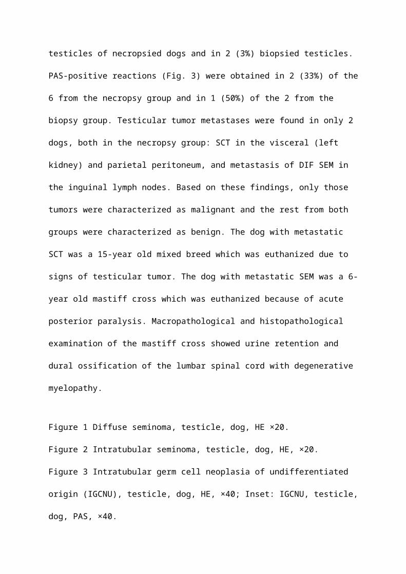
Figure 1 Diffuse seminoma, testicle, dog, HE ×20.
Figure 2 Intratubular seminoma, testicle, dog, HE, ×20.
Figure 3 Intratubular germ cell neoplasia of undifferentiated origin (IGCNU), testicle, dog,
HE, ×40; Inset: IGCNU, testicle, dog, PAS, ×40.
Immunohistochemical analysis
Of testicles with neoplastic changes (including IGCNU), 16% in the necropsy group and 26%
in the biopsy group expressed c-KIT (Table 2). Expression of c-KIT was predominantly
cytoplasmic and membranous, with moderate intensity. In both groups, SEM and the
seminomatous components of mixed tumors were most often c-KIT positive. In the necropsy
group, 3 (27%) of 11 SEM (Fig. 4) [2 (50%) of 4 diffuse SEM; 1 (14%) of 7 intratubular
SEM), 1 (16%) of 6 IGCNU, 1 (50%) of 2 mixed LCT/diffuse SEM, 1 (33%) of 3 mixed
LCT/intratubular SEM, 1 (50%) of 2 mixed SCT/diffuse SEM, 1 (50%) of 2 mixed LCT/SCT,
and 1 (5%) of 19 LCT] were c-KIT positive. The SEM that had metastasized was c-KIT
positive. The percentage of positive cells had a range of 90% in diffuse SEM to 5–20% in
intratubular SEM, IGCNU, and mixed tumors. In the biopsy group, c-KIT expression was as
follows: 9 (40%) of 22 SEM [9 (45%) of 20 diffuse SEM, 0 (0%) of 2 intratubular SEM], 1
(6%) of 16 SCT, 1 (100%) of 1 teratoma, and 4 (100%) of 4 mixed LCT/SEM. The
percentage of positive cells was 43% in diffuse SEM, 26% in mixed LCT/SEM, 20% in
teratoma, and 5% in SCT.
Immunohistochemical analysis showed PLAP expression in 23% of testicular tumors
(including IGCNU) from necropsied dogs and 26% of tumors (including IGCNU) from the
biopsy group (Table 2). PLAP expression was predominantly cytoplasmic and membranous,
with moderate intensity. The distribution of neoplastic cells with PLAP expression was
mostly focally intratubular. Expression of PLAP was most common in the necropsy group
IGCNU (2/6, 33%) (Fig. 6), biopsied intratubular SEM (1/3, 33%), biopsied teratoma (1/2,
50%), and in some mixed tumors from both groups (25–100%). The incidence of PLAP-
positive diffuse SEM was 25% in the necropsy group and 15% in the biopsy group. Both
IGCNU tumors (100%) from the biopsy group showed PLAP expression.
There were 5% PLAP-positive cells in SEM and mixed SCT/diffuse SEM in the
necropsy group and 5% PLAP-positive cells in biopsied IGCNU. The percentage of PLAP-
positive cells in biopsied intratubular SEM and mixed LCT/intratubular SEM was 10%, and
the percentage was 11% in biopsied diffuse SEM. In other tumor types, expression was lower
than 5%, or absent.
Co-expression of c-KIT and PLAP was seen in 2 (10%) of 20 diffuse SEM in the
biopsy group and in 1 (25%) of 4 in the necropsy group. Only 1 mixed intratubular SEM/LCT
tumor from the biopsy group showed simultaneous co-expression of c-KIT and PLAP.
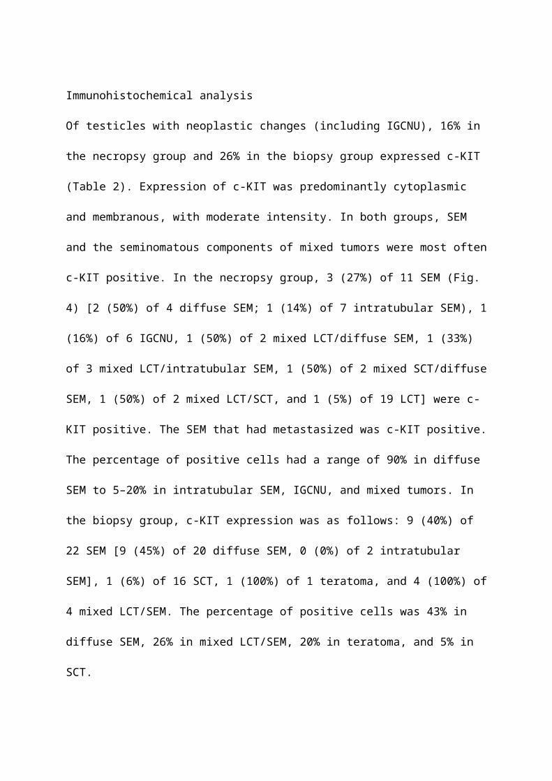
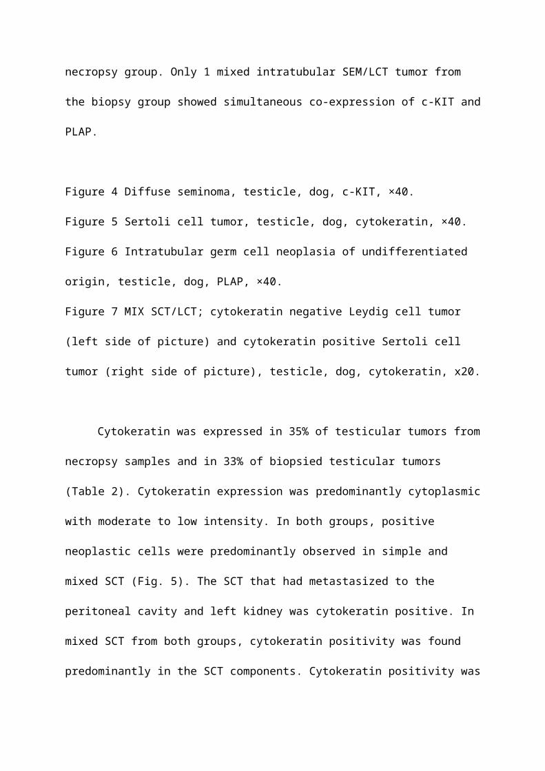
Figure 4 Diffuse seminoma, testicle, dog, c-KIT, ×40.
Figure 5 Sertoli cell tumor, testicle, dog, cytokeratin, ×40.
Figure 6 Intratubular germ cell neoplasia of undifferentiated origin, testicle, dog, PLAP, ×40.
Figure 7 MIX SCT/LCT; cytokeratin negative Leydig cell tumor (left side of picture) and
cytokeratin positive Sertoli cell tumor (right side of picture), testicle, dog, cytokeratin, x20.
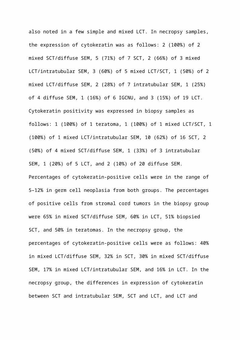
Cytokeratin was expressed in 35% of testicular tumors from necropsy samples and in
33% of biopsied testicular tumors (Table 2). Cytokeratin expression was predominantly
cytoplasmic with moderate to low intensity. In both groups, positive neoplastic cells were
predominantly observed in simple and mixed SCT (Fig. 5). The SCT that had metastasized to
the peritoneal cavity and left kidney was cytokeratin positive. In mixed SCT from both
groups, cytokeratin positivity was found predominantly in the SCT components. Cytokeratin
positivity was also noted in a few simple and mixed LCT. In necropsy samples, the expression
of cytokeratin was as follows: 2 (100%) of 2 mixed SCT/diffuse SEM, 5 (71%) of 7 SCT, 2
(66%) of 3 mixed LCT/intratubular SEM, 3 (60%) of 5 mixed LCT/SCT, 1 (50%) of 2 mixed
LCT/diffuse SEM, 2 (28%) of 7 intratubular SEM, 1 (25%) of 4 diffuse SEM, 1 (16%) of 6
IGCNU, and 3 (15%) of 19 LCT. Cytokeratin positivity was expressed in biopsy samples as
follows: 1 (100%) of 1 teratoma, 1 (100%) of 1 mixed LCT/SCT, 1 (100%) of 1 mixed
LCT/intratubular SEM, 10 (62%) of 16 SCT, 2 (50%) of 4 mixed SCT/diffuse SEM, 1 (33%)
of 3 intratubular SEM, 1 (20%) of 5 LCT, and 2 (10%) of 20 diffuse SEM. Percentages of
cytokeratin-positive cells were in the range of 5–12% in germ cell neoplasia from both
groups. The percentages of positive cells from stromal cord tumors in the biopsy group were
65% in mixed SCT/diffuse SEM, 60% in LCT, 51% biopsied SCT, and 50% in teratomas. In
the necropsy group, the percentages of cytokeratin-positive cells were as follows: 40% in
mixed LCT/diffuse SEM, 32% in SCT, 30% in mixed SCT/diffuse SEM, 17% in mixed
LCT/intratubular SEM, and 16% in LCT. In the necropsy group, the differences in expression
of cytokeratin between SCT and intratubular SEM, SCT and LCT, and LCT and mixed
LCT/intratubular SEM were significant (p < 0.05). In the biopsy group, the difference in
expression of cytokeratin between SCT and diffuse SEM was statistically significant (p <
0.05).
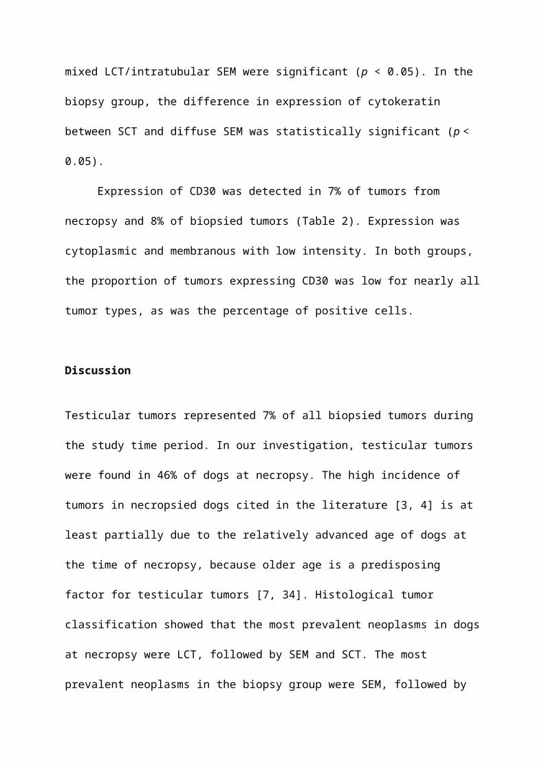
Expression of CD30 was detected in 7% of tumors from necropsy and 8% of biopsied
tumors (Table 2). Expression was cytoplasmic and membranous with low intensity. In both
groups, the proportion of tumors expressing CD30 was low for nearly all tumor types, as was
the percentage of positive cells.
Discussion
Testicular tumors represented 7% of all biopsied tumors during the study time period. In our
investigation, testicular tumors were found in 46% of dogs at necropsy. The high incidence of
tumors in necropsied dogs cited in the literature [3, 4] is at least partially due to the relatively
advanced age of dogs at the time of necropsy, because older age is a predisposing factor for
testicular tumors [7, 34]. Histological tumor classification showed that the most prevalent
neoplasms in dogs at necropsy were LCT, followed by SEM and SCT. The most prevalent
neoplasms in the biopsy group were SEM, followed by SCT and LCT. In both groups, other
simple tumors were rarely diagnosed but the incidence of mixed tumors was relatively high.
The relative incidence of tumors in the biopsy group, the age of dogs with tumors, and
the results of histological classification from both groups are consistent with results from
earlier reports [1, 4, 5, 35, 36].
The higher incidence of SEM and SCT in the biopsied group can be attributed to the
fact that these tumor types result in more obvious clinical signs (testicular enlargement in
SEM, hormonal imbalance in SCT) [4].
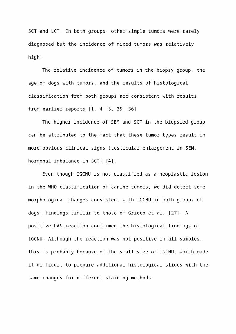
Even though IGCNU is not classified as a neoplastic lesion in the WHO classification
of canine tumors, we did detect some morphological changes consistent with IGCNU in both
groups of dogs, findings similar to those of Grieco et al. [27]. A positive PAS reaction
confirmed the histological findings of IGCNU. Although the reaction was not positive in all
samples, this is probably because of the small size of IGCNU, which made it difficult to
prepare additional histological slides with the same changes for different staining methods.
The small number of metastatic changes found confirms that testicular tumors in dogs
have predominantly benign biological behavior, although they can have malignant
histological appearance [26].
Our immunohistochemical analysis showed that the investigated markers are useful for
differentiation of testicular tumors in dogs. Cytokeratin AE1/AE3 showed particularly good
results in differentiating stromal tumors, especially SCT, from germ cell tumors.
Low expression of CD30 in all tumors showed that embryonal carcinoma, which is
mostly CD30-positive in people [37, 38], did not appear in either group and is a very rare
testicular neoplasm in dogs. Focal CD30 positivity in some germ cell tumors (SEM and
teratoma) is an interesting finding and should be considered for the possibility of transforming
neoplastic cells into embryonal carcinoma cells, as described in human patients [39].
Our findings of positive c-KIT expression in 27% of SEM from the necropsy group
(50% of diffuse SEM, 14% of intratubular SEM) and 40% of biopsied SEM (45% of diffuse
SEM, 0% of intratubular SEM) are consistent with literature reports [4, 10, 14], in which
germ cell tumors can express c-KIT. Higher expression of c-KIT in diffuse SEM than in
intratubular SEM may be related to differences in the biological behavior of the different
types. In humans c-KIT expression is highly correlated to biological behavior. CSEM are
malignant type of SEM because they originate from undifferentiated c-KIT positive
transformed primordial germ cells and gonocytes (prespermatogonia and spermatogonia). In
contrast to CSEM, SSEM are c-KIT negative and have benign behavior, which is in
accordance with their origin from differentiated germ cells, mostly spermatocytes [15, 20-25].
We hypothesize that in dogs the more aggressive diffuse type of tumor, like in human
CSEM, expresses c-KIT more frequently because of its different cellular origin than the less
invasive intratubular SEM. The higher percentage of c-KIT-positive SEM in the biopsy group
can be explained by the higher incidence of diffuse SEM in that group because of its more
obvious clinical symptoms (testicular enlargement) in contrast to intratubular SEM, which
generally does not cause enlargement.
Expression of PLAP and to a lesser degree c-KIT in IGCNU in both groups supports
the histological findings and the hypothesis that IGCNU can be found as one variety of
neoplastic change in canine testicles.
The lower expression of PLAP compared with c-KIT in SEM is consistent with a
study published by Yu et al. [10], in which the number of PLAP-positive SEM was much
lower than c-KIT-positive tumors. The low co-expression of c-KIT and PLAP in SEM could
result from the different cellular origin of seminomas, because c-KIT is expressed in both
spermatogonia and prespermatogonia whereas PLAP is expressed only in prespermatogonia,
while in people both cells give rise to CSEM [10, 11, 17, 20, 27, 29, 31]. Based on our results
and those of Yu et al. [10], canine CSEM are probably predominantly derived from
spermatogonia and to a lesser degree from prespermatogonia, given the expression of c-KIT
and PLAP.
Conclusions
Our histopathological and immunohistochemical analyses (c-KIT and PLAP
expression) indicate that, just as in human classification, some canine SEM can be classified
as CSEM. We conclude that c-KIT positive SEM, or at least tumors with simultaneously
expressed c-KIT and PLAP can definitively be classified as CSEM. On the basis of these
results, canine SEM can be divided in two groups: the less prevalent CSEM and the more
prevalent SSEM.
Although canine SEM are mostly benign and rarely metastasizes [26], differentiation
of canine SEM in to CSEM and SSEM on the basis of c-KIT expression may be clinically
significant. Considering the more aggressive behavior of c-KIT positive CSEM in men, close
clinical monitoring would be advisable for dogs with this type of tumor. However further
studies are necessary to establish whether c-KIT expression is correlated with a higher
metastatic rate in canine SEM.
Abbreviations
ANOVA: analysis of variance; CSEM: classical seminoma; HE: hematoxylin and eosin;
IGCNU: intratubular germ cell neoplasia of undifferentiated origin; IHC:
immunohistochemistry; LCT: Leydig cell tumors; MET. T. LYMPH: metastatic tumor –
lymphoma; PAS: periodic acid-Schiff; PLAP: placental alkaline phosphatase; SD: standard
deviation; SEM: seminoma; SSEM: spermatocytic seminoma; TERAT: teratoma; WHO:
World Health Organization.
Competing interests
The authors declare that they have no competing interests.
Authors' contributions
MH participated in the design of this study, performed histopathological and IHC analysis,
performed necropsies, evaluated IHC, and prepared the manuscript. KS performed statistical
and image analysis. BA and ŽB participated in the design of the study and histopathological
analysis and necropsies. AGK and AB performed histopathological analysis and necropsies.
ICŠZ performed necropsies and IHC analysis. All authors read and approved the final
manuscript.
AcknowledgmentsWe would like to thank Doc. dr. Marjana Ćorić, from the Department of Clinical
Pathology, Zagreb Clinical Hospital Centre for conducting the immunohistochemical analysis.
This study was supported by a project of the Ministry of Science and Technology,
Republic of Croatia: Comparative diagnostic, morphometry, and analysis of human and
animal tumors (053-05322642260, principal investigator: prof. dr. sc. Željko Grabarević).

References
1. Nødtvedt A, Gamlem H, Gunnes G, Grotmol T, Inderbø A, Moe L: Breed differences in
the proportional morbidity of testicular tumours and distribution of histopathologic
types in a population-based canine cancer registry. Vet Comp Oncol 2011, 9: 45-54.
2. North S, Banks T, Straw R: Tumors of the urogenital tract. U: Small Animal Oncology,
an introduction. Edited by North S, Banks T, Straw R. Edinburgh, London, New York,
Oxford, Philadelphia, St. Louis, Sydney, Toronto: Elsevier Saunders; 2009: 151-172.
3. Dow C: Testicular tumours in the dog. J Comp Pathol 1962, 72, 247-265.
4. Grieco V, Riccardi E, Greppi GF, Teruzzi F, Iermano V, Finazzi M: Canine Testicular
Tumours: a Study on 232 Dogs. J Comp Path 2008, 138: 86-89.
5. Gamlem H, Nordstoga K, Glattre E: Canine neoplasia-introductory paper. APMIS 2008,
Suppl: 5-18.
6. Vascellari M, Baioni E, Ru G, Carminato A, Mutinelli F: Animal tumour registry of two
provinces in northern Italy: incidence of spontaneous tumours in dogs and cats. BMC
Vet Res 2009, 5: 39.
7. Fan TM, De Lorimer LP: Tumors of the male reproductive system. In Withrow &
MacEwen's Small Animal Clinical Oncology. 4th edn. Edited by Withrow S.J, Vail DM. St.
Louis: Saunders; 2007: 799-804.
8. Maclachlan NJ, Kennedy PC: Tumors of the Genital systems. In Tumors in Domestic
Animals, 4th ed. Edited by Meuten DJ. Ames, Iowa:Iowa Sate Press; 2002: 547-574.
9. Kennedy PC, Cullen JM, Edwards JF, Goldschmidt MH, Larsen S: Histological
Classification of Tumors of the Genital System of Domestic Animals, 2nd Series,
Washington DC: WHO, Armed Forces Institute of Pathology; 1998.
10. Yu CH, Hwanq DN, Yhee JH, Kim JH, Im KS, Nho WG, Lyoo YS, Sur JH:
Comparative immunohistochemical characterization of canine seminomas and Sertoli
cell tumors. J Vet Sci 2009, 10:1-7.
11. Woodward PJ, Heindereich A, Looijenga LHJ, Osterhuis JW, McLeod DG, Møller H,
Manivel JC, Mostofi FK, Hailemariam S, Parkinson MC, Grigor K, True L, Jacobsen GK,
Oliver TD, Talerman A, Kaplan GW, Ulbright TH, Sesterhenn IA, Rushton HG, Michael H,
Reuter VE: Germ cell tumours. In World Health Organization Classiffication of Tumors.
Pathology & Genetics. Tumors of the Urinary System and Male Genital Organs. Edited by
Eble JN, Sauter G, Epstein JI, Sesterhenn IA. Lyon, France: IARC Press; 2004: 221-249.
12. Emerson RE, Ulbright TM: The use of immunohistochemistry in the differential
diagnosis of tumors of the testis and paratestis. Semin Diagn Pathol 2005, 22, 33-50.
13. Grieco V, Banco B, Giudice C, Mosca F, Finazzi M: Immunohistochemical Expression
of the KIT Protein (CD117) in Normal and Neoplastic Canine Testes. J Comp Path 2010,
142: 213-217.
14. Owston MA, Ramos-Vara JA: Histologic and immunohistochemical Characterization
of a Testicular Mixed Germ Cell Sex Cord-Stromal Tumor and Leydig Cell Tumor in a
Dog. Vet Pathol 2007, 44:936-943.
15. Bush JM., Gardiner DW, Palmer JS, Rajpert-De Meyts E, Veeramachaneni DN:
Testicular germ cell tumours in dogs are predominantly of spermatocytic seminoma
type and are frequently associated with somatic cell tumours. Int J Androl 2011, 34: 288-
295;doi: 10.1111/j.1365-2605.2011.01166.x
16. Thorvaldsen TE, Nødtvedt A, Grotmol T, Gunnes G: Morphological and
immunohistochemical characterisation of seminomas in Norwegian dogs.
Acta Vet Scand 2012, 54:52. doi: 10.1186/1751-0147-54-52.
17. Grieco V, Riccardi E, Rondena M, Ciampi V, Finazzi M: Classical and spermatocytic
seminoma in the dog: histochemical and immunohistochemical findings. J Comp Pathol
2007, 137: 41-46.
18. Cheville JC, Rao S, Iczkowski KA, Lohse CM, Pankratz VS: Cytokeratin expression in
seminoma of the human testis. Am J Clin Pathol 2000, 113, 583-588.
19. Banco B, Giudice C, Veronesi MC, Gerosa E, Grieco V: An immunohistochemical
study of normal and neoplastic canine sertoli cells. J Comp Pathol 2010, 143, 239-247.
20. Kim JH, Yu CH,Yhee JY, Im KS, Kim NH, Sur JH: Canine classical seminoma: a
specific malignant type with human classifications is highly correlated with tumor
angiogenesis. BMC Cancer 2010, 10:243.
21. Skakkebaek NE, Berthelsen JG, Giwercman A, Müller J: Carcinoma-in-situ of the
testis: possible origin from gonocytes and precursor of all types of germ cell tumours
except spermatocytoma. Int J Androl 1987, 10(1):19-28.
22. Goriely A, Hansen RM, Taylor IB, Olesen IA, Jacobsen GK, Mcgowan SJ, Pfeifer SP,
Mcvean GA, Rajpert-De Meyts E, Wilkie AO: Activating mutations in FGFR3 and HRAS
reveal a shared genetic origin for congenital disorders and testicular tumors. Nat Genet
2009, 41(11):1247-52. doi: 10.1038/ng.470.
23. Looijenga LH, Hersmus R, Gillis AJ, Pfundt R, Stoop HJ, Van Gurp RJ, Veltman J,
Beverloo HB, Van Drunen E, Van Kessel AG, Pera RR, Schneider DT, Summersgill B,
Shipley J, Mcintyre A, Van Der Spek P, Schoenmakers E, Oosterhuis JW: Genomic and
expression profiling of human spermatocytic seminomas: primary spermatocyte as
tumorigenic precursor and DMRT1 as candidate chromosome 9 gene. Cancer Res 2006,
66(1): 290-302.
24. Looijenga LH, Stoop H, Hersmus R, Gillis AJ, Wolter Oosterhuis J: Genomic and
expression profiling of human spermatocytic seminomas: pathogenetic implications. Int
J Androl 2007, 30(4):328-336.
25. Nochomovitz LE, Rosai J: Current concepts on the histogenesis, pathology, and
immunochemistry of germ cell tumors of the testis. Pathol Annu 1978, 13(1):327-362.
26. Foster RA, Ladds PW: Male genital system. In Jubb, Kennedy, and Palmer's pathology
of Domestic Animals. 5th ed. Vol. 3. Edited by Grant Maxie M. Philadelphia: Elsevier
Saunders; 2007: 565-619.
27. Grieco V, Riccardi E, Veronesi MC, Giudice C, Finazzi M: Evidence of testicular
dysgenesis syndrome in the dog. Theriogenology 2008, 70: 53-60.
28. Saegusa Y, Hayashi H, Taniai E, Imaoka M, Ohishi T, Wang L, Mitsumori K, Shibutani
M: Spermatocytic seminoma with neuroectodermal differentiation and sertoli cell tumor
in a dog. Vet Pathol 2011, 48:1024-1028.
29. Gaskel TL, Esnal A, Robinson LL, Anderson RA, Saunders PT: Immunohistochemical
profiling of germ cells within the human fetal testis: identification of three
subpopulations. Biol Reprod 2004, 71: 2012-2021.
30. Rajpert-De Meytes E, Bartkova J, Samson M, Hoi-Hansen CE, Frydelund-Larsen L,
Bartek J, Skakkanaek NE: The emerging phenotype of the testicular carcinoma in situ
germ cell. APMIS 2003, 111:267-278.
31. Mauduit C, Hamamah S, Benahmed M: Stem cell factor/c-kit system in
spermatogenesis. Hum Reprod Update 1999, 5:535-545.
32. World Health Organisation: Histological Classification of Tumors of the Genital
System of Domestic Animals. 1999.
[http://www.afip.org/consultation/vetpath/who/whogenet.html] Accessed 4 December 2009.
33. Blackhall FH, Pinitilie M, Michael M, Leihg N, Feld R, Tsao MS, Shepherd FA:
Expression and prognostic significance of kit, protein kinase B, and mitogen-activated
protein kinase in patients with small cell lung cancer. Clin Cancer Res 2003, 9:2241-2247.
34. Peters MA, De Rooij DG, Teerds KJ, Van Der Gaag I, Van Sluijs FJ: Spermatogenesis
and testicular tumours in ageing dogs. J Reprod Fertil 2000, 120: 443-452.
35. Dobson JM, Samuel S, Milstein H, Rogers K, Wood JL: Canine neoplasia in the UK:
estimates of incidence rates from a population of insured dogs. J Small Anim Pract 2002,
43, 240-246.
36. Liao AT, Chu PY, Yeh LS, Lin CT, Liu CH: A 12-year retrospective study of canine
testicular tumors. J Vet Med Sci 2009, 71(7): 919-23.
37. Gopalan A, Dhall D, Olgac S, Fine SW, Korkola JE, Houldsworth J, Chaganti RS, Bosl
GJ, Reuter VE, Tickoo SK: Testicular mixed germ cell tumors: a morphological and
immunohistochemical study using stem cell markers, OCT3/4, SOX2 and GDF3, with
emphasis on morphologically difficult-to-classify areas. Mod Pathol 2009, 22: 1066-1074.
38. Leroy X, Augusto D, Leteurtre E, Gosselin B: CD30 and CD117 (c-kit) used in
combination are useful for distinguishing embryonal carcinoma from seminoma. J
Histochem Cytochem 2002, 50:283-285.
39. Hitmair A, Rogatsch H, Hobisch A, Mikuz G, Feichtinger H: CD30 expression in
seminoma. Hum Pathol 1996, 27, 1166-1171.
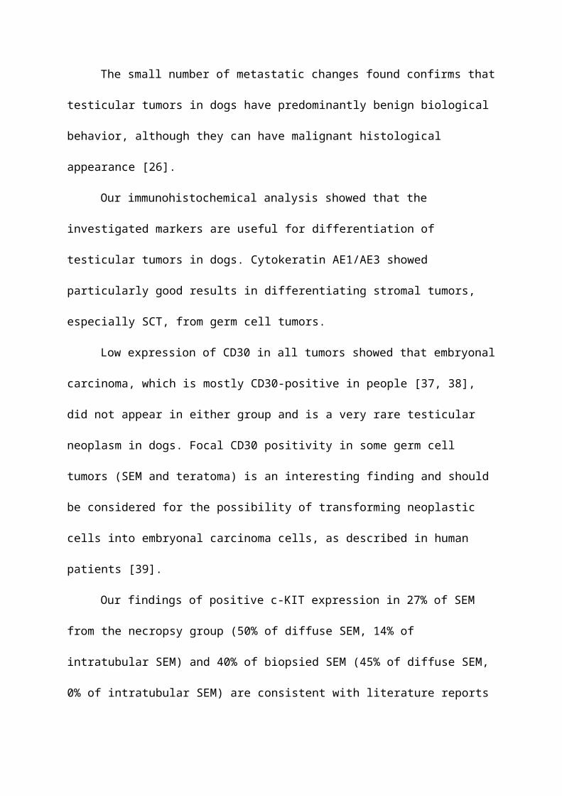
Figure legends
Figure 1 Diffuse seminoma, testicle, dog, HE, ×20.
Figure 2 Intratubular seminoma, testicle, dog, HE, ×20.
Figure 3 Intratubular germ cell neoplasia of undifferentiated origin (IGCNU), testicle, dog,
HE, ×40; Inset: IGCNU, testicle, dog, PAS, ×40.
Figure 4 Diffuse seminoma, testicle, dog, c-KIT, ×40.
Figure 5 Sertoli cell tumor, testicle, dog, cytokeratin, ×40.
Figure 6 Intratubular germ cell neoplasia of undifferentiated origin, testicle, dog, PLAP, ×40.
Figure 7 MIX SCT/LCT; cytokeratin negative Leydig cell tumor (left side of picture) and
cytokeratin positive Sertoli cell tumor (right side of picture), testicle, dog, cytokeratin, x20.
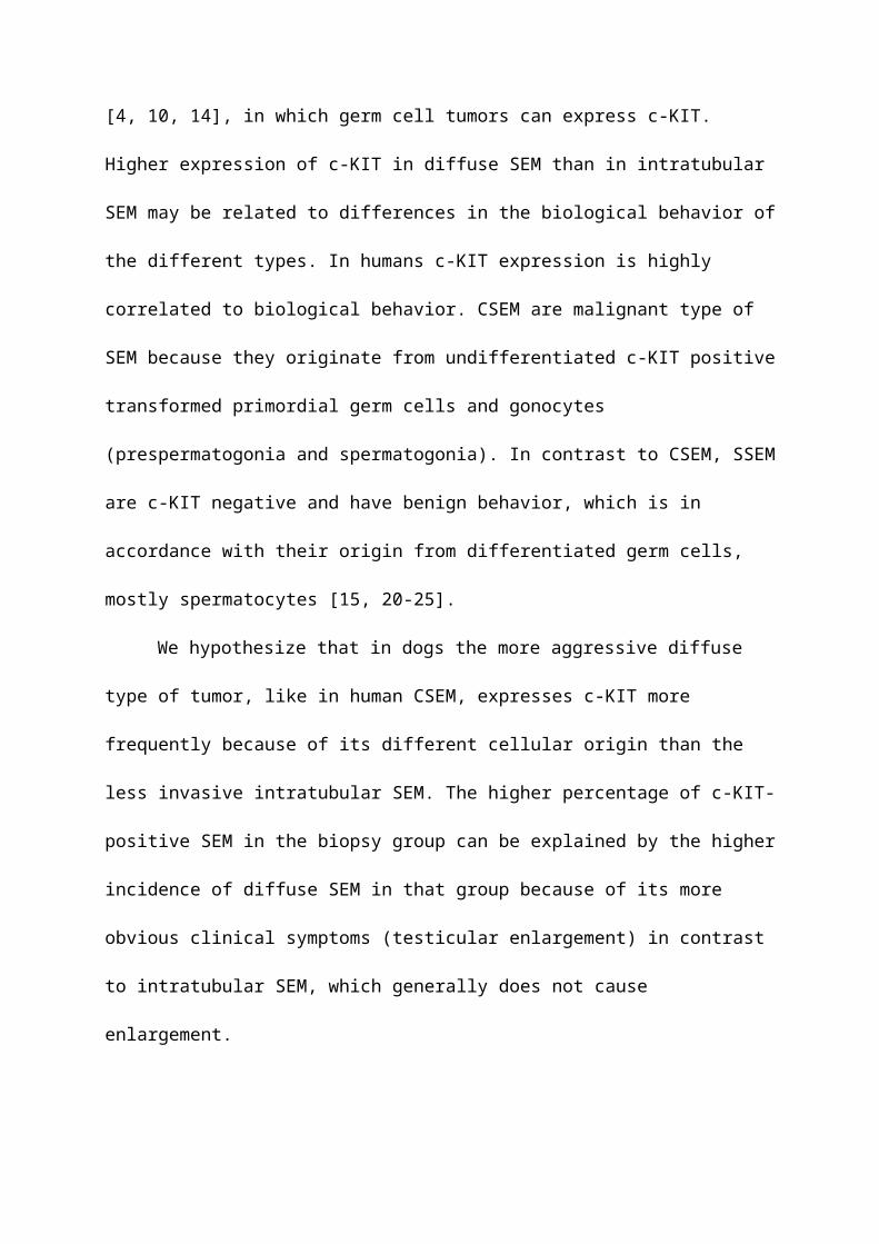
Table 1 Incidence of histological types of testicular tumors diagnosed
HISTOLOGICAL TYPE OF
TUMOR
NECROPSY GROUP BIOPSY GROUP
Number of
tumors
Percentage of all
tumors / %
Number of
tumors%
SIMPLE TUMORS (∑) 37 74 46 84
LCT 19 38 5 9
SCT 7 14 16 29
IT SEM 7 14 2 4
DIF SEM 4 8 20 36
PNST 0 0 2 4
Teratoma 0 0 1 2
MIXED TUMORS (∑) 12 24 9 16
LCT/SCT 5 10 1 2
LCT/ IT SEM 3 6 1 2
SCT/DIF SEM 2 4 4 7
LCT/DIF SEM 2 4 3 5
MET. T. LYMPH 1 2 0 0
TOTAL 50 100 55 100
Note: ∑, sum; DIF SEM, diffuse seminoma; IGCNU, intratubular germ cell neoplasia of
undifferentiated origin; IT SEM, intratubular seminoma; LCT, Leydig cell tumors; MET. T.
LYMPH, metastatic tumor – lymphoma; PNST, peripheral nerve sheat tumor; SCT, Sertoli
cell tumor
Table 2 Expression of IHC markers by diagnosed histological type of testicular tumor
HIST
OL
OG
ICA
L T
YPE
OF T
UM
OR
NECROPSY GROUP BIOPSY GROUP
c-KIT positive
tumors/ %
% of c-K
IT positive cells
PLAP positive
tumors/%
% of PLA
P positive cells
Cytokeratin
positive tumors/%
% of C
ytokeratin positive cells
CD
30 positive tum
ors/ %
% of C
D 30
positive cells
c-KIT positive
tumors/ %
% of c-K
IT positive cells
PLAP positive
tumors/ %
% of PLA
P positive cells
Cytokeratin
positive tumors/%
% of C
ytokeratin positive cells
CD
30 positive tum
ors/ %
% of C
D 30
positive cells
SIMPLE
TUM
OR
S (∑)
11 39 23 <5 27 19 7 8 22 40 27 <6 33 43 8 23
MIX
ED
TUM
OR
S (∑)
33 23 25 <5 66 25 8 5 44 26 22 <7 44 46 11 30
LCTsimple 5 5 21 <5 15 16 5 10 0 0 16 <5 20 60 6 30
withSCT 20 20 40 <5 60 23 20 5 0 0 0 0 100 50 0 0
with IT SEM 33 10 0 0 60 17 0 0 100 20 100 10 100 5 0 0
with DIF SEM
50 60 0 0 40 40 0 0 75 28 0 0 0 0 0
SCTsimple 0 0 28 <5 71 32 14 10 6 5 31 <5 62 51 12 27
withLCT 20 20 40 <5 60 23 20 5 0 0 0 0 100 50 0 0
withDIF SEM
50 5 50 5 100 30 0 0 0 0 25 5 50 65 25 30
IT SEMsimple 14 5 14 5 28 5 0 0 0 0 33 10 33 5 0 0
withLCT 33 10 0 0 60 17 0 0 100 20 100 10 100 5 0 0
DIF SEMsimple
50 90 25 5 25 5 25 5 45 43 15 11 10 12 0 0
withSCT 50 5 50 5 100 30 0 0 0 0 25 5 50 65 25 30
withLCT 50 60 0 0 40 40 0 0 75 28 0 0 0 0 0
PNST 0 0 0 0 0 0 0 0 0 0 0 0 0 0 0 0
TERAT 0 0 0 0 0 0 0 0 0 20 0 <5 100 50 100 10
IGCNU 16 33 <5 16 5 0 0 0 0 100 5 0 0 0 0
MET. T.LYMPH 0 0 0 0 0 0 0 0 0 0 0 0 0 0 0 0
TOTAL 16 32 23 <5 35 21 7 7 26 36 26 <6 29 44 8 25
Note: ∑, sum; DIF SEM, diffuse seminoma; IGCNU, intratubular germ cell neoplasia of
undifferentiated origin; IT SEM, intratubular seminoma; LCT, Leydig cell tumors; MET. T.
LYMPH, metastatic tumor – lymphoma; PLAP, placental alkaline phosphatase; PNST,
peripheral nerve sheat tumor; SCT, Sertoli cell tumor; TERAT, teratoma