abdomen-pelvis ct protocols€¦ · disclosures dianna: ge healthcare – “in kind” research...
TRANSCRIPT
Abdomen-Pelvis CT protocols
Dianna Cody, Ph.DUniversity of Texas MD Anderson Cancer Center
Rendon C. Nelson, M.D.Duke University
DISCLOSURES
Dianna:
GE Healthcare – “In Kind” research project (Dual-Energy CT)
Rendon:
GE Healthcare - Consultant
Approach
• Routine abdomen-pelvis CT & kidney stone CT
• Review ACR CT Dose Index Registry Data
• Review audience sample protocols
• Compare to AAPM & speaker’s protocols
• Data specific to one vendor (GE)
• General tips
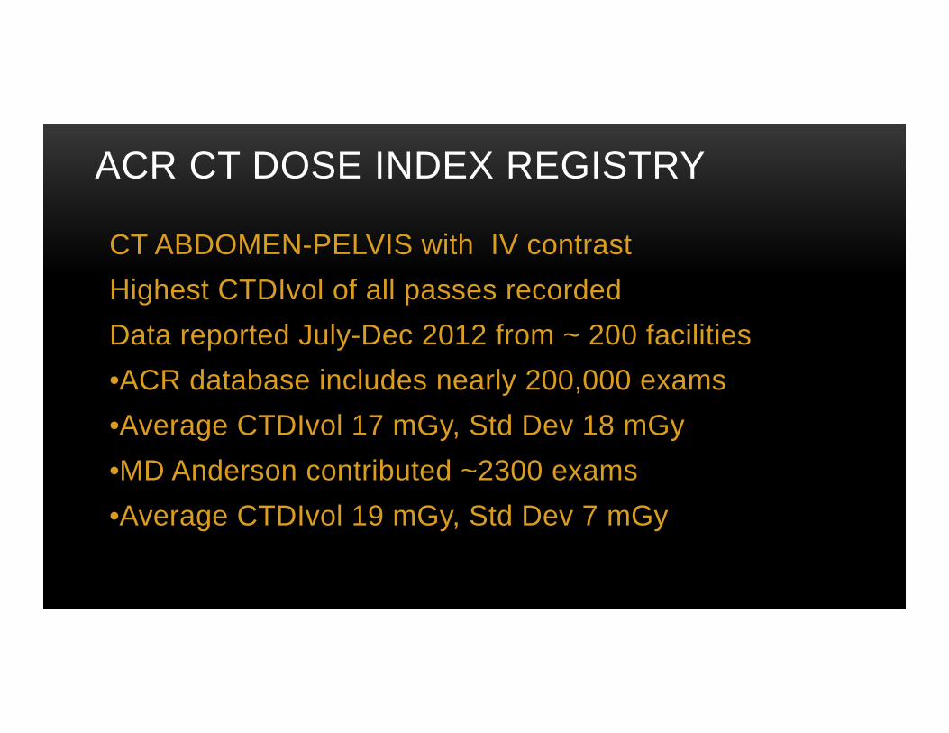
ACR CT DOSE INDEX REGISTRY
CT ABDOMEN-PELVIS with IV contrastHighest CTDIvol of all passes recordedData reported July-Dec 2012 from ~ 200 facilities•ACR database includes nearly 200,000 exams•Average CTDIvol 17 mGy, Std Dev 18 mGy•MD Anderson contributed ~2300 exams•Average CTDIvol 19 mGy, Std Dev 7 mGy

ACR CT DOSE INDEX REGISTRYCT ABD PELVIS KIDNEY CALCULUS
Highest CTDIvol of all passes recorded
Data reported July-Dec 2012 from ~ 200 facilities
•ACR database includes nearly 22,000 exams
•Average CTDIvol 16 mGy, Std Dev 9 mGy
•MD Anderson contributed 6 exams
•Average CTDIvol 19 mGy, Std Dev 4 mGy
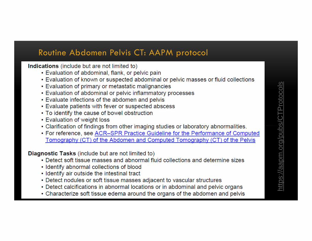
Routine Abdomen Pelvis CT: AAPM protocol
http
s://a
apm
.org
/pub
s/C
TPro
toco
ls
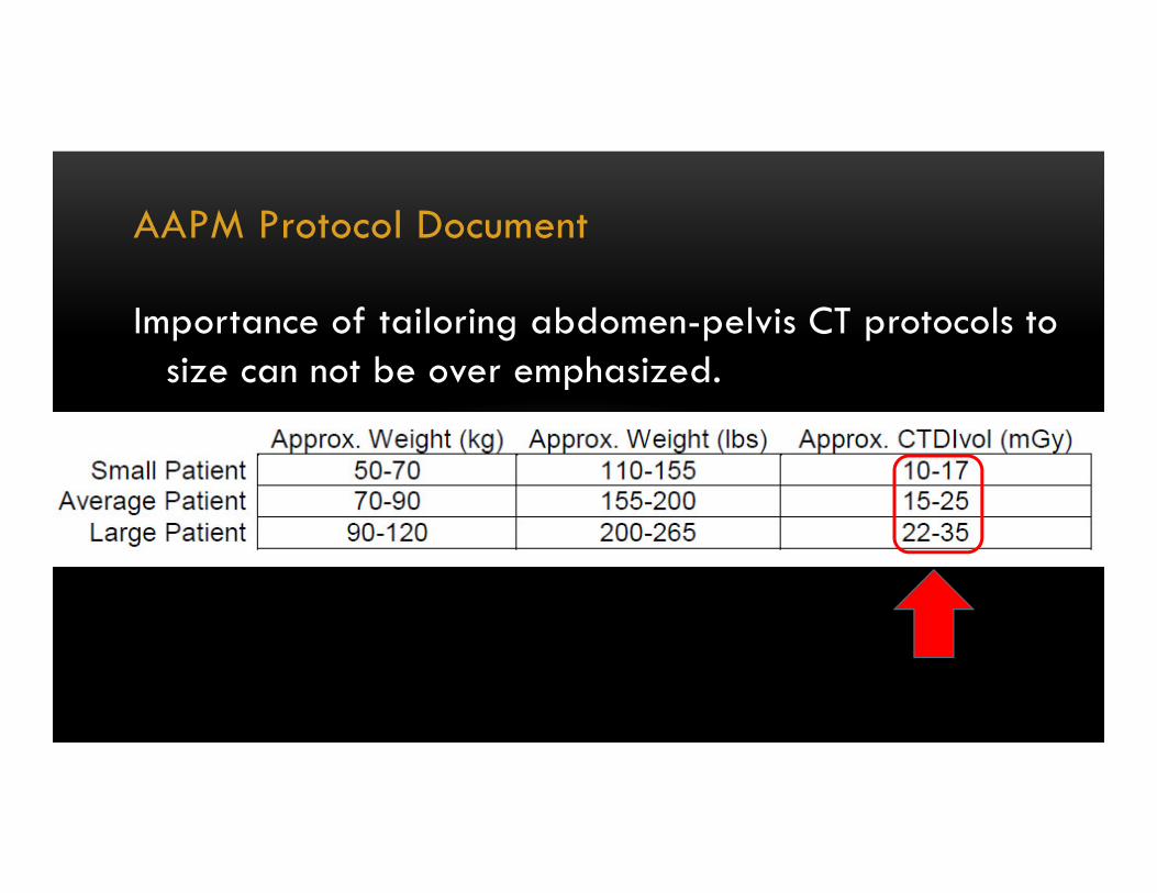
AAPM Protocol Document
Importance of tailoring abdomen-pelvis CT protocols to size can not be over emphasized.
GE User (n=1): Routine abdomen/pelvis CT protocol
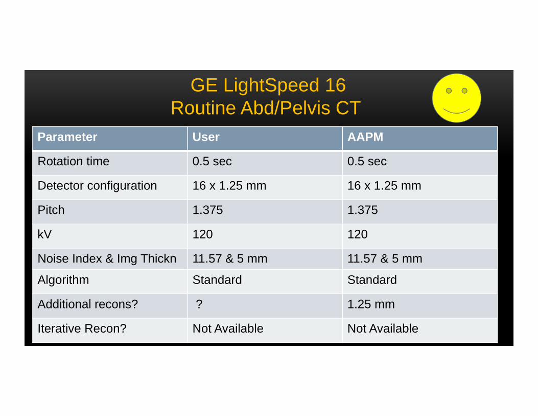
Parameter User AAPM
Rotation time 0.5 sec 0.5 sec
Detector configuration 16 x 1.25 mm 16 x 1.25 mm
Pitch 1.375 1.375
kV 120 120
Noise Index & Img Thickn 11.57 & 5 mm 11.57 & 5 mm
Algorithm Standard Standard
Additional recons? ? 1.25 mm
Iterative Recon? Not Available Not Available
GE LightSpeed 16Routine Abd/Pelvis CT
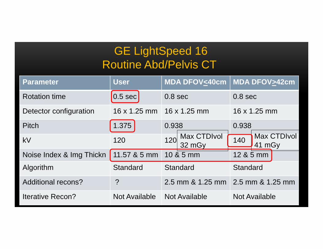
Parameter User MDA DFOV<40cm MDA DFOV>42cm
Rotation time 0.5 sec 0.8 sec 0.8 sec
Detector configuration 16 x 1.25 mm 16 x 1.25 mm 16 x 1.25 mm
Pitch 1.375 0.938 0.938
kV 120 120 140
Noise Index & Img Thickn 11.57 & 5 mm 10 & 5 mm 12 & 5 mm
Algorithm Standard Standard Standard
Additional recons? ? 2.5 mm & 1.25 mm 2.5 mm & 1.25 mm
Iterative Recon? Not Available Not Available Not Available
GE LightSpeed 16Routine Abd/Pelvis CT
Max CTDIvol41 mGy
Max CTDIvol 32 mGy
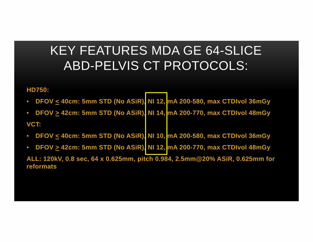
KEY FEATURES MDA GE 64-SLICE ABD-PELVIS CT PROTOCOLS:
HD750:
• DFOV < 40cm: 5mm STD (No ASiR), NI 12, mA 200-580, max CTDIvol 36mGy
• DFOV > 42cm: 5mm STD (No ASiR), NI 14, mA 200-770, max CTDIvol 48mGy
VCT:
• DFOV < 40cm: 5mm STD (No ASiR), NI 10, mA 200-580, max CTDIvol 36mGy
• DFOV > 42cm: 5mm STD (No ASiR), NI 12, mA 200-770, max CTDIvol 48mGy
ALL: 120kV, 0.8 sec, 64 x 0.625mm, pitch 0.984, 2.5mm@20% ASiR, 0.625mm for reformats
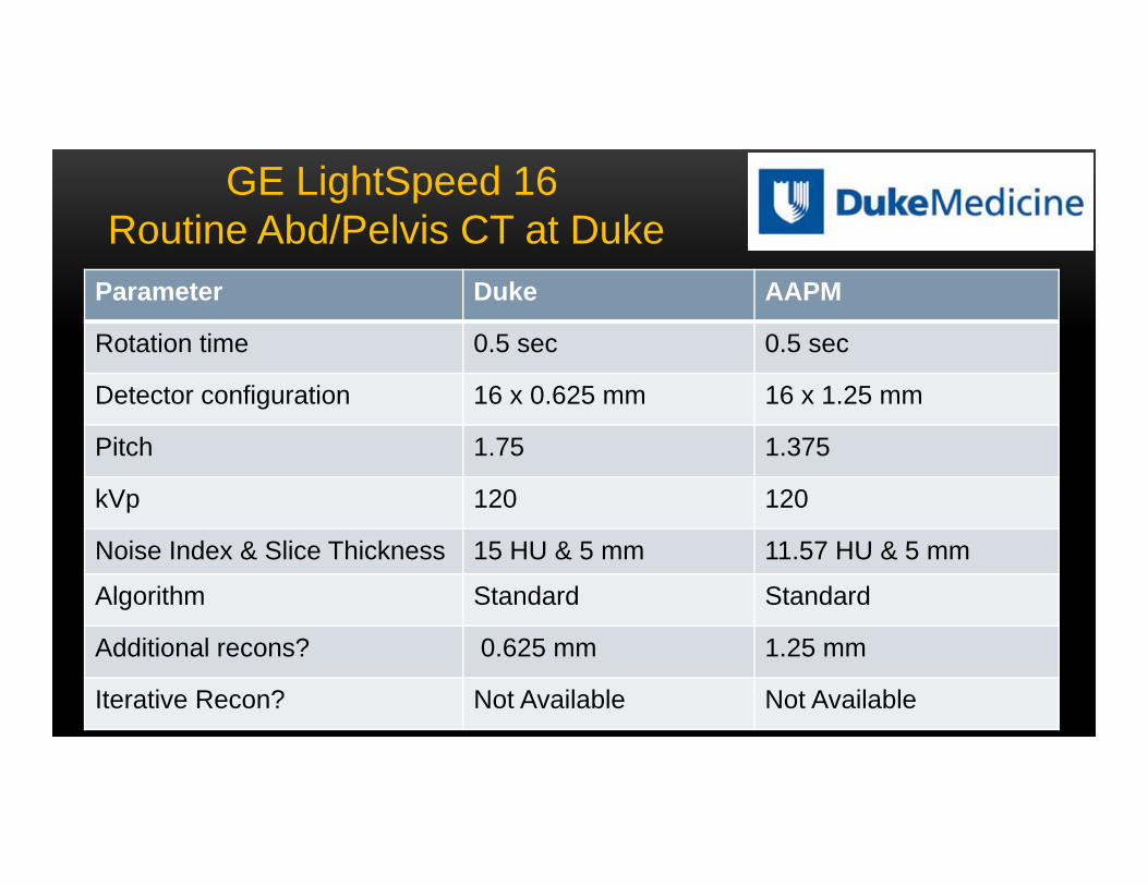
Parameter Duke AAPM
Rotation time 0.5 sec 0.5 sec
Detector configuration 16 x 0.625 mm 16 x 1.25 mm
Pitch 1.75 1.375
kVp 120 120
Noise Index & Slice Thickness 15 HU & 5 mm 11.57 HU & 5 mm
Algorithm Standard Standard
Additional recons? 0.625 mm 1.25 mm
Iterative Recon? Not Available Not Available
GE LightSpeed 16Routine Abd/Pelvis CT at Duke
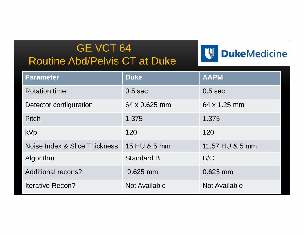
Parameter Duke AAPM
Rotation time 0.5 sec 0.5 sec
Detector configuration 64 x 0.625 mm 64 x 1.25 mm
Pitch 1.375 1.375
kVp 120 120
Noise Index & Slice Thickness 15 HU & 5 mm 11.57 HU & 5 mm
Algorithm Standard B B/C
Additional recons? 0.625 mm 0.625 mm
Iterative Recon? Not Available Not Available
GE VCT 64Routine Abd/Pelvis CT at Duke

Parameter Duke AAPM
Rotation time 0.5 sec 0.5 sec
Detector configuration 64 x 0.625 mm 64 x 1.25 mm
Pitch 1.375 0.984
kVp 120 120
Noise Index & Slice Thickness 22 HU & 5 mm 25.2 HU & 5 mm
Algorithm Standard B B/C
Additional recons? 0.625 mm 0.625 mm
Iterative Recon? ASiR (40% Blend) ASiR (50% Blend)
GE 750 HD 64Routine Abd/Pelvis CT at Duke
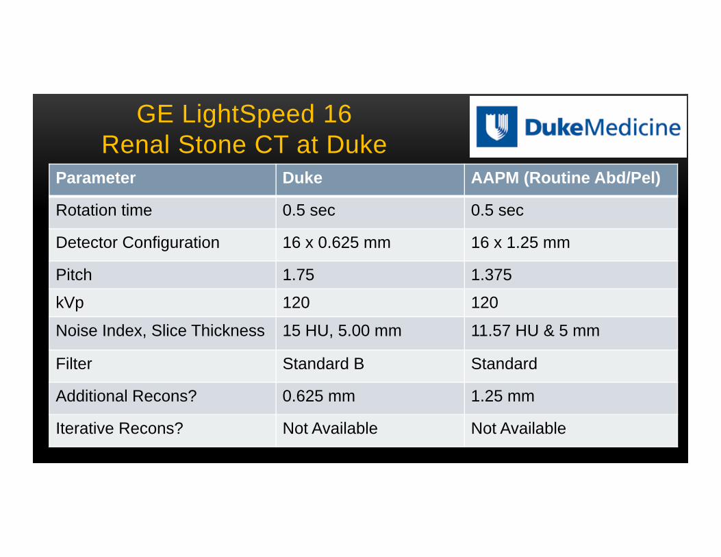
GE LightSpeed 16Renal Stone CT at Duke
Parameter Duke AAPM (Routine Abd/Pel)
Rotation time 0.5 sec 0.5 sec
Detector Configuration 16 x 0.625 mm 16 x 1.25 mm
Pitch 1.75 1.375
kVp 120 120
Noise Index, Slice Thickness 15 HU, 5.00 mm 11.57 HU & 5 mm
Filter Standard B Standard
Additional Recons? 0.625 mm 1.25 mm
Iterative Recons? Not Available Not Available
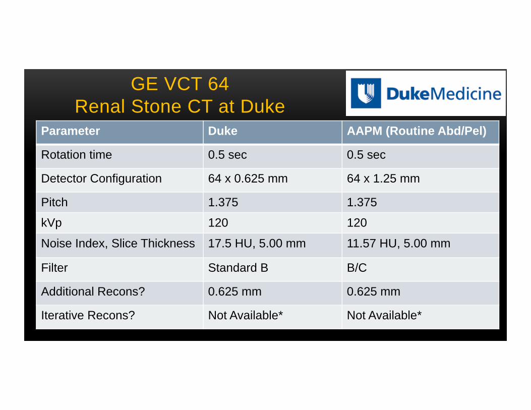
GE VCT 64Renal Stone CT at Duke
Parameter Duke AAPM (Routine Abd/Pel)
Rotation time 0.5 sec 0.5 sec
Detector Configuration 64 x 0.625 mm 64 x 1.25 mm
Pitch 1.375 1.375
kVp 120 120
Noise Index, Slice Thickness 17.5 HU, 5.00 mm 11.57 HU, 5.00 mm
Filter Standard B B/C
Additional Recons? 0.625 mm 0.625 mm
Iterative Recons? Not Available* Not Available*
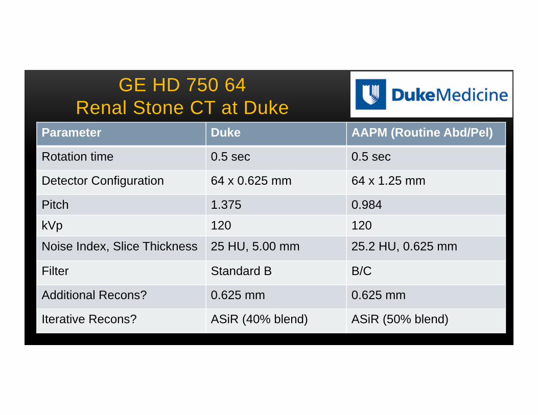
GE HD 750 64Renal Stone CT at Duke
Parameter Duke AAPM (Routine Abd/Pel)
Rotation time 0.5 sec 0.5 sec
Detector Configuration 64 x 0.625 mm 64 x 1.25 mm
Pitch 1.375 0.984
kVp 120 120
Noise Index, Slice Thickness 25 HU, 5.00 mm 25.2 HU, 0.625 mm
Filter Standard B B/C
Additional Recons? 0.625 mm 0.625 mm
Iterative Recons? ASiR (40% blend) ASiR (50% blend)
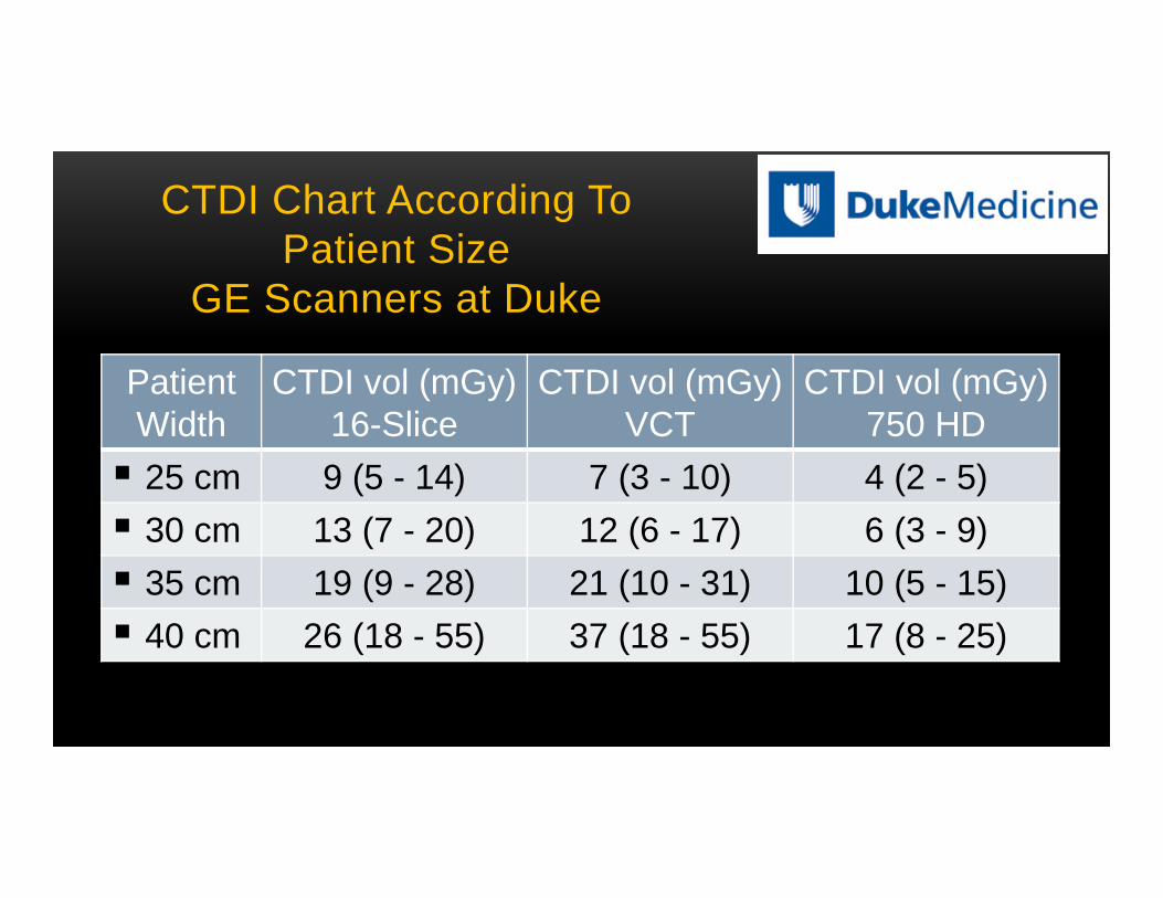
CTDI Chart According ToPatient Size
GE Scanners at Duke
PatientWidth
CTDI vol (mGy)16-Slice
CTDI vol (mGy)VCT
CTDI vol (mGy)750 HD
25 cm 9 (5 - 14) 7 (3 - 10) 4 (2 - 5) 30 cm 13 (7 - 20) 12 (6 - 17) 6 (3 - 9) 35 cm 19 (9 - 28) 21 (10 - 31) 10 (5 - 15) 40 cm 26 (18 - 55) 37 (18 - 55) 17 (8 - 25)
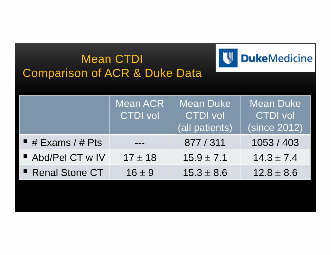
Mean CTDIComparison of ACR & Duke Data
Mean ACRCTDI vol
Mean DukeCTDI vol
(all patients)
Mean DukeCTDI vol
(since 2012) # Exams / # Pts --- 877 / 311 1053 / 403 Abd/Pel CT w IV 17 18 15.9 7.1 14.3 7.4 Renal Stone CT 16 9 15.3 8.6 12.8 8.6

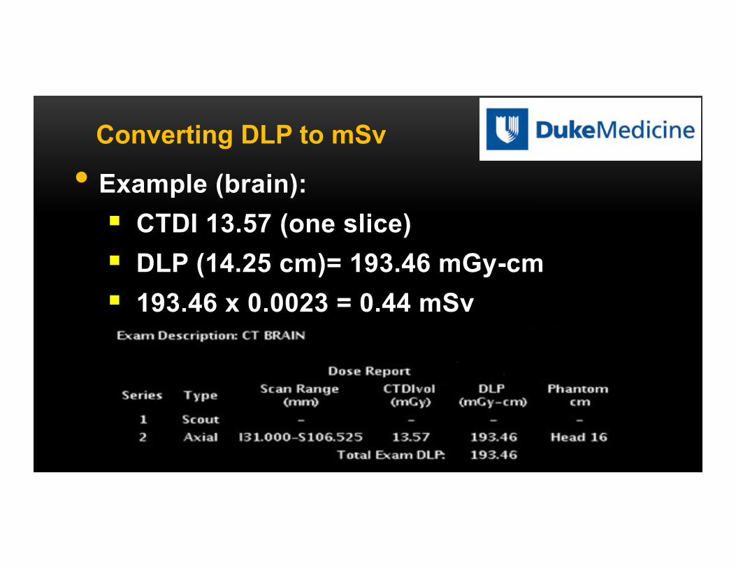
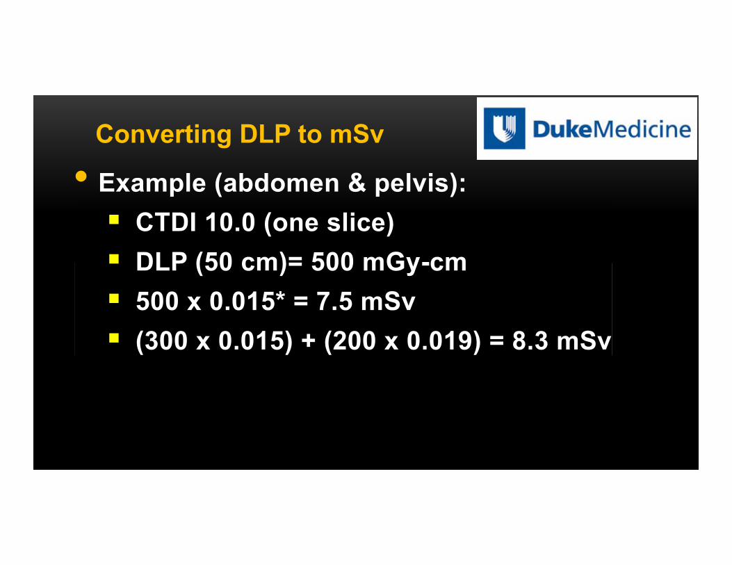
Region of body Normalized effective doseEDLP (mSv mGy-1 cm-1)
Head 0.0023 Neck 0.0054 Chest 0.017 Abdomen 0.015 Pelvis 0.019
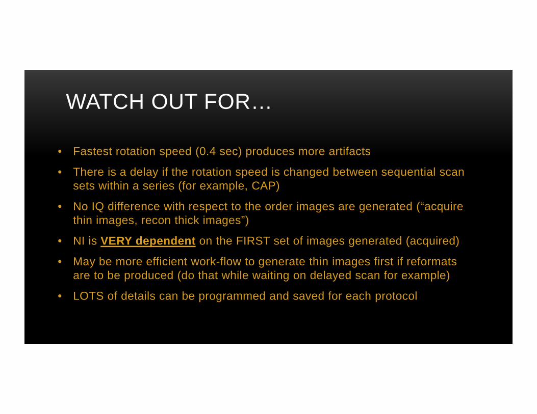
WATCH OUT FOR…
• Fastest rotation speed (0.4 sec) produces more artifacts
• There is a delay if the rotation speed is changed between sequential scan sets within a series (for example, CAP)
• No IQ difference with respect to the order images are generated (“acquire thin images, recon thick images”)
• NI is VERY dependent on the FIRST set of images generated (acquired)
• May be more efficient work-flow to generate thin images first if reformats are to be produced (do that while waiting on delayed scan for example)
• LOTS of details can be programmed and saved for each protocol
Philips Users: Routine abdomen/pelvis (n=3) & kidney stone (n=1) CT protocols
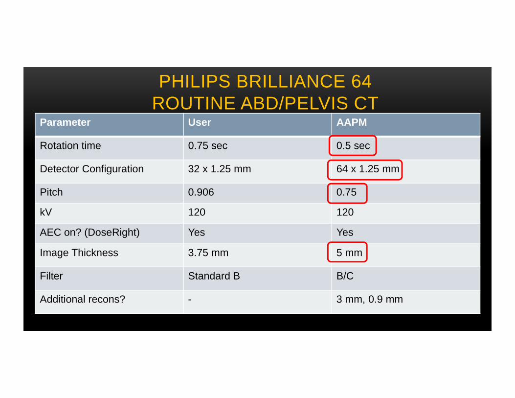
PHILIPS BRILLIANCE 64ROUTINE ABD/PELVIS CT
Parameter User AAPM
Rotation time 0.75 sec 0.5 sec
Detector Configuration 32 x 1.25 mm 64 x 1.25 mm
Pitch 0.906 0.75
kV 120 120
AEC on? (DoseRight) Yes Yes
Image Thickness 3.75 mm 5 mm
Filter Standard B B/C
Additional recons? - 3 mm, 0.9 mm
Siemens Users:Routine abd/pelvis CT (n=6) &
Kidney stone CT (n=4) Protocols
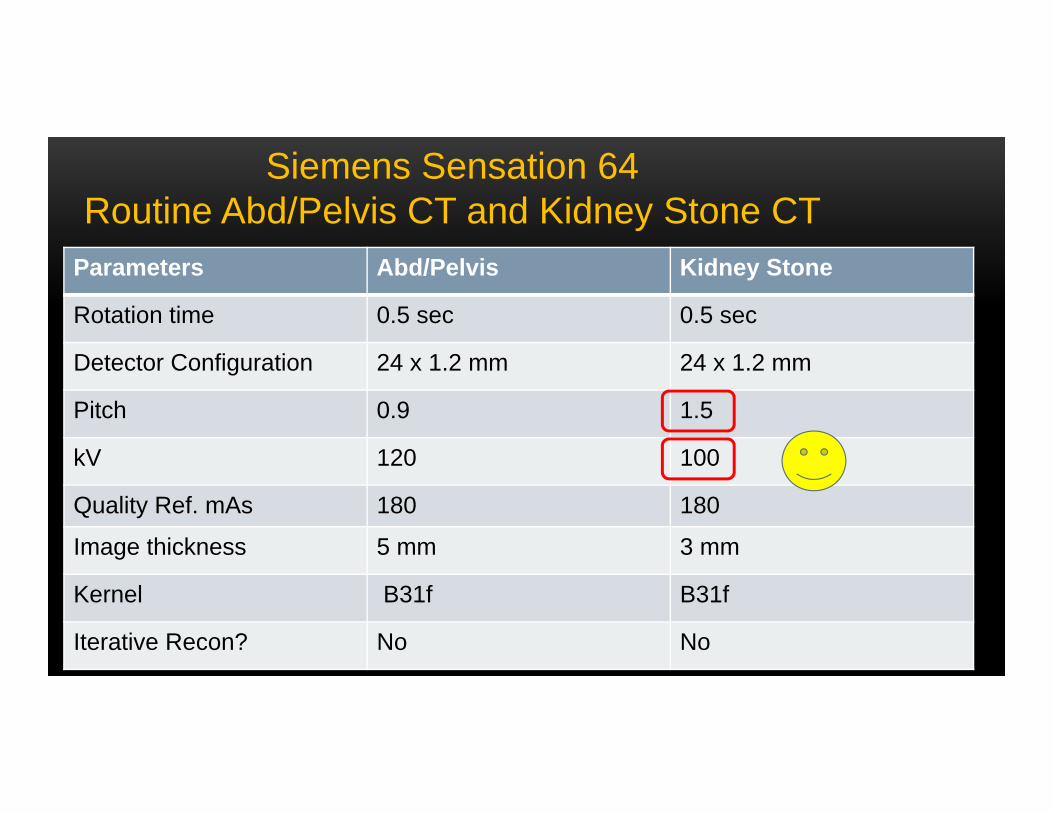
Parameters Abd/Pelvis Kidney Stone
Rotation time 0.5 sec 0.5 sec
Detector Configuration 24 x 1.2 mm 24 x 1.2 mm
Pitch 0.9 1.5
kV 120 100
Quality Ref. mAs 180 180
Image thickness 5 mm 3 mm
Kernel B31f B31f
Iterative Recon? No No
Siemens Sensation 64Routine Abd/Pelvis CT and Kidney Stone CT

Parameters Duke AAPM
Rotation time 0.5 sec 0.5 sec
Detector Configuration 64 x 0.6 mm 64 x 0.6 mm
Pitch 0.8 0.6
kVp 120 120
Quality Ref. mAs 200 210, 150 (with IR)
Image thickness 5.0 mm, 3.0 mm, 0.6 mm 5.0 mm, 1.0 mm
Kernel I31 B30f
Iterative Recon? Safire (Power 2.0) Safire (Power 3.0)
Siemens Definition FLASH 64Routine Abdomen/Pelvis CT at Duke
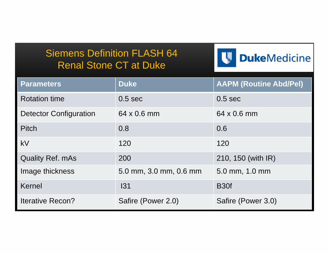
Parameters Duke AAPM (Routine Abd/Pel)
Rotation time 0.5 sec 0.5 sec
Detector Configuration 64 x 0.6 mm 64 x 0.6 mm
Pitch 0.8 0.6
kV 120 120
Quality Ref. mAs 200 210, 150 (with IR)
Image thickness 5.0 mm, 3.0 mm, 0.6 mm 5.0 mm, 1.0 mm
Kernel I31 B30f
Iterative Recon? Safire (Power 2.0) Safire (Power 3.0)
Siemens Definition FLASH 64Renal Stone CT at Duke
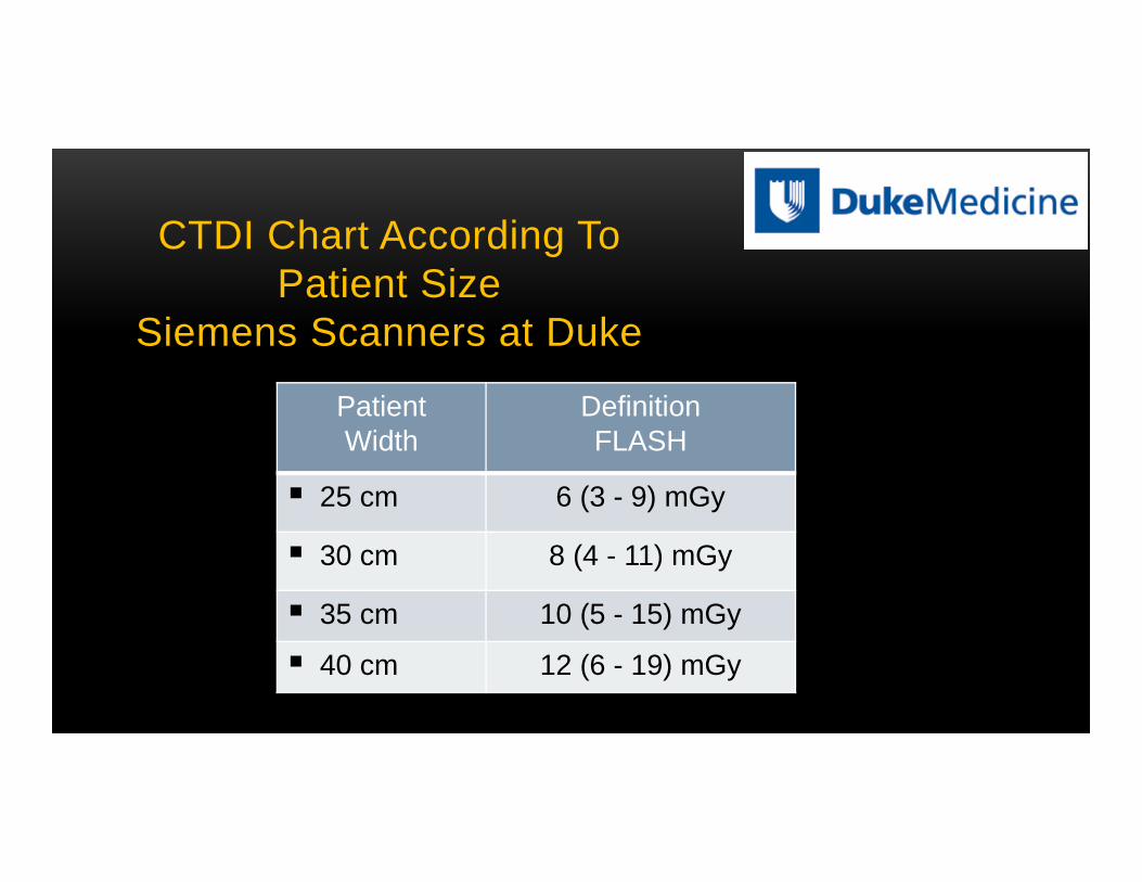
CTDI Chart According ToPatient Size
Siemens Scanners at Duke
PatientWidth
DefinitionFLASH
25 cm 6 (3 - 9) mGy
30 cm 8 (4 - 11) mGy
35 cm 10 (5 - 15) mGy
40 cm 12 (6 - 19) mGy
Toshiba users:Abd/Pelvis CT (n=1)
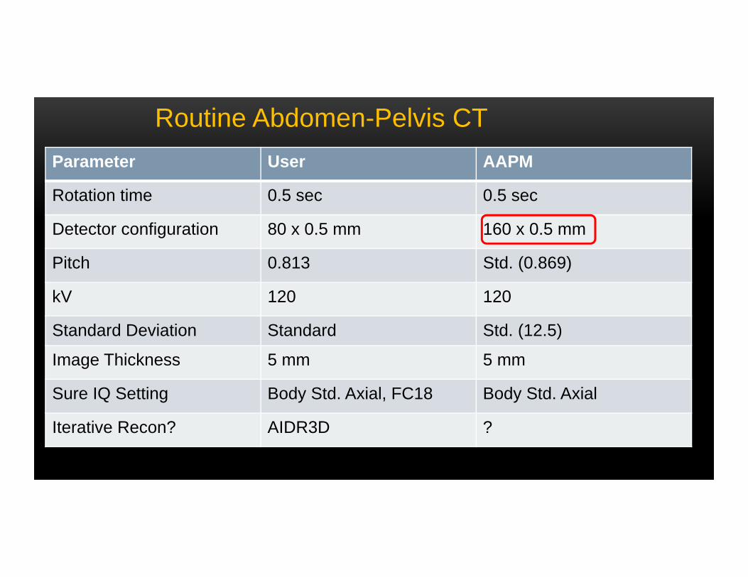
Routine Abdomen-Pelvis CTParameter User AAPM
Rotation time 0.5 sec 0.5 sec
Detector configuration 80 x 0.5 mm 160 x 0.5 mm
Pitch 0.813 Std. (0.869)
kV 120 120
Standard Deviation Standard Std. (12.5)
Image Thickness 5 mm 5 mm
Sure IQ Setting Body Std. Axial, FC18 Body Std. Axial
Iterative Recon? AIDR3D ?
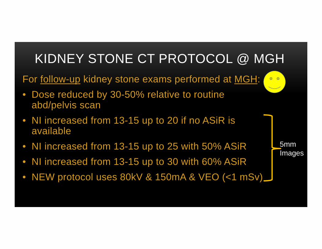
KIDNEY STONE CT PROTOCOL @ MGHFor follow-up kidney stone exams performed at MGH:• Dose reduced by 30-50% relative to routine
abd/pelvis scan• NI increased from 13-15 up to 20 if no ASiR is
available• NI increased from 13-15 up to 25 with 50% ASiR• NI increased from 13-15 up to 30 with 60% ASiR• NEW protocol uses 80kV & 150mA & VEO (<1 mSv)
5mm Images
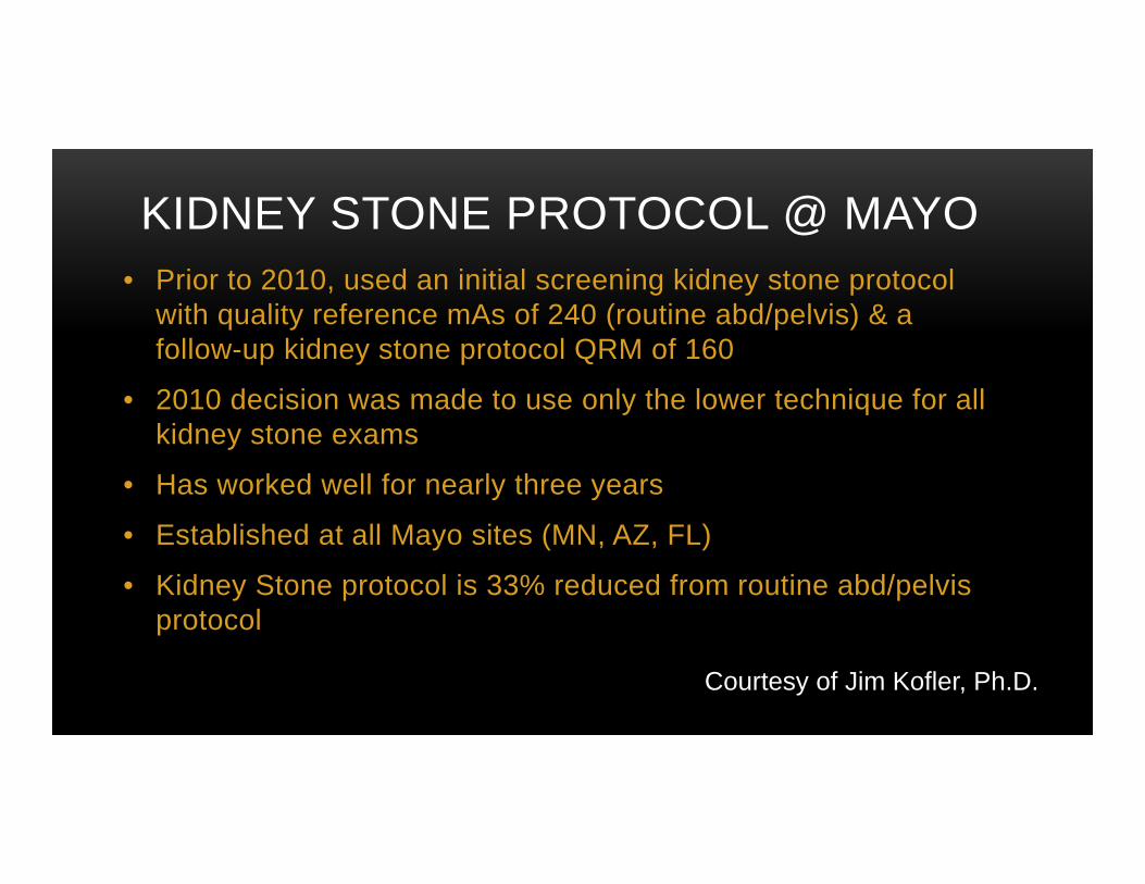
KIDNEY STONE PROTOCOL @ MAYO• Prior to 2010, used an initial screening kidney stone protocol
with quality reference mAs of 240 (routine abd/pelvis) & a follow-up kidney stone protocol QRM of 160
• 2010 decision was made to use only the lower technique for all kidney stone exams
• Has worked well for nearly three years
• Established at all Mayo sites (MN, AZ, FL)
• Kidney Stone protocol is 33% reduced from routine abd/pelvis protocol
Courtesy of Jim Kofler, Ph.D.
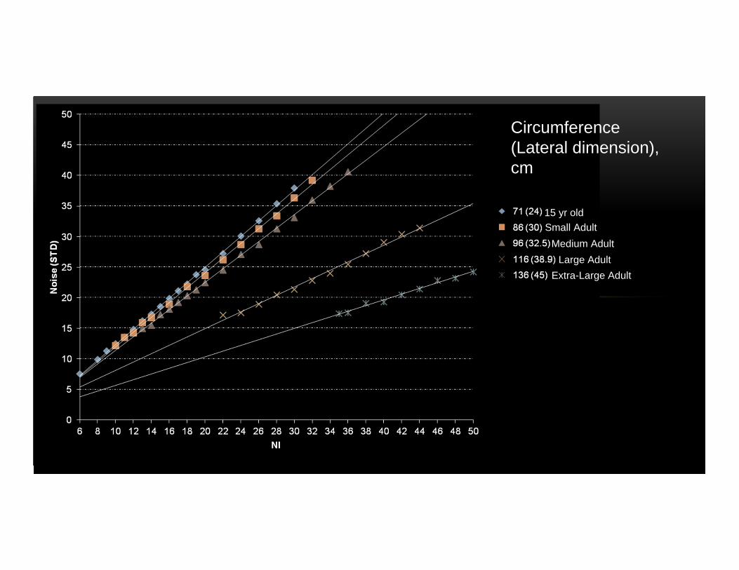
Circumference(Lateral dimension), cm
15 yr oldSmall Adult
Medium AdultLarge Adult
Extra-Large Adult
Salient points in Abdomen-Pelvis CT protocols
• Strongly recommend AEC for routine abd-pelvis CT
• Be aware of potential pitfalls (TCM talk!)
• Be stingy with extra phases like routine delayed phase
• Both in doing it and in the technique used
• Apply iterative reconstruction if available
• May not improve IQ until dose is decreased substantially
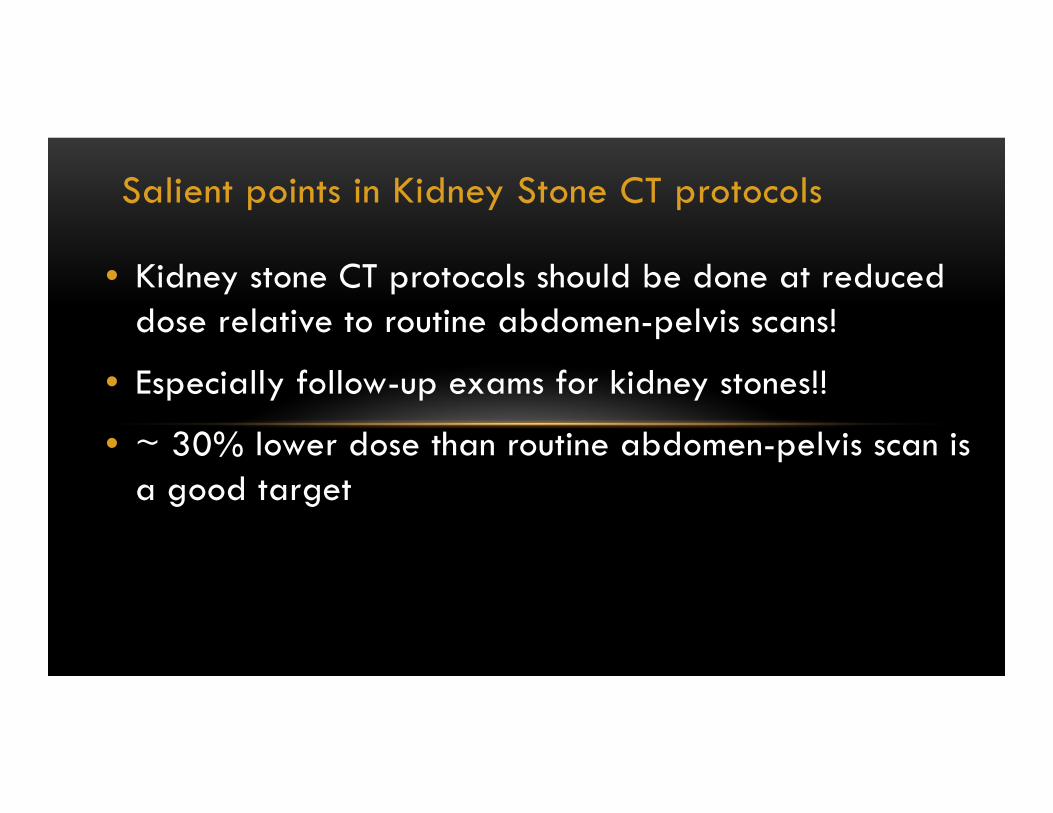
Salient points in Kidney Stone CT protocols
• Kidney stone CT protocols should be done at reduced dose relative to routine abdomen-pelvis scans!
• Especially follow-up exams for kidney stones!!
• ~ 30% lower dose than routine abdomen-pelvis scan is a good target