abdominal & gu trauma october 10, 2002 moritz haager dr. michael betzner
Post on 21-Dec-2015
216 views
TRANSCRIPT

Abdominal & GU Trauma
October 10, 2002
Moritz Haager
Dr. Michael Betzner
Objectives
• Anatomical review
• Examine relationship between mechanism of injury, and resultant injury patterns
• Review diagnostic & therapeutic options
• Develop an approach to abdominal trauma
My 2 ¢..
• Trauma is highly variable in presentation, extent of injury, and examination
• Easy to lose sight of the forest for the trees
• A structured (i.e. ATLS) approach helps
• 3 ideas to keep in mind:– Clinical suspicion avoids missed injuries– Frequent reassessment avoids missed injuries– Know the limitations of your tests
Case
• 22 yo male, roll-over MVA
• GCS 12, HR 120, BP 100/60, RR 24
• CHI, RUQ contusion, obvious R lower leg #
• Resuscitated with 2L NS, vitals improve to GCS 12, HR 80, BP 115/70, RR 18
• CT shows grade IV liver lac’n
• Does he need OR?
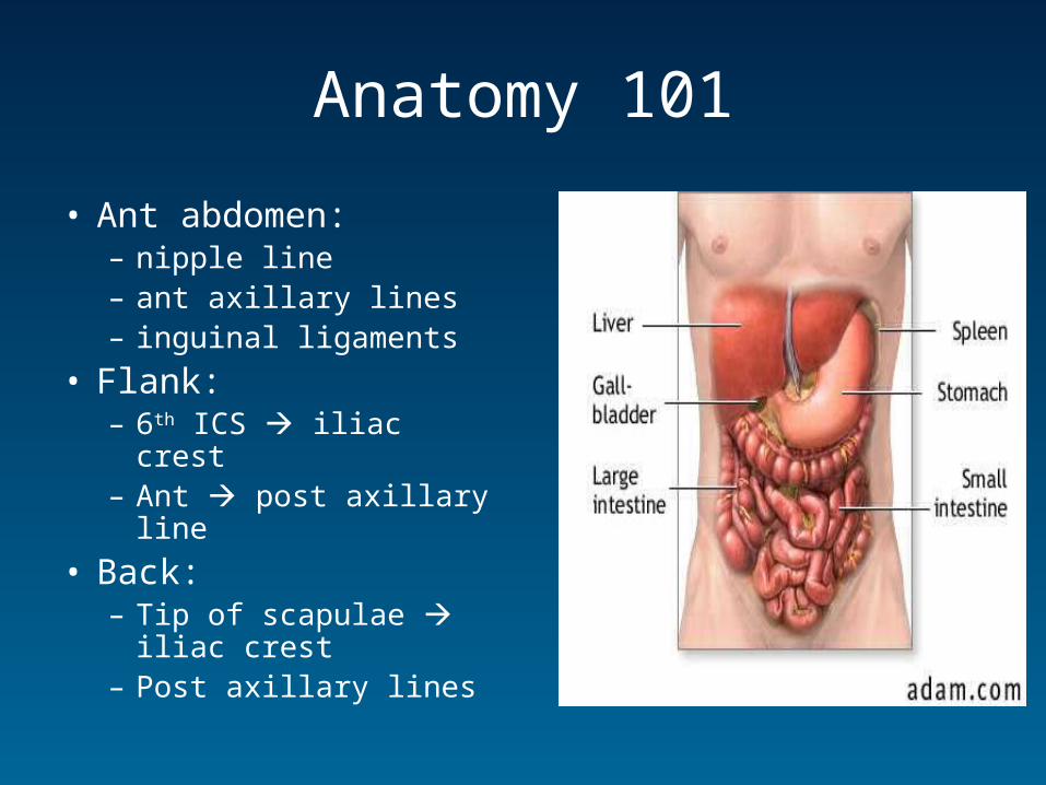
Anatomy 101
• Ant abdomen:– nipple line – ant axillary lines– inguinal ligaments
• Flank:– 6th ICS iliac crest– Ant post axillary line
• Back:– Tip of scapulae iliac
crest– Post axillary lines
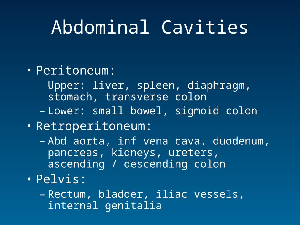
Abdominal Cavities
• Peritoneum:– Upper: liver, spleen, diaphragm, stomach,
transverse colon– Lower: small bowel, sigmoid colon
• Retroperitoneum:– Abd aorta, inf vena cava, duodenum, pancreas,
kidneys, ureters, ascending / descending colon
• Pelvis:– Rectum, bladder, iliac vessels, internal genitalia
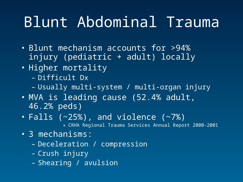
Blunt Abdominal Trauma
• Blunt mechanism accounts for >94% injury (pediatric + adult) locally
• Higher mortality – Difficult Dx– Usually multi-system / multi-organ injury
• MVA is leading cause (52.4% adult, 46.2% peds)• Falls (~25%), and violence (~7%)
» CRHA Regional Trauma Services Annual Report 2000-2001
• 3 mechanisms:– Deceleration / compression– Crush injury– Shearing / avulsion
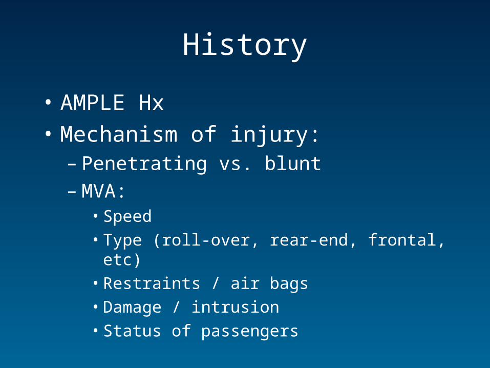
History
• AMPLE Hx
• Mechanism of injury:– Penetrating vs. blunt– MVA:
• Speed
• Type (roll-over, rear-end, frontal, etc)
• Restraints / air bags
• Damage / intrusion
• Status of passengers
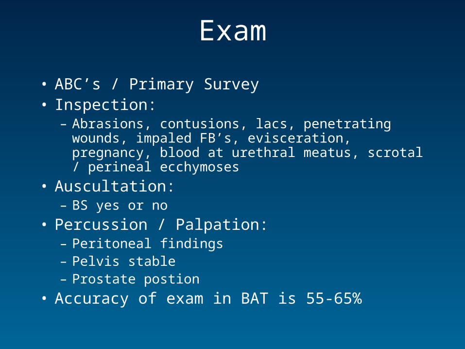
Exam
• ABC’s / Primary Survey • Inspection:
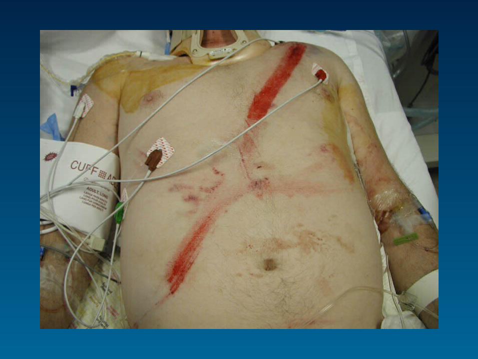
– Abrasions, contusions, lacs, penetrating wounds, impaled FB’s, evisceration, pregnancy, blood at urethral meatus, scrotal / perineal ecchymoses
• Auscultation:– BS yes or no
• Percussion / Palpation:– Peritoneal findings– Pelvis stable– Prostate postion
• Accuracy of exam in BAT is 55-65%
Injury patterns in BAT
• Spleen is most commonly injured organ• Seatbelt injuries
– Rib fractures– Abdominal injuries
• Mesenteric lacerations hemoperitoneum• Bowel contusions / perforations delayed S/S
– Diaphragmatic rupture– Abd aortic dissection (rare)
• Iatrogenic– Ventilation GI distention / rupture– CPR solid organ injury– Tube thoracostomy solid organ injury
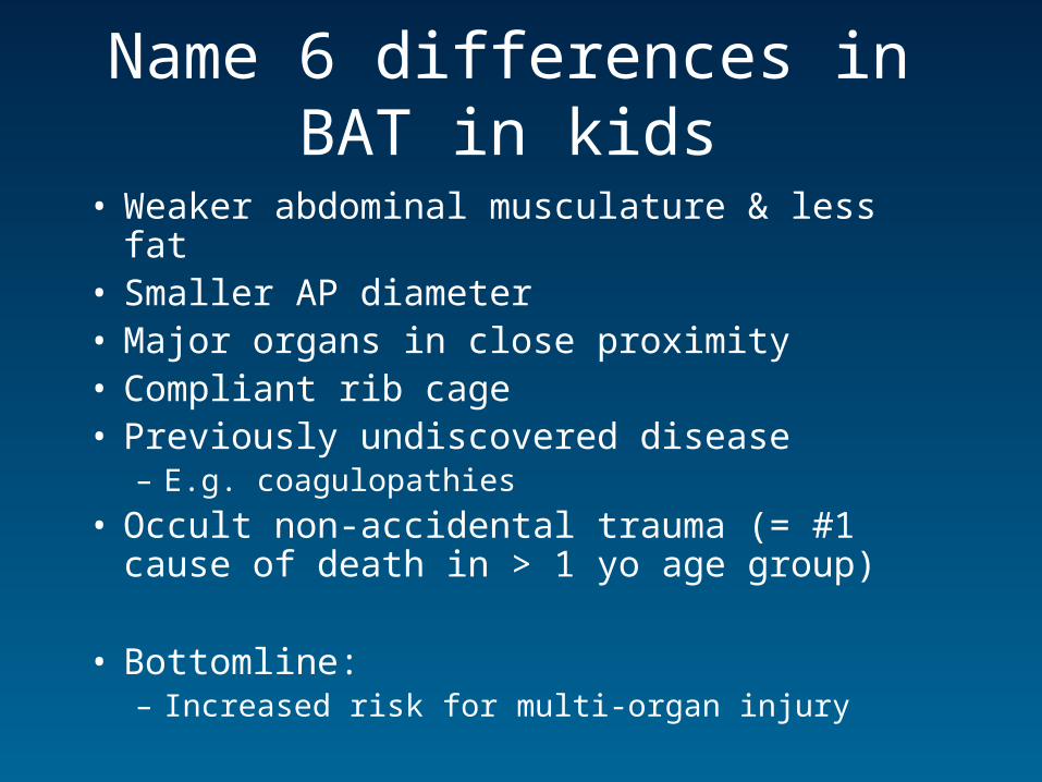
Name 6 differences in BAT in kids
• Weaker abdominal musculature & less fat• Smaller AP diameter • Major organs in close proximity• Compliant rib cage• Previously undiscovered disease
– E.g. coagulopathies
• Occult non-accidental trauma (= #1 cause of death in > 1 yo age group)
• Bottomline:– Increased risk for multi-organ injury
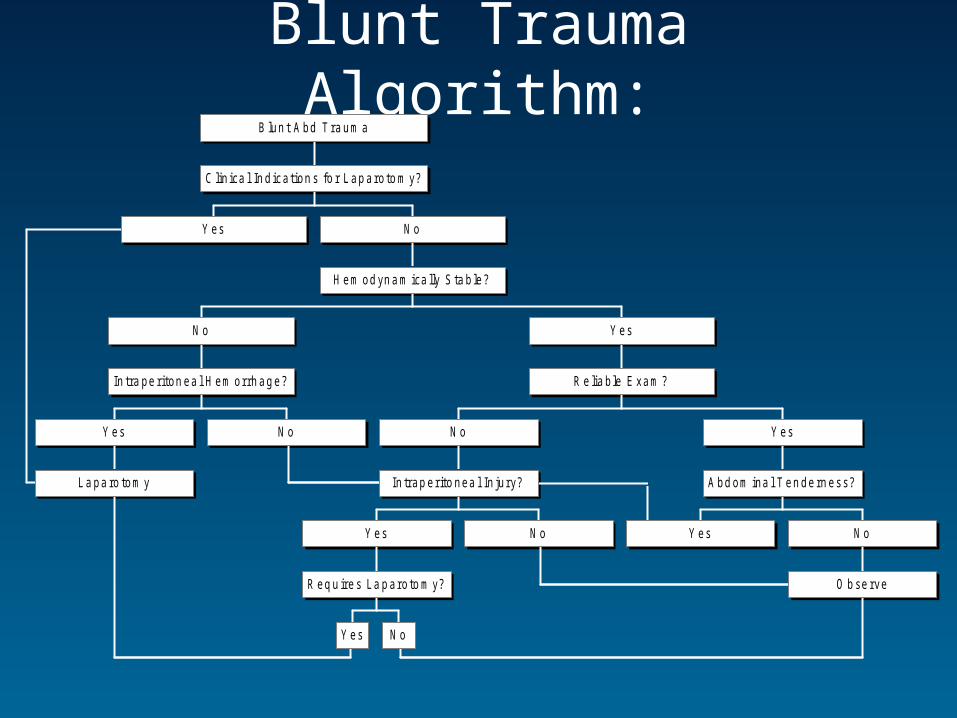
Blunt Trauma Algorithm:
Y es
L a pa ro tom y
Y es N o
In tra pe ri ton ea l H em orrh ag e?
N o
Y es N o
R eq u ire s La p aro to m y?
Y es N o
In trap er ito nea l In ju ry?
N o
Y es
O b se rve
N o
A bd om ina l T en de rn ess?
Y es
R e lia b le E xam ?
Y es
H em od yn a m ica lly S ta b le?
N o
C lin ica l In d ica tion s fo r L ap aro to m y?
B lun t A bd T ra um a
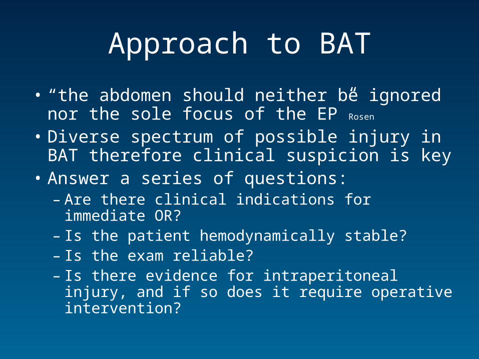
Approach to BAT
• “the abdomen should neither be ignored nor the sole focus of the EP”Rosen
• Diverse spectrum of possible injury in BAT therefore clinical suspicion is key
• Answer a series of questions:– Are there clinical indications for immediate OR?– Is the patient hemodynamically stable?– Is the exam reliable?– Is there evidence for intraperitoneal injury, and if
so does it require operative intervention?
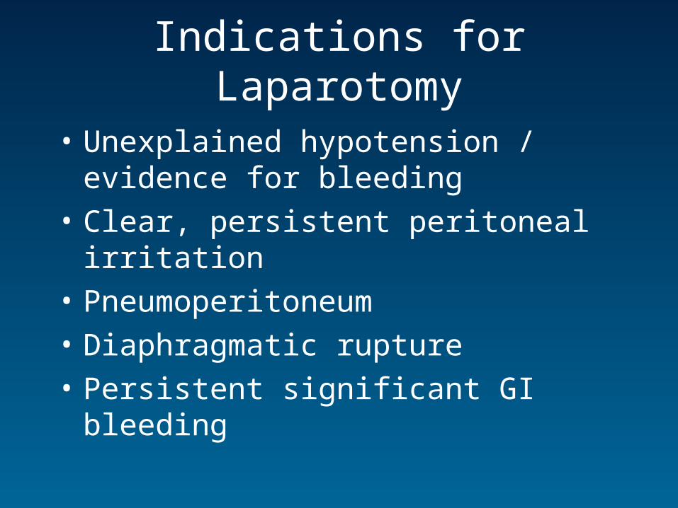
Indications for Laparotomy
• Unexplained hypotension / evidence for bleeding
• Clear, persistent peritoneal irritation
• Pneumoperitoneum
• Diaphragmatic rupture
• Persistent significant GI bleeding
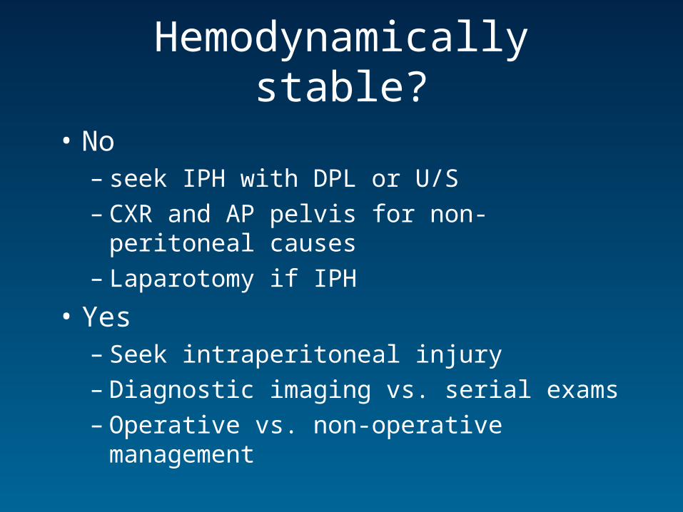
Hemodynamically stable?
• No – seek IPH with DPL or U/S– CXR and AP pelvis for non-peritoneal causes– Laparotomy if IPH
• Yes– Seek intraperitoneal injury– Diagnostic imaging vs. serial exams– Operative vs. non-operative management
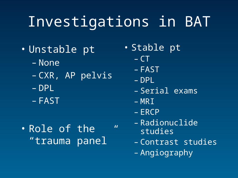
Investigations in BAT
• Unstable pt– None– CXR, AP pelvis– DPL– FAST
• Role of the “trauma panel”
• Stable pt– CT– FAST– DPL– Serial exams– MRI– ERCP– Radionuclide studies– Contrast studies– Angiography
The Trauma Panel
• What’s usually ordered:– CBC, ‘lytes, Cr, BUN, PT, PTT, T /S, T/C, UA,
EtOH, ABG,
• What there is actually evidence for:– Mostly retrospective studies– Most demonstrate abnormalities, but fail to
indicate any change in management– Review concludes most useful tests are T/S,
T/C, CBC, PT/PTT , ABG, and tox screen» Asimos. Emerg Med Reports. 1997
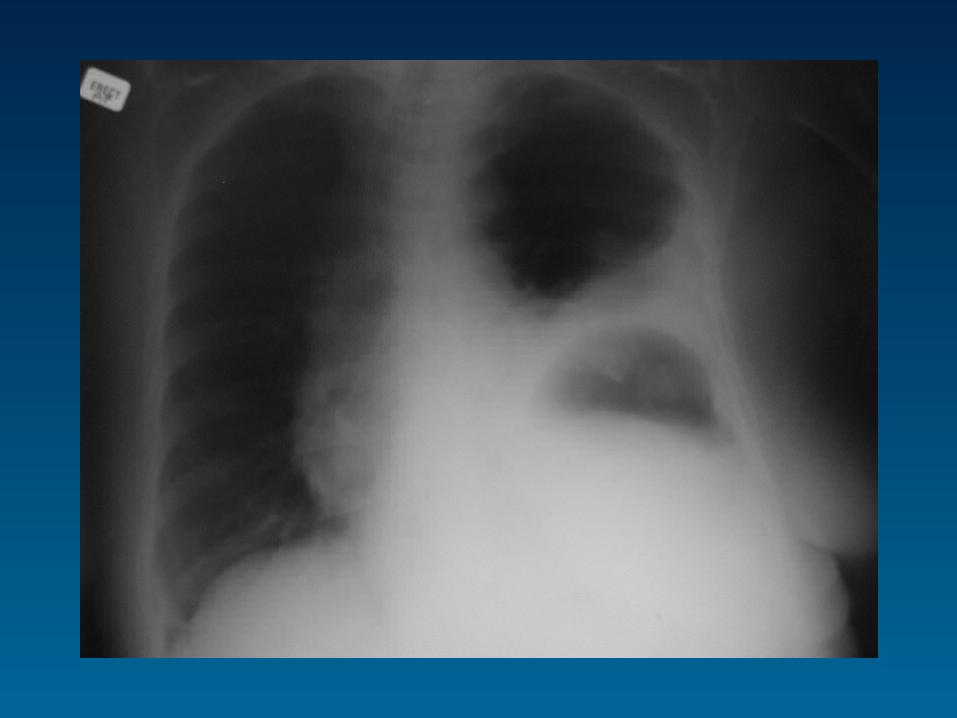
Plain Radiography
• CXR– Ruptured hemidiaphragm, pneumoperitoneum,
loss of psoas shadow, retroperitoneal air
• AP Pelvis– Pelvic fracture– Routine use in awake pt with stable, non-tender
pelvis may be unnecessary
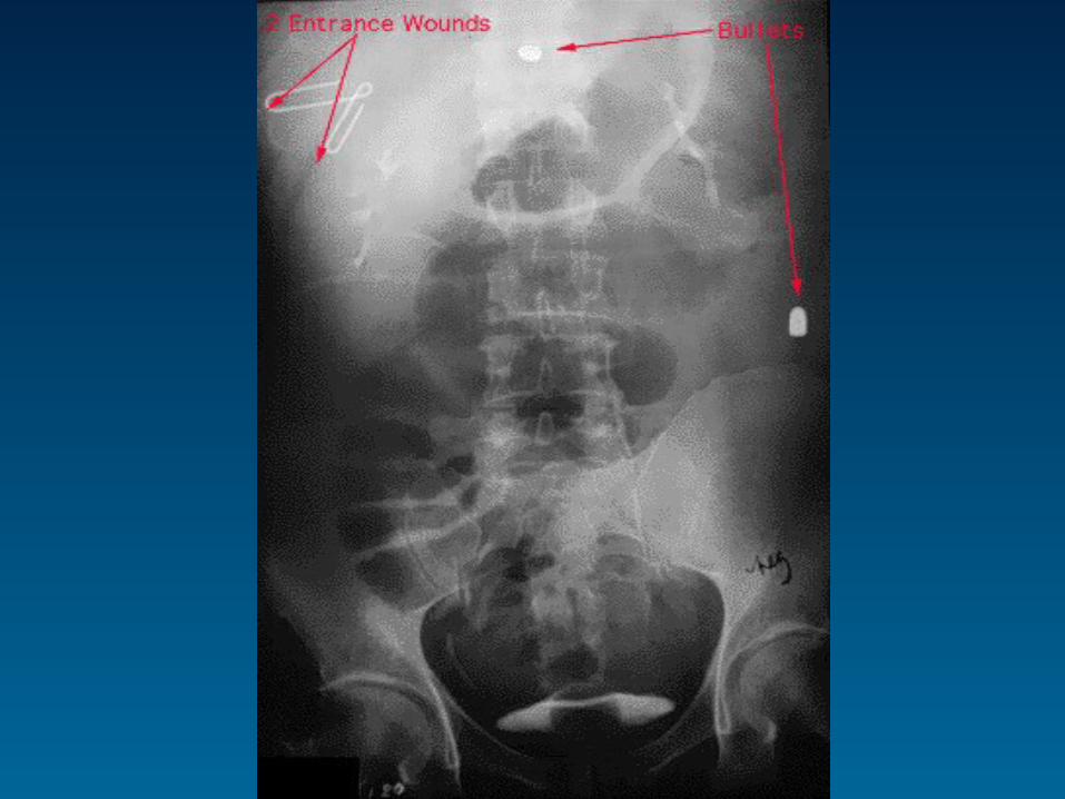
• AXR– Not routinely indicated in BAT– May show tract, or retained missiles in PAT
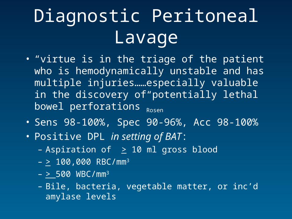
Diagnostic Peritoneal Lavage
• “virtue is in the triage of the patient who is hemodynamically unstable and has multiple injuries……especially valuable in the discovery of potentially lethal bowel perforations”Rosen
• Sens 98-100%, Spec 90-96%, Acc 98-100%• Positive DPL in setting of BAT:
– Aspiration of > 10 ml gross blood
– > 100,000 RBC/mm3
– > 500 WBC/mm3
– Bile, bacteria, vegetable matter, or inc’d amylase levels

Diagnostic Peritoneal Lavage
• Advantages– Rapid– Aids operative decision-making– Good for detecting hollow viscus injury
• Disadvantages– Samples only peritoneal cavity– Invasive– False positive rate of 2% (Inc’s laps)– Non-specific
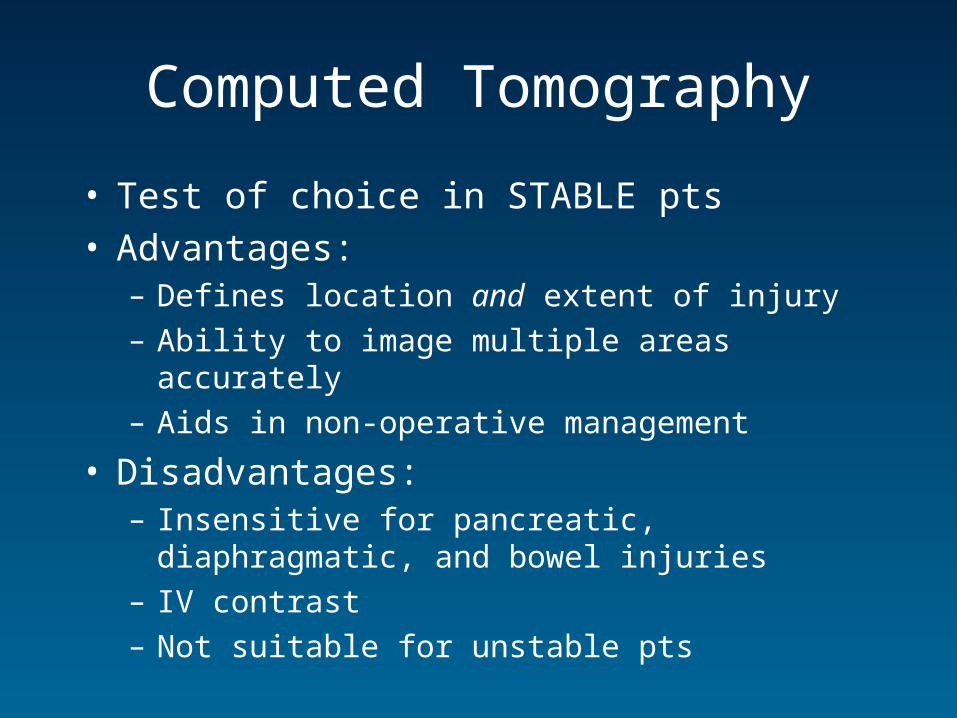
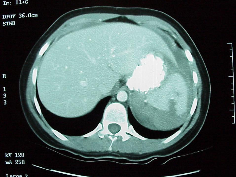
Computed Tomography
• Test of choice in STABLE pts• Advantages:
– Defines location and extent of injury
– Ability to image multiple areas accurately
– Aids in non-operative management
• Disadvantages:– Insensitive for pancreatic, diaphragmatic, and bowel
injuries
– IV contrast
– Not suitable for unstable pts
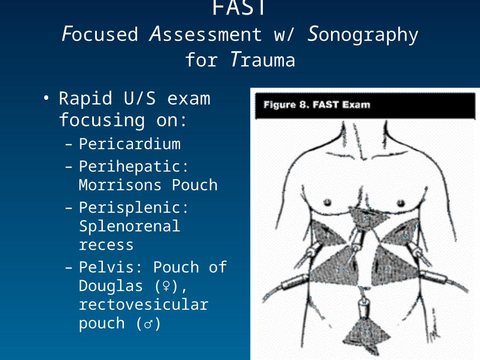
FASTFocused Assessment w/ Sonography for Trauma
• Rapid U/S exam focusing on:– Pericardium
– Perihepatic: Morrisons Pouch
– Perisplenic: Splenorenal recess
– Pelvis: Pouch of Douglas (♀), rectovesicular pouch (♂)
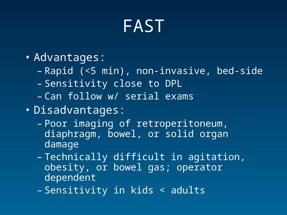
FAST
• Advantages:– Rapid (<5 min), non-invasive, bed-side– Sensitivity close to DPL– Can follow w/ serial exams
• Disadvantages:– Poor imaging of retroperitoneum, diaphragm,
bowel, or solid organ damage– Technically difficult in agitation, obesity, or
bowel gas; operator dependent– Sensitivity in kids < adults
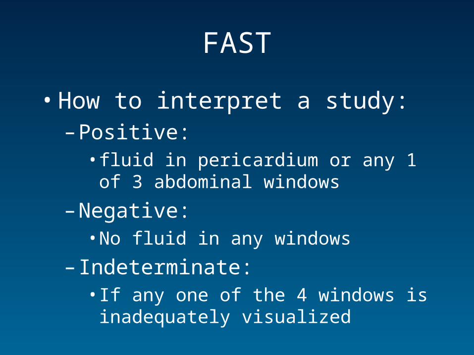
FAST
• How to interpret a study:– Positive:
• fluid in pericardium or any 1 of 3 abdominal windows
– Negative:• No fluid in any windows
– Indeterminate:• If any one of the 4 windows is inadequately
visualized
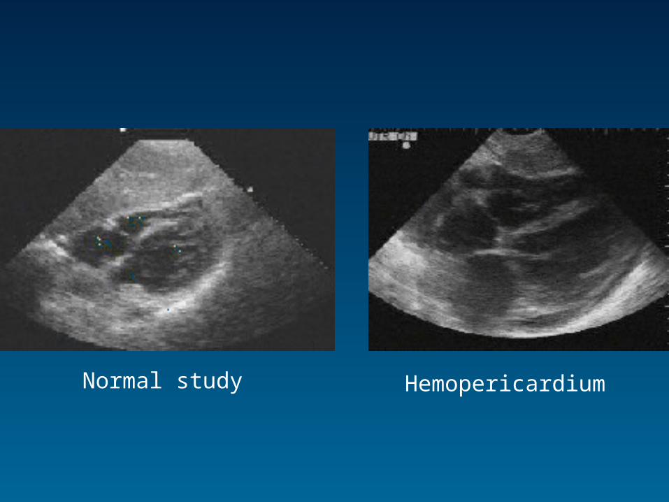
Normal study Hemopericardium

Normal Study
Perihepatic fluid
Perisplenic fluid

FAST• How good is it?
– Hemoperitoneum (Sens: 78-99%, Spec: 97-100%)– Hemopericardium (Sens: 100%, Spec: 97%)
• Assumes important injuries will have ass’d free fluid• However recent meta-analysis found NPV for IPFF and organ
injury to be 0.78-0.94 + 0.72-0.99 respectively• By calculating LR’s this means assuming pre-test prob of
50%, the post-test prob after –ve FAST remains 25%• Concluded that a negative study does not adequately exclude
IP injury» Stengel et al. Br J Surg. 88: 901-912. 2001
• Not adequately studied in pediatric trauma» Jones. Trauma Reports. 2000
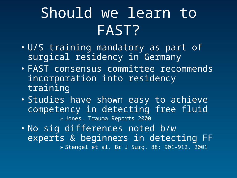
Should we learn to FAST?
• U/S training mandatory as part of surgical residency in Germany
• FAST consensus committee recommends incorporation into residency training
• Studies have shown easy to achieve competency in detecting free fluid
» Jones. Trauma Reports 2000
• No sig differences noted b/w experts & beginners in detecting FF
» Stengel et al. Br J Surg. 88: 901-912. 2001
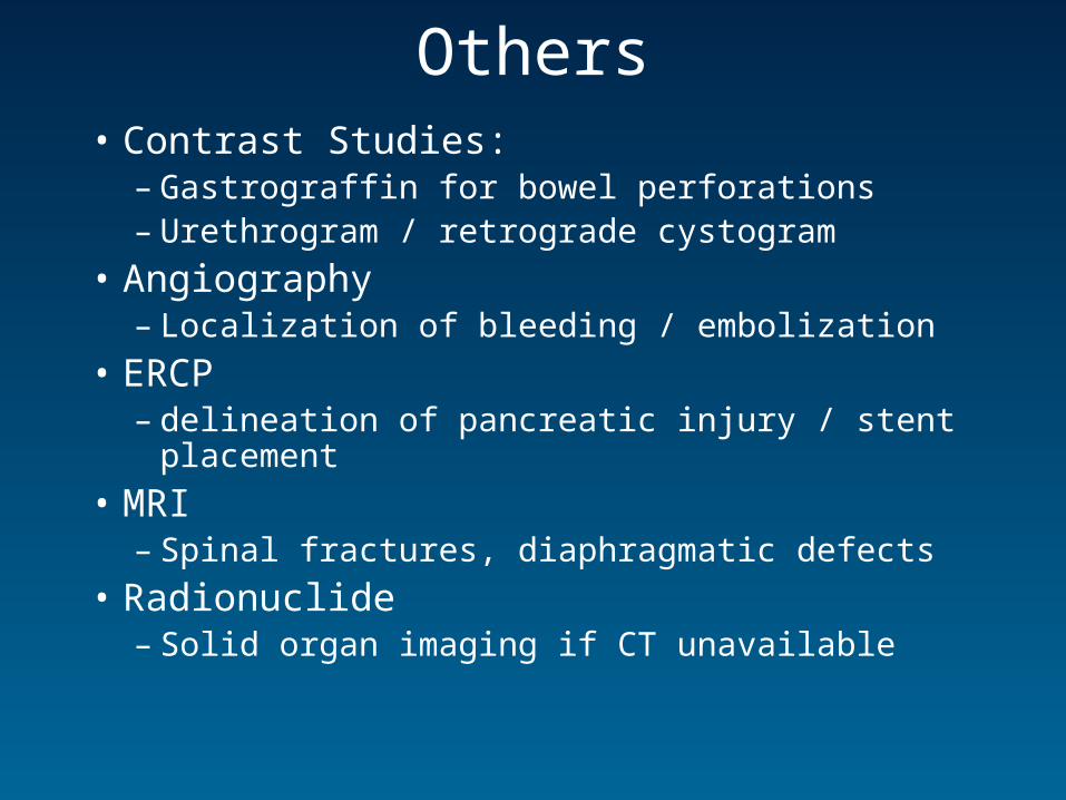
Others• Contrast Studies:
– Gastrograffin for bowel perforations– Urethrogram / retrograde cystogram
• Angiography– Localization of bleeding / embolization
• ERCP– delineation of pancreatic injury / stent placement
• MRI– Spinal fractures, diaphragmatic defects
• Radionuclide– Solid organ imaging if CT unavailable
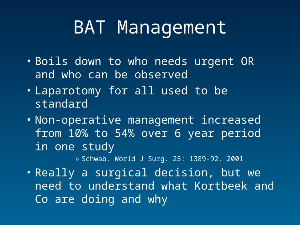
BAT Management
• Boils down to who needs urgent OR and who can be observed
• Laparotomy for all used to be standard
• Non-operative management increased from 10% to 54% over 6 year period in one study
» Schwab. World J Surg. 25: 1389-92. 2001
• Really a surgical decision, but we need to understand what Kortbeek and Co are doing and why
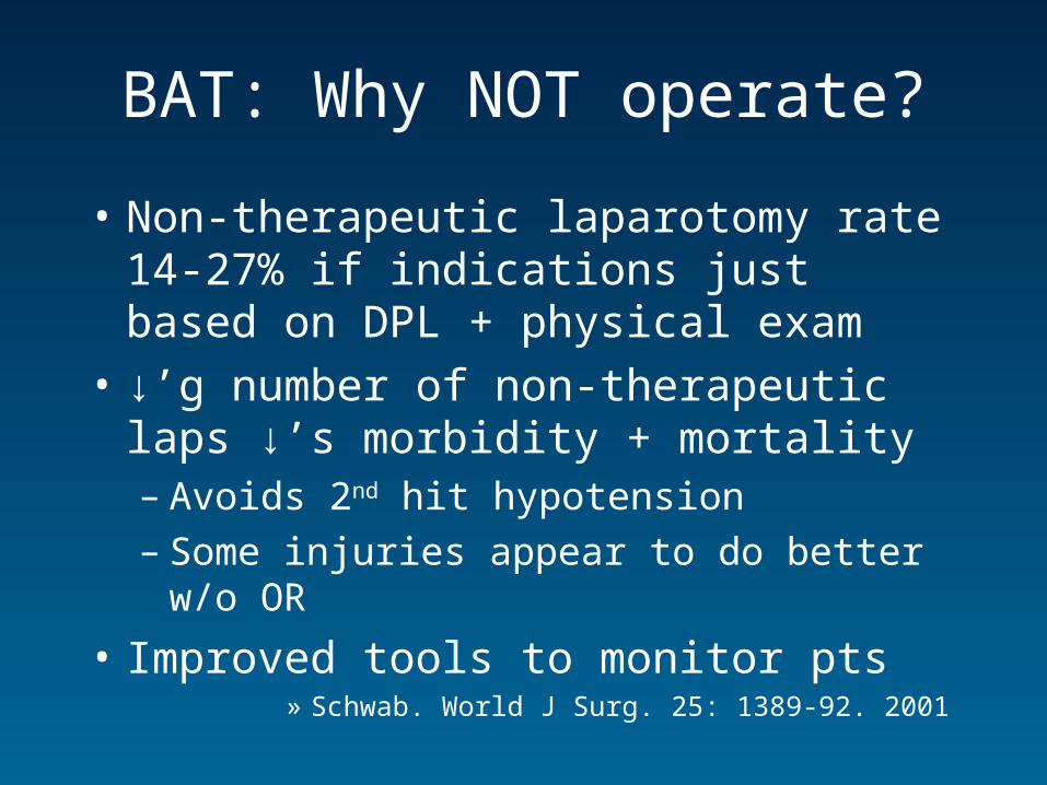
BAT: Why NOT operate?
• Non-therapeutic laparotomy rate 14-27% if indications just based on DPL + physical exam
• ↓’g number of non-therapeutic laps ↓’s morbidity + mortality– Avoids 2nd hit hypotension– Some injuries appear to do better w/o OR
• Improved tools to monitor pts» Schwab. World J Surg. 25: 1389-92. 2001
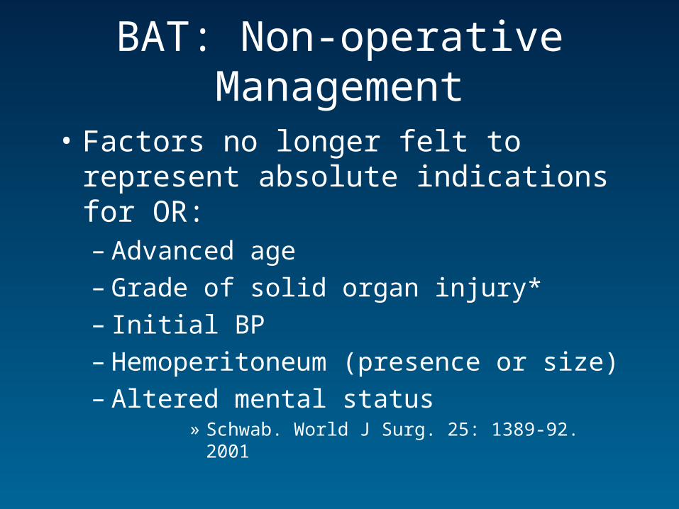
BAT: Non-operative Management
• Factors no longer felt to represent absolute indications for OR:– Advanced age– Grade of solid organ injury*– Initial BP– Hemoperitoneum (presence or size)– Altered mental status
» Schwab. World J Surg. 25: 1389-92. 2001
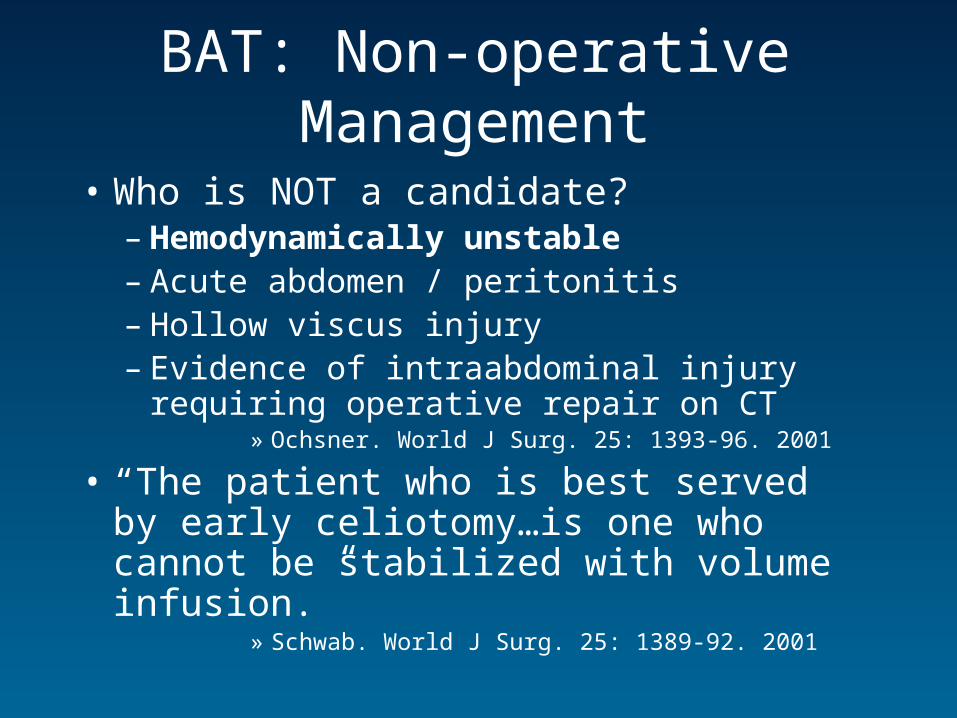
BAT: Non-operative Management
• Who is NOT a candidate?– Hemodynamically unstable – Acute abdomen / peritonitis– Hollow viscus injury– Evidence of intraabdominal injury requiring
operative repair on CT» Ochsner. World J Surg. 25: 1393-96. 2001
• “The patient who is best served by early celiotomy…is one who cannot be stabilized with volume infusion.”
» Schwab. World J Surg. 25: 1389-92. 2001
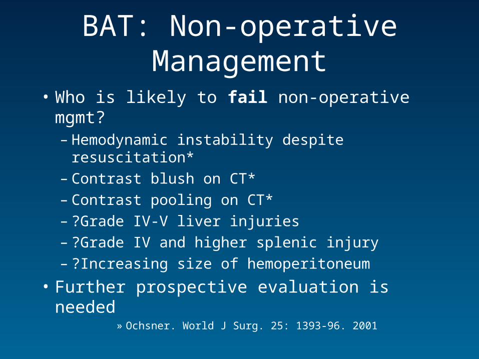
BAT: Non-operative Management
• Who is likely to fail non-operative mgmt?– Hemodynamic instability despite resuscitation*– Contrast blush on CT*– Contrast pooling on CT*– ?Grade IV-V liver injuries – ?Grade IV and higher splenic injury– ?Increasing size of hemoperitoneum
• Further prospective evaluation is needed» Ochsner. World J Surg. 25: 1393-96. 2001
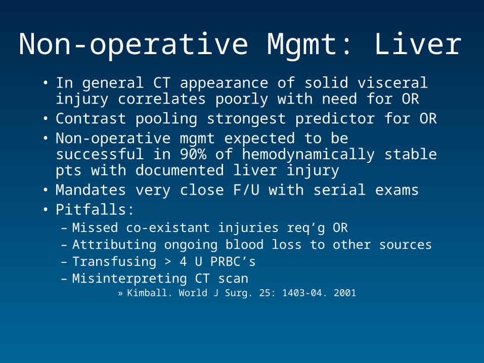
Non-operative Mgmt: Liver• In general CT appearance of solid visceral injury
correlates poorly with need for OR • Contrast pooling strongest predictor for OR• Non-operative mgmt expected to be successful in
90% of hemodynamically stable pts with documented liver injury
• Mandates very close F/U with serial exams• Pitfalls:
– Missed co-existant injuries req’g OR– Attributing ongoing blood loss to other sources– Transfusing > 4 U PRBC’s– Misinterpreting CT scan
» Kimball. World J Surg. 25: 1403-04. 2001
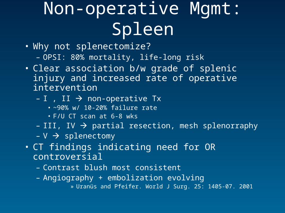
Non-operative Mgmt: Spleen
• Why not splenectomize?– OPSI: 80% mortality, life-long risk
• Clear association b/w grade of splenic injury and increased rate of operative intervention– I , II non-operative Tx
• ~90% w/ 10-20% failure rate• F/U CT scan at 6-8 wks
– III, IV partial resection, mesh splenorraphy– V splenectomy
• CT findings indicating need for OR controversial– Contrast blush most consistent– Angiography + embolization evolving
» Uranüs and Pfeifer. World J Surg. 25: 1405-07. 2001
Penetrating Abdominal Trauma
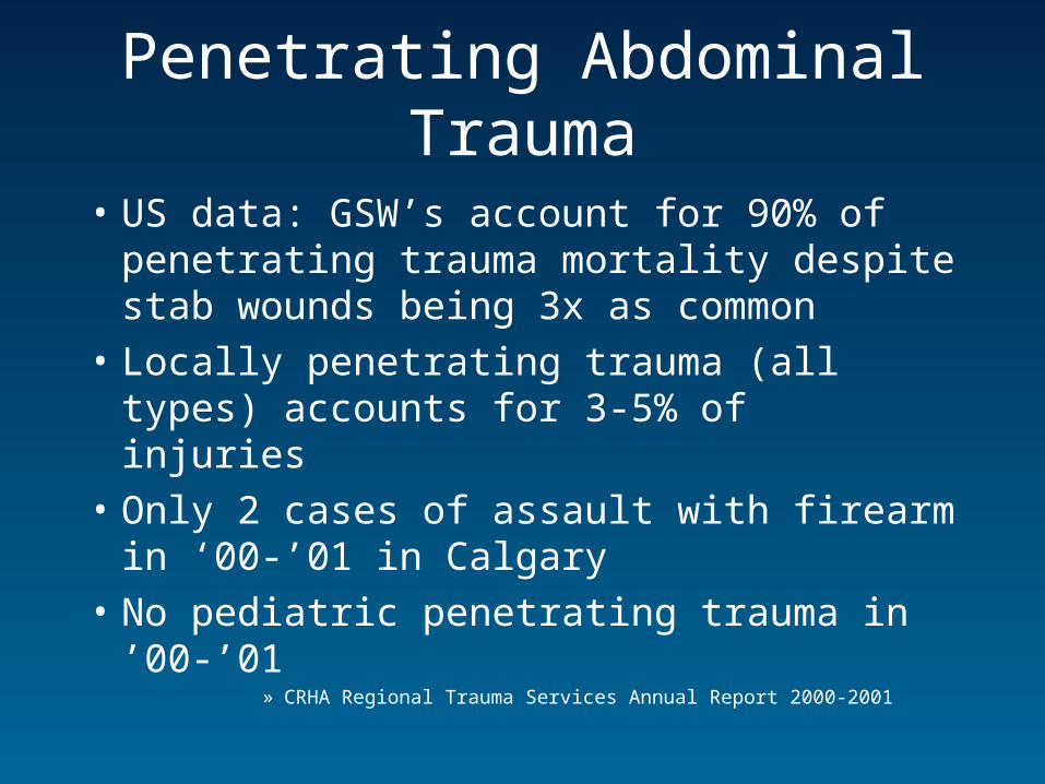
• US data: GSW’s account for 90% of penetrating trauma mortality despite stab wounds being 3x as common
• Locally penetrating trauma (all types) accounts for 3-5% of injuries
• Only 2 cases of assault with firearm in ‘00-’01 in Calgary
• No pediatric penetrating trauma in ’00-’01» CRHA Regional Trauma Services Annual Report 2000-2001

My view on gun control:

..and the NRA perspective:
Penetrating Abdominal Trauma
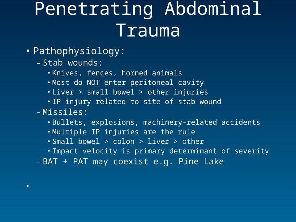
• Pathophysiology:– Stab wounds:
• Knives, fences, horned animals• Most do NOT enter peritoneal cavity• Liver > small bowel > other injuries• IP injury related to site of stab wound
– Missiles: • Bullets, explosions, machinery-related accidents• Multiple IP injuries are the rule• Small bowel > colon > liver > other• Impact velocity is primary determinant of severity
– BAT + PAT may coexist e.g. Pine Lake
•
Penetrating Abdominal Trauma
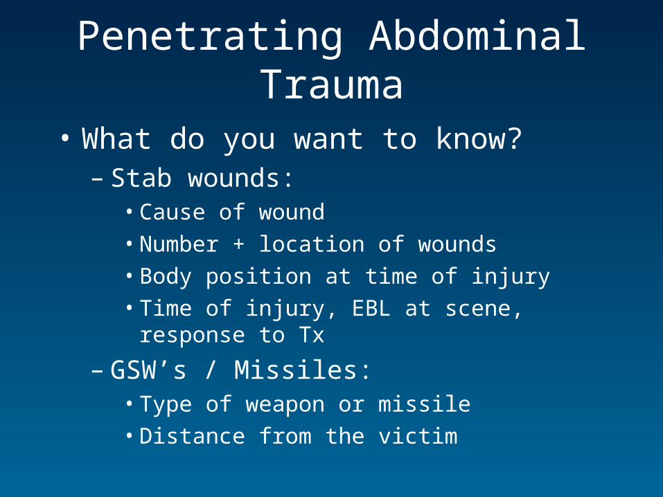
• What do you want to know?– Stab wounds:
• Cause of wound
• Number + location of wounds
• Body position at time of injury
• Time of injury, EBL at scene, response to Tx
– GSW’s / Missiles:• Type of weapon or missile
• Distance from the victim
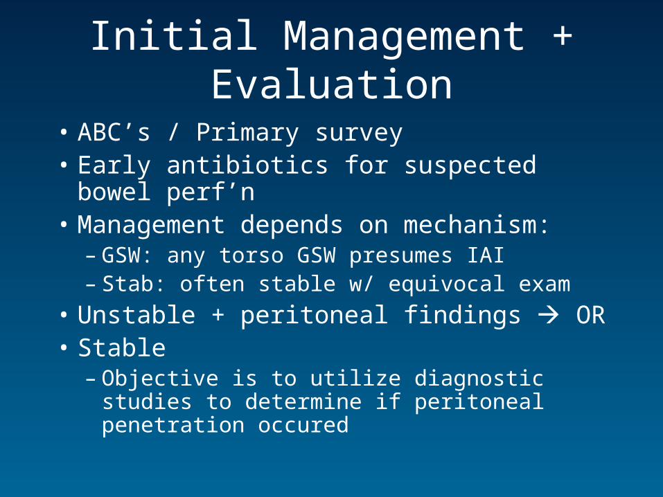
Initial Management + Evaluation
• ABC’s / Primary survey• Early antibiotics for suspected bowel perf’n• Management depends on mechanism:
– GSW: any torso GSW presumes IAI– Stab: often stable w/ equivocal exam
• Unstable + peritoneal findings OR• Stable
– Objective is to utilize diagnostic studies to determine if peritoneal penetration occured
Investigations
• Trauma panel
• Plain films
• DPL
• FAST
• CT
• Local Wound Exploration
• Laparoscopy

DPL• Same technique and diagnostic criteria as in
BAT, except RBC criteria depending on location:
Location / TypePositive
(RBC/mm3)Indeterminate (RBC/mm3)
Ant abd stab > 100,000 20,000-100,000
Flank stab > 100,000 20,000-100,000
Back stab > 100,000 20,000-100,000
Low chest stab > 5000 1000-5000
GSW’s > 5000 1000-5000
Local Wound Exploration
• Primary use in single anterior stab wounds to r/o peritoneal penetration
• Not used in:– Multiple stab wounds– entry over thorax
• Inability to clearly visualize end of wound tract presumes peritoneal violation
Laparoscopy
• Limited utility– Usually requires GA– Risk of tension pneumothorax / gas embolus– High false negative rate– No info on post peritoneum / retroperitoneum
• Primary use:– Isolated stab injuries of ant abd can d/c if -
ve
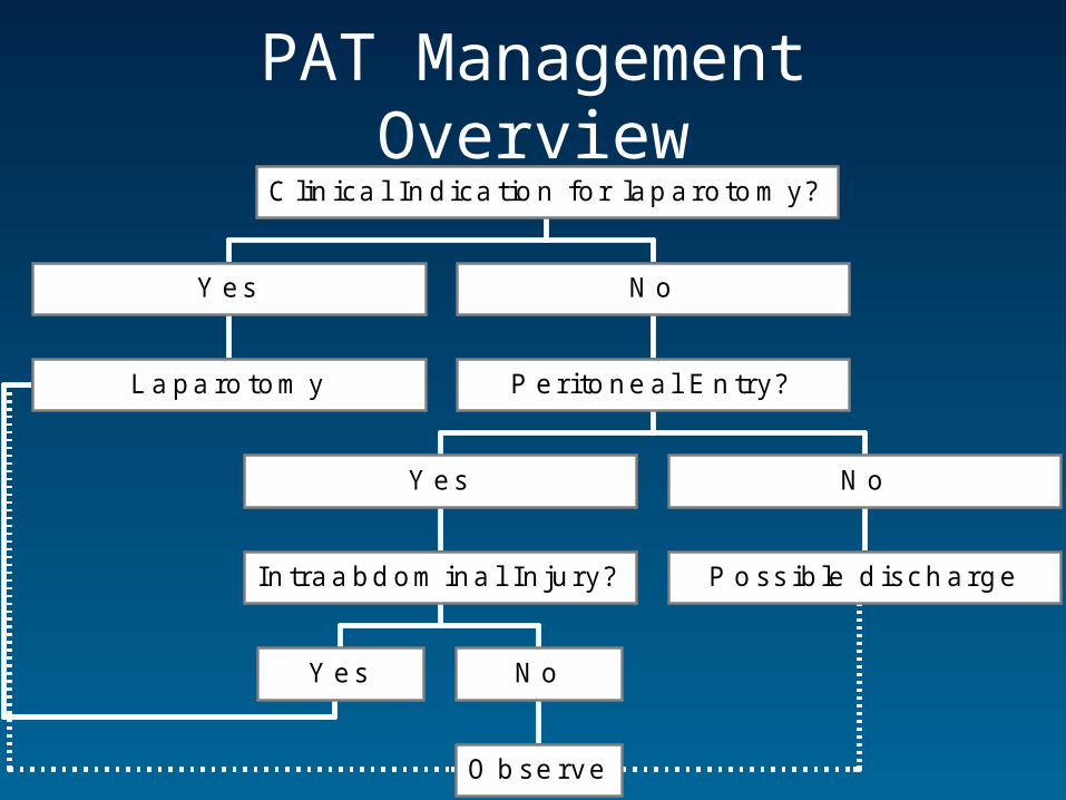
PAT Management Overview
L a pa ro tom y
Y es
Y es
O b se rve
N o
In traa bd om ina l In ju ry?
Y es
P o ss ib le d isch arge
N o
P er i to ne a l E n try?
N o
C lin ica l In d ica tio n fo r lap aro to m y?
PAT: Overview• Approach to GSW + stab wound similar but:
– Stab: • Low incidence of peritoneal penetration• Operative intervention for all would result in
unacceptable non-therapeutic lap rate
– GSW:• High incidence of peritoneal entry and injury (>80%)• Selective management is increasing but << stab
wounds
• Approach outlined by 3 basic questions:– 1. Does this person need OR now?– 2. Has peritoneal penetration occurred– 3. Does an IAI exist, and does it need the OR?
Clinical Indications for Laparotomy in PAT
• Hemodynamic instability
• Peritoneal signs
• Evisceration
• Diaphragmatic injury
• GI or vaginal hemorrhage
• Implement in situ
• Intraperitoneal air
Rule Out Peritoneal Penetration
• Stab wounds:– DPL– LWE– CT– Laparoscopy– U/S
• GSW’s:– Missile path– Plain films– LWE– U/S– Laparoscopy– CT

Is there an injury requiring OR?
• Stab wounds:– DPL– CT– Serial examinations
(min 24 hrs)
• GSW’s:– Serial exams– DPL– Laparoscopy
GSW + Non-operative Mgmt • Highly controversial• Non-therapeutic lap rate 15-25% w/
traditional approach• Significant number of pts w/o peritoneal
penetration, or minimal IAI• 9 trials of non-OR mgmt:
– No deaths – ~300 pts managed non-operatively– Heterogenous, 6 studies from same center
• Non-operative Tx for GSW is evolving, but laparotomy remains standard for most
» Saadia and Degiannis. Br J Surg. 87: 393-397. 2000
Other Situations:
• Thoracoabdominal wounds:– FAST to r/o hemopericardium– DPL: lower criteria↑’s sensitivity for
diaphragm lacerations
• Back / Flank wounds:– ↑’d risk of retroperitoneal + diaphragm injury– LWE, CT, serial exams, OR for most GSW’s
• Implements in situ– Should be removed in OR– May need angiogram or CT to r/o vascular
involvement
GU Trauma
• Rarely life-threatening
• Most common BAT injuries in kids
• Should be identified as part of secondary survey
• More likely to be occult injuries
• Investigated in retrograde fashion:– Urethra bladder ureter kidney
• Hematuria is hallmark sign
GU Exam
• Following increase likelihood of GU injury & demand investigation:– Abdo tenderness or PAT– Pelvic fracture / tenderness– Blood at urethral meatus / vaginal introitus– Penile / scrotal / perineal ecchymoses or pain– Abnormal prostate / rectal exam– Any color urine other than clear yellow
• DON’T even think about placing a foley

Hematuria in Trauma• Indications for investigation:
– Adults:• Gross hematuria• Microhematuria & shock (sys BP <90)• Microhematuria in setting of sig other injuries
» Tze et al. Clin Radiol 56: 268-77. 2001
– Kids:• Gross hematuria• Microhematuria > 50 RBC/hpf
» Morey et al. J Urol. 156: 2014-18. 1996
• Other studies & clinicians disagree; feel any microhematuria should be investigated
» Christopher. Ped Emerg Med Reports. 2000
Foley Cath: Do’s + Don’t’s
• Do:– Place foley in absence of previous S & S– Single attempt in documented partial tears– Place suprapubic cath in complete tears
• Don’t:– Place foley in suspected injury until retrograde
urethrogram has been performed– Remove a in situ foley
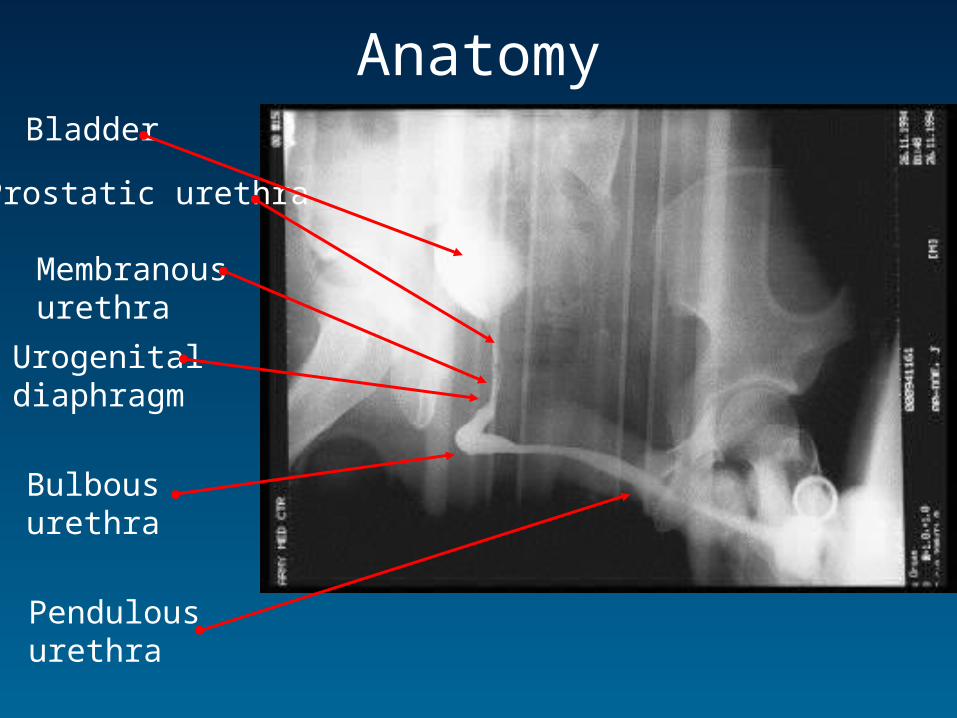
AnatomyBladder
Prostatic urethra
Urogenitaldiaphragm
Bulbousurethra
Pendulousurethra
Membranousurethra
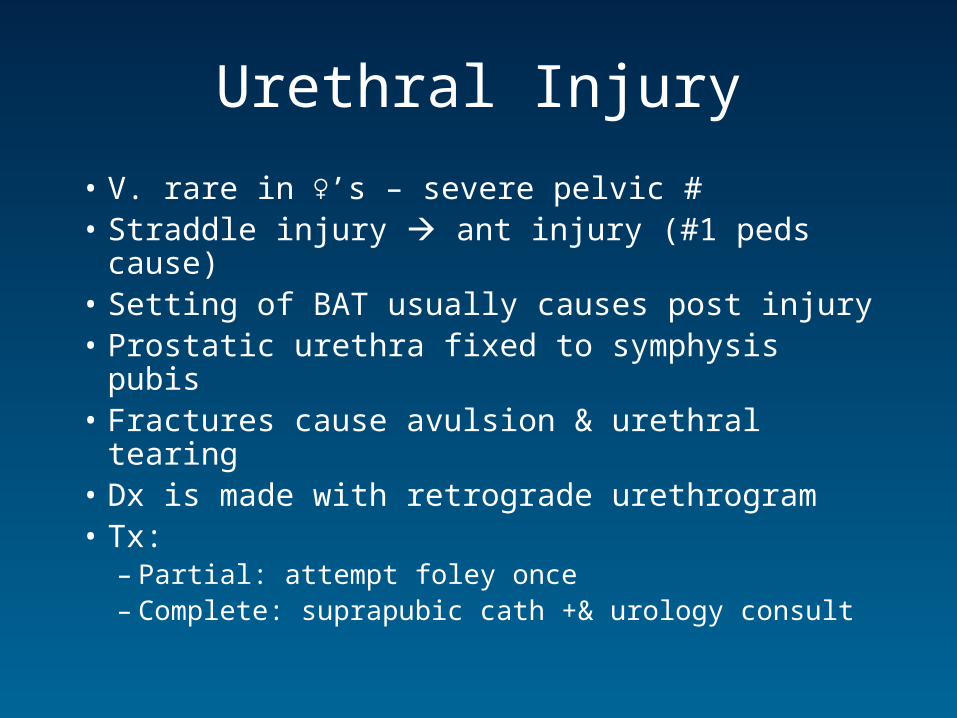
Urethral Injury
• V. rare in ♀’s – severe pelvic #• Straddle injury ant injury (#1 peds cause)• Setting of BAT usually causes post injury• Prostatic urethra fixed to symphysis pubis• Fractures cause avulsion & urethral tearing• Dx is made with retrograde urethrogram• Tx:
– Partial: attempt foley once– Complete: suprapubic cath +& urology consult
Bladder Rupture• Intrapelvic (empty) umbillicus (full)• Kids > adults b/c bladder more intraabdominal• 3 muscle layers arranged at angles seal small
perforations• Ass’d w/ severe trauma (>90%) & high mortality• Extraperitoneal & intraperitoneal• Present w/ abd or pelvic pain, inability to void, &
gross hematuria (>95%)• Investigations:
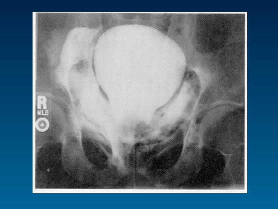
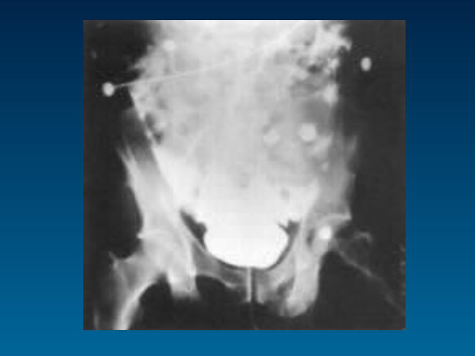
– Retrograde cystography– Retrograde CT cystography– Standard CT abd/pelvis is NOT adequate
Bladder Rupture
• Extraperitoneal (80-90%):– Ass’d w/ pelvic #– Flame-shaped extravasation of contrast in
obturator region / prevesicular space of Retzius– Tx: > 20 F foley x7-14 days
• Intraperitoneal (10-20%):– Ass’d w/ blunt trauma & full bladder– Intraperitoneal organs outlined by contrast– Tx: surgical repair
Renal Trauma
• Most common traumatic GU injury• Most common organ injury in pediatric BAT • Associated 1oly w/ BAT (85-90%)• Penetrating mechanism has ~80% rate of
associated intraperitoneal injury• Tend to be recognized late• 1o complication: renovascular HTN (~1%)
– ↓RBF renin release from JGA AII vasoconstriction & aldosterone release

Anatomy
• Retroperitoneal• Enclosed by Gerota’s
fascia• Suspended by pedicle &
surrounding fat1. Interlobular v.2. Interlobular a.3. Renal pelvis4. Renal a.5. Renal v.6. Ureter7. Cortex8. Medulla9. Arcuate v.10. Arcuate a.
Diagnosis of Renal Injury• S +S:
– Flank pain, abd pain, pelvic #, Grey-Turner’s, Cullen’s, low post rib #’s, L-spine #’s:
– Gross hematuria, Microhematuria & shock (sys BP <90), Microhematuria in setting of sig other injuries, Microhematuria > 50 RBC/hpf alone in kids
• Enhanced CT is study of choice if STABLE• IVP – lacks sensitivity for other injuries• U/S – no info on function (uptake)• Angiography – no advantage over CT
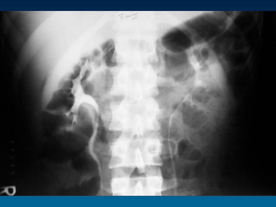
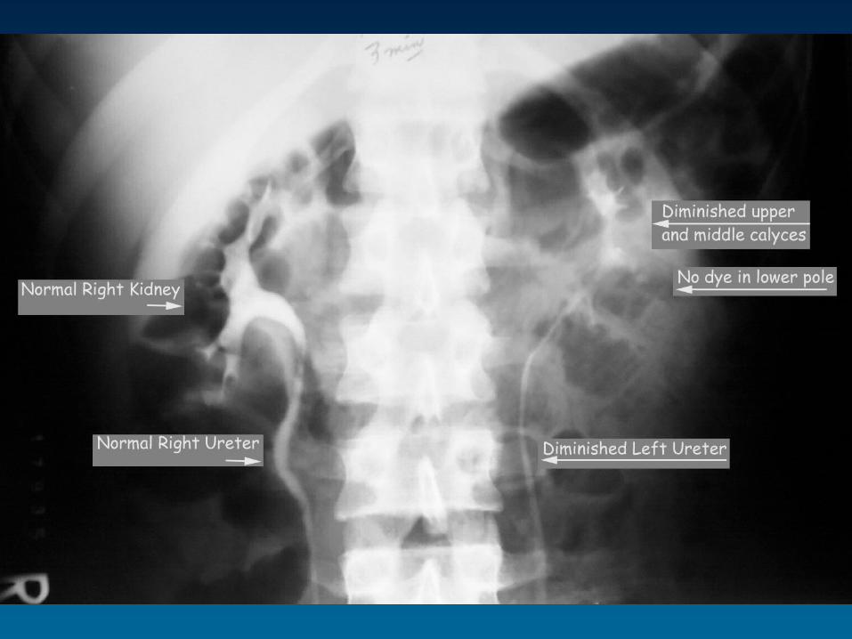
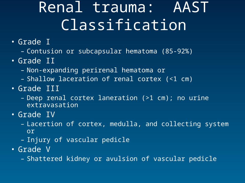
Renal trauma: AAST Classification
• Grade I – Contusion or subcapsular hematoma (85-92%)
• Grade II– Non-expanding perirenal hematoma or– Shallow laceration of renal cortex (<1 cm)
• Grade III– Deep renal cortex laneration (>1 cm); no urine extravasation
• Grade IV– Lacertion of cortex, medulla, and collecting system or– Injury of vascular pedicle
• Grade V– Shattered kidney or avulsion of vascular pedicle
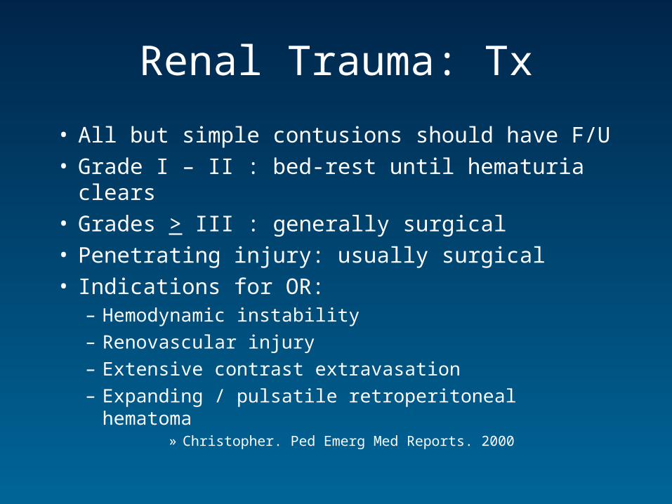
Renal Trauma: Tx
• All but simple contusions should have F/U• Grade I – II : bed-rest until hematuria clears• Grades > III : generally surgical• Penetrating injury: usually surgical• Indications for OR:
– Hemodynamic instability
– Renovascular injury
– Extensive contrast extravasation
– Expanding / pulsatile retroperitoneal hematoma» Christopher. Ped Emerg Med Reports. 2000

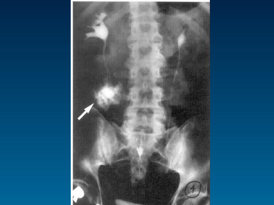
Ureteral Trauma
• Rare (1%)
• PAT > BAT
• Tends to involve proximal 1/3
• Often present later ~ 10 days post-injury w:– Hematuria, flank pain / mass, fever / chills
• Dx: enhanced CT, IVP, or retrograde pyelogram
• Tx: surgical

?