about the specialized myocardial conducting tissue … · about the specialized myocardial...
TRANSCRIPT

A
R
A
A
a
b
c
R
1h
Document downloa
rch Cardiol Mex. 2013;83(4):278---281
www.elsevier.com.mx
EVIEW ARTICLE
bout the specialized myocardial conducting tissue
lfredo de Micheli Serraa,∗, Pedro Iturralde Torresb, Alberto Aranda Fraustroc
Research Fallow, Instituto Nacional de Cardiología Ignacio Chávez, Tlalpan, DF, MexicoDepartment of Electrocardiology, Instituto Nacional de Cardiología Ignacio Chávez, Tlalpan, DF, MexicoDepartment of Patology, Instituto Nacional de Cardiología Ignacio Chávez, Tlalpan, DF, Mexico
eceived 5 November 2012; accepted 21 March 2013
KEYWORDSMyocardialspecializedconducting system;Left ventricularconducting system;Right ventricularconducting system;Mexico
Abstract The chronological succession of discoveries on the location and structure of theatrio-ventricular conducting system are described. The starting point of this system is locatedin the sinus atrial node, identified by the English scientists A. Keith and M. W. Flack in 1907.The atrioventricular conducting system was pointed out by the Swiss physician Wilhelm His Jr.in 1893. The atrioventricular node (AV) was first identified by the Japanese pathologist SumaoTawara and his German professor Ludwig Aschoff in 1906.
Likewise the structure and routes of the three internodal bundles are described. These bun-dles include: Bachmann’s bundle (1916) connecting the right with the left atrium and the AVnode; the middle Wenckebach’s bundle (1910) and the posterior or Thörel’s bundle (1910),extending from the region of the sinus atrial node towards the posterior margin of the AV node.
Lastly, the ventricular left and right conduction systems are detailed. These include the maintrunk and their peripheral subdivisions with respective networks. Regarding the controversialexistence of the left middle subdivision, it can exist in animal and human hearts. Nevertheless,an intermediate left septal network of specialized fibers seems to act as a functional equivalentof this subdivision.© 2012 Instituto Nacional de Cardiología Ignacio Chávez. Published by Masson Doyma MéxicoS.A. All rights reserved.
PALABRAS CLAVE Acerca del tejido miocárdico especializado de excitoconducción
ded from http://zl.elsevier.es, day 29/01/2014. This copy is for personal use. Any transmission of this document by any media or format is strictly prohibited.
Sistema cardiaco deexcitoconducción;Sistema deconducciónventricular izquierdo;
Resumen Se describe, en orden cronológico, la sucesión del descubrimiento de la localizacióny la estructura de los componentes del sistema de conducción auriculoventricular. El haz deconducción AV fue descrito por el médico suizo Wilhelm His Jr. en 1893. El punto de origende dicho sistema se halla en el nodo sinoauricular, identificado por los ingleses A. Keith y
∗ Corresponding author at: Juan Badiano, No. 1, Col. Sección XVI, Tlalpan, México DF, C.P. 14080, Mexico.E-mail address: [email protected] (A. de Micheli Serra).
405-9940/$ – see front matter © 2012 Instituto Nacional de Cardiología Ignacio Chávez. Published by Masson Doyma México S.A. All rights reserved.ttp://dx.doi.org/10.1016/j.acmx.2013.03.002

About the specialized myocardial conducting tissue 279
Sistema deconducciónventricular derecho;México
M.W. Flack en 1907. El nodo auriculoventricular (AV) fue identificado por el patólogo japonésSunao Tawara y su maestro, el alemán Ludwig Aschoff, en 1906.
Asimismo se relatan la estructura y los recorridos de los 3 haces internodales: el anterioro de Bachmann (1916), que conecta la aurícula derecha con la izquierda y el nodo AV; el medio ohaz de Wenckebach (1910) y el posterior o haz de Thörel (1910), que se dirige desde la regióndel nodo sinoauricular hacia la aurícula izquierda y el margen de atrás del nodo AV.
Se presentan asimismo, de forma esquemática, los sistemas de conducción ventricularizquierdo y derecho, que comprenden el tronco principal y las subdivisiones periféricas con susrespectivas redes de Purkinje. Respecto a la controvertida existencia de un fascículo izquierdomedio, éste sí puede existir en corazones humanos y de animales. Pero la red septal intermediade fibras especializadas parece ser un equivalente funcional de dicho fascículo.© 2012 Instituto Nacional de Cardiología Ignacio Chávez. Publicado por Masson Doyma México
ervad
stsdisrc
L
Tbtlwv
dtwo subdivisions. This network appears to be the functionalequivalent of a middle bundle, distributed in the left sep-tal mass of the medial-third of the interventricular septum12
(Fig. 1). Each of the left subdivisions end in the network of
Figure 1 Staining of the left ventricular conduction systemwith Lugol’s solution in a canine heart. LBB, left bundle branch;
Document downloaded from http://zl.elsevier.es, day 29/01/2014. This copy is for personal use. Any transmission of this document by any media or format is strictly prohibited.
S.A. Todos los derechos res
The specialized myocardial excitoconducting tissueincludes several sections. The first component is the sinoa-trial (SA) node, the primum movens (first mover) of normalcardiac activity, described by the English A. Keith and M.W.Flack in 1907.1 It is located in the sulcus terminalis andextends from the recess of the right atrial appendage to theintercaval band.2 The atrioventricular conduction systemoriginates in the homonymous node (atrioventricular node)identified by Tawara and Aschoff.3 The anatomy of the afore-mentioned node has been carefully studied by James.4,5
The AV node is found in the inferior portion of the inter-atrial septum or, to put it more accurately, the AV node liesin what can be defined as the fibromuscular atrioventricu-lar septum. It is located between the orifice of the coronarysinus in the right atrium and the septal leaflet of the tricus-pid valve, to the right of the central fibrous body (trigonumfibrosum) where it joins the mitral annulus.5
In the human heart, there are three connections betweenthe SA and the AV nodes, containing Purkinje fibers as well asnon-specialized myocardial fibers.6,7 The anterior bundle,8
otherwise known as Bachmann’s bundle, connects the SAnode with the left atrium where it turns backwards anddescends to the interatrial septum towards the AV node. Themiddle internodal or Wenckebach’s bundle9 extends fromthe dorsal and posterior margins of the SA node and runsbehind the superior vena cava through the intercaval sul-cus towards the crest of the interatrial septum. From there,it extends towards the AV node, intersecting with fibersproceeding from the anterior bundle as it approaches thenode in the inferior portion of the interatrial septum or,what might be better called, the fibromuscular atrioven-tricular septum. This means, it lies between the entry ofthe coronary sinus in the right atrium and the septal leafletof the tricuspid valve. It lies, thus, to the right of the cen-tral fibrous body. The posterior internodal bundle describedby Thörel10 follows the crista terninalis from the AV node tothe Eustachian ridge and, from there, runs along the sameridge to the posterior margin of the AV node.
Atrioventricular conduction system
The atrioventricular conduction system originates in the AVnode with the bundle of His. The bundle of His was describedby the Swiss doctor, Wilhelm His Jr. (1863---1934) and com-prises the common trunk and the left and right ventricular
Aomc
os.
ystems.11 The first, evidenced by Sunao Tawara, using his-ological staining with Chinese ink in the human heart andeveral animal species, has been localized in recent times byifferent staining methods. In the canine heart, the bundles evidenced using iodine-potassium iodide solution (Lugol’solution), because the specialized tissue it contains is veryich in glycogen that has high affinity for iodine. This systeman also be stained by Lugol’s solution in the human heart.
eft ventricular conduction system
his system is composed by the main trunk of the leftranch, which is short (2 or 3 mm) and soon gives rise towo subdivisions: the anterior subdivision located in theeft superior septal mass, and the posterior subdivisionith branches that run to the posterior wall of the leftentricle.12
In the canine heart, an entanglement of fibres (interme-iary network), branches off from this main trunk and the
S, anterior subdivision; PS, posterior subdivision. A networkf fibers branches off between these subdivisions, from theain trunk and both the anterior and posterior subdivisions. It
onstitutes the functional equivalent of a middle left fascicle.
280 A. de Micheli Serra et al.
Figure 2 The single canine heart (out of eleven) in whichta
ebs
aeti
ows
chHiaftsdhttpemowdsdt
R
Tft
FsT
tpaschhofrbdtseptal mass with numerous fillets that are distributed inthe posterior, and medial and inferior lateral regions of thehomolateral free ventricular wall. Each subdivision ends inthe corresponding Purkinje network. Fig. 4 illustrates the
Document downloaded from http://zl.elsevier.es, day 29/01/2014. This copy is for personal use. Any transmission of this document by any media or format is strictly prohibited.
he left medial fascicle could be identified as a well-definednatomical structure. A, anterior; P, posterior.
ndocardial and subendocardial specialized fibers observedy J.E. Purkinje in 1839 and identified histologically by him-elf a few years later.13
Through the fibers that form the intermediate network,ctivation impulses may reach the left septal mass and itsndocardial surface where the process of ventricular activa-ion starts.14 This process manifests as the first septal vector,.e., the first vector resulting from ventricular activation.14
Regarding the ‘‘left middle subdivision’’, which is thebject of current controversies, it is worth pointing out thate have been able to identify it as a well-defined anatomical
tructure in only one of eleven canine hearts (Fig. 2).In all of these cases, we stained the left ventricular
onduction systems with concentrated Lugol’s solution.12 Weave not intentionally searched for it in the human heart.owever, it should be recalled that Tawara himself had
dentified this fascicle in human heart using Chinese ink,15
nd the Belgian cardiologists Demoulin and Kulbertus16 haveound it through appropriate septal dissections in eleven ofwenty human hearts. These authors reported that using theame procedure they were able to identify the middle sub-ivision in another series of 49 cases in 33 (67%) humanearts.17 In 17 cases, the fascicle branched off the mainrunk of the left ventricular system, in 7 cases it arose fromhe anterior subdivision, and in 9 cases it branched off theosterior subdivision (Fig. 3). Thus, the middle fascicle doesxist in human and animal hearts, but activation impulsesay also be transmitted through the intermediate network
f specialized fibers to activate the medial left septal masshere the first vector resulting from ventricular myocardialepolarization originates. This event would explain the per-istent beginning of a normal activation process in hearts,evoid of this fascicle and even in the presence of so-calledrifascicular blocks.18,19
ight ventricular conduction system
he main trunk of the right branch of the bundle of Hisollows a longer horseshoe shaped trajectory with its ini-ial portions running quite superficially. Its medial portions
Fos
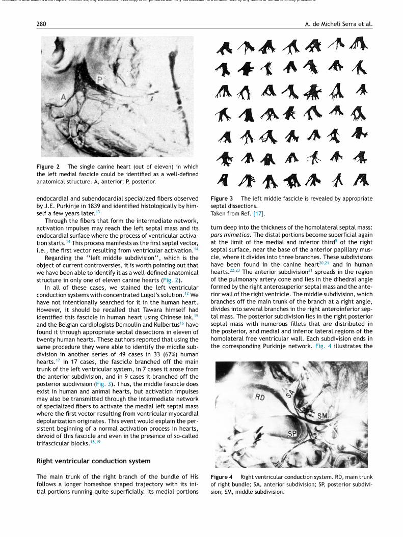
igure 3 The left middle fascicle is revealed by appropriateeptal dissections.aken from Ref. [17].
urn deep into the thickness of the homolateral septal mass:ars mimetica. The distal portions become superficial againt the limit of the medial and inferior third5 of the righteptal surface, near the base of the anterior papillary mus-le, where it divides into three branches. These subdivisionsave been found in the canine heart20,21 and in humanearts.22,23 The anterior subdivision21 spreads in the regionf the pulmonary artery cone and lies in the dihedral angleormed by the right anterosuperior septal mass and the ante-ior wall of the right ventricle. The middle subdivision, whichranches off the main trunk of the branch at a right angle,ivides into several branches in the right anteroinferior sep-al mass. The posterior subdivision lies in the right posterior
igure 4 Right ventricular conduction system. RD, main trunkf right bundle; SA, anterior subdivision; SP, posterior subdivi-ion; SM, middle subdivision.

1
1
1
1
1
1
1
1
1
1
2
2
2
2
2
2
2
2
Mexicana; 1971. p. 34.
Document downloaded from http://zl.elsevier.es, day 29/01/2014. This copy is for personal use. Any transmission of this document by any media or format is strictly prohibited.
About the specialized myocardial conducting tissue
right ventricular conduction system in a canine heart. In1913 the existence of the Kent bundle24 was identified.
Conclusions
Peripheral on distal left blocks that may hide the coex-istence of a non-activable myocardium25 can be easilyrecognized by the diagnostic approach proposed, in duetime, by the Mexican School of electrovectorcardiogra-phy. The correct diagnosis of a peripheral left ventricularconduction disorder together with pertinent clinical datashould suggest the probable association of a non-activablemyocardium.25
On the other hand, right distal peripheral blocks gen-erally do not hide the electrical signs of a non-activablemyocardium.26 These blocks enhance the manifestations oflater occurring electromotive forces resulting from rightventricular activation. Therefore, they do not interfere withthe development of ventricular electromotive forces in thezones affected by myocardial injury. This happens both inthe presence of a right anterior subdivision block, whichincreases by at least 10 ms the duration of basal electro-motive forces that normally develop in 64---72 ms27 and inthe presence of a right posterior subdivision block. This lat-ter block prolongs by an average of 12 ms the electromotiveforces originating in the posterior and medial and inferiorposterolateral septal regions of the right ventricular freewall, which become activated in 30---45 ms.28
The middle left fascicle may in fact exist in humanand animal hearts, but its presence does not appear to beindispensable for transmission of activation impulses to themedial left septal mass.
Acknowledgment
The authors greatly appreciate the careful collaborationsecretarial of Mrs. Blanca Lilia Ochoa.
References
1. Keith A, Flack MW. The form and nature of the muscular connec-tions between primary divisions of the vertebrate hearts. AnatPhysiol. 1907;41:172---89.
2. Lev M. The normal anatomy of the conductive system in manand its pathology in atrioventricular block. Ann N Y Acad Sci.1964;111:817---29.
3. Tawara S. Das Reizleitungssystem des Säugetierherzens. Jena:Gustav Fisher ed.; 1906.
4. James TN. Anatomy of the human sinus node. Anat Rec.1961;141:109---39.
5. James TN. Morphology of the human atrio-ventricular node,with remarks pertinent to its electrophysiology. Am Heart J.1961;62:756---71.
6. James TN. The connecting pathways between the sinus nodeand the A---V node and the A---V node and the right andleft atrium in the human heart. Am Heart J. 1963;66:498---508.
2
281
7. James TN, Sherf L. Specialized tissues and preferential conduc-tion in the atria of the heart. Am J Cardiol. 1971;23:371---427.
8. Bachmann G. The interauricular time interval. Am J Physiol.1916;41:309---20.
9. Wenckebach KF. Beiträge zur Kenntnis der menschlikenHerztätigkeit. Arch Anat Physiol. 1907;1---2:1---24.
0. Thörel C. Über den Aufbau des Sinusknotens und seineVerbindung mit der cava superior und den WenckebachschenBundein. Munchen Med Wschr. 1910;57:183---6.
1. His Jr W. Die Thätigkeit des embryonen Herzens und derenBedeutung für die Lehre von der Herzbewegung beim Erwach-senen. Arbeiten aus med Klin zu Leipzig. 1893:14---50.
2. Medrano GA, Brenes C, de Micheli A, Sodi Pallares D. El bloqueosimultáneo de las subdivisiones anterior y posterior de la ramaizquierda del haz de His (bloqueo bifascicular), y su asociacióncon bloqueo de la rama derecha (bloqueo trifascicular). ArchInst Cardiol Mex. 1970;40:752---70.
3. Purkinje JE. Mikrosckopisch neurologische Beobachtungen. ArchAnatom Physiol Wissenchaliche Med. 1845;12:281.
4. Sodi Pallares D, Bisteni A, Medrano GA. Electrocardiografía yVectocardiografía deductivas. México. La Prensa Méd Mexicana.1964:103.
5. Akiyamuri T. Sunao Tawara: discoverer of the atrioventricularconduction system of the heart. Cardiol J. 2010;17:428---33.
6. Demoulin JC, Kulbertus HE. Histopathological examination ofconcept of left hemiblock. Br Heart J. 1972;34:307---14.
7. Demoulin JC, Kulbertus HE. Pathological correlates of intra-ventricular conduction. In: Masoni A, Alboni P, editors. Cardiacelectrophysiology today. Londres, UK: Academic Press Inc; 1982.p. 427---35.
8. Medrano GA, de Micheli A, Brenes C, Sodi Pallares D. Experi-mental bases for diagnosis of left bifascicular and trifascicularblocks. G Ital Cardiol. 1975;5:8---18.
9. De Micheli A. Los bloqueos parciales de la rama izquierda delhaz de His. Arch Inst Cardiol Mex. 1971;41:625---8.
0. Uhley HN, Rivkin LM. Peripheral distribution of the canine A---Vconduction system. Observations on gross morphology. Am JCardiol. 1960;5:688---91.
1. Medrano GA, de Micheli A. Contribución experimental al diag-nóstico de los bloqueos fasciculares derechos. Arch Inst CardiolMex. 1975;45:704---15.
2. Lev M. The conduction system. In: Gould S, editor. Pathology ofthe heart and blood vessels. Springfield, IL: Charles C. Thomas;1968. p. 185.
3. Mahaim J. Les maladies organiques du faisceau de His-Tawara.Paris: Ed. Masson & Cie; 1931.
4. Kent AFS. A neuromuscular mechanism in the heart. Trans InternCongr Med Londres. 1913;Section III:102---8.
5. De Micheli A, Medrano GA. Bloqueos periféricos. In: de MicheliA, Medrano GA, Iturralde P, editors. Diagnóstico electrovecto-cadiográfico en clínica. México: Méndez Editores. S.A.; 1992. p.44---52.
6. Medrano GA, de Micheli A, Iturralde P. Peripheral heart blocksassociated with myocardial infarcts. Clinical diagnosis based onexperimental findings. Curr Cadiol Rev. 2008;4:140---7.
7. De Micheli A, Medrano GA. Electrocardiograma y vectocadio-grama en el infarto del miocardio. México: La Prensa Médica
8. Gallagher JJ, Sealy WL, Richet LE, Lan W, Wallace AG. Epicardialmapping in the WPW syndrome. In: Welsen AA, editor. Reviewof contemporary laboratory methods. Am Heart Ass. Inc; 1980.