abstract book...
TRANSCRIPT


} Abstract Book CIOCV2019
} Page 2

} Abstract Book CIOCV2019
} Page 3
Copyright © 2019 | Comissão Organizadora CIOCV2019 ISBN | 978-989-54154-1-0 Edição / Edition | Comissão Organizadora do 16º Congresso Internacional de Optometria e Ciências da Visão (CIOCV’2019); Membros/Members Madalena Lira, Jorge M. Jorge, João Linhares, Paulo Fernandes, António Queirós. Coordenação /Coordination | Paulo Fernandes Distribuição / Distribution | Secretaria do Congresso Internacional de Optometria e Ciências da Visão Departamento de Física Universidade do Minho Campus de Gualtar 4710-057 Braga (Portugal) Telf: +351253604320 Fax: +351253604061 e-mail: [email protected] URL: http://ciocv.fisica.uminho.pt Capa / Cover | Paulo Gomes, Portugal. Impressão / Printing | Paulo Gomes, Portugal - [email protected] Advertência Legal / Legal Warning |. Reservados todos os direitos. É proibida a duplicação, total ou parcial desta obra, sob quaisquer formas ou por quaisquer meios (electrónico, mecânico, gravação, fotocopiado, fotográfico ou outros) sem autorização expressa por escrito do editor / All rights reserved. Reproduction in part or as a whole by any process or in any media (electronic, mechanical, recording, copying, photographic or others) is extrictly forbidden without the written authorization of the editor.

} Abstract Book CIOCV2019
} Page 4
Welcome Message

} Abstract Book CIOCV2019
} Page 5

} Abstract Book CIOCV2019
} Page 6
Dear Colleagues
The International Conference in Optometry and Visual Science (CIOCV’2019) will take place in the next 4th and 5th of May, 2019. The previous editions of the conference involved a large number of participants pushing this new edition to a new place: The Altice Fórum Braga. Please do accept this positive impulse and sense the great pleasure we are feeling by organizing this meeting. Join us into building a great conference of very high scientific quality and human importance.
The CIOCV has been an example of what can be achieved by creating value through knowledge by internationalizing it and by embodying it into the daily clinical practice. The CIOCV is a place that provides opportunities for the development and exchange of this knowledge while providing a space for nurturing useful networking and collaborations among researchers, professionals and students.
New challenges demand greater skills and greater ability to seize in full these opportunities.
Like previous years the scientific program will include recent developments in the field of vision science and topics that will provide support the permanent development needs of the participants.
We are expecting you in Braga to enrich this sharing of scientific and clinical knowledge and please expect some new information from us very soon.
Welcome to the 16th International Conference in Optometry and Visual Science!
With Kind regards,
The organizing committee of the CIOCV’2019
Follow us at: Facebook: www.facebook.com/ciocv/ Webpage: ciocv.fisica.uminho.pt

} Abstract Book CIOCV2019
} Page 7
Index

} Abstract Book CIOCV2019
} Page 8
Página/Page
Welcome Message __________________________________________ Organizing/Scientific Committees_______________________________ Program __________________________________________________ Lectures __________________________________________________ Free Papers _______________________________________________ Posters___________________________________________________ Conference Area____________________________________________ Sponsors__________________________________________________
4 10 14 21 35 46 94 96

} Abstract Book CIOCV2019
} Page 9

} Abstract Book CIOCV2019
} Page 10
Organizing/Scientific Committee

} Abstract Book CIOCV2019
} Page 11
} Abstract Book CIOCV2019
} Page 12
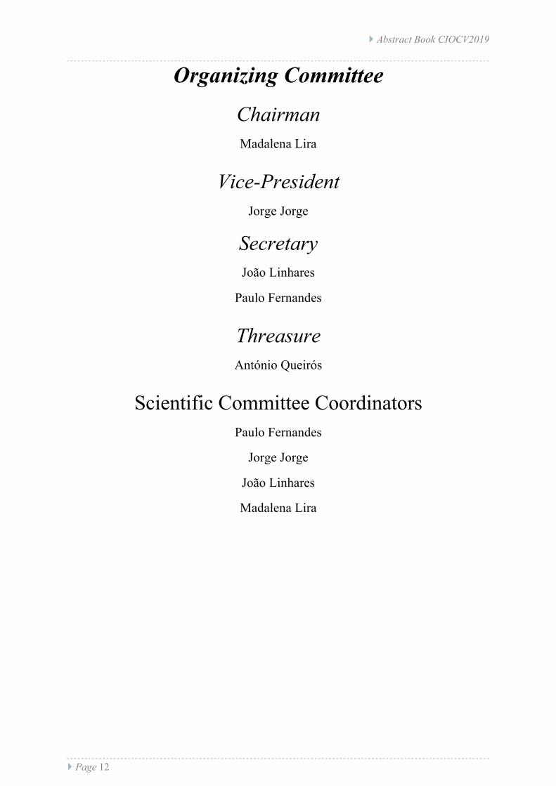
Organizing Committee
Chairman Madalena Lira
Vice-President Jorge Jorge
Secretary João Linhares
Paulo Fernandes
Threasure António Queirós
Scientific Committee Coordinators
Paulo Fernandes
Jorge Jorge João Linhares
Madalena Lira

} Abstract Book CIOCV2019
} Page 13
Scientific Committee
Local Scientific Committee
Amélia Fernandes Nunes, PhD, Portugal
António Manuel Gonçalves Baptista, PhD, Portugal
António Filipe Teixeira Macedo, PhD, Portugal
António Manuel Marques Queirós Pereira, PhD, Portugal
Elisabete Maria S.C. Coutinho, PhD, Portugal
Francisco Miguel Pereira Brardo Ferreira, PhD, Portugal
João Manuel Maciel Linhares, PhD, Portugal
Jorge Manuel Martins Jorge, PhD, Portugal
José Alberto Diaz Rey, MD, PhD, Portugal
José Manuel González- Meijome, PhD, Portugal
Madalena Madalena Cunha Faria Lira, PhD, Portugal
Maria Elisabete Cunha Dias Real Oliveira, PhD, Portugal
Miguel Faria Ribeiro, PhD, Portugal
Paulo Rodrigues Botelho Fernandes, PhD, Portugal
Pedro Miguel Lourenço Monteiro, PhD, Portugal
Sandra Maria Braga Franco, PhD, Portugal
Sérgio Miguel Cardoso Nascimento, PhD, Portugal

} Abstract Book CIOCV2019
} Page 14
Program
- Lectures - Free Papers - Posters

} Abstract Book CIOCV2019
} Page 15
} Abstract Book CIOCV2019
} Page 16
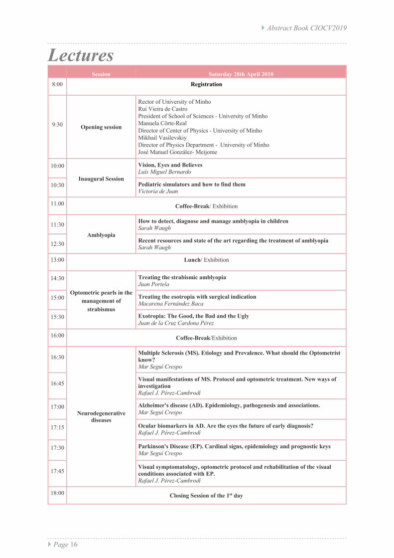
Lectures Session Saturday 28th April 2018
8:00 Registration
9:30 Opening session
Rector of University of Minho Rui Vieira de Castro President of School of Sciences - University of Minho Manuela Côrte-Real Director of Center of Physics - University of Minho Mikhail Vasilevskiy Director of Physics Department - University of Minho José Manuel González- Meijome
10:00
Inaugural Session
Vision, Eyes and Believes Luís Miguel Bernardo
10:30 Pediatric simulators and how to find them Victoria de Juan
11.00 Coffee-Break/ Exhibition
11:30
Amblyopia
How to detect, diagnose and manage amblyopia in children Sarah Waugh
12:30 Recent resources and state of the art regarding the treatment of amblyopia Sarah Waugh
13:00 Lunch/ Exhibition
14:30
Optometric pearls in the management of
strabismus
Treating the strabismic amblyopia Juan Portela
15:00 Treating the esotropia with surgical indication Macarena Fernández Baca
15:30 Exotropia: The Good, the Bad and the Ugly Juan de la Cruz Cardona Pérez
16:00 Coffee-Break/Exhibition
16:30
Neurodegenerative diseases
Multiple Sclerosis (MS). Etiology and Prevalence. What should the Optometrist know? Mar Seguí Crespo
16:45 Visual manifestations of MS. Protocol and optometric treatment. New ways of investigation Rafael J. Pérez-Cambrodí
17:00 Alzheimer's disease (AD). Epidemiology, pathogenesis and associations. Mar Seguí Crespo
17:15 Ocular biomarkers in AD. Are the eyes the future of early diagnosis? Rafael J. Pérez-Cambrodí
17:30 Parkinson's Disease (EP). Cardinal signs, epidemiology and prognostic keys Mar Seguí Crespo
17:45 Visual symptomatology, optometric protocol and rehabilitation of the visual conditions associated with EP. Rafael J. Pérez-Cambrodí
18:00 Closing Session of the 1st day

} Abstract Book CIOCV2019
} Page 17
Session Saturday 29th April 2018 8:30 Registration
9:00 Free Papers
Results of Cross-Linking in a patient with keratoconus, 4 years and 10 months after surgery. Nora León Rodríguez New metrics for scleral lens fitting and evaluation. Rute J, Macedo-de-Araújo The Influence of the illumination intensity on Ocular Accommodation Raquel Moreira Alteration in tear film break up time after instillation of artificial tears Celeste Lago Visual quality changes with soft contact lenses after different hyaluronic acid eye drops instillation Carlos Carpena Torres Ocular surface temperature in dry eye and healthy eyes using non-contact infrared thermography Javier Ruiz-Alcocer Diagnosis of dry eye assisted by gold nanoparticles Carlos Carpena Torres Improve of reading with vision rehabilitation Laura Hernandez-Moreno Changes to grating orientation and spatial frequency in two different tests to assess the electrophysiological response of the retina Ana Amorim-de-Sousa Addition and Pupil Size effect on the visual performance of a Novel Extended-Depth-of-Focus Contact Lens and a Center-Near design Javier Ruiz-Alcocer
10:30 Clinical cases I Andres Gene Sanpedro Universidade de Valência
11:00 Coffee-Break/ Exhibition
11:30 New technologies in refraction
Today’s state of the art in subjective and objective refraction Mikel Aldaba Arévalo
13:00 Lunch/ Exhibition
14:30 Clinical cases II César Villa Universidade Europeia de Madrid
15:00
Ophthalmology
Refractive surgery with the femtosecond laser Alberto Parafita-Fernández
15:30 New approaches to glaucoma Paulo Ribeiro
16:00 Awards and Certificates Ceremony
17:00 Closing Session
} Abstract Book CIOCV2019
} Page 18
Posters Ner AUTHOR(S) TITLE
001 Ana Rita Coelho Pinheiro, João Manuel Maciel Linhares, António Filipe Teixeira Macedo
Using the MARS test to measure visual contrast sensitivity in children
002 Avelino Mazuze; João Viriato Mazalo; Tomasina Fernando Nchuaki; Dulnerio Sengo.
Promotion and education actions on visual and eye health in the rural community, Mozambique - Africa: report of an experience
003
Ana Rita Oliveira Vaz, Daniela Lopes-Ferreira, Ana Amorim-de-Sousa, Rute J. Macedo-de-Araújo, António Queirós, José Manuel González-Méijome
Ocular biometry and refractive error of a young portuguese subjects during 3 years of university enrolment.
004 Andreia E. Gomes, João M. M. Linhares, Ricardo J. F. Pereira, Sérgio M. C. Nascimento
Estimation of the best daylight illumination for optimal viewing of human skin.
005 Adrián Pérez Baladrón, Isabel Fambuena Muedra
Is contact lens fitting a valuable alternative in low vision patients?
006 Teresa Calderón González Adaptation of strabismus therapy in learning disorders
007 Nelva de Luísa David Sixpene, Sérgio Latorre Arteaga
Visual and sociodemographic factors that influence the road accident in the city of Nampula-Mozambique
008 Tatiana Rodrigues, Maria João Batista, Pedro Monteiro, Amélia Fernandes Nunes
Anisometropia in the 5th and 6th school years
009 Hugo Pena-Verdeal, Rosa Calo-Santiago, Covadonga Vazquez-Sanchez, Carlos García-Resúa, Maria J. Giraldez
Analysis of the relationship between convergence insufficiency symptoms and AC/A ratio
010 Rosa Calo-Santiago, Hugo Pena-Verdeal, Gonzalo Garcia-Dominguez, Covadonga Vazquez-Sanchez, Maria J. Giraldez
Comparison between variable anaglyphs and aperture rule results in a group of young healthy subjects
011 Carracedo G, Espinosa-Vidal TM, Martínez-Alberquilla I, Batres L.
The topographical effect of optical zone diameter in orthokeratology contact lenses in high myopes
012 Jéssica Gomes, Sandra Franco Real time measurement of ocular aberrations changes with accommodation in myopic subjects
013
Albertos Arranz, Henar; Cervera Sánchez, Zaíra; Martínez Abad, Antonio; Amesty Morello, Alejandra; Plaza Luche, Ana Belén; Díez de la Uz, Rosa; Cantó Cerdán, Mario
Comparative analysis between different refractive error measurement systems conducted in children with and without cycloplegia
014 Reis, Clarisse; Fonseca, Elsa; Ferreira, Francisco
Study of biometric changes and intraocular scattering in diabetic population
015 María Inmaculada Vera Alarcón Ocular Refraction with exposure time at the myopization
016 João Cunha, Sara Marinho, João M. M. Linhares, Sérgio M. C. Nascimento
The influence of the aging lens on the perception of the Ishihara test plates
017 Noemí Olcoz, Javier Blasco, Alejandro Blasco
Rheological characterization methods for commercial artificial tears
018 Silvia García-Montero, Dolores Ferreiro, Hugo Pena-Verdeal, Jacobo Garcia-Queiruga, Eva Yebra-Pimentel
Assessment of the correlation between symptomathology, tear meniscus height and phenol red inter-eye differences
019 Rico-del-Viejo L, Llorens-Quintana C, Ruiz-Alcocer J, Martínez- Alberquilla I, García-Montero M, Madrid-Costa D
The relationship between new morphological and objective Meibomian glands parameters and relevant ocular surface parameters
} Abstract Book CIOCV2019
} Page 19
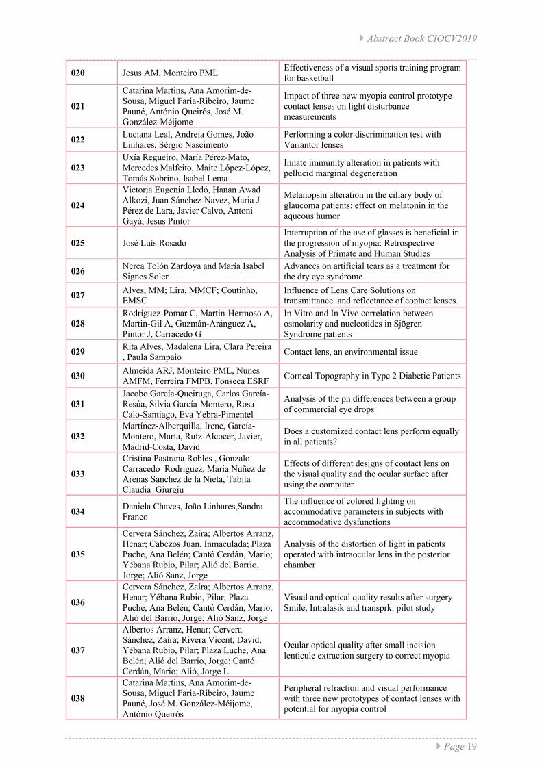
020 Jesus AM, Monteiro PML Effectiveness of a visual sports training program for basketball
021
Catarina Martins, Ana Amorim-de-Sousa, Miguel Faria-Ribeiro, Jaume Pauné, António Queirós, José M. González-Méijome
Impact of three new myopia control prototype contact lenses on light disturbance measurements
022 Luciana Leal, Andreia Gomes, João Linhares, Sérgio Nascimento
Performing a color discrimination test with Variantor lenses
023 Uxía Regueiro, María Pérez-Mato, Mercedes Malfeito, Maite López-López, Tomás Sobrino, Isabel Lema
Innate immunity alteration in patients with pellucid marginal degeneration
024
Victoria Eugenia Lledó, Hanan Awad Alkozi, Juan Sánchez-Navez, Maria J Pérez de Lara, Javier Calvo, Antoni Gayà, Jesus Pintor
Melanopsin alteration in the ciliary body of glaucoma patients: effect on melatonin in the aqueous humor
025 José Luís Rosado Interruption of the use of glasses is beneficial in the progression of myopia: Retrospective Analysis of Primate and Human Studies
026 Nerea Tolón Zardoya and María Isabel Signes Soler
Advances on artificial tears as a treatment for the dry eye syndrome
027 Alves, MM; Lira, MMCF; Coutinho, EMSC
Influence of Lens Care Solutions on transmittance and reflectance of contact lenses.
028 Rodríguez-Pomar C, Martin-Hermoso A, Martin-Gil A, Guzmán-Aránguez A, Pintor J, Carracedo G
In Vitro and In Vivo correlation between osmolarity and nucleotides in Sjögren Syndrome patients
029 Rita Alves, Madalena Lira, Clara Pereira , Paula Sampaio Contact lens, an environmental issue
030 Almeida ARJ, Monteiro PML, Nunes AMFM, Ferreira FMPB, Fonseca ESRF Corneal Topography in Type 2 Diabetic Patients
031 Jacobo García-Queiruga, Carlos García-Resúa, Silvia García-Montero, Rosa Calo-Santiago, Eva Yebra-Pimentel
Analysis of the ph differences between a group of commercial eye drops
032 Martínez-Alberquilla, Irene, García-Montero, María, Ruíz-Alcocer, Javier, Madrid-Costa, David
Does a customized contact lens perform equally in all patients?
033
Cristina Pastrana Robles , Gonzalo Carracedo Rodriguez, Maria Nuñez de Arenas Sanchez de la Nieta, Tabita Claudia Giurgiu
Effects of different designs of contact lens on the visual quality and the ocular surface after using the computer
034 Daniela Chaves, João Linhares,Sandra Franco
The influence of colored lighting on accommodative parameters in subjects with accommodative dysfunctions
035
Cervera Sánchez, Zaíra; Albertos Arranz, Henar; Cabezos Juan, Inmaculada; Plaza Puche, Ana Belén; Cantó Cerdán, Mario; Yébana Rubio, Pilar; Alió del Barrio, Jorge; Alió Sanz, Jorge
Analysis of the distortion of light in patients operated with intraocular lens in the posterior chamber
036
Cervera Sánchez, Zaíra; Albertos Arranz, Henar; Yébana Rubio, Pilar; Plaza Puche, Ana Belén; Cantó Cerdán, Mario; Alió del Barrio, Jorge; Alió Sanz, Jorge
Visual and optical quality results after surgery Smile, Intralasik and transprk: pilot study
037
Albertos Arranz, Henar; Cervera Sánchez, Zaíra; Rivera Vicent, David; Yébana Rubio, Pilar; Plaza Luche, Ana Belén; Alió del Barrio, Jorge; Cantó Cerdán, Mario; Alió, Jorge L.
Ocular optical quality after small incision lenticule extraction surgery to correct myopia
038
Catarina Martins, Ana Amorim-de-Sousa, Miguel Faria-Ribeiro, Jaume Pauné, José M. González-Méijome, António Queirós
Peripheral refraction and visual performance with three new prototypes of contact lenses with potential for myopia control

} Abstract Book CIOCV2019
} Page 20
039 Silvia Duran, Alima Amuzá, Alvaro Pons
Evaluation of contrast sensivity function response in patients with HIV in Mozambique.
040
Francisca Sena, Amélia Fernandes Nunes, Rita Tuna, Ana Paula Gonçalves, Rui Calado, Maria dos Anjos Esperança , Pedro Monteiro
Reduced Visual Acuity in 5-year-old children
041 Dolores Ferreiro, Silvia García-Montero, Eva Punin, Covadonga Vázquez, Eva Yebra-Pimentel
Analysis of Interchangeability between two corneal topographers for the eccentricity assessment
042 Gisela Ferreira, Silvia Duran, Alvaro Pons
Validation of the new sensitivity contrast test by computer in Mozambique
043 Suellen Cristine Haensch e Angelita Fatima Beloto Dutra de Lima
Evaluation of visual perception with the test of visual perceptual skills (tvps-3) in children aged 6 to 14 years with learning disorders
044 Tiago Machado, João Linhares, Sandra Franco
The influence of colored lighting on binocular vision
045 Carlos García-Resúa, Jacobo Garcia-Queiruga, Hugo Pena-Verdeal, Dolores Ferreiro, Maria J. Giraldez
Osmolality measurement of a group of lens care solutions by a freezing point depression osmometer
046 Rute J, Macedo-de-Araújo, Eef van der Worp, José M. González-Méijome
Visual performance with scleral lenses: report of a one-year prospective study.
047 Ana Filipa Mota, Ana Amorim de Sousa, Jaume Pauné, Barcelona,José González-Méijome,António Queirós Pereira
Evaluation of visual performance at high and low contrast with contact lenses prototypes for control of myopia progression
048 María Serramito Blanco, Juan Gonzalo Carracedo Rodríguez
Differences between the optic section with slit lamp and optical coherence tomography during scleral lens fitting
} Abstract Book CIOCV2019
} Page 21
Lectures
} Abstract Book CIOCV2019
} Page 22
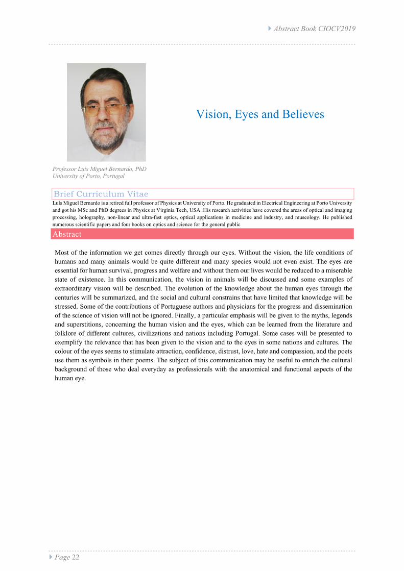
Vision, Eyes and Believes
Professor Luis Miguel Bernardo, PhD University of Porto, Portugal
Abstract Most of the information we get comes directly through our eyes. Without the vision, the life conditions of humans and many animals would be quite different and many species would not even exist. The eyes are essential for human survival, progress and welfare and without them our lives would be reduced to a miserable state of existence. In this communication, the vision in animals will be discussed and some examples of extraordinary vision will be described. The evolution of the knowledge about the human eyes through the centuries will be summarized, and the social and cultural constrains that have limited that knowledge will be stressed. Some of the contributions of Portuguese authors and physicians for the progress and dissemination of the science of vision will not be ignored. Finally, a particular emphasis will be given to the myths, legends and superstitions, concerning the human vision and the eyes, which can be learned from the literature and folklore of different cultures, civilizations and nations including Portugal. Some cases will be presented to exemplify the relevance that has been given to the vision and to the eyes in some nations and cultures. The colour of the eyes seems to stimulate attraction, confidence, distrust, love, hate and compassion, and the poets use them as symbols in their poems. The subject of this communication may be useful to enrich the cultural background of those who deal everyday as professionals with the anatomical and functional aspects of the human eye.
Brief Curriculum Vitae Luís Miguel Bernardo is a retired full professor of Physics at University of Porto. He graduated in Electrical Engineering at Porto University and got his MSc and PhD degrees in Physics at Virginia Tech, USA. His research activities have covered the areas of optical and imaging processing, holography, non-linear and ultra-fast optics, optical applications in medicine and industry, and museology. He published numerous scientific papers and four books on optics and science for the general public
} Abstract Book CIOCV2019
} Page 23
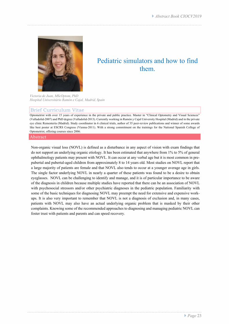
Pediatric simulators and how to find them.
Victoria de Juan, MScOptom, PhD Hospital Universitário Ramón e Cajal, Madrid, Spain
Abstract Non-organic visual loss (NOVL) is defined as a disturbance in any aspect of vision with exam findings that do not support an underlying organic etiology. It has been estimated that anywhere from 1% to 5% of general ophthalmology patients may present with NOVL. It can occur at any verbal age but it is most common in pre-pubertal and pubertal-aged children from approximately 8 to 14 years old. Most studies on NOVL report that a large majority of patients are female and that NOVL also tends to occur at a younger average age in girls. The single factor underlying NOVL in nearly a quarter of these patients was found to be a desire to obtain eyeglasses. NOVL can be challenging to identify and manage, and it is of particular importance to be aware of the diagnosis in children because multiple studies have reported that there can be an association of NOVL with psychosocial stressors and/or other psychiatric diagnoses in the pediatric population. Familiarity with some of the basic techniques for diagnosing NOVL may preempt the need for extensive and expensive work-ups. It is also very important to remember that NOVL is not a diagnosis of exclusion and, in many cases, patients with NOVL may also have an actual underlying organic problem that is masked by their other complaints. Knowing some of the recommended approaches to diagnosing and managing pediatric NOVL can foster trust with patients and parents and can speed recovery.
Brief Curriculum Vitae Optometrist with over 15 years of experience in the private and public practice. Master in “Clinical Optometry and Visual Sciences” (Valladolid-2007) and PhD degree (Valladolid-2013). Currently working in Ramón y Cajal University Hospital (Madrid) and in the private eye clinic Rementería (Madrid). Study coordinator in 6 clinical trials, author of 35 peer-review publications and winner of some awards like best poster at ESCRS Congress (Vienna-2011). With a strong commitment on the trainings for the National Spanish College of Optometrist, offering courses since 2006.
} Abstract Book CIOCV2019
} Page 24
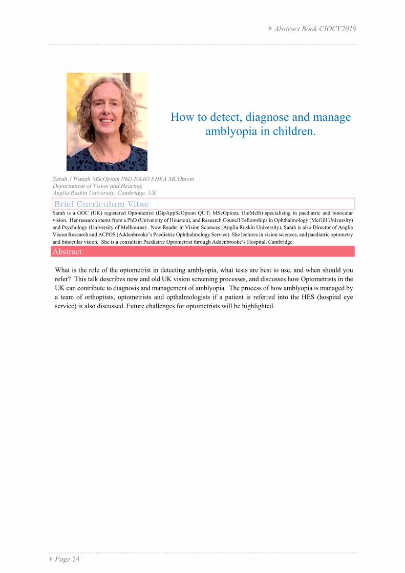
How to detect, diagnose and manage amblyopia in children.
Sarah J Waugh MScOptom PhD FAAO FHEA MCOptom Departament of Vision and Hearing, Anglia Ruskin University, Cambridge, UK
Abstract What is the role of the optometrist in detecting amblyopia, what tests are best to use, and when should you refer? This talk describes new and old UK vision screening processes, and discusses how Optometrists in the UK can contribute to diagnosis and management of amblyopia. The process of how amblyopia is managed by a team of orthoptists, optometrists and opthalmologists if a patient is referred into the HES (hospital eye service) is also discussed. Future challenges for optometrists will be highlighted.
Brief Curriculum Vitae Sarah is a GOC (UK) registered Optometrist (DipAppScOptom QUT; MScOptom, UniMelb) specialising in paediatric and binocular vision. Her research stems from a PhD (University of Houston), and Research Council Fellowships in Ophthalmology (McGill University) and Psychology (University of Melbourne). Now Reader in Vision Sciences (Anglia Ruskin University), Sarah is also Director of Anglia Vision Research and ACPOS (Addenbrooke’s Paediatric Ophthalmology Service). She lectures in vision sciences, and paediatric optometry and binocular vision. She is a consultant Paediatric Optometrist through Addenbrooke’s Hospital, Cambridge.
} Abstract Book CIOCV2019
} Page 25
Recent resources and state of the art regarding the treatment of
amblyopia.
Sarah J Waugh MScOptom PhD FAAO FHEA MCOptom Departament of Vision and Hearing, Anglia Ruskin University, Cambridge, UK
Abstract With the advent of new electronic technology comes the potential for new treatments of amblyopia. Amblyopia is characterised by reduced visual acuity even with best refractive correction, combined with reduced binocularity. New treatments are able to better target binocularity training, but how do results with them compare to those obtained with traditional monocular (patching) treatments? Where might new treatments aim to go next? What should we be measuring to assess outcomes?
Brief Curriculum Vitae Sarah is a GOC (UK) registered Optometrist (DipAppScOptom QUT; MScOptom, UniMelb) specialising in paediatric and binocular vision. Her research stems from a PhD (University of Houston), and Research Council Fellowships in Ophthalmology (McGill University) and Psychology (University of Melbourne). Now Reader in Vision Sciences (Anglia Ruskin University), Sarah is also Director of Anglia Vision Research and ACPOS (Addenbrooke’s Paediatric Ophthalmology Service). She lectures in vision sciences, and paediatric optometry and binocular vision. She is a consultant Paediatric Optometrist through Addenbrooke’s Hospital, Cambridge.

} Abstract Book CIOCV2019
} Page 26
Treating the strabismic amblyopia
Juan Portela, PhD Clínica Begira, Bilbao, Spain
Abstract
La ambliopía estrábica presenta características que la hacen especialmente compleja. En muchas ocasiones el factor ambliogénico (el estrabismo) no desaparece después del tratamiento mediante corrección óptica y posterior oclusión y/o penalización del ojo director por lo que el paciente no recupera la visión binocular. Esta presencia del fator ambliogénico provoca además un alto porcentaje de recidivas. En los últimos años, se han propuesto tratamientos mediante juegos serios y estimulación dicóptica que pretenden mejorar el resultado binocular en este grupo de pacientes. Lamentablemente, los resultados son semejantes a los obtenidos con oclusión y/o penalización. Durante la conferencia se presentará un modelo de tratamiento basado en la corrección prismática de la desviación estrábica. Con la finalidad de eliminar el prisma se propone un programa de terapia vergencial en un formato Random Dot en el domicilio del paciente.
Brief Curriculum Vitae Optometrist specializing in strabismus, amblyopia at the Begira Clinic and the Ikusgune Optometry Center in Donosti. Doctor in Optometry by the European University UEM, Madrid. Master's Degree in Health Research. European University of Madrid (UEM). Master in Clinical Optometry. Pennsylvania College of Optometry, (Philadelphia, United States).

} Abstract Book CIOCV2019
} Page 27
Treating the esotropia with surgical indication
Macarena Fernández Baca, PhD Universidade Europeia de Madrid, Spain
Abstract Los estrabismos producen anomalías musculares y sensoriales que pueden afectar al pronóstico del tratamiento. El manejo de los estrabismos puede ir dirigido a conseguir una mejoría en el aspecto estético, o puede ir dirigido a obtener una visión binocular normal. Para reducir la necesidad de segundas cirugías y lograr una cura funcional tras la cirugía de estrabismo, es fundamental el papel que puede jugar la optometría bien con ayuda de lentes, prismas, oclusiones a terapia visual. Describiremos el papel del optometrista y los pasos a seguir según el tipo de estrabismo para asegurar el éxito de la cirugía posterior.
Brief Curriculum Vitae PhD in Optometry from the University of Houston. Diplomate in Binocular Vision Perception and Pediatric Optometry. She has been vice chair of the International Admissions Committee of the American Academy of Optometry, and previously a member of the committee. External teacher at the Universidad Europea de Madrid. She participates in postgraduate courses in Optometry, and has been coordinator and professor of the postgraduate courses of the Boston Center of Optometry and the Camilo José Cela University, Madrid; Adjunct Assistance Professor at the University of Houston College of Optometry; and Adjunct Clinical Faculty Member, The New England College of Optometry, Boston.

} Abstract Book CIOCV2019
} Page 28
Exotropia: The Good, the Bad and the Ugly
Juan de la Cruz Cardona Pérez, PhD Optics Departament, University of Granada, Spain
Abstract Divergent strabismus in their different patterns (Excess divergence, Basic Exotropia or Insufficiency of Convergence) are the ones that have the best prognosis of functional recovery (85% of the cases). In pediatrics, the majority are intermittent due to the control absence of divergent fusion or an absence of positive vergence in near vision. However, in adults/geriatrics they are usually associated with the destabilization of an exoforia due to an increase in it (or a casual decrease in compensatory fusional vergence). Other cases to be taken into account are exotropias secondary to an endotropic surgery (XTSS), which are complicated due to the sensory adaptation that may had at the moment of surgery, complicating the subsequent treatment in therapy due to deep suppressions, anomalous, inharmonious or paradoxical correspondences. In any case, and once the sensory adaptation has been overcome, they tend to respond favorably to passive and active therapy treatments. Abnormal correspondences are infrequent (except in XTSS), but the suppressions that develop can be very active and deep, even in intermittent misalignments. As soon as the deviation begins, the suppression is activated immediately. For this reason, it is essential to work on two very important aspects: pathological diplopia and controlled divergence. During this speech we will analyze the most appropriate treatments in a practical way on real cases.
Brief Curriculum Vitae Juan de la Cruz Cardona Pérez (Optometrist, MSc and PhD) is Associate Professor at the University of Granada and Head of the “Gabinete de Optometría y Rehabilitación Visual”. His recent interest researching in the Laboratory of Biomaterials Optics include performing the optical characterization of artificial corneal substitutes generated by tissue engineering and stem cells. He is currently teaching at the Degree in Optics and Optometry and directs the Master in Clinical Optometry and Advanced Optics..

} Abstract Book CIOCV2019
} Page 29
Neurodegenerative diseases and vision
Maria del Mar Seguí-Crespo, PhD University of Alicante, Spain Rafael J. Pérez-Cambrodí, PhD University of Valencia, Spain
Abstract The optometrist, as a primary health professional, is able to evaluate the structural integrity of the eye and the performance of the visual system. In the last few years, there is an increasing risk of developing neurodegenerative diseases associated with ageing. The diagnosis of those pathologies has been frequently complex and invasive and thus, practitioners and researchers have highlighted the importance of more simple biomarkers in order to monitorize the progression and efficacy of the available treatments. The visual system may be affected in the course of those neurodegenerative diseases and the availability of advanced and high-resolution imaging techniques and devices surely will become a key factor to help in the diagnosis and in the evaluation of the progression of the diseases. This fact opens a perspective to develop preventive and also interventional programs where optometrists can add their konowledge and clinical experience. The main objective of this sesión is, from a double perspective (Public Health and Optometry) to provide the professional the adequate tools to recognize the visual signs and symptoms of prevalent conditions as Multiple Sclerosis, Alzheimer´s disease and Parkinson´s disease, to recommend a clinical protocol to help the neurological diagnosis and to establish a follow-up in order to evaluate the progression and efficacy of the medical treatment..
Brief Curriculum Vitae Maria del Mar Seguí-Crespo, PhD in Public Health, Diploma in Optics and Optometry and Degree in Documentation. Professor at the University of Alicante, where she teaches in the Degree in Optics and Optometry, and also in the Master in Advanced Optometry and Visual Health. Since 2008 she belongs to the Public Health Research Group of this university. She is the author of articles published in impact journals in the Ophthalmology and Public, Environmental & Occupational Health categories. Rafael J. Pérez-Cambrodí, PhD in Optometry and Vision Science (University of Valencia). Master of Sciences in Optometry and Research (UEM). Bachelor of Sciences in Optometry (NEWENCO). Degree in Optics and Optometry (University of Alicante). Assistant professor at the University of Valencia. Private practice in Optometric Clinic. Evaluator in the Spanish Research Agency. Author and co-author of 73 articles published in impact journals in the Ophthalmology and Optometry categories. Reviewer of journals in the Optometry and Ophthalmology fields. Vice-president of COOCV

} Abstract Book CIOCV2019
} Page 30
When is convenient to prescribe prisms to the patient: Clinical Cases
Andres Gené Sampedro, PhD University of Valencia, Spain
Abstract Nuestra labor asistencial profesional está encaminada principalmente a solucionar problemas refractivos, binoculares, y a la prevención para mantener la salud visual. Muchas veces, tras detectar algún problema binocular, la única opción de tratamiento para optimizar la visión del sujeto es la prescripción de prismas. Los prismas pueden ser muy útiles para tratar pacientes con trastornos de la visión binocular sintomáticos. Sin embargo, las opiniones varían ampliamente acerca de cómo determinar cual es la mejor cantidad a prescribir. De hecho, los métodos comunes para valorar la cuantía prismática pueden dar lugar a diferentes magnitudes de prisma a recomendar para el mismo paciente. Esta falta de consenso, junto con pautas generales y reglas aparentemente contradictorias, puede disuadir a algunos optometristas a prescribirlos. Es por ello que, el objetivo general de esta comunicación, es actualizar nuevos conocimientos relacionados con la aplicación de prismas para el tratamiento y mejora de las disfunciones del sistema binocular y acomodativo. Siendo los objetivos específicos mediante el análisis de diversos casos clínicos: • Entender como se puede afectar la binocularidad y cuales son sus tratamientos. • Saber en que casos se debe prescribir prismas, en uno o los dos ojos repartidos, que permitan mejorar el estado sensorial, y vergencial del paciente. • Conocer que sujetos pueden mejorar significativamente con la compensación prismática. Con esta comunicación se mejora y disminuye la inseguridad de como aplicar las prescripciones prismáticas adecuadamente. El beneficio que se obtiene es doble, por un lado, la adquisición de confianza de los ópticos-optometristas, en cómo medirlo y qué cuantía poner en cada ojo, en base a los resultados obtenidos. Por otro lado, la mejora que se producirá en los pacientes afectos de disfunciones binoculares y acomodativas que requieran prismas, como tratamiento de elección para mejorar su problema visual. En esta comunicación se profundiza en el desarrollo aplicado de la batería de pruebas, afianzando las bases para aprender a valorar las capacidades binoculares y a cuantificar las anomalías. A la vez que se dota de los conocimientos necesarios para saber en que casos se debe poner prismas viendo el cómo realizarlo y que información extraer de cada prueba, y la valoración de casos clínicos, que permita coger confianza en el manejo y las soluciones a aplicar a los sujetos en el que una prescripción prismática es una opción de ayuda optométrica. Finalmente, como ópticos-optometristas debemos educar a los usuarios sobre como optimizar su visión, y hacer que la población vea lo mejor posible, siendo importante que dispongan de unas buenas funciones visuales para conseguir ser eficaces en las tareas a realizar. Los prismas son una buena opción, siendo a veces la única válida para restituir una visión binocular cómoda para el paciente.
Brief Curriculum Vitae Profesor Titular de Universidad en Óptica, y Optometría y Ciencias de la Visión, Universidad de Valencia. Investigador del INTRAS (Instituto de Tráfico y Seguridad Vial), Universidad de Valencia. Diplomado en Óptica Universidad Complutense Madrid, 1988. Diplomado en Óptica y Optometría Universidad de Alicante, 1996. Grado en Óptica y Optometría, Universidad de Alicante, 2014. Bachelor in Optometry, Pennsylvania College of Optometry, Philadelphia (EEUU), 2000. Doctor por la Universidad de Valencia, 2016. Miembro Fundador de la European Academy of Optometry and Optics (EAOO). Presidente del Colegio de Ópticos Optometristas de la Comunidad Valenciana 2018-Actualidad. Presidente de la Sociedad de Optometría y Contactología de la Comunidad Valenciana 2006-Actualidad. Coordinador en España de la Red Epidemiológica Iberoamericana en Salud Visual y Ocular (REISVO) 2010-2014. Secretario de la Comisión de Deontología y Ética del Consejo General de Colegios de Ópticos Optometristas de España.

} Abstract Book CIOCV2019
} Page 31
Today’s state of the art in subjective and objective refraction
Mikel Aldaba Arévalo, PhD Instruments and Systems Development (CD6) Universitat Politecnica de Catalunya, Spain
Abstract We will start this talk introducing the role that refraction plays on research and industry. We will address the question of “Why refractive error is important?”. After that, the presentation will follow with a review of basic concepts of ocular refraction such as its definition and measurement, including the state-of-the-art of subjective, objective and automated refraction methods and ophthalmic devices. We will address questions such as “Why do objective and automated refraction methods have not replaced the clinician yet?”, “How much accuracy is needed for that purpose?” or “What does it really mean to have an accurate refraction device?”. Finally, some of the new lines of research that have recently shown up will be presented including a brief mention to new startup companies that have introduced the misleading concept of “online refraction”.
Brief Curriculum Vitae He received a degree in Optics and Optometry in 2003 from the University of Valladolid in Spain and a BSc in Optometry and Vision Science in 2005 from the University of Minho in Braga, Portugal. Received a PhD in Optical Enginnering from the Technical University of Catalonia in 2012. He works in the physiological optics area, with research interest on accommodation, refraction, optometric and ophthalmic instrumentation, and dry eye diagnose.

} Abstract Book CIOCV2019
} Page 32
Clinical Cases:
1- Evaluation of myopia in two adolescent brothers
2 - Clinical management of corneal ectasia in adolescent
César Villa Collar, PhD Universidad Europea de Madrid, Spain
Abstract Nº1. EVOLUCIÓN DE LA MIOPÍA EN DOS HERMANOS ADOLESCENTES En este caso se analizará la diferente evolución de la miopía en la adolescencia de dos hermanos miopes que fueron, por varias razones que se comentarán, manejados de forma distinta. Uno de ellos fue tratado con lentes de ortoqueratología (OKN) de forma ininterrumpida durante más de 10 años. Por el contrario, su hermana, que también comienza con OKN, debe de abandonar su uso y compensó su miopía con lentes de contacto hidrofílicas esférica hasta la actualidad. El caso nos permitirá revisar brevemente las técnicas empleadas, y su evidencia científica, para el control de miopía escolar en la niñez y adolescencia. Nº2. MANEJO CLÍNICO DE ECTASIA CORNEAL EN ADOLESCENTE Este caso trata sobre un adolescente al que se le diagnostica queratocono en ambos ojos en la adolescencia. Se expondrá y discutirá el tratamiento realizado consistente en la realización, en primer lugar, de un cross linking epi-off y posteriormente de la adaptación de lentes de contacto esclerales. El caso nos permitirá revisar brevemente los protocoles actuales en la detección y manejo de ectasias corneales que aparecen en las primeras décadas de la vida, así como revisar las distintas opciones de tratamiento. Se prestará especial atención a los criterios seguidos en la recomendación y selección del tipo de cross linking a realizar y de las lentes de contacto (que, cuando y como adaptar).
Brief Curriculum Vitae Doctor en Óptica, Optometría y Visión por la Universidad Complutense de Madrid con Premio Extraordinario de Doctorado. Es Managing Editor de Journal of Optometry y Fellow de la Academia Americana de Optometría y de la Academia Europea de Optometría y Óptica. Con más de 40 años de experiencia clínica en lentes de contacto es actualmente catedrático de la Universidad Europea de Madrid e investigador principal de su Grupo de Investigación en Visión (GIV).

} Abstract Book CIOCV2019
} Page 33
Refractive surgery with the femtosecond laser.
Alberto Parafita-Fernández, MD, PhD Complexo Hospitalario Universitario de Pontevedra, Clinica Oftalmológica Dr Parafita, Coruña, Spain
Abstract In the recent years, femtosecond lasers are becoming popular with their application in cataract and corneal refractive surgery. This photodisruptive lasers operate in the infrared spectrum and ionize the tissue, releasing free electrons and creating plasma (electrically charged particles). If the energy is enough, gas cavitation bubbles are created close to each other in short pulses, which allow for LASIK flap creation in the cornea. Since the first Intralase system appeared more than a decade ago, other manufacturers developed their own devices. Despite using the same technology for the same purpose, are they really all the same in terms of accuracy and effectiveness? Are these differences, if there is any, clinically relevant?
Brief Curriculum Vitae Degree in Medicine from the University of Navarra in 2012, he made his specialty in ophthalmology in the Hospital of Pontevedra (2013-2017) and took a PhD in Vision Science in the University of Santiago de Compostela (2019). Author of 10 indexed articles and speaker at AAO, ESCRS and ISRS meetings, he is currently developing his clinical activity in the Hospital of Pontevedra and in his private practice along with his father and mentor, Prof. Manuel Parafita Mato.

} Abstract Book CIOCV2019
} Page 34
New approaches to glaucoma.
Paulo Ribeiro, MD Clinica Oftalmológica Dr. Rufino Ribeiro, Portugal
Abstract Glaucoma is considered to be the principal cause of irreversible blindness throughout the world. It is an illness characterized by alterations in the optical nerve that leads to irreversible damage to the nerve fibres. This presentation reviews the main risk factors for the development of glaucoma as well as diagnostic techniques and treatment options. What strategies can be implemented in order to obtain an earlier diagnosis? Intra-ocular pressure is a significant risk factor however it is not alone sufficient to diagnose glaucoma. Glaucoma is frequently not diagnosed or diagnosed too late. Early detection is crucial for treatment of this condition.
Brief Curriculum Vitae Degree in Medicine-1969 at the Faculty of Medicine, University of Coimbra.Study and training for the specialty of Ophthalmology: Barraquer Clinic in Barcelona, Spain. Works as ophthalmologist in Clinica Oftalmológica Dr. Rufino Ribeiro, Lda. since 1974 (later called Clinica OftalmologicaRibeiro-Barraquer SA and currently ClinicaOfthalmologicaRufino Ribeiro, SA.)Since then works as ophthalmologist and medical director of the institution.

} Abstract Book CIOCV2019
} Page 35
Free Papers

} Abstract Book CIOCV2019
} Page 36
Free Paper
#001
Results of Cross-Linking in a patient with keratoconus, 4 years and 10 months after surgery
Nora León Rodríguez, Germano Kerber
Abstract “Objectives To evaluate the effectiveness of cross-linking (CXL) in a patient with keratoconus (KT) over four years and ten months after surgery. To analyze the quantitative changes in the topography of the reflection and elevation before and after the operation, to determine if there was ectasia arrest; evaluate the progression of ectasia in the unoperated eye. Materials and Methods A documentary and descriptive case study of a 15-year-old male patient diagnosed with bilateral KT greater OD at the ClinicaUniverstitaria de Saúde Visual UnC-Brazil , and later submitted to CXL OD at the Sadalla Amin Ghanem Hospital in Joinville, Brazil. Variables studied/ Medmont Studio: SimKs, K, KMax, p, delta K. Orbscan AO: BFSs Anterior and Posterior, MediaSimK's, KMax, pachymetry (center point / thinnest point), pre and postoperative. Results Topography of Reflection OD. decrease of: SimK1 2.2 Dpt, SimK2 2.50Dpt; K 3.6 Dp, KMax 2.00 Dpt; p: 0.09 (V) / 0.12 (H), Delta K increased 0.3 OS increase of: Simk1 1.1Dpt, SimK2 1.3Dpts, K 1,4Dpts, KMax 3.4 Dpt; deltaK 0.2, p 0.17 (V), decrease p in 0.2 (H). Topography of Elevation OD. Decrease: Mean Simks 4.25 Dpts, KMax 3.1, BFA 1.7, BFP 0.7. Central Pachymetry 133micras, thinnest point 112 micras, Astigmatism corneal 0.3 OS Increase: MeanSimks 0,3Dpts, KMax 3,0Dpts, BFA 0,7, BFP 0,5 Central Pachymetry 15micras; Corneal Astigmatism 0.4, decrease thinnest point 54micras Conclusion: The CXL in the OD was effective according to the parameters studied showing hesitation of the ectasia and slow progression in the unoperated OS was observed.”

} Abstract Book CIOCV2019
} Page 37
Free Paper
#002
New metrics for scleral lens fitting and evaluation.
Rute J, Macedo-de-Araújo, Ana Amorim-de-Sousa, Eef van der Worp, José M. González-Méijome
Abstract “Purpose: To report three new approaches for scleral lens (SL) fitting and evaluation: optic biometer (IOLMaster) and an image processor software (ImageJ) for central corneal clearance measurement (CCC); a scleral topographer to measure conjunctival changes after SL wear; and outcomes from corneal topographer to help to choose the best sagittal height (SAG) for SL. Methods: CCC measurements with IOLMaster, ImageJ and slitlamp were performed in 61 eyes enrolled in a clinical trial. Scleral shape (tangent angles and SAGs) of 19 eyes were measured with Eye Surface Profiler (ESP) at different chord lengths before and after 3h of SL wear. Corneal topographies (Medmont E300) were measured in 126 eyes before initial SL fitting and several parameters were correlated with the SAG of the lens that subjects were wearing. Results: Measurements of CCC were 238.66±95.94μm, 250.16±124.31μm and 263.15±90.60μm, for the IOLMaster, SlitLamp and ImageJ, respectively with high correlations; ImageJ vs SlitLamp (r=0.891), IOL vs SlitLamp (r = 0.748) and IOL vs ImageJ (r=0.745). Some changes were seen in sclero-conjuctival shape after SL wear: namely a reduction in tangent angles at specific chord lengths, which were coincidental with the landing zone of the lens. The parameter from Medmont that best correlated with the SL-SAG was the Estimated Height (EH) parameter measured for a chord equal to the diameter of the SL (r>0.60, p<0.001). Conclusions: IOLMaster, ESP and EH_Chord attributes from Medmont might be valuable techniques for CCC measurement, quantify the scleral shape changes after SL wear and to choose the best SL-SAG, respectively.”
} Abstract Book CIOCV2019
} Page 38
Free Paper
#003
The Influence of the illumination intensity on ocular accommodation
Raquel Moreira, João Linhares, Sandra Franco
Abstract “The illumination that is used in our daily tasks plays an important role in our vision quality. Using the correct light source at proper location and adjusted to the specific visual requirements in use are paramount for proper visual tasks. Nevertheless, even when all these factors are accounted for, the impact of the illumination intensity on specific ocular properties other than proper accomplishment of visual tasks may still be unaccounted for. The aim of this work was to assess the influence of the illumination intensity on ocular accommodation and its response with the lighting used. Fifteen subjects, with ages from 18 to 35 years old, participated in the study. All subjects had 6/6 or better corrected visual acuity, normal colour vision and no history of ocular disease or eye-surgery. A white LED light source was used at varying intensities (20lx, 150lx and 400lx) while the accommodative amplitude and the monocular and binocular accommodative facility were measured, under normal viewing conditions. It was found a decrease of 1,00 D for the ocular accommodation and a decrease of 4 cycles per minute for the accommodative facility when measured with 20 lx and compared against the data obtained for 400 lx, with a statistically significant difference of (p<0.05) in both cases. These results seem to indicate that maintaining the illumination type, location and orientation but changing the illumination intensity may be sufficient to impair ocular accommodation and accommodative facility.”

} Abstract Book CIOCV2019
} Page 39
Free Paper
#004
Alteration in tear film break up time after instillation of artificial tears
Celeste Lago, Madalena Lira
Abstract “The purpose of this work was to evaluate the dynamic changes in the stability of the tear film based on parameters obtained through corneal topography after and instilling artificial tears. Through these parameters, the duration of the activity and the differences between the artificial tears under study were characterized. Methods: In this study it was used the Medmont E-300 topographer (Medmont Pty., Ltd, Melbourne, Australia), with the lacrimal tear film analysis software that provides lacrimal surface quality assessment indexes, such as Tear Film Surface Quality (TFSQ), non-invasive tear break up time (NIBUT) and lacrimal rupture area. The tears used in this study were Systane Ultra and Systane Ultra Plus (Alcon, Novartis Company) and HyalDrop (Bausch & Lomb Company). Results: The results show that after instillation of artificial tear, the TFSQ index changes immediately, showing an increase in tear film quality (p <0.001). The tears Systane Ultra and Systane Ultra Plus present better results immediately after the instillation, however after 5 minutes of application all the tears lose their effectiveness in terms of improvement of lacrimal stability. Conclusion: Although the results show that there are improvements in these indexes immediately after the installation of artificial tear, their duration is quite limited.”
} Abstract Book CIOCV2019
} Page 40
Free Paper
#005
Visual quality changes with soft contact lenses after different hyaluronic acid eye drops instillation
Carlos Carpena Torres, Cristina Pastrana Robles, Candela Rodríguez Pomar, María Serramito Blanco, Juan Gonzalo Carracedo Rodríguez
Abstract “Purpose: To evaluate visual function and corneal high-order aberrations (HOA) during soft contact lenses (SCL) wear after the instillation of different hyaluronic acid eye drops. Methods: A prospective, contralateral, randomized and single-masked study was performed. Twenty healthy participants (25.40 ± 2.64 years) were evaluated before and after the instillation of eye drops with different hyaluronic acid concentrations (saline as control, 0.1%, 0.2% and 0.3%) at different times (PRE, 1, 3, 5, 10, 20 and 30 minutes). Ocufilcon D (hydrogel) and Somofilcon A (silicone-hydrogel) SCL were randomly assigned to both eyes of the same participant. High-contrast visual acuity (VA), low-contrast VA and root mean square (RMS) HOA were measured. Results: With hydrogel SCL, high-contrast VA improved after the instillation of 0.1%, 0.2% and 0.3% during at least 20 minutes (P < 0.05). Low-contrast VA improved with 0.2% and 0.3% after 30 and 20 minutes respectively (P < 0.05), but it decreased with 0.3% for the first 5 minutes (P < 0.05). RMS HOA increased with all concentrations for 30 minutes (P < 0.05). With silicone-hydrogel SCL, high-contrast VA decreased with 0.3% for the first 3 minutes (P < 0.05). Low-contrast VA decreased with 0.1% after 5 minutes and with 0.3% for 30 minutes (P < 0.05). RMS HOA showed no differences with any concentration (P < 0.05). Conclusions: Instillation of 0.1% and 0.2% hyaluronic acid eye drops could improve VA during hydrogel SCL wear. However, instillation of 0.3% hyaluronic acid eye drops could decrease VA, especially during silicone-hydrogel SCL wear.”

} Abstract Book CIOCV2019
} Page 41
Free Paper
#006
Ocular surface temperature in dry eye and healthy eyes using non-contact infrared thermography
Javier Ruiz-Alcocer; Irene Martínez-Alberquilla; Laura Rico-del-Viejo; Clara Llorens-Quintana; David Madrid-Costa
Abstract " Purpose: To assess the ocular surface temperature (OST) changes and the effect of blinking between dry eye (DED) and healthy eyes using infrared thermography. Material and Methods: Non-contact infrared thermography camera (FLIR A325; FLIR Systems Inc., USA) was used to register the OST. Data acquisitions were done with a frame rate of 30 Hz blinking naturally for 40 seconds. The infrared images were analysed using a custom-built algorithm developed in Matlab (R2017b v9.3, MathWorks Inc, USA) and several OST metrics were generated. In order to evaluate the temporal OST changes, the first and last complete inter-blink interval (IBI) were assessed. Results: A total of 86 participants, 38 DED and 48 healthy eyes were enrolled in this study. Findings showed that differences between healthy eyes and DED in OST metrics and tear evaporation along 40 seconds of thermal recording were not statistically significant (p>0.05). Despite this, statistically differences were found in OST metrics in the first and the last complete IBI between DED and healthy eyes (p<0.05). Conclusions: In this study, the assessment of the IBI has provided valuable information about the thermal changes between DED and healthy eyes. Additionally, it was observed that the blink act as a thermal regulator, being more determinant in DED eyes.”
} Abstract Book CIOCV2019
} Page 42
Free Paper
#007
Diagnosis of dry eye assisted by gold nanoparticles
Carlos Carpena Torres, Joana Rafaela Guerreiro, Cristina Pastrana Robles, Marta Prado Rodríguez, Lorenzo Pastrana Castro, Juan Gonzalo Carracedo Rodríguez, Jesús Pintor Just
Abstract “Diadenosine tetraphosphate (Ap4A) is a biomarker molecule that is abnormally elevated in tears of patients diagnosed with dry eye. The detection of Ap4A allows the diagnosis with a sensitivity of 74% and a specificity of 96%. Gold nanoparticles (GNP) have been extensively explored due to their optical properties, which make them attractive for optical biosensing applications. GNP color change according to their aggregation state, red when disperse and purple upon aggregation. Here, it is purposed the colorimetric detection of Ap4A biomarker for dry eye diagnosis. GNP were functionalized with peptides acting as recognition elements which interact specifically with Ap4A providing a color change. The interaction between the peptide and the Ap4A was evaluated with circular dichroism (CD) spectroscopy. The increasing concentration of Ap4A caused a slight conformational change in the peptide indicating the specific interaction. Spherical GNP were synthesized and characterized by ultraviolet-visible spectroscopy and dynamic light scattering (DLS). These GNP showed a maximum extinction of 526.41 ± 0.03 nm (red color) and a diameter between 30 and 40 nm. Upon characterization, the GNP surface was loaded with peptide though a sulfur-gold bond. Peptide loading on GNP caused an increase of 3.87 ± 0.79 nm of the particle average size determined by DLS. At this stage GNP/peptide functionalized nanoparticles were red, which would only turn purple in the presence of Ap4A due to the aggregation of the GNP. The specificity of the system, based on peptide and Ap4A interaction, will allow to develop as colorimetric system for dry eye diagnosis.”

} Abstract Book CIOCV2019
} Page 43
Free Paper
#008
Improve of reading with vision rehabilitation
Laura Hernandez-Moreno, António Filipe Macedo
Abstract “Purpose: The aim of this work was to characterize the impact of vision rehabilitation in reading ability. Methods: Fifteen patients with low vision caused by DR and AMD participated in this study, median age was 73 years (95CI=62.51,79.76) and median time since low vision onset was 2 years (95CI=1.59,3.54), 60% were male and 67% had DR. Our rehabilitation consisted in dispensing new glasses or magnifiers with minimal training and instructions provided. Visual acuity, reading parameters (Rp), functional status and years with low vision were collected. Rp were collected before the intervention (BI), immediately after (IAI) and three months after the intervention (3M). Results: The mean best-corrected distance visual acuity in the better eye was 0.63logMAR(SD=0.20). BI the mean reading acuity (RA) was 0.72logMAR(SD=0.16), IAI was 0.35logMAR(SD=0.08) and 3M was 0.36logMAR(SD=0.06). The mean critical print size (CPS) BI, IAI and 3M was, respectively, 1.02logMAR(SD=0.11), 0.65logMAR(SD=0.15) and 0.61logMAR(SD=0.11). BI the mean maximum reading speed (MRS) was 50words/min(SD=31), this value improves 24% IAI and 39% at 3M. The differences for RA and CPS between BI and IAI were statistically significant. Between BI and 3M there were statistically significant differences for all 3 reading parameters. For the functional status the mean visual ability score was -0.02logits(SD=1.17) BI and 1.04logits(SD=1.39) IAI, t(14)=-2.326, p=0.036. Discussion: Our results indicate that vision rehabilitation improves reading and the effects remain over time. Reading improvements are confirmed by the improvement in the functional status. Visual rehabilitation reduces the reading difficulties caused by loss vision. Acknowledgments: The ophthalmic lenses and part of the magnifiers are supported by Essilor Portugal. Trial ISRCTN10894889; https://www.isrctn.com/ISRCTN10894889.”

} Abstract Book CIOCV2019
} Page 44
Free Paper
#009
Changes to grating orientation and spatial frequency in two different tests to assess the electrophysiological response of the retina
Ana Amorim-de-Sousa, Rute Macedo-de-Araújo, Ana Filipa Mota, André Amorim, Paulo Fernandes, António Queirós, José Manuel González-Méijome
Abstract “Purpose: We aimed to evaluate the changes in multifocal (mfERG) and pattern (PERG) electroretinography with different grating orientations and spatial frequency (SF). Method: mfERG and PERG of 5 fully-dilated right eyes of healthy subjects (28.6±7.2years) were recorded with RETI-port/scan21 (Roland Consult, Germany) using a DTL-plus electrode, following the ISCEV guidelines. mfERG stimulus consisted of 61-hexagonal segments scaled-with-eccentricity, and PERG stimulus was composed by a reversal black-white checkerboard, displayed on a monitor, at 28cm and 1meter, respectively. Seven evaluations with each methodology were performed using 6 black-white square gratings (98% contrast) of different orientation (horizontal, vertical, 45º and 135º) and SF (1.2cpd and 4.8cpd) and baseline measurement. Peak-times and amplitudes of each peak-wave were evaluated. Results: Overall baseline mfERG peak-times did not change with any grating orientation, while amplitudes decreased (>60.37±38.68nV, p<0.044). At horizontal and vertical retinal meridians, mfERG amplitude decreased with vertical and oblique gratings (>213.89±172.64nV, p<0.045), while at oblique retinal meridians the amplitude decreased with gratings of the same orientation, horizontal and vertical (p<0.036). All gratings orientation and SF delayed PERG peak-times and decreased peak-amplitudes, although not significantly. Low SF increased PERG peak-times and decreased peak-amplitudes more than high SF. Conclusion: mfERG results suggest a sensitivity of opposite meridians versus grating’s orientation, especially at horizontal and vertical meridians. These results suggest that oblique astigmatism generated along the posterior retina influences the retinal cell's response to resolution stimuli. PERG results suggest that macula is more sensitive to low SF gratings rather than high SF, which can be related to the usual contrast sensitivity function in healthy subjects.”

} Abstract Book CIOCV2019
} Page 45
Free Paper
#010
Addition and Pupil Size effect on the visual performance of a Novel Extended-Depth-of-Focus Contact Lens and a Center-Near design
Javier Ruiz-Alcocer; Irene Martínez-Alberquilla; Laura Rico-del-Viejo; David Madrid-Costa
Abstract “PURPOSE: To evaluate the visual performance of a novel “Extended Depth-of-Focus” (EdoF) soft contact lens (CL) and a soft “Center-Near” (CN) CL as a function of pupil size and addition power. METHODS: Fourteen patients (mean age 36.1 ± 13.2 years) were fitted with an EdoF prototype (Etafilcon A) and a CN design (Filcon V3). Three different pupil apertures (3, 4 and 5 mm) after pupil dilation and three different addition powers (0.75D, 1.5D and 2.25D) were randomly considered. Monocular defocus curves were obtained for all conditions. RESULTS: The EdoF lens showed no significant differences in VA at 0D defocus position as a function of the addition for any aperture. As a function of pupil size, statistically significant differences were found for 0.75D (p=0.042), 1.5D (p=0.035) and 2.25D addition (p=0.016), being the results better for smaller apertures. Conversely, CN outcomes showed significant differences in VA at 0D defocus position for all apertures as a function of addition power (p<0.05 in all cases), being the results better for lower additions. As a function of pupil size, no significant differences were showed for 0.75D addition (p=0.062). For higher additions, the best results were obtained for 3mm of aperture (p=0.018 for 1.5D and p=0.023 for 2.25D). Defocus curves showed more variability for the CN lens. CONCLUSION: Although the general results showed that both lenses performed better for smaller apertures and lower additions, the Edof design presented significant more robust visual outcomes than the CN lens, showing the CN more pupil and addition dependence.”
} Abstract Book CIOCV2019
} Page 46
Posters

} Abstract Book CIOCV2019
} Page 47
Poster
#001
Using the MARS test to measure visual contrast sensitivity in children
Ana Rita Coelho Pinheiro, João Manuel Maciel Linhares, António Filipe Teixeira Macedo
Abstract “The aim of this work was to determine normal values for the MARS test in children with good vision. This information can be used to compare with the performance of children with visual impairment. A total of 162 children participated in this study. Participants attended 2nd to 10th grades and we gather measures of a minimum of 31 per grade. Visual acuity was assessed using the ETDRS charts and contrast sensitivity was assessed using the MARS test. Information about reading difficulties and eye diseases was collected. Different grades defined the groups used for comparisons. The median and interquartile range values of binocular contrast sensitivity were: 2nd grade= 1.71±0.04; 4th grade= 1.72±0.08; 6th grade= 1.72±0.04; 8th grade=1.72±0.08; 10th grade= 1.72±0.04. We found differences in contrast sensitivity, using the MARS test, between groups for the non-dominant eye (KW test, p=0.011) and for binocular measures (0.014). We also found a different in contrast sensitivity measured with different MARS charts (KW test, p<0.001). There was a small but significant correlation between binocular contrast sensitivity and acuity of the dominant eye, r=-0.18 (p=0.023) and non-dominant eye, r=-0.16 (p=0.044). In conclusion, our results show that there are subtle differences in contrast between groups and charts in the MARS test. These differences need to be taken in consideration when comparing clinical measures with normal values.”

} Abstract Book CIOCV2019
} Page 48
Poster
#002
Promotion and education actions on visual and eye health in the rural community, Mozambique - Africa: report of an experience
Avelino Mazuze; João Viriato Mazalo; Tomasina Fernando Nchuaki; Dulnerio Sengo
Abstract “The purpose of this article is to describe the actions of visual and ocular health promotion and education developed by the "Moçambique Te Vejo" program of the University of Lurio in 4 districts (Malema, Mogovolas, Rapale, Meconta) in the province of Nampula, in the northern region of Mozambique, in 2018. The work planning process counted on the collaboration of several actors, namely: Faculty of Health Sciences - Department of Optometry; local representatives (district education director and secondary school principals); representatives of the Ministry of Health of Mozambique (department of Ophthalmology) and community participation. The activities were organized in two stages: 1) visual health promotion and promotion of the course; 2) clinical evaluation. A descriptive study with a research-action type design was carried out in 6 secondary schools in Nampula province, northern of Mozambique. Results: Of the 543 students evaluated, with a mean age of 18.1 (SD = 6.2), about 24% presented symptoms; 17% had a low visual acuity (AV VL) both eyes; 10% with changes in the fundus of the eye. Conclusions: The results, obtained from the "Moçambique Te Vejo" program, point to the need for periodic evaluations in the field of visual and ocular health and that actions for the promotion and education of visual and ocular health represent an important tool in the early detection of visual problems and ocular diseases, especially in populations with problems of access to health services, such as Mozambique.”

} Abstract Book CIOCV2019
} Page 49
Poster
#003
Ocular biometry and refractive error of a Young Portuguese Subjects during 3 years of University Enrolment.
Ana Rita Oliveira Vaz, Daniela Lopes-Ferreira, Ana Amorim-de-Sousa, Rute J. Macedo-de-Araújo, António Queirós, José Manuel González-Méijome
Abstract “Purpose: To evaluate the ocular corneal curvature, axial length and refractive error in young Portuguese students by gender and age in 3 consecutive years, during the university enrollment. Methods: Subjects aged among 17 to 25 years were evaluated in 2016, 2017 and 2018 during their 1st university enrollment at University of Minho, Braga, Portugal. Corneal curvature, axial length and non-cycloplegic refractive error were measured by IOLMaster Optical Biometer and by Grand Seiko WAM-5500 Open-Field Autorefractor, respectively. Age and gender of subject were obtained by application of an inquiry. Results A total of 821, 835 and 773 subjects were evaluated at 2016, 2017 and 2018, respectively. Mean age of subjects was 18.82±1.26, 18.61±1.00, 18.54±1.00 years (p<0.05), being 39%, 36% and 33% male, respectively in 2016, 2017 and 2018. Comparing subjects by age, no statistical differences were found to neither variable, in any of the year. Exception to subjects with age 18 that presented Kflat of RE (mean diff 0.16±0.04mm, p=0.001) and LE (0.14±0.04mm, p=0.012) flatter than subjects with 20, in 2017. In 2016, males were slightly older (p=0.047), presenting REs longer (0.44mm) and Kflat of RE and LEs flatter (mean diff 0.13mm and 0.12mm) than female. Similarly, in 2017 and 2018, male presented both eyes longer (0.51mm, 0.51mm // 0.58mm, 0.59mm, p<0.001) and Kflat of both eyes flatter (0.14mm, 0.13mm // 0.14mm, 0.14mm, p<0.001). At 2018 male also presented greater myopia at LE (mean diff 0.15D, p=0.03). Conclusions: Male subjects evaluated manifested a tendency to present longer eyes and flatter corneas, and in some cases greater degree of myopia than females.”

} Abstract Book CIOCV2019
} Page 50
Poster
#004
Estimation of the best daylight illumination for optimal viewing of human skin.
Andreia E. Gomes, João M. M. Linhares, Ricardo J. F. Pereira, Sérgio M. C. Nascimento
Abstract " It is possible to infer from the colour of the human skin information related to health condition, emotional status and beauty and changes to the expected natural viewing of the human skin induces an observers’ reaction. For example, dermatologists use colour clues to diagnose and treat skin conditions. As the chromatic perception of the human skin changes with the illumination it is paramount to estimate the light that observers prefer to visualize natural human skin. The purpose of this work is to estimate the best daylight illumination for viewing natural human skin of human faces. Hyperspectral images of 11 human faces were imaged and the reflectance spectra estimated from the acquired data. Then converted into radiances assuming daylight illumination from 4000K to 25000K in 41 steps. Images were presented on a colour calibrated CRT monitor in a circular endless loop with a random starting point. With an average luminance of 12 cd/m2, images were presented at 1 meter from the observer. The observers’ task was to select the image that produced the best visual impression. Twelve naive observers with normal colour vison performed the experiment. It was found that observers’ preference peaked at a CCT of about 5590K. If black and white skin is considered independently, the preferences changed into 5800K and 5460K, respectively. These results seem to suggest that observers prefer a smaller CCT daylight illumination when compared with the average daylight D65 and small differences exist if black and white skin are considered independently."
} Abstract Book CIOCV2019
} Page 51
Poster
#05
Differences between the optic section with slit lamp and optical coherence tomography during scleral lens fitting
María Serramito Blanco, Juan Gonzalo Carracedo Rodríguez
Abstract " Introduction: Scleral contact lenses (ScCL) are rigid gas permeable with a large diameter. They have gained renewed interest during the last decade and have become an important tool in visual rehabilitation of patients with an irregular corneal surface from conditions such as keratoconus, keratoglobus, penetrating keratoplasty and severe ocular surface disorders such as Sjogren´s syndrome, exposure keratopathy and Steven- Johnson´s Syndrome. Scleral lenses offer great advantages because they are supported by the sclera without ever bearing the cornea, thus remaining stable in a center position and providing nonsurgical option for visual correction in irregular corneas. At present, there are several ways to fit, the most common ways to calculate the meniscus post-lens or vault is using optical coherence tomography or optical section with slit lamp. The objective of this study was to evaluate the differences to fit a scleral lens using the otic section from the slit lamp and using optical coherence tomography. Material and methods: Experimental pilot study was conducted in 30 subjects with keratoconus. The patients were fitting with Ivault® scleral lens using the "iVault Lens Fitting Application®" calculator. The same optometrist evaluated the post lens tear meniscus vault at different points with optical coherence tomography and with slit lamp, separately. To compare the vault, the measurement was taken in the center, at 9 mm and 11.6 mm from the center in nasal and temporal, and at 12.8 mm from the center in the four quadrants, nasal, temporal, superior and inferior. Results: In all cases, the difference between the vaults measured in the same meridian taken with optical coherence tomography was lower in the nasal-superior quadrant, it was up to 200 microns. The subjective measurement with slit lamp was practically the same in all the quadrants. As to the difference between meridians, the lens has a difference in elevation between meridians of 200 microns which could be insufficient. There are no differences between slit lamp and optical coherence tomography in the center vault. Also, there are not differences between slit lamp and optical coherence tomography at 9 mm nasal but exist great difference in the vault temporal. On the other hand, there is a great difference between slit lamp and optical coherence tomography in nasal and temporal vault at 11.6 mm, and in the vault at 12.8 mm Conclusion: The asymmetry in the same meridian is higher measured with optical coherence tomography. To fit Ivault® lens is convenient to use the nasal and temporal mean measurements at 9 and 11.6 mm, and only nasal and superior for 12.8 mm. The vault measurement with slit lamp is similar to the optical coherence tomography for the central point, but different for the temporal and inferior points."
} Abstract Book CIOCV2019
} Page 52
Poster
#006
Adaptation of strabismus therapy in learning disorders
Teresa Calderón González
Abstract " Case Report. 6-year-old boy with partially accommodative esotropia was referred to start Visual Therapy (VT). Hyperopia and astigmatism in both eyes compensated with glasses. With them, distance right esotropia 25PD / near 35PD ET OD, that decreases to 14PD with ADD+3.00D. OD suppression and oculomotor dysfunction. He dislikes to read or write, even though he's very good at math. He doesn't concentrate on homework and always needs help, including dressing, is messy and irresponsible. Behaviour during evaluation restless, naughty and with clumsy movements and walk on tiptoes. We decided to prescribe refraction in bifocals, Fresnel prism and begin VT. Beginning VT: We find rejection to writing and reading, challenging behaviour, absence of attention and interest, kicks and annoyances. Negative attitude and always use blackmail. We referred him to the School Orientation Team, but they think their evaluation is unnecessary because he pass each course. So, we are forced to adapt the usual VT protocol for this possible SLD and absence of collaboration. Adapted VT: In an initial stage, we adjusted the fixation exercises to his level of fine psychomotor, avoiding writing letters and words, replace them with handicraft activities, promoting order, correct distribution in the task and reinforcing fine motor manipulation and coordination. In an advanced stage, we introduced separate words with anti-suppression/fixation exercises, rhythm in tasks and spelling, self-correction and memory activities. Conclusion. It is necessary to have knowledge of the different SLD, and that will allow us to adapt our working protocols in this kind of patients.”

} Abstract Book CIOCV2019
} Page 53
Poster
#007
Visual and sociodemographic factors that influence the road accident in the city of Nampula-Mozambique
Nelva de Luísa David Sixpene, Sérgio Latorre Arteaga
Abstract "Objective: To assess the state of visual and sociodemographic acuity of car drivers in the city of Nampula and the interaction with the number of self-reported traffic accidents. Methods: A retro-prospective cross-sectional study with a sample of 160 conductors performed at the Center for Environmental Hygiene and Medical Examinations in the city of Nampula, in October 2013. The Visual Acuity was performed using LogMAR scale optotypes. A questionnaire was used to extract information on sociodemographic variables, occupation, alcohol and / or tobacco consumption, number of kilometers travelled / week, years of driving, visual inspection and the occurrence of traffic accidents in the last 24 months. The univariate and multivariate Binary Logistic Regression was performed with the IBM SPSS Statistics version 24. Results: The professional driver was a driver that had 0.16 times less chance of making a road accident, compared to those who have another profession. Statistically significant that every one year of driving, this would be 1.50 times more likely to have a car accident and every kilometer is more traveled would give the driver 1.0 times more chances of having accidents. The part of the sample that corresponded to the Reduced Visual acuity which was 22% committed a total of 35% of accident, the number of accidents exceeded in 13% the sample in this group. Conclusion: The visual examinations to obtain the driving license are essencial and very important in the elderly whose revision should be more frequent. Reliable visual parameters are required to provide driving skills."
} Abstract Book CIOCV2019
} Page 54
Poster
#008
Anisometropia in the 5th and 6th school year
Tatiana Rodrigues, Maria João Batista, Pedro Monteiro, Amélia Fernandes Nunes
Abstract "Purpose: To estimate the frequency of anisometropia in children of the 2nd cycle of Basic Education. Methods: A total of 519 children attending the 5th and 6th school years, from Covilhã schools, from urban and rural areas, aged between 9 and 14 years (10.8 ± 0.8 years) were enrolled in the study. The refractive error was measured with a paediatric auto refractometer (Plusoptix), without cycloplegic and in binocular conditions. Anisometropia was defined as the interocular difference in spherical equivalent or cylindrical, greater than 1.00 D and a separate analysis for values greater than 2.00 D. Results: The sample was symmetrically divided into genders (50.9% Male), between school grade (53% 5th year) and higher in urban areas (70.1%). The prevalence of anisometropia with cut-off points of 1.00D and 2.00D was 12.3% and 5.0%, respectively. There was a higher prevalence among males, in rural areas and in 6th grade. The Chi-square test (ꭓ2) shows that the difference is statistically significant only between years of schooling, with a higher prevalence in the 6th grade (p = 0.001). Conclusions: There was a slightly higher prevalence of spherical and cylindrical anisometropia (5% and 12.3%) than is reported in the literature (rates between 4.4% and 9.4%). The 6th, presented rates significantly higher than the 5th year, which points out that anisometropia increases with age, as was also advocated by other authors. Visual screening programs in adolescence for the detection of anisometropia are fundamental, since their timely correction allows to safeguard the binocular vision. "
} Abstract Book CIOCV2019
} Page 55
Poster
#009
Analysis of the relationship between convergence insufficiency symptoms and AC/A ratio
Hugo Pena-Verdeal, Rosa Calo-Santiago, Covadonga Vazquez-Sanchez, Carlos García-Resúa, Maria J. Giraldez
Abstract " Purpose: Convergence insufficiency (CI) is a condition characterised by a receded near point of convergence, reduced positive fusional vergence (PFV), and a low accommodative convergence to accommodation (AC/A) ratio with a reported prevalence of about 3%-5% of the population. The aim of the present study was to investigate the correlation between CI symptoms and the AC/A ratio in a university students’ population. Methods: First, the Convergence Insufficiency Symptom Survey (CISS) was administered to a group of 30 university students aged 19 to 22 (mean ± SD = 20.4 ± 1.79 years) with no accommodative or binocular complaints or disorders. Second, the gradient AC/A was determined by measuring the phoria at near two times, with and without a −1.00 D lens (the change in the phoria, with the additional minus, was the AC/A ratio). Results: AC/A ratio showed a statistically negative weak correlation with the CISS total score (p = 0.035, r = - 0.335). In addition, when the test was analysed by questionnaire areas, there was a statistically negative moderate correlation (p = 0.013, r = -0.406) and no correlation (p = 0.276) with the eye- and performance-related symptoms subscales respectively. Conclusions: Clinicians should perform a targeted history that addresses general symptomatology related to CI, as well as both performance- and eye-related symptoms to help identify patients with symptomatic CI.”

} Abstract Book CIOCV2019
} Page 56
Poster
#010
Comparison between variable anaglyphs and aperture rule results in a group of young healthy subjects
Rosa Calo-Santiago, Hugo Pena-Verdeal, Gonzalo Garcia-Dominguez, Covadonga Vazquez-Sanchez, Maria J. Giraldez
Abstract " Purpose: Vectogram red/green anaglyphs and the Aperture Rule are visual therapy material used to practice image fusion and improvement of convergence and divergence skills (base-out and base-in vergence respectively). The aim of this study was to compare the results of Variable Anaglyph based on circles and the Aperture Rule on a group of young healthy subjects. Material and methods: 95 young subjects were recruited among students attending to the Optometry Clinic of the Optometry Faculty (USC, Spain). All of them have a good general health and were free of any accommodative or binocular problems. Following manufacturer’s instructions, the subjects performed in the same session in a random way one Variable Demand Anaglyph based on circles (VDA) and the Aperture Rule exercise. On the VDA, subjects were asked to indicate the maximum value where the image fusion was loss, both base-out and base-in. On the Aperture Rule, subjects were instructed to indicate the higher number card where the image fusion was clear, both in base-out and base-in exercises. Results indicated in prismatic dioptres. Results: No statistical difference was found on both base-out and base-in measurements between the VDA and the Aperture Rule (paired t-test: all ≥ 0.202). There was found a statistically correlation between the results of the VDA and the Aperture base-out and base-in (Spearman Rho Correlation: p ≤ 0.002; r ≥ 0.296). Conclusion: There is a close relationship between the performance of Variable Demand Anaglyphs and the Aperture Rule.”

} Abstract Book CIOCV2019
} Page 57
Poster
#011
The topographical effect of optical zone diameter in orthokeratology contact lenses in high myopes
Carracedo G, Espinosa-Vidal TM, Martínez-Alberquilla I, Batres L
Abstract " Purpose: To evaluate the effect of the optical zone diameter (OZ) in orthokeratology contact lenses regarding the topographical profile in patients with high myopia (-4.00 D to -7.00 D) and to study its effect over the visual quality. Materials and methods: Twelve patients (18 eyes) were fitted with overnight orthokeratology (OrthoK) with a randomized 6 mm or 5 mm OZ lens worn for 2 weeks, followed by a 2-week washout period, between both designs. Keratometry (K) readings, optical zone treatment diameter (OZT), peripheral ring width (PRW), higher-order aberrations (HOA), high (HC) and low contrast (LC) visual acuity, and subjective vision and comfort were measured at baseline and after 2 weeks of OrthoK lens wear of each contact lens. Results: No significant differences were found between any measurements for the same subject at both baselines (p value > 0.05). There was no difference between OZ lens designs found in refraction, subjective vision or comfort, and HC and LC visual acuity. Contrast sensitivity was decreased in the 5 mm OZ lens design compared with 6 mm OZ design (p-value < 0.05). 5 mm OZ design provoked a greater flattening, more powerful midperipheral ring and 4th-order corneal and total spherical aberration than the 6 mm OZ design, being statistically significant after 7 days, for corneal aberration, and 15 days, for corneal and total, of wearing the lens (p-value < 0.05). The OZT obtained were 2.8 ± 0.2 mm and 3.1 ± 0.1 mm for 5 mm and 6 mm OZ design, respectively (p-value < 0.05). Regarding PRW, the 5 mm OZ design had a wider ring width in both the nasal and temporal zones (p-value < 0.05). Conclusions: A smaller diameter optical zone (5 mm) in orthokeratology lenses produces a smaller treatment area and a larger and more powerful midperipheral ring, increasing the 4th-order spherical aberration that affects only the contrast sensitivity but without differences in visual acuity and subjective vision compared with a larger OZ diameter (6 mm)."
} Abstract Book CIOCV2019
} Page 58
Poster
#012
Real time measurement of ocular aberrations changes with accommodation in myopic subjects
Jéssica Gomes, Sandra Franco
Abstract " Aberrometry is one of the techniques available for the measurement of the optical quality of the eye. In previous study it was developed a methodology to permits in vivo and in real time accommodative stimulation and simultaneous acquisition of the ocular aberrations. Algorithms were developed to calculate accommodative parameters, and thus to obtain detailed information about the ocular accommodation behavior under different viewing conditions. Results showed that the methodology developed is effective and with several applications, for instance for studying the variations of the optical proprieties of the eye and detection of accommodative problems. This methodology was previously performed only in emmetropic subjects. Some studies claim that myopic eyes have lower accommodative amplitudes and significant differences in some Zernike terms compared with emmetropic eyes. The objective of this study is to evaluate the variations of wavefront aberrations with accommodation in real time and obtain details on accommodative behavior in myopic subjects. It was first performed an optometric evaluation, with analysis of ocular health, measurement of visual acuity, objective and subjective refractive exam and accommodative and binocular evaluation. For this study only myopic subjects (equal or higher than -0.75 D) without presbyopia and never submitted to surgical intervention and orthokeratology were considered. The ocular accommodation was stimulated with negative lenses with different demands, while Zernike coefficients up to 6th order were obtained every 0.4 seconds. The results obtained for both ocular aberrations and accommodative parameters computed from it are compared with those obtained for emmetropic subjects."

} Abstract Book CIOCV2019
} Page 59
Poster
#013
Comparative analysis between different refractive error measurement systems conducted in children with and without cycloplegia
Albertos Arranz, Henar; Cervera Sánchez, Zaíra; Martínez Abad, Antonio; Amesty Morello, Alejandra; Plaza Luche, Ana Belén; Díez de la Uz, Rosa; Cantó Cerdán, Mario
Abstract " Purpose. Comparison of objective refraction with multiple autorefractometers and subjective refraction of children with and without cycloplegia. Methods. Prospective, observational, cross-sectional study including children (2-17 years) that completed subjective and multiple autorefraction measurements with and without cycloplegia. Patients with any active ocular pathology and/or loss of transparency were excluded. Data were collected on age, gender, visual acuity with and without best correction, objective refraction with the Nidek autorefractometer (ARK-510A, NIDEK) and the Vision Screener Welch Allyn (VS100, Welch Allyn) and subjective refraction. The values of the sphere, cylinder, axis and spherical equivalent (SE) were included. The sample was divided in groups according to gender, ametropia and value of astigmatism (<1,25 diopters (D) and >1,50 D). Results. Overall, 136 eyes (n=136), of which 64 (47%) were of male gender and 72 (53%) were female, were included. 103 (75%) were hyperopic and 33 were myopic (25%). There were no significant differences in sphere or SE between the ARK-510A and VS100. There were significant differences (p<0.050) in sphere and SE between subjective refraction and Nidek as well as Welch Allyn system, respectively. There was no significant different between the subjective and objective refraction in cylinder in the astigmatism ≤1,25 D group. There were significant differences in cylinder (p <0.050) in the astigmatism ≥1,5 D group. Conclusion. The sphere, SE and astigmatism ≤1,25 D group cylinder with the ARK-510A and VS100 auto refractometers are reliable but not interchangeable with the subjective refraction. Cylinder measurements in the astigmatism ≥1,50 D group using the Welch Allyn are not accurate.”

} Abstract Book CIOCV2019
} Page 60
Poster
#014
Study of Biometric Changes and Intraocular Scattering in Diabetic Population
Reis, Clarisse; Fonseca, Elsa; Ferreira, Francisco
Abstract " Objective: To evaluate the influence of type 2 diabetes on the quality of retinal image by measuring the intraocular straylight and correlating it with the biometric changes on the cornea and the lens caused by this pathology. Methods: A case-control study in which the study sample carries 54 type 2 diabetic patients (62,7± 7,2 years) and the control sample carries 27 control patients (64,6±7,1 years). Both groups performed straylight measurements with C-Quant, measurement of corneal thickness and backscattering with a Scheimpflug camera (Pentacam HR), and corneal and lens thickness with the Lenstar LS900 biometer in the right eye. Results: A slight increase in straylight was observed in the diabetic group (+0,043 log(s)), however with no statistical significance (p=0,430. There were no significant differences in ocular biometry of diabetic patients compared to control patients. However, a significant positive correlation between straylight and corneal thickness was observed in diabetic patients when measured with the biometer (r= 0,281; p= 0,039). There was only a positive correlation between straylight and intraocular pressure (r= 0,298; p= 0,038) and did not occur with the remaining risk factor (duration of DM, body mass índex, and HbA1c level). As for corneal backscatter, there were only a significant difference between the groups in the most central ring zone (0 to 2mm). There was also a significant negative correlation between backscatter and intraocular pressure (r= -0,366; p= 0,010). Conclusions: In relation to the control group, diabetic patients did not present significant increase in straylight. The biometric variables are within the normal values, therefore there no sources of increase of straylight.“
} Abstract Book CIOCV2019
} Page 61
Poster
#015
Ocular refraction with exposure time at the myopization
María Inmaculada Vera Alarcón
Abstract " This is a never done study about Refraction Subjective non-invasive. "How it affects a strict methodology and the exposure time of the fogging, to the refractive results and accommodation". We will see how with this methodology we control more the no-activation of the accommodation during the process of refraction. We will compare it with refractive results and Accommodation Amplitude, one of the three methods of Refraction Subjective existing: Maximum Positive Maximum Visual Acuity. We will do the study in seventeen people (men-women), aged between 18-35, emetropes and ametropes. Starting point: retinoscopy-far. We will continue with the first methodology MPMVA: we get refractive outcome and its Accommodation Amplitude. We will finish with the methodology Ocular Refraction with Exposure Time at The Myopiztation: we get refractive outcome and its Accommodation Amplitude. We will discuss the following innovations: amount of fogging and time to wait with it, what to do in each not-clear AV line got, in what line to do a new and first spherical adjustment and how, the same for the final spherical adjustment, how to supplement the cylindrical adjustment. We will check that with Ocular Refraction with Exposure Time in The Myopiztation: - From 5 emetropes we go to 8, from 7 myopic to 5 and from zero hypermetropic to 2. - The 67´65% increased the Accommodation Amplitude, 20´59% remained equal and the 11´76% decreased. Conclusion: “Is it not enough a fogging for the no-activation of accommodation. We begin a New Methodology”.

} Abstract Book CIOCV2019
} Page 62
Poster
#016
The influence of the aging lens on the perception of the Ishihara test plates
João Cunha, Sara Marinho, João M. M. Linhares, Sérgio M. C. Nascimento
Abstract “As the human lens ages becomes yellowish increasing the absorption of the short wavelengths of the light spectrum changing the color perceived. The purpose of this work was to assess the changes with age in the color perception of Ishihara plates. Twenty-two images of Ishihara plates where imaged with a hyperspectral imaging system. They were then represented on a computer screen calibrated in color and luminance. The influence of the age on the lens was simulated from 20 to 80 years old in steps of 2 years. Thirty observers with normal color vision performed the psychophysical experiment. The observers? task was to visualize a sequence of images simulated as seen through the lens with different ages and select the image when a chromatic change was noticeable and, if occurred, when the legibility of the plate changed. It was found that the age of the lens at which a chromatic change is perceived is at 50 years old when considering the normalized cumulative frequency progression of the answers and a threshold of 50% of the total answers for all 30 observers and all 22 images. If a threshold of answers of 75% was considered the chromatic change was perceived at 60 years old. None of the observers reported a change in the legibility of the image of a plate. These results seem to suggest that despite the changes in the chromatic perception of the Ishihara plates with age, their legibility still holds.“

} Abstract Book CIOCV2019
} Page 63
Poster
#017
Rheological characterization methods for commercial artificial tears
Noemí Olcoz, Javier Blasco, Alejandro Blasco
Abstract " Artificial tears are usually being commercialized as health products for different treatments, such us Dry Eye Disease. Within its use, it is expected to emulate biological tear behaviour. However, the usage of a prescription with sporadic instillation and consisting of the whole ensemble of biological components, taking also into account their degradation, is still not possible. Deepen research in this field points to preservative-free compositions, in order to avoid any toxicities in the ocular surface. In this article, we present a rheological characterization of several artificial tears commercialized in Spain. Surface tension has been determined through Du Noüy’s method, and viscosity has been measured using a rheometer with cone-plate sensor system. Results show that surface tension values are highly dependent on artificial tears’ active substances and excipients. In terms of viscosity, if it remains constant among the time, fluids behave like non-Newtonians, pseudoplastic or dilatants. When viscosity varies during time, fluids become non-Newtonian thixotropics."
} Abstract Book CIOCV2019
} Page 64
Poster
#018
Assessment of the correlation between symptomatology, tear meniscus height and phenol red inter-eye differences
Silvia García-Montero, Dolores Ferreiro, Hugo Pena-Verdeal, Jacobo Garcia-Queiruga, Eva Yebra-Pimentel
Abstract "Purpose: Inter-eye variability in tear film characteristics has been found to be a hallmark of dry eye disease. The purpose of this study was whether inter-eye TMH and phenol red (PR) inter-eye differences are related between them and with dry eye symptomatology. Methods and methods: 50 subjects were recruited among patients of the Optometry Clinic of the Optometry Faculty (USC, Spain). Previously to any test, OSDI was filled out. First, lower TMH was videotaped in both patients´ eyes (in a 10-15 min. lapse) by a digital camera attached to a slit-lamp. Ten minutes later, a PR was performed in both patient eyes (in a 5 min. lapse). After the study, TMH was measured by a masked observer in each image by using open source software (ImageJ). TMH as well as PR inter-eye differences were calculated as the absolute difference between values obtained from both patient eyes (TMH|OD-OS| and PR|OD-OS| respectively). Results: There was a positive correlation between OSDI score and both parameters of inter-eye absolute difference (both Spearman Rho: p≤ 0.040, r ≥ 0.249). PR|OD-OS| showed a negative correlation with the TMH results of both eyes (both Spearman Rho: p ≤ 0.030, r ≥ - 0.332), while TMH|OD-OS| showed no correlation with PR results (both Spearman Rho: p ≥ 0.329). Conclusion: There is a correlation between inter-eye difference in tear film volume and symptomatology."
} Abstract Book CIOCV2019
} Page 65
Poster
#019
The relationship between new morphological and objective Meibomian glands parameters and relevant ocular surface parameters
Rico-del-Viejo L, Llorens-Quintana C, Ruiz-Alcocer J, Martínez- Alberquilla I, García-Montero M, Madrid-Costa1 D
Abstract " Purpose: To study the relationship between new objective Meibomian gland (MG) morphology parameters and the ocular surface parameters. Methods: A total of 149 meibography images from the upper eyelid were analysed using an automated algorithm to obtain objective information about the MG morphology. The parameters obtained were: objective dropout area (DOA), gland length and gland width. A complete ocular examination was carried out including: tear film osmolarity; automated measurements with the Keratograph 5M, a slit-lamp examination including an assessment of the eyelid and finally, Schirmer test performed with topical anaesthesia. Results: A total of 149 subjects (80 females and 69 males; mean age 42 ± 17 years) were enrolled in this study. Significant positive correlations were observed between DOA and tear meniscus height, redness and corneal staining (r = 0.2 p=0.002; r = 0.1, p=0.02 and r = 0.1, p=0.01, respectively). Also, a significant positive correlation was observed between MG length and tear film stability (TBUT) (r = 0.1, p=0.03) and a negative correlation with lid margin thickness (r = -0.1, p=0.04). Additionally, a significant positive correlation was observed between MG width and TBUT (fair; r = 0.1, p=0.03) and negative correlations with lid margin thickness, foam secretion and conjunctival staining (r = -0.1, p=0.04; r = -0.1, p=0.03 and r = -0.1, p=0.02, respectively). Conclusions: These findings provide new knowledge about the MG morphology and its relationship with several ocular surface parameters which can help clinicians to interpret and carry out a better dry eye diagnosis."

} Abstract Book CIOCV2019
} Page 66
Poster
#020
Effectiveness of a visual sports training program for basketball
Jesus AM, Monteiro PML
Abstract " Objectives: To study the effectiveness of a visual training program, applied to basketball, in improving sports performance. Methods: We analysed 38 federated basketball players from the sub 16 levels in Covilhã. The performance evaluation in basketball was measured by 10 free throws. Two groups were randomly assigned, the control group and the study group, to which a visual training program in sports was applied. Visual training exercises were carried out and later loading activities were introduced over a 6 weeks’ period in the study group. Basketball performance was reevaluated at the end of training in the 2 groups and the results compared. Results: A Mann-Whitney test demonstrated that there was no statistically significant difference (p> 0.05) between the free throws converted in the study group (3.4 ± 1.1) and the control group (3.6 ± 1, 4). At the end of the visual training program, after 6 weeks, a Mann-Whitney test demonstrated that there was a statistically significant difference (p <0.01) between the free throws converted in the study group (5.4 ± 1, 6) and in the control group (3.3 ± 1.6). Comparing the results of each group in the beginning and end of the training period, a Wilcoxon test indicated an improvement in the study group results (p <0.01, Δ = 1.9 ± 1.1) and with no statistically significant difference for the group control group (p> 0.05; Δ = -0.1 ± 0.8). Conclusions: A visual training program in sports has improved the effectiveness of basketball players.."

} Abstract Book CIOCV2019
} Page 67
Poster
#021
Impact of three new myopia control prototype contact lenses on light disturbance measurements
Catarina Martins, Ana Amorim-de-Sousa, Miguel Faria-Ribeiro, Jaume Pauné, António Queirós, José M. González-Méijome
Abstract " Purpose: To evaluate the impact of different multifocal contact lenses (MCL) designed for myopia control on light disturbance (LD) measurements and to understand how the higher-order aberrations induced by different designs may influence the perception of photic phenomena. Methods: This is a non-dispensing cross-over, double blind and controlled study where 30 right eyes of myopic subjects worn 3 Test Lenses (Lens 1, Lens 2 and Lens 3) and 1 Control Lens (Monofocal) in random order. Subjects had an age of 21.96±2.23 [18 to 30] (years) and mean spherical equivalent refraction M=-2.23±1.50 [-0.75 to -5.50] (D) with refractive astigmatism below -0.75D. LD was evaluated with the Light Distortion Analyzer (LDA) in natural conditions. LD measurements have been recorded under monocular conditions. The impact of spherical aberration (SA) induced by MCL on light disturbance size was also studied. Ocular aberrations were evaluated with a Hartman-Shack aberrometer (IRx3, ImaginEyes, France) under physiological conditions and rescaled for 3.00 and 5.00 mm. Results: Relatively to light disturbance index (LDI) that represents the halo size, there was an increase for all multifocal contact lens compared to the control lens, but the larger disturbance was observed with Lens 1. Lens 1 presents a statistically significant increase compared to the other lenses (p<0.001, ANOVA - Bonferroni post hoc test). The irregularity parameters (BFCIrreg and BFCIrregSD) did not differ significantly between the test lenses and the control (p>0,05, ANOVA – Bonferroni post hoc test). SA showed a higher increase on positive SA to Lens 1 (center-distance design) compared to Lens 2 and 3 (center-near design) and the differences between Lens 1 and the other lenses was statistically significant (p<0.001, Kruskal-Wallis - Bonferroni post hoc test). There was a correlation between LDI and SA RMS and Z40 for a 5 mm pupil, for the Lens 2 and 3 (Pearson and Spearman correlation, respectively). Conclusions: Lenses with lower treatment power and larger areas for distance vision interfere less with light disturbance size and induce lower values of higher order aberrations and image degradation. Furthermore, positive values of spherical aberration improved by center-distance designs has a negative impact on light disturbance size compared to center-near design that improve an increase less positive or an increase of negative spherical aberration."
} Abstract Book CIOCV2019
} Page 68
Poster
#022
Performing a color discrimination test with Variantor lenses
Luciana Leal, Andreia Gomes, João Linhares, Sérgio Nascimento
Abstract " The chromatic diversity experienced by color vision deficient observers (CVD) is impaired when compared to the normal observers. In a color-coded world, it is paramount to design and built color information that CVD observers could also perceive and extract the desired coded information. Variantor colored lens simulates for normal color vision observers the chromatic impairment experienced by CVD observers, proving to be a useful tool when inclusion is a major factor when using chromatic information. The purpose of this work was to compare the chromatic discrimination of simulated CVD observers with the one obtained by normal observers using the Variantor lens. Five normal observers wearing Variantor lens for simulating protanopia and deuteranopia performed two computer color discrimination tests with dynamic luminance noise masking (CAD and CVAbyUMinho). Discrimination thresholds were obtained for different hues after a stair case procedure and the color difference (DE) to the neutral point estimated and plotted as a function of the hue in use. The results found were compared with the ones averaged across the results for deuteranope and protanope observers by comparing the DE as a function of the hue in use for both cases. The Similarity Index was used as a metric of the results similarity. It was found that Variantor lens for simulating deuteranopia performs better than the one for simulating protanopia, when comparing with real observers.”
} Abstract Book CIOCV2019
} Page 69
Poster
#023
Innate immunity alteration in patients with pellucid marginal degeneration.
Uxía Regueiro, María Pérez-Mato, Mercedes Malfeito, Maite López-López, Tomás Sobrino, Isabel Lema
Abstract " Purpose: To compare expression of Toll-like receptors 2 (TLR2) and 4(TLR4) in cells of corneal and conjunctival epithelium between pellucid marginal degeneration (PMD) patients and control subjects. Methods: Cross-sectional study including 15 patients with PMD (29 eyes) and 36 control subjects (72 eyes). All participants underwent an ophthalmological examination and corneal topography and tomography studies. TLR2 and TLR4 expression was measured by flow cytometry. The results were expressed in arbitrary fluorescence units (AFUs). Furthermore, tear concentrations of cellular fibronectin (cFn) and lactoferrin (Lf), both inmunomodulatory ligands of TLR2 and TLR4, were measured by ELISA. Results: Mean expression of TLR2, in both corneal (1057±510vs.752±629, p<0.05) and conjunctival (1038±609vs.806±412, p<0.05) cells, was significantly higher in PDM compared to control group. Likewise, mean expression of TLR4 was also significantly higher in PMD compared to control group in both corneal (2779±1027vs.1768±1163, p<0.0001) and conjunctival (3969±894vs.2669±1441, p<0.0001) cells. Age, gender and allergy were not correlated to the expression of these receptors. On the other hand, tear levels of cFn and Lf was altered in patients with PDM (p <0.05). cFn levels were positively correlated with conjunctival TLR2 expression in patients with PDM (r=0.301, p=0.013). By contrast, Lf levels were inversely associated with conjunctival TLR4 expression (r=-0.291, p=0.003) and with both TLR2(r=-0.203,p=0.029) and TLR4 (r=-0.254,p=0.008) corneal expression. Conclusions: TLR2 and TLR4 expression in corneal and conjunctival epithelial cells is higher expressed in PDM, and is correlated with the tear levels of some immunomodulatory ligands for both TLRs. These findings suggest that TLRs may play an important role in the pathophysiology of PDM."
} Abstract Book CIOCV2019
} Page 70
Poster
#024
Melanopsin alteration in the ciliary body of glaucoma patients: effect on melatonin in the aqueous humor
Victoria Eugenia Lledó, Hanan Awad Alkozi, Juan Sánchez-Navez, Maria J Pérez de Lara, Javier Calvo, Antoni Gayà, Jesus Pintor
Abstract " Purpose: To study the possible presence of melanopsin in different ocular structures other than the retina and the effect of glaucoma over the expression of this photopigment. Finally, the relationship between melatonin in the aqueous humor and melanopsin. Methods: Human donor eyes were used in this study, 6 healthy and 4 glaucomatous eyes were processed for different immunohystochemistry assays. Human aqueous humor samples were taken during cataract surgery of normotensive patients and patients with elevated intraocular pressure and further processed to quantify melatonin by high performance liquid chromatography analysis. Results: Results showed the presence of melanopsin in the non-pigmented ciliary body epithelial cells. Moreover, melanopsin staining was significantly decreased in the ciliary processes of glaucoma donors (reduction of 76.30 ± 9.29%, p<0.0001, n=4). Analysis of melatonin levels in the aqueous humor showed a 3-fold increment in patients with elevated intraocular pressure. However, a significant drop of melatonin receptors expression with glaucoma was demonstrated. Finally, the effect of melatonin instillation was done in a glaucomatous model and it showed that higher amounts of melatonin can still act as a hypotensor agent. Conclusion: Melanopsin in the ciliary body and the crystalline lens acts as a local control for melatonin synthesis to the aqueous humor. A decrease in its expression leaded to an increment in both, the key enzyme for melatonin synthesis AANAT, and melatonin levels in the aqueous humor. However, although melatonin receptors suffered a decrease with glaucoma, high amounts of melatonin are still able to decrease IOP.”

} Abstract Book CIOCV2019
} Page 71
Poster
#025
Interruption of the use of glasses is beneficial in the progression of myopia - Retrospective analysis of primate and human studies
José Luís Rosado
Abstract " In view of some control over the evolution of myopia, in the early myopia whose ametropia value is prescribed, some clinicians choose to recommend permanent and continuous use of the glasses, while others recommend its use only when necessary (school, television, etc.) and in the remaining time, the use should not be made. Purpose: Although there are practically no human studies to answer this question, and knowing that the human eye responds to imposed myopic and hypermetropic blurs¹ , it may be possible to extrapolate the results from primate studies²³ Results: The imposition of hypermetropic blurs by the imposition of negative lenses leads to the development of myopia. If during this development, the use of the negative lenses is interrupted, either permanently² or for brief periods³ the evolution of myopia is controlled. Conversely the constant presence of hypermetropic blurring leads to greater development of myopia¹'²'³. Conclusion: The use of correction should be interrupted for daily periods.” 1- Human Optical Axial Length and Defocus Scott A. Read, Michael J. Collins, and Beata P. Sande Invest Ophthalmol Vis Sci. 2010; 51:6262– 6269 2- Eyes in Various Species Can Shorten to Compensate for Myopic Defocus Xiaoying Zhu, Neville A. McBrien, Earl L. Smith III, David Troilo, and Josh Wallman Invest Ophthalmol Vis Sci.2013 3 - Temporal Constraints on Experimental Emmetropization in Infant Monkeys Chea-su Kee, Li-Fang Hung, Ying Qiao-Grider, Ramkumar Ramamirtham, Jonathan Winawer,Josh Wallman, and Earl L. Smith III Invest Ophthalmol Vis Sci. 2016;57:3949–3960.
} Abstract Book CIOCV2019
} Page 72
Poster
#026
Advances on artificial tears as a treatment for the dry eye syndrome
Nerea Tolón Zardoya, María Isabel Signes Soler
Abstract " The aim of this study is to evaluate the effectiveness of the artificial tears used in the treatment of the dry eye syndrome doing a systematic review. The dry eye syndrome is a multifactorial disease of the tear film and the ocular surface, which can cause different degrees and types of dry eye. A large number of qualitative signs can be analysed for the definition of the type of dry eye. There are different treatments for this disease, but we are going to focus on the artificial tears because it is the first line of treatment and the more used one. We realized a systematic review on the effectiveness of the artificial tears for the treatment of the dry eye syndrome searching on PubMed, EMBASE, Web of Science and Scopus. This review includes articles published between 2013 and 2018, written on English or Spanish, done with humans. We included trials that compare two types of artificial tears, and the ones which analyse the symptoms and signs. We didn’t analyzed the ones that were about the dry eye syndrome caused by surgery or contact lenses, the ones which were done in vitro or in animals, and the ones that didn’t specify the type of artificial tear used. Evidences were found for improving the symptoms and signs with artificial tears based on lipids, artificial tears with lubricin, with sodium hyluronate, with diquafasol, with carboxymethylcellulose, with hyluronate acid and trehalosa and with dextran and hypomelose. Also Visaid, Systane and LiquiGel refresh were analysed."
} Abstract Book CIOCV2019
} Page 73
Poster
#027
Influence of Lens Care Solutions on transmittance and reflectance of contact lenses
Alves, MM; Lira, MMCF; Coutinho, EMSC
Abstract " Relevance: The transmittance is an optical property of contact lenses that represents the amount of refracted light. This attribute displays interest in issues related to protection against ultraviolet radiation and visual performance of lenses. Purpose: To investigate the effects of lens care solutions on transmittance and reflectance of contact lenses, analyzing the lenses before and after storage. Methods: From a cohort study, triplicate measurements of transmittance and reflectance of CLs was evaluated before and after 8 hours, 1 day and 1 week of storage with three multi-purpose solutions (ReNu MultiPlus®, Biotrue™ and Optifree® PureMoist®) and one hydrogen peroxide system (AOSept® Plus). The lenses used in this study were Acuvue Oasys™, Air Optix Aqua™, Purevision® 2, Biofinity™ and one conventional hydrogel material, Proclear™. The outcomes were provided by Shimadzu UV3101-PC UV-vis-NIR spectrophotometer, between 200 - 700 nm. Results: After immersed in the different solutions, all the materials exhibited a greater or lesser statistically significant differences on study variables over time. The Comfilcon A showed the poorest UVA & UVB attenuation. Balafilcon A and Lotrafilcon B displayed a considerable suppression of UV radiation. Senofilcon A was effective in UVR protection. The materials showed a general trend to decrease of reflectance after storage in UVR spectra. After storage, Lotrafilcon B displayed greater changes in the variables when compared with the other materials. Conclusion: Significant differences on transmittance were found after storage, probably due to the interactions with the products. The changes exhibited in the visible spectrum have no implications on visual performance."
} Abstract Book CIOCV2019
} Page 74
Poster
#028
In Vitro and In Vivo correlation between osmolarity and nucleotides in Sjögren Syndrome patients
Rodríguez-Pomar C, Martin-Hermoso A, Martin-Gil A, Guzmán-Aránguez A, Pintor J, Carracedo G
Abstract "Purpose: To evaluate the correlation between osmolarity, concentrations of diadenosine tetraphosphate(Ap4A) and matrix metalloproteinase 9 (MMP-9) in Sjögren Syndrome patients compared with healthy subjects. Methods: Twelve patients of primary Sjögren Syndrome (46.64 ± 13.34 years) and twenty volunteers (41.38 ± 9.67 years) participated in the present study. All participants in the study were women. Osmolarity, Ap4A and MMP-9 concentrationwere evaluated. To perform In Vitro assays, conjunctival cells were incubated overnight in two different osmotic solutions (290 mOsm/L and 320 mOS/L) and supernatants were collected. Diadenosine tetraphosphate and MMP-9 concentration was evaluated for each solution. Results: Osmolarity and Ap4A were higher in Sjögren Syndrome group than in control group (p<0.05), but MMP-9 showed a non-significant difference between both groups (p=0.244). A positive correlation between osmolarity and Ap4A were found (R= 0.451; p=0.016). In Vitro assays showed that Ap4A was statistically higher, two-folds, in the hyperosmotic solution, showing astrong correlation was found between Ap4A and osmolarity (R=0.984; p < 0.001) Conclusion: Osmolarity and diadenosine tetraphosphate have a positive correlation in both studies performed, In Vivo and In Vitro. The relationship between these objective markers of dry eye seems to corroborate the diagnostics role of Ap4A in the Sjögren Syndrome."

} Abstract Book CIOCV2019
} Page 75
Poster
#029
Contact lens, an environmental issue
Rita Alves, Madalena Lira, Clara Pereira, Paula Sampaio
Abstract " Contact lenses (CL) are more popular than ever and convenience and hygiene issues make disposable CL the first choice. However, they can have a high adverse environmental impact resulting from the need to discard many lenses and lens packaging material. In a questionnaire prepared for this work, it was verified that 53% of respondents know that residues from CL are harmful to the environment and 47% do not know this. The majority (75.8%) eliminate their CL in the organic waste, 25.8% in the toilet and 10.6% in the recycling waste. Since there aren’t studies that show if CL have an environmental impact or if they are degraded in nature, this study aims to find out if microorganisms can degrade or deteriorate them. Six unicellular fungi were selected (Pichia orientalis, Pichia fermentans, Candida humilis, Candida intermedia, Candida tropicalis, and Wickerhamomyces anomalous) to incubate with the Nesofilcon A (Bausch&Lomb) and Senofilcon A (Johnson&Johnson) CL and evaluated fungi growth over four days. Results indicated that the presence of CL in the growth medium did not affect yeast growth. Also, at the end of the incubation period, the CL refractive index (RI) and water content (WC) were analysed with the refractometer CLR 12-70 (Index Instruments) and the values did not change when compared with the new lens. CLs were also placed in soil and in a soil extract and the lens RI and WC analysed after 1, 2, 4, 6 and 8 weeks. It was verified that the lens RI and WC did not change significantly over the weeks in both conditions tested. It was concluded that CL are not affected by fungi growth and 8 weeks in soil or soil extract doesn’t seem to degrade or deteriorate CL, therefore CL must be considered an environmental problem.."

} Abstract Book CIOCV2019
} Page 76
Poster
#030
Corneal topography in type 2 diabetic patients
Almeida ARJ, Monteiro PML, Nunes AMFM, Ferreira FMPB, Fonseca ESRF
Abstract " Objectives: Characterize the corneal topography of type 2 diabetic patients who participated in the screening for diabetic retinopathy. Methods: A total of 333 patients with type 2 diabetes who participated in the screening for diabetic retinopathy, referred by family physicians in the region of influence of ACES Cova da Beira, were analyzed. One eye was randomly selected from each patient for the study, excluding cases of eyes with ocular trauma and cases that underwent refractive surgery. The patients were analyzed with slitlamp and with the Nidek OPD Scan III Corneal Topography system / Aberrometer and classified according to the results of the instrument classifier system and corneal topography indexes. Results: A total of 149 (44.7%) cases of corneal alterations were verified, with 141 (42.3%) of these presenting changes in corneal topography. Seven (2.1%) cases of keratoconus, 40 (12%) cases of suspected keratoconus, 1 (0.3%) case of pellucid marginal degeneration, 7 (2.1%) cases of patterns similar to refractive surgery, 78 (23.4%) cases of unclassified corneal alterations and 8 (2.4%) cases of altered topographic indexes with normal corneal classification. Conclusions: A significant percentage (42.3%) of changes in corneal topography were found in subjects with type 2 diabetes, who participated in the study. The most frequent corneal topography change (23.4% of the total and 55.3% of the alterations) was in non-classifiable forms."
} Abstract Book CIOCV2019
} Page 77
Poster
#031
Analysis of the pH differences between a group of commercial eye drops
Jacobo García-Queiruga, Carlos García-Resúa, Silvia García-Montero, Rosa Calo-Santiago, Eva Yebra-Pimentel
Abstract " Purpose: Eye drops are the principal recommendation to manage the dry eye (DE) symptoms. Laboratories do not usually provide the pH information of their eye drops, however, this is a physical property that must be taking into account to prescribe the most appropriate to manage each DE patient individually. The purpose of the present study was to compare pH value between eye drops. Material and Methods: Six commercial eye drops from different laboratories were measured in this study: Avizor Moisture Unidose, Avizor Ocu-dry 0.3 Unidose, Disop Acuaiss Unidose, Opto Idro A+ Unidose, Alcon Systane Ultra and Alcon Systane Ultra Hydration. A total of 6 measurements of the pH from each eye drop were performed by one investigator with the pH-meter Crison micropH 2000 following manufacturer’s indications. During all the measurements, room temperature, illumination and humidity were maintained under similar conditions. Results: pH mean values ranged from 7.11 to 7.99 being all of them statistically different (ANOVA: p < 0.001). Tukey post hoc shows that the pH values were statistically different from each other (all p < 0.001), with the exception of Avizor Moisture Unidose vs. Opto Idro A+ Unidose (p = 0.183) and Alcon Systane Ultra vs. Alcon Systane Ultra Hydration (p = 0.071). Conclusions: pH of eye drops are different and should be considered by practitioners for individually management of DE patients."

} Abstract Book CIOCV2019
} Page 78
Poster
#032
Does a customized contact lens perform equally in all patients?
Martínez-Alberquilla, Irene; García-Montero, María; Ruíz-Alcocer, Javier; Madrid-Costa, David
Abstract " Purpose: This study aims to verify that a customized contact lens (CL) design behaves in the same way in standard and non-standard patients and to evaluate the symptomatology and the integrity of the ocular surface. Methods: A total of 16 participants (12 women, 4 men; mean age 23.6+1.6 years) wore Filcon V3 soft monthly disposable CL for a four-week longitudinal study. Conjunctival indentation was captured on the temporal edge of the CL using Optical Coherence Tomography (OCT). Objective Non-Invasive Keratograph Break-Up Time (NIKBUT), NIKBUT average, Tear Meniscus Height (TMH), bulbar and limbal ocular redness (BR and LR, respectively) were evaluated (Keratograph 5M) after 1 day, 1 week, 2 weeks and 1 month of 8 hours of wear. Symptoms were assessed using the Contact Lens Dry Eye Questionnaire (CLDEQ-8) and conjunctival staining was graded at every visit. Results: No statistically significant differences were found for TMH (p=0.21), NIKBUT (p=0.19), NIKBUT average (p=0.06), BR (p=0.38) and LR (p=0.50). Values of conjunctival indentation did not vary over time (p=0.60). Significant differences were found in upper bulbar and total bulbar conjunctival staining (p=0.03 both) between the first week and the rest of the visits. The comfort assessed with CLDEQ-8 increased significantly in the 4th week (p=0.03). Conclusion: The outcomes showed that customized CLs with a knife edge design respect the integrity of the ocular surface, maintain user comfort levels and even improve the symptomatology associated with their use and are as suitable for patients with standard ocular parameters as for those outside the norm.”
} Abstract Book CIOCV2019
} Page 79
Poster
#033
Effects of different designs of contact lens on the visual quality and the ocular surface after using the computer
Cristina Pastrana Robles , Gonzalo Carracedo Rodriguez, Maria Nuñez de Arenas Sanchez de la Nieta, Tabita Claudia Giurgiu
Abstract " Purpose: to evaluate the influence of a soft contact lens (SCL) designed to the use of digital devices, in comfort, visual function and quality of the ocular surface compared with other contact lens not exclusively designed for that purpose. Methods: an experimental short-term pilot study was conducted. Twenty healthy patients (25.40 ± 2.64 years) were evaluated before and after two hours using the computer. The study was carried out in two different days evaluating two types of contact lenses per day. The contact lenses we used were: Biofinity Energys® with a Digital Zone Optics® design and Biofinity, both with same material (Comfilcon A). Visual Acuity, near point of convergence (NPC ), tear break-up time (TBUT), corneal staining and vision with the visual analogue scale (VAS) (score from 0 to 10, being 10 better comfort and vision) were evaluated. Results: With Biofinity, there was a statistically significant increase of near point of convergence and ocular symptomatology (OSDI) (p < 0.05) and a significant decrease in TBUT (p < 0.05). On the other hand, no differences were found for all these parameters with Biofinity Energys contact lens (p > 0.05). There were no statistically significant changes of comfort and visual acuity for both (p > 0.05). The corneal staining was statistically greater for both contact lens. However, this increase was statistically higher with Biofinity than Energy (p < 0.05). CONCLUSIONS: Energy contact lens keeps the stability and the integrity of the ocular surface better than Biofinity contact lens after using the computer.”

} Abstract Book CIOCV2019
} Page 80
Poster
#034
The influence of coloured lighting on accommodative parameters in subjects with accommodative dysfunctions
Daniela Chaves, João Linhares ,Sandra Franco
Abstract " In modern times people are increasingly dependent on artificial lighting to perform any type of task, in particular visual tasks. If the artificial lighting is not adequate it may cause symptomatology such as general and ocular fatigue and ocular stress. The aim of this work was to assess the influence of coloured lighting on ocular accommodation in subjects with accommodative dysfunctions. Twenty subjects, with ages from 18 to 35 years old, participated in the study. All subjects had 6/6 or better corrected visual acuity, normal colour vision and no history of ocular disease or eye surgery. All subjects were previously diagnosed with an ocular accommodative dysfunction. A tunable LED light source was used as a white LED light source and as a coloured LED light source, tuned to blue, green and red lighting. The accommodation amplitude and accommodative facility were measured under normal conditions using the white LED light source and the outcomes compared against the coloured LED light source tuned to the different colours. Variations on the parameters analysed on the coloured LED over the reference white LED were identified. Improvements were assumed as better lighting conditions."
} Abstract Book CIOCV2019
} Page 81
Poster
#035
Analysis of the distortion of light in patients operated with intraocular lens in the posterior chamber
Cervera Sánchez, Zaíra; Albertos Arranz, Henar; Cabezos Juan, Inmaculada; Plaza Puche, Ana Belén; Cantó Cerdán, Mario; Yébana Rubio, Pilar; Alió del Barrio, Jorge; Alió Sanz, Jorge
Abstract " Purpose: To analyze the distortion produced by a point of light in patients operated with posterior chamber IOL. Methods: A prospective study was carried out in 30 eyes which were divided into three groups: monofocal IOL, refractive and diffractive MIOL. The light distortion was quantified by the Light Distortion Analizer® (CEORLab, University of Minho, Portugal). A descriptive analysis of the VA was made without and with correction in distance (UDVA and CDVA, respectively) and near vision (UNVA, CNVA) and light distortion parameters: distortion index (DI), distortion area (DA), circle radius better adjusted (BFCR) and best-fit circle irregularity (BFCI). Results: Monofocal IOL group values were UDVA 0.75 ± 0.27 and UNVA 0.41 ± 0.13. In the diffractive MIOL group, results were UDVA 0.67 ± 0.22 and UNVA 0.64 ± 0.12. Refractive IOL group values were UDVA 0.93 ± 0.08 and UNVA 0.75 ± 0.11. The analysis of light distortion in monofocal group was: DI 18.94 ± 8.34%, DA 3194 ± 1829 mm2, BFCR 34.92 ± 7.30 mm and BFCI 0.41 ± 0.23 mm. In diffractive MIOL group was DI 29.09 ± 14.05%, DA 5849 ± 2847 mm2, BFCR 43.19 ± 9.63 mm and BFCI 0.45 ± 0.21 mm and in refractive MIOL group was DI 18.97 ± 6.10%, DA 3816 ± 1227 mm2, BFCR 35.12 ± 5.90 mm and BFCI 0.62 ± 0.33 mm. Conclusion: Diffractive MIOL cause more light distortion than refractive MIOL, which in turn cause more light distortion than monofocal lenses."
} Abstract Book CIOCV2019
} Page 82
Poster
#036
Visual and optical quality results after surgery Smile, Intralasik and TransPRK: pilot study
Cervera Sánchez, Zaíra; Albertos Arranz, Henar; Yébana Rubio, Pilar; Plaza Puche, Ana Belén; Cantó Cerdán, Mario; Alió del Barrio, Jorge; Alió Sanz, Jorge
Abstract “Purpose: To compare the optical quality of the eyes with the Optical Quality Analyzer System (OQAS) in myopic patients operated on with SMILE, INTRALASIK and TransPRK surgery. Methods: The study included patients aged between 20-47 years. They were divided in groups according to the surgical technique (SMILE (n=30 eyes), INTRALASIK (n=16 eyes) and TransPRK (n=10 eyes)). Visual acuity with and without correction and the spherical equivalent were performed. The measurement of optical quality and the influence of the tear film on optical quality was carried out using the OQAS. Results: Overall, 56 eyes (n=56) were included. SMILE postoperative values (decimal scale) were UDVA 0.98 ± 0.23, BCVA 1.06 ± 0.13 and SE -0.08 ± 0.52 D. INTRALASIK were UDVA 1.06 ± 0.09, BCVA 1.06 ± 1.10, SE -0.02 ± 0.12 D. TransPRK postoperative UDVA were 0.94 ± 0.09, BCVA 1.00 ± 0.01, SE -0.04 ± 0.84 D. Regarding the analysis of optical quality, the measurement of ocular Strehl ratio postoperative in SMILE was 0.169 ± 0.044, INTRALASIK was 0.194 ± 0.028 and TransPRK was 0.158 ± 0.019. In the postoperative analysis of the spatial frequency of cut when the MTF is zero in SMILE was 29.98 ± 9.79 cycles/degree (cpg), INTRALASIK was 33.66 ± 6.52 cpg and TransPRK was 26.67 ± 4.83 cpg. No significant differences were found in the analysis pre and postoperative of the influence of the tear film on optical quality. Conclusion: These surgical techniques for the correction of myopia provide similar visual and optical quality results.”

} Abstract Book CIOCV2019
} Page 83
Poster
#037
Ocular optical quality after small incision lenticule extraction surgery to correct myopia
Albertos Arranz, Henar; Cervera Sánchez, Zaíra; Rivera Vicent, David; Yébana Rubio, Pilar; Plaza Luche, Ana Belén; Alió del Barrio, Jorge; Cantó Cerdán, Mario; Alió, Jorge L
Abstract " Purpose: To evaluate visual outcomes and ocular optical quality after small incision lenticule extraction surgery (SMILE) to correct myopia. Methods: This prospective consecutive observational case series comprised 24 eyes of 12 patients who underwent SMILE to correct myopia. Visual outcomes, refractive status, corneal topography and aberrometry, ocular aberrations with pyramidal aberrometer and ocular optical quality with the third generation of the Optical Quality Analysis System (OQAS) were evaluated after surgery. Results: Mean preoperative spherical equivalent (SE) was -6.43±1.85D and mean postoperative SE was -0.36±0.61D, (p<0.01). 75% and 92% of eyes had a spherical equivalent within ±0.50D and ±1.00D, respectively. Mean postoperative uncorrected visual acuity (UVA) was 0.05 LogMAR. The efficacy index was 0.87 and the safety index was 1.02. Preoperative corneal root mean square (RMS) high order aberrations (HOA) was 0.36±0.11µm and postoperative RMS HOA was 0.72±0.25µm (p<0.01). Mean postoperative ocular Strehl Ratio was 0.182±0.052 and mean modulation transfer function (MTF) cut-off point was 33.02±11.09 cycles per degree (cpd). Conclusion: The SMILE surgery is an effective and safe technique to correct myopia and provides adequate postoperative ocular optical quality.”

} Abstract Book CIOCV2019
} Page 84
Poster
#038
Peripheral Refraction and Visual Performance with three new prototypes of contact lenses with potential for myopia control
Catarina Martins, Ana Amorim-de-Sousa, Miguel Faria-Ribeiro, Jaume Pauné, José M. González-Méijome, António Queirós
Abstract " Purpose: The present study aims to evaluate the peripheral refraction with three new prototypes of contact lenses intended to control axial elongation of the eye before they are considered for clinical trials. The main goal of this study was to compare the differences in relative peripheral refraction between the Test lenses (Lens 1, Lens 2 and Lens 3) and a monofocal contact lens (Control lens). Methods: This is a non-dispensing cross-over, double blind and controlled study where 30 right eyes of myopic subjects worn 3 test lenses and 1 control lens in random order. Subjects had an age 21.96±2.23 [18 to 30] (years) and mean spherical equivalent refraction M=-2.23±1.50 [-0.75 to -5.50] (D) with refractive astigmatism below -0.75D. Along with a complete set of examination procedures to assess suitability for treatment, the central and peripheral refractions were measured along the horizontal meridian up to 35° of eccentricity in the nasal and temporal retinal area in 10° steps. Peripheral refraction was measured with an open-field autorrefractometer (WAM5500, Grand Seiko, Japan). The difference between the central refraction and the peripheral refraction at eccentricities of 10º, 20º and 30º in the nasal and temporal retina was considered the outcome measure of relative peripheral refractive error (RPRE) and compared between each test lens and control lens. To evaluate the impact of optical design on visual performance was also evaluated high and low contrast visual acuity (Precision Vision, IL, USA). Results: All lenses changed significantly the RPRE (myopic defocus), in the temporal retina at 20 and 30º of eccentricity compared with the Control (p<0.001). Lens 1 inducing the stronger changes, particularly in the temporal retina (nasal visual field). Visual acuity under high contrast conditions was similar for all lenses. Under low contrast conditions, Lens 1 and Lens 2 performed significantly worse than Control (Bonferroni post-hoc correction, p<0.001). Conclusions: The lenses evaluated were successful to induce relative peripheral myopic defocus for a wide range of central myopic refractive errors. Lens 3 is the one with the best performance in terms of peripheral myopic defocus in the temporal retinal field. Practitioners should be aware that must assume a trade-off in terms of visual acuity as the RPRE obtained increases.”

} Abstract Book CIOCV2019
} Page 85
Poster
#039
Evaluation of contrast sensitivity function response in patients with HIV, in Mozambique
Silvia Duran, Alima Amuzá, Alvaro Pons
Abstract " Introduction: Of the 36.9 million people living with HIV / AIDS in the world, almost 90% live in developing countries, of the total population with HIV 35.1 million are adults and 1.8 million are children, 83% of all HIV deaths occur in Africa, in sub-Saharan African countries such as Botswana, Namibia, Mozambique and Zimbabwe, HIV has a prevalence of 6.1% in the population aged 15 to 49 years, the tendency to present the disease and more common in rural areas, according to UNAIDS, it was shown that by 2017 59% of people with HIV had access to ART, these figures have increased compared to previous years; HIV is still a worldwide public health problem, which presents many visual and ocular complications, these factors causing loss of colour vision and contrast sensitivity. In view of the complications that these patients may have at the visual level, we evaluated the contrast sensitivity function response in patients with HIV in ART at the 1 May Health Centre in the city of Nampula during the year 2017. Methods: A descriptive, cross - sectional and quantitative study was carried out, where demographic and clinical variables of 80 HIV - positive patients were evaluated, which met the inclusion criteria and the ethical norms established in the Helsinky Declaration; Statistical analysis was performed using the MATLAB program; frequency tables and graphs were made in the Microsoft Excel program. Results: Patients presented good visual acuity far as near in both eyes, low contrast sensitivity was observed with respect to normal patients. Conclusion: Contrast sensitivity values were significantly reduced in HIV-positive patients at all frequencies, including asymptomatic patients with corrected refractive errors. Keywords: Contrast sensitivity, HIV, ART, computer, Pelli-Robson, Mozambique."
} Abstract Book CIOCV2019
} Page 86
Poster
#040
Reduced Visual Acuity in 5-year-old children
Francisca Sena, Amélia Fernandes Nunes, Rita Tuna, Ana Paula Gonçalves, Rui Calado, Maria dos Anjos Esperança, Pedro Monteiro
Abstract "Purpose: To assess visual acuity in 5-year-old children and to estimate the frequency of reduced visual acuity (VA). Methods: Study aimed at children who attend the last year of pre-school in the Kindergarten and Private Social Solidarity Institutions (IPSS) covered by ACES of Médio Tejo. All children participated with the permission of their parents. LEA charts were used at 3 meters and the presentation monocular VA´s were measured. The following classification criteria were used: VA normal if both eyes have VA equal or better than 0.1 LogMAR and VA reduced if at least one eye has VA worse than 0.1 LogMAR. Results: The total sample consisted of 2184 children, 1059 girls and 1125 boys. The mean VA was 0.1 LogMAR (±0,08 SD) for right eye and for left eye. According to established classification criteria, the total number of children with normal VA in both eyes was 1517 (~70%) and children who had reduced VA in at least one eye were 667 (~30%). Conclusions: This study shows that the number of children with reduced VA in at least one eye is quite high. The main causes of reduced VA in childhood are amblyopia and uncorrected refractive errors, and these abnormalities affect the child's development, especially at the educational level. Reduced VA affects the performance of a number of major tasks in the learning process. Therefore, it is important to develop and implement strategies to identify these deficits and their solution, before the beginning of the school year."
} Abstract Book CIOCV2019
} Page 87
Poster
#041
Analysis of Interchangeability between two corneal topographers for the eccentricity assessment
Dolores Ferreiro, Silvia García-Montero, Eva Punin, Covadonga Vázquez, Eva Yebra-Pimentel
Abstract "Purpose: Topography is a fundamental method for the corneal anterior surface assessment. Eccentricity (e) is an important indicator for the adequate selection of the parameters of a contact lens. The purpose of the present study was to compare the eccentricity values provided by two commercially available topographers, the Oculus Easygraph and the multi-diagnostic platform (VX120). Material and methods: A group of 56 subjects (Mean age ±SD = 22.05 ±4.24 years) were randomly selected from those treated at the Optometric Clinic of the University of Santiago de Compostela (USC), Only the right eye from each subject was used for the study. Subjects were excluded if they had a history of conjunctival, scleral, or corneal disease, prior eye surgery, glaucoma, diabetes mellitus, a thyroid disorder, or wore contact lenses. Eccentricity values were obtained in all subjects with the Oculus Easygraph topographers and VX120. Measurements were performed following manufacturer’s instructions always in the same order by one expert optometrist who was not aware of the results of the other device Measurements were performed three times on each device and the data were averaged. Results: There was a substantial positive correlation between the eccentric values obtained by the two topographers (Pearson Correlation: r = 0.623, p < 0.001). Contrary, there was found a statistical difference in the eccentricity value obtained by the two topographers (paired t-test: p < 0.001). Conclusion: The present study showed that both topographers, the Oculus Easygraph and the VX120, are not interchangeable for eccentricity measurements in the daily clinic.”

} Abstract Book CIOCV2019
} Page 88
Poster
#042
Validation of the new sensitivity contrast test by computer in Mozambique
Gisela Ferreira, Silvia Duran, Alvaro Pons
Abstract " Introduction: Contrast sensitivity is one of the visual functions besides visual acuity (AV) that we normally measure in optometry consultations; contrast sensitivity (CS) is defined as the ability to discriminate between light and dark in two adjacent areas of the retina ; We have reported cases of people with different ages and ocular pathologies that present different and low contrast sensitivity, contrast sensitivity measurement allows us to know the visual performance, quality of vision of each person, and helps us to detect and monitor of visual and ocular diseases; such as cataract, glaucoma, retinal, choroidal and optic nerve pathologies, as well as other pathologies of the anterior segment. The AV evaluates macular vision through high contrast objects (black and white) in different sizes, from the largest to the smallest and under optimal lighting conditions, the contrast sensitivity test (CSF) evaluates the different spatial frequencies, providing a measure of low and intermediate frequencies and high frequencies with different contrasts. The objective was to carry out a study to measure FSC through a computer program, an innovative study since it is the first time it is done in Mozambique. Methodology: A descriptive, cross-sectional, quantitative study was applied to 266 eyes of the student population at the health sciences Faculty in Lurio University; The test was created through the matlab mathematical program, in which the forced-size psychophysical stimulus and decreasing color, a non-periodic pattern (letters in this case, E directional) similar to the Pelli-Robson SC test, which was once used as the standard for the function of contrast sensitivity in other studies, was used by patients to meet the criteria for inclusion of the study, the data were analyzed in the matlab program, the study has the approval of the bioethics committee of the Lurio University and if the norms established in the declaration of Helsinky have been fulfilled. Conclusions: The results of this study correspond to those of a sample of healthy patients who met the inclusion criteria of the study and the contrast sensitivity curve is within the parameters of normality compared to the gold standard. Keywords: Contrast Sensitivity, Visual Acuity, Visual Function, Letter, Computer."

} Abstract Book CIOCV2019
} Page 89
Poster
#043
Evaluation of visual perception with the test of visual perceptual skills (tvps-3) in children aged 6 to 14 years with learning disorders
Suellen Cristine Haensch, Angelita Fatima Beloto Dutra de Lima
Abstract "Introdução: As habilidades perceptuais visuais fornecem a capacidade de organizar e interpretar estímulos visuais, dando sentido ao que é visto. Estes são atributos importantes para todas as situações de aprendizagem e contribuem com o desempenho acadêmico. OBJETIVOS: Avaliar através do teste TVPS-3 a percepção visual de alunos com idades entre 6 e 14 anos com transtornos de aprendizagem, tendo como objetivos específicos identificar quais habilidades perceptuais visuais se mostrarão alteradas, além de classificar através dos sinais e sintomas o transtorno específico e correlacionar as alterações das habilidades perceptuais visuais com o transtorno de aprendizagem. Materiais e métodos: Estudo transversal, descritivo em 46 crianças com transtornos de aprendizagem. Os alunos foram avaliados com o teste TVPS-3, e os resultados foram apresentados em discriminação visual, memória visual, relação espacial, constância da forma, memória sequencial, figura-fundo, fechamento visual e pontuação global. As variáveis foram comparadas com os valores normais descritos na literatura. Foi realizado o teste de significância estatística utilizando a versão T-Student, bicaudal. O erro estatístico considerado foi o de 5%. Resultados: A pontuação global em percentil apresentou valores abaixo ou muito abaixo da média em 98% da amostra. Os resultados dos testes de significância encontrados para discriminação visual foi de 0,9254, memória visual 0,95944, relação espacial 0,880999, constância da forma 0,917427, memória sequencial 0,698865, figura-fundo 0,834213 e fechamento visual 0,888885. Conclusões: Os resultados estatísticos indicam uma forte relação entre os valores muito baixos encontrados no desempenho de cada habilidade de percepção visual e o transtorno de aprendizagem. Palavras-Chave: TVPS-3. Percepção visual. Habilidades Perceptuais Visuais. Transtorno de Aprendizagem."

} Abstract Book CIOCV2019
} Page 90
Poster
#044
The influence of coloured lighting on binocular vision
Tiago Machado, João Linhares, Sandra Franco
Abstract "Increasingly, one depends on lighting for performing different visual tasks. This is often inadequate, leading to an overexertion of our visual system, causing symptoms as eye strain, headaches and blurred vision. The aim of this work was to evaluate the influence of coloured lighting on the binocular vision and how it changes with the lighting used. Thirty emmetropic subjects with ages from 18 to 35 years old participated in the study, recruited from students of the University of Minho. All subjects had 6/6 or better corrected visual acuity, normal binocular vision, normal colour vision and no history of ocular disease or eye surgery. Near point of convergence, near phoria and fusional vergences were measured under normal viewing conditions with a white LED and with a coloured LED light source with different peak wavelengths. The binocular vision parameters measured under different conditions of coloured illumination were analysed and compared, assuming the white LED as the reference light source. Variations on the parameters analysed on the coloured LED over the reference white LED were identified. Improvements were assumed as better lighting conditions."

} Abstract Book CIOCV2019
} Page 91
Poster
#045
Osmolality measurement of a group of lens care solutions by a freezing point depression osmometer
Carlos García-Resúa, Jacobo Garcia-Queiruga, Hugo Pena-Verdeal, Dolores Ferreiro, Maria J. Giraldez
Abstract " Purpose: The physical properties of the contact lens (CL) care solution may have clinical implications regarding initial in-eye comfort in CL wearers. However, most parameters such as the osmolality of the different commercially available solutions are not always provided by manufacturers. The purpose of this study was to measure and compare osmolality of a group of commercially available lens care solutions. Material and Methods: Eight different lens care solutions divided in three groups were used in the present study: two versions of a Saline Solution (Avizor Saline Multi- and Unidose), four new generation multipurpose solutions (Opti-Free Express, Opti-Free Pure Moist, Alvera and Biotrue) and two RPG care solutions (Boston Simplus and GP Conditioner Avizor). From each solution, ten osmolality measurements were obtained by a freezing point depression technique osmometer (Fiske 110). All measurements were performed by the same investigator. During all the process, room temperature, illumination and humidity were controlled. Results: Osmolality Mean±SD values ranged from 221.90±1.95 to 302.70±2.06 mOsm/Kg, being all of them statistical difference (ANOVA: p < 0.001). Tukey post hoc shows that the osmolality values were statically different from each other (all p < 0.001), with the exception of Avizor Saline vs. Biotrue (MD±SD = 2.23±1.07 mOsm/Kg, p = 0.429), Avizor Saline UD vs. Alvera (MD±SD = 2.30±1.04 mOsm/Kg, p = 0.355), and Opti-Free Express vs. Opti-Free Pure Moist (MD±SD = 3.10±1.04 mOsm/Kg, p = 0.071). Conclusions: Osmolality of lens care solutions are different and should be considered on CL fitting and maintenance by the clinicians."

} Abstract Book CIOCV2019
} Page 92
Poster
#046
Visual performance with scleral lenses: report of a one-year prospective study
Rute J, Macedo-de-Araújo, Eef van der Worp, José M. González-Méijome
Abstract "Purpose: Report the 1-year visual and optical performance with scleral lenses (SL) in irregular and regular cornea patients. Methods: Sixty-nine patients completed the 1-year follow-up. Considering their corneal condition, patients were divided into two groups: patients with irregular corneas (ICGroup, 99 eyes) and regular corneas (RCGroup, 27 eyes). Visual quality assessments were performed in all patients at Baseline, lens dispensing visit and follow-up visits of 1, 3, 6 and 12 months. Visual acuity was assessed in logMAR scale; the size (LDI, %) and shape (BFCIrregSD, mm) of night vision disturbances was assessed with Light Disturbance Analyzer (LDA); aberrometry was assessed with IRX3 (ImaginEyes); symptoms were assessed with quality of vision (QoV) questionnaire. Results: HCVA improved significantly in IC group (from +0.35±0.32 to +0.08±0.14, p<0.001) and RC Group (+0.17±0.23 « to +0.10±0.23, p<0.05) without statistically and clinically significant differences up to the 12-months. LCVA presented the same performance. LDI decreased significantly from 13.85±13.99% to 5.75±4.51% in ICGroup (p<0.001) and 6.16±5.38 to 3.99±3.05 in RCGroup (p<0.05). BRCIrregSD also decreased significantly (mean decrease of 51% on ICGroup and 21% on RCGroup). Frequency, severity and bothersome of the vision-related symptoms also had a statistically significant decrease with SL (p<0.05). There were statistically significant reductions in several low and higher order polynomials, namely on ICGroup. Conclusions: Scleral lenses promote a better subjective and objective visual quality, namely on patients with irregular cornea. The results also suggest that additional measurements such as night vision disturbances, aberrometry and subjective visual perception should be considered."

} Abstract Book CIOCV2019
} Page 93
Poster
#047
Evaluation of High and Low Contrast Visual Performance with Contact Lenses Prototypes for Myopia Progression Control
Ana Filipa Mota, Braga, Ana Amorim de Sousa, Jaume Pauné, José González-Méijome, António Queirós Pereira
Abstract " Purpose: The main goal of this study was to evaluate the influence of 3 myopia control contact lens (CL) prototypes on the high and low-contrast visual performance, compared to a monofocal CL. Methods: High (HCDVA - 100%) and low contrast (LCDVA - 10%) distance visual acuity were evaluated in 20 young adult subjects with age between 17 and 24 years (20.95±1.67years), best corrected VA of 0.00 LogMAR units or better, without ocular pathologies or ocular surgery. VA was measured with the ETDRS chart (Precision Vision. IL) at 4m. Considering the impact of HOA’s, mainly spherical aberration, on the visual performance of MFCL, aberrations were measured using the IRx3 Hartmann-Shack aberrometer (ImaginEyes, France) for 3 and 5mm pupil. These measurements were made monocularly with the monofocal CL and 3 prototypes CL with different designs. Results: The VA with prototypes was lower compared with monofocal CL. However, these changes were only statistically significant for LCDVA (p<0.001, Kruskal-Wallis test). Prototype 3 presented the best HCDVA and LCDVA (0.00±0.12; 0.26±0.16, respectively), then prototype 1 (0.06±0.15; 0.39±0.20) and prototype 2 (0.09±0.15; 0.42±0.20). Regarding spherical aberration, prototype 1 presented higher value (0.43±0.07µm - 5mm) and prototype 2 lower value (0.16±0.05µm - 5mm). Conclusion: The results from this experimental study suggest that prototypes for myopia control used can reduce the visual performance, especially in low contrast conditions, compared to a monofocal CL. The decreased in visual performance might be dependent on the power distribution of distance and near vision area and add power."

} Abstract Book CIOCV2019
} Page 94
Conference Area

} Abstract Book CIOCV2019
} Page 95
} Abstract Book CIOCV2019
} Page 96
Sponsors

} Abstract Book CIOCV2019
} Page 97
Premium Sponsors
ALCON® VISION CARE
Divisão: Vision Care Alcon Portugal - Produtos e Equipamentos Oftalmológicos, Lda. Avenida Professor Doutor Cavaco Silva, n.º 10E Taguspark 2740-255 Porto Salvo – PORTUGAL TEL: 214 400 330 FAX: 214 400 315 Email: [email protected]
INDO PORTUGAL SA
Lentes oftálmicas, equipamentos para ótica, optometria e oftalmologia Tel : 00 351 219609030 Web : www.indo.pt
ESSILOR PORTUGAL, SOCIEDADE INDUSTRIAL DE ÓPTICA, LDA
Lentes oftálmicas, armações de óptica e sol e equipamentos para óptica, optometria e oftamologia TEL 219 179 800
Sponsors
VISIONAL
Rua dos Moinhos 63, 4585-177 Gandra-PRD www.visional.pt
DMDI. ARTIGOS ÓTICOS LDA
E.n 1 km 144, nº 22, CARRINHOS 3100-323 Pombal TEL (+351) 236 200 760 EMAIL: [email protected] http://dmdi.pt/
ASTUTO SOFTWARE
Rua Magalhães de Lima, Nº23 3430-064 Carregal do Sal – Viseu, PORTUGAL TEL.: +351 232 962 632 (Geral) TELM.: 961 755 675 / 915 885 059 (Gerais) FAX.: +351 232 962 634 Email.: [email protected] http://astuto.pt/
} Abstract Book CIOCV2019
} Page 98
BAUSCH & LOMB, SA
Lentes de contacto e produtos de manutenção Serviço de Atendimento ao Cliente: TEL 214 241 510
GRANDVISION PORTUGAL UNIPESSOAL, LDA
Rua do Carmo 102 1249-063 Lisboa Telf. +351 213 234 500 Fax. +351 213 234 597 www.multiopticas.pt [email protected] [email protected]
COOPERVISION
Fabrico e distribuição de todo tipo de lentes de contacto e líquidos de manutenção Apoio ao cliente: TEL 800 263 263 EMAIL:[email protected]
i3O - Oftalmologia, Óptica, Optometria
Travessa da Presa Cachana, nº25, Frac. D, Lj 4 3720-265 Oliveira de Azeméis TEL/FAX: (+351) 256 690 270 Email.: [email protected] Web.: www.i3o.pt
Hoya Lens Iberia, S.A.
Avenida 5 de Outubro, 293 5º Piso 1600 Lisboa TEL.: +351 217 929 600 http://www.hoya.pt
Well's - Óptica
Modelo Continente Hipermercados, S.A. Edificio 2D - 5º piso Lugar do Espido - Via Norte 4471-909 Maia http://www.wells.pt
} Abstract Book CIOCV2019
} Page 99
Zeiss
Carl Zeiss Vision Portugal ZEISS Group Av. D. João II, 9-I Torre B Piso 3 1990-077 Lisboa, Portugal TEL (351) 218981182 FAX (351) 218981152/3 www.zeiss.pt/vision
Mais Optica
Dep.RH [email protected] Web: https://www.maisoptica.pt/
Fibo - Fábrica Ibérica de Óptica, Lda.
Rua da Fábrica S/n 5000-662 Vila Real, PORTUGAL TEL.: +351 259 378 187 FAX.: +351 259 347 602 Email.: [email protected] http://www.fibo.pt
F3M - Information System
Edifício F3M Rua de Linhares, 4715-435 BRAGA Tel: (+351) 253 250 300 Fax: (+351) 253 613 561 Email: [email protected]
Institutoptico - Comercio de Óptica, Lda.
Rua Jorge Barradas, 16B 1500-270 Lisboa, PORTUGAL TEL.: +351 21 351 53 50 FAX.: +351 21 315 15 17 Email.: [email protected] https://www.institutoptico.pt
Lusíadas, Distribuição de Ópticas, Lda
Avenida da República, 207 4450-241 Matosinhos PORTUGAL TEL.: +351 22 936 33 91 FAX.: +351 22 937 71 71 Email.: [email protected] http://www.lusiadas-lda.com

} Abstract Book CIOCV2019
} Page 100
CECOP - Globalize Comercio Internacional e Serviços, Unipessoal Lda.
Rua Marcos Portugal, 14A 1495-091 - Lisboa PORTUGAL TEL.: +351 214 136 937 Email.: [email protected] http://www.cecop.pt/
CECOP - Globalize Comercio Internacional e Serviços, Unipessoal Lda.
Av. Eng Duarte Pacheco, Torre Dois, 13º letra A Empreendimento das Amoreiras 1099 - 042 Lisboa PORTUGAL TEL.: +351 214 170 225 Email.: [email protected] http://www.luneautech.pt/
NEOUM
NEOUM Núcleo de Estudantes de Optometria da Universidade do Minho Universidade do Minho Campus de Gualtar, Braga Web : https://www.facebook.com/neouminho/ Email : [email protected]