abstract - clinical neuropsychiatry - journal of treatment ... human brain (ortigue et al. 2010 for...
TRANSCRIPT

3
Clinical Neuropsychiatry (2012) 9, 1,
© 2012 Giovanni Fioriti Editore s.r.l.
3-13
SUBMITTED JANUARY 2012, ACCEPTED FEBRUARY 2012
SOCIAL NEUROSCIENCE OF LOVE
Stephanie Cacioppo, Francesco Bianchi-Demicheli,Elaine Hatfield, Richard L. Rapson
Abstract
Although philosophers, psychologists, artists, and poets have been interested in the nature and origin of passionatelove throughout the ages, only in the 1960s have social psychologists begun to systematically investigate its complexity(Berscheid & Hatfield 1969, Hatfield & Rapson 1993, Hatfield & Rapson 2009). And in the last decade did socialneuroscientists begin to contribute to a better understanding of passionate love by unraveling its specific network inthe human brain (Ortigue et al. 2010 for review). In the present article, we review what social psychologists and socialneuroscientists have learned about the complex phenomenon of passionate love, present the most relevant data onhuman brain network (as shown by electroencephalogram and/or functional magnetic resonance imaging), which isthought to be involved in the physiology of passionate love, and compare the neuroimaging results with other types oflove (such as maternal love). Based on recent neuroimaging findings, passionate love does not only activate subcorticalbrain areas mediating basic emotions, reward or motivation, but also higher-order cortical brain areas that are involvedin social cognition, attention, memory, mental associations, and self-representation.
Key words: passionate love, maternal love, neuroimaging, social cognition, attention, memory, mental associations,and self-representation
Declaration of interest: none
Stephanie Cacioppo1,2
Francesco Bianchi-Demicheli1,Elaine Hatfield3, Richard L. Rapson3
1. University of Geneva2. Syracuse University3. University of Hawaii
Corresponding authorProf. Stephanie Cacioppo, Ph.D., Section of Psychology, 4172, FPSE, University of GenevaBd du Pont-d’Arve 40 - 1211 Geneva 4, Switzerland.Ph +41 (0)22 379 9163, fax +41 (0)22 379 9020, [email protected]
view with other types of love (such as maternal love)and other biological drives (such as sexual desire). Webegin with the paper’s main point and often a commonsource of confusion i.e., the definition of love.
Definition of passionate love
Love carries many definitions, but the one usedhere is the existence of an emotional state involvingchemical, cognitive, rewarding and goal-directedbehavioral components. Passionate love is defined as“a state of intense longing for union with another”(Hatfield & Rapson 1987) that is characterized by amotivated and goal-directed mental state (Hatfield &Sprecher 1986, Hatfield & Rapson 2009)
For years, most social psychologists have agreedthat passionate love is an emotion.
In a seminal article, Kurt W. Fischer and hiscolleagues (1990) characterized emotions this way:
At dawn of its 20th anniversary, social neuroscienceis emerging as a strong and essential multidisciplinaryfield dedicated to the study of the complexity of the socialbrain and relationships (Cacioppo & Berntson 1992,2005; Cacioppo et al. 2000; Hatfield et al. 1994). Amongthe specific questions that social neuroscientists areactively investigating are the effects of social factors onbrain and biological functioning; the biologicalmechanisms underlying social cognition and emotions,social connections, social interactions; the supposedexistence of specialized circuits for social functions; andthe nature of interdependencies between genes and socialenvironments (Cacioppo & Ortigue 2011). Accordingly,social neuroscience constitutes an astonishing field forthe study of complex phenomena such as the nature andorigin of love in the human brain (Cacioppo & Ortigue2011). In the present article, we report what theorists,psychologists, and researchers in social neurosciencehave learned about the complex phenomenon ofpassionate love over the past decades, and compare this

4
Stephanie Cacioppo et al.
Clinical Neuropsychiatry (2012) 9, 1
“Emotions are complex functional wholes includingappraisals or appreciations, patterned physiologicalprocesses, action tendencies, subjective feelings,expressions, and instrumental behaviors” (p. 85).
That said, neuroscientists are still divided as towhether passionate love is an emotion (see Bartels &Zeki 2000, Hatfield & Rapson 2002) or is not anemotion (see Diamond 2004, Diamond 2003). Toaddress this question, scholars have interviewed menand women from a variety of cultures and of differentages using prototype analyses, and taken a socialcategorical approach (see Hatfield & Rapson 2009 forreview of this work). Several cross-cultural studiesshowed that love is indeed classified as an emotion,among other basic emotions such as joy, anger, sadness,and fear, independently of the languages (e.g., English,Italian, Basque, and Indonesia; see Hatfield & Rapson2009 for review; Shaver et al. 2001; Shaver et al. 1987).
Yet, recently, another debate appeared inpsychology as to whether passionate love should beclassified as a basic or a complex emotion (Ekman &Cordaro 2011, Russell et al. 2011). Most scientistsaccept the idea that all basic emotions share three maincharacteristics, as they are all: 1) basic, 2) universal,and 3) short-lasting human emotions, debates are ragingto define which emotions are basic and which arecomplex (Ekman 1992, Ekman & Cordaro 2011, Ekman1999); whereas complex emotions tend to last longer.Darwin differentiated complex emotions from basicemotions, as follows: “Parental love, romantic love,envy, or jealousy last for much longer periods-months,years, a lifetime for love and at least hours or days forenvy or jealousy” (Darwin 1872, p. 83). Similarly,Ekman differentiates love from basic emotion basedon the fact that there is no facial expression for love (amain criterion of basic emotions) (Sabini & Silver2005). By demonstrating that love in the brain may becharacterized by a specific network involving brainareas that mediate complex cognitive functions (ratherthan basic emotions only), social neuroscientistsprovide further evidence towards love as a complexemotion rather than a basic emotion only. Based on theabove, one can define passionate love as follows: “arewarding emotional state that includes basic emotionsand also complex emotions, goal-directed motivations,and cognition” (Ortigue et al. 2010).
To measure the cognitive, emotional andbehavioral indicants of passionate love, Hatfield andSprecher developed the Passionate Love Scale (PLS;Hatfield & Sprecher 1986). The PLS has been found tobe a useful measure of passionate love for men andwomen of all ages, in a variety of cultures, and tocorrelate well with certain well-defined patterns ofneural activation (see Hatfield & Rapson 2009, Ortigueet al. 2010 for review).
The brain network of passionate love
Central and peripheral electrophysiologicalindices of love in the human brain
To our knowledge, the first modern-day neuro-
scientists to study passionate love were Niels Birbaumerand his Tübingen colleagues (1993), who performed aseries of electroencephalogram (EEG) recordings (from15 different brain locations on the surface of the scalp)in healthy participants during love-related imaging tasks(imagining a time in their past in which they had beenjoyously in love [without sexual imagery] andimagining the same scene [with sexual imagery])compared to sensory tasks - such as determining whichof two pieces of sandpaper was the smoothest(Birbaumer et al. 1993). The authors suggested (on thebasis of their EEG assessments) that the frontal andposterior groupings showed similar dimensions on theromantic imagery tasks, whereas smaller dimensionswere found in the frontal as compared to the posteriorelectrode sites on the four sensory tasks. The authorsthen concluded that passionate imagery involves asignificantly higher brain complexity than does sensorystimulation at all brain sites, but particularly at frontalregions (Birbaumer et al. 1993).
In another experiment, Birbaumer and his groupcompared electrophysiological responses from studentspassionately in love (as assessed by the Passionate LoveScale described earlier) with a matched group of 10people, who were not emotionally involved withanyone. In this experiment, participants were asked toimagine a joyous scene with a beloved partner, a sceneof intense jealousy, and a neutral scene (an empty livingroom), while their electric brain activity was recordedfrom the midline (Fz, Cz, Pz) and its fractal dimensionswere estimated (using the method described by Graf &Elbert 1988). No meaningful between group or contentdifferences resulted from these specific analyses.Overall, nevertheless, the fractal dimensionality of theEEG indicated that passionate imagery employedanatomically more widespread (“less localized”,(Birbaumer et al. 1993, p. 133) activity and morecomplex brain processes than sensory tasks. Frontallobe mechanisms, in particular, seemed to add toimagery-related chaos compared to tactile or visualstimulation. Images, they note, may be “more than justpictures in the head” (Birbaumer et al. 1993, p. 134).
A decade later, other experiments are reported insocial neuroscience of love. For instance, in 2008, Basaret al. investigated the oscillatory brain dynamics of loveusing facial stimuli of a “loved person” in 20 women(Basar et al. 2008). Their main results showed that aspecific frequency band generated by the brain (i.e.,the delta band here) may be evoked by the photo of a“loved person”, and showed significantly higheramplitude values in comparison with an “unknownperson”, and also with the picture of the “appreciatedperson” (Basar et al. 2008). More recently, in 2010,Vico and colleagues also investigated central andperipheral electrophysiological indices associated withthe perception of loved faces. In Vico et al.’sexperiment, 30 female undergraduate students (rangingin age between 20 and 27 years), viewed black-and-white photographs of faces that belonged to one of fivecategories: loved ones, famous people (pre-selected bythe participants), unknown people, babies from theIAPS, and neutral faces from the Ekman and Friesensystem. Subcategories of loved faces included romanticpartner, parents, siblings, second-degree relatives, andfriends. Participants were informed that the purpose of

5
Social neuroscience of love
Clinical Neuropsychiatry (2012) 9, 1
the study was to examine physiological responses tofamiliar faces (Vico et al. 2010). One of the selectioncriteria was that participants were required to have acurrent romantic relationship and to reside in closeproximity to five loved ones, including the partner, soas to be able to take their photograph. Pictures werepresented in two separate blocks, differing in viewingtime (0.5 s vs. 4 s), inter-stimulus interval (1.2 s vs.18s), and number of face presentations (200 vs. 50).
Figure 1. Skin conductance (top), zygomatic activity (middle), and heart rate (bottom) as a function of facecategory (loved, babies, neutral, famous, and unknown; Vico et al. 2010)
Heart rate, skin conductance, electromyography of thezygomatic muscle, and event-related potentials (ERPs)were obtained while participants passively viewed thepictures. Subjective picture ratings of valence, arousal,and dominance were obtained at the end of theexperiment. Both central and peripheral electro-physiological measures differentiated faces of lovedones from all other categories by eliciting higher heartrate, skin conductance, and zygomatic activity, as well
Time (in seconds)Ski
nC
ondu
ctan
ceC
hang
e(inµS
iem
ens)
Cha
nge
inE
MG
(inµV
olts
)H
eart
Rat
eC
hang
e(in
bpm
)
Time (in seconds)
Time (in seconds)
LOVED
BABIES
UNKNOWN
FAMOUS
NEUTRAL
LOVED
BABIES
UNKNOWN
FAMOUS
NEUTRAL
LOVED
BABIES
UNKNOWN
FAMOUS
NEUTRAL

6
Stephanie Cacioppo et al.
Clinical Neuropsychiatry (2012) 9, 1
Figure 2. Event-related potentials (ERPs) for the Slow (left column) and Fast (right column) Blocks as a functionof Face Category and Electrode Location: Fz (top), Cz (middle), and Pz (bottom); Vico et al. 2010
as larger amplitudes of the late ERP components P3and LPP - see figure 1; Vico et al. 2010).
In Vico et al.’s experiment, additional differenceswere found among subcategories of loved faces. Forinstance, faces of romantic partners elicited higherphysiological (skin conductance and zygomaticactivity) and higher subjective (emotional arousal)responses than parents, siblings, or friends, suggestingthat looking at the image of someone we love evokesstrong positive affect and emotional/cognitive arousalthat go beyond a feeling of familiarity or simplerecognition. Accordingly, the electroencephalogram(EEG) recordings done at the Fz, Cz, and Pz electrodesites of the 10-20system showed that faces of loveevoked larger positive responses than the other fourtypes of faces (figure 2). For instance, the authors notedthat approximately 300 ms after the onset of a face:
“A component (named P3)’s amplitude wasaffected by Face Categories(F(4, 96)=13.18, p < .001),being largest when evoked by the faces of loved onesthan by all other face categories (all p-values < .001).No significant differences were found between babies
and neutral faces, or between famous and unknownfaces. The largest amplitudes of the P3 were found atPz and Cz (F(2, 48)=70.49, p < .0001), with bothelectrodes sites differing significantly from the Fzlocation (p < .001). Similar differences, that we don’tdescribe here) were also observed at later stages ofinformation processing (after 400 ms) as a function offace categories. In the past two years, Vico andcolleagues performed a series of three otherexperiments attempting to tease apart the role offamiliarity, arousal, and valence during the facialprocessing of loved ones (Vico et al. 2010, Guerra etal. 2011). Their results all support the conclusion that“viewing the faces of familiar loved ones elicits anintense positive emotional reaction that cannot beexplained either by familiarity or arousal alone”(Guerra et al. 2011).
Although these studies provide interesting resultsabout the speed of love at the central and peripherallevel, the poor spatial resolution of standard EEG whenrecorded from only a few locations on the surface ofthe scalp does not allow researcher to know exactlywhere love is mediated in the brain.
-100 0 100 200 300 400 500 600 700 800 900Time (in ms)
-100 0 100 200 300 400 500 600 700 800 900Time (in ms)
-100 0 100 200 300 400 500 600 700 800 900Time (in ms)
-100 0 100 200 300 400 500 600 700 800 900Time (in ms)
-100 0 100 200 300 400 500 600 700 800 900Time (in ms)
-100 0 100 200 300 400 500 600 700 800 900Time (in ms)
Am
plitu
de(in
µVol
ts)
LOVED
BABIES
UNKNOWN
FAMOUS
NEUTRALS
LOVED
BABIES
UNKNOWN
FAMOUS
NEUTRALS
LOVED
BABIES
UNKNOWN
FAMOUS
NEUTRALS
LOVED
BABIES
UNKNOWN
FAMOUS
NEUTRALS
LOVED
BABIES
UNKNOWN
FAMOUS
NEUTRALS
LOVED
BABIES
UNKNOWN
FAMOUS
NEUTRALS
-8
-4
0
4
8
12
16
20
Am
plitu
de(in
µVol
ts)
-12-10-8-6-4-202468
10
Am
plitu
de(in
µVol
ts)
-8
-4
0
4
8
12
16
20
Am
plitu
de(in
µVol
ts)
-12-10-8-6-4-202468
10
Am
plitu
de(in
µVol
ts)
-8
-4
0
4
8
12
16
20
Am
plitu
de(in
µVol
ts)
-12-10-8-6-4-202468
10
SLOW TASK Fz
SLOW TASK Cz
SLOW TASK Pz
FAST TASK Fz
FAST TASK Cz
FAST TASK Pz

7
Social neuroscience of love
Clinical Neuropsychiatry (2012) 9, 1
Figure 3. Subcortical brain network of passionate love
Figure 4. Cortical brain network of passionate love
Where does love happen in the human brain?fMRI (functional magnetic resonance imagery)evidence
Using high-spatial resolution neuroimagingtechniques, such as fMRI, researchers have devotedincreasing attention to neurobiological substrates andneurological processes of close relationships and loveduring the past decade. In brief, fMRI studies of lovepresent changes in blood flow and metabolismassociated with the presentation of partner-relatedstimuli fMRI measures, changes in blood flow andoxygenation (hemodynamic response) that are producedin the brain in response to the presentation of a broadvariety of stimuli. These stimuli can theoretically bevisual, auditory, olfactory, or tactile. To date, however,mostly visual stimuli (i.e., faces, names, pictures, video-
clips) have been used in fMRI studies of love. The firstneuroscientists to use an fMRI approach to attempt toidentify the brain regions associated with passionatelove were Andreas Bartels and Semir Zeki (2000). Thescientists put up posters around London, advertisingfor men and women who were “truly, deeply, and madlyin love”. They also recruited participants via theinternet. Seventy young men and women from 11countries and several ethnic groups responded.Respondents were asked to write about their feelingsof love and to complete the Passionate Love Scale(PLS). Seventeen men and women, ranging in age from21-37, were selected for the study. Participants werethen placed in an fMRI scanner (Bartels & Zeki 2000).Bartels and Zeki (2000) gave each participant a colorphotograph of their beloved to gaze at, alternating thebeloved’s picture with pictures of a trio of casual

8
Stephanie Cacioppo et al.
Clinical Neuropsychiatry (2012) 9, 1
friends. They then digitally compared the scans takenwhile the participants viewed their beloved’s picturewith those taken while they viewed a friend’s picture,creating images that represented the brain regions thatbecame more (or less) active in both conditions. Theseimages, the researchers argued, revealed for the firsttime the brain regions involved when a personexperiences passionate love. Not surprisingly, theBartels and Zeki (2000, 2004) research sparked acascade of fMRI research. Since 2000, a growing bodyof fMRI studies of passionate love has been performedin social neuroscience. To provide readers with asynthetized view of the specific brain network of lovefound from these studies, we recently performed amultilevel kernel density analysis (Cacioppo et al.2012, Ortigue et al. 2010). Results are summarizedhere below.
Subcortical brain network of passionate love
Overall, fMRI studies on passionate love show thatpassion sparked increased activity in the subcorticalbrain areas that are associated with euphoria, reward,and motivation (figure 3).
Notably, subcortical activity is reported in theventral tegmental area, caudate nucleus, and theputamen, all bilaterally (figure 3). The activation ofthese subcortical dopaminergic-rich areas duringexperiences of passionate love is in line with psy-chological studies defining love as a rewarding, positi-ve and motivating experience. Most of these regionswere those that are active when people are under theinfluence of euphoria-inducing drugs such as opiatesor cocaine. Apparently, both passionate love and thosedrugs activate a “blissed-out” circuit in the brain.
Blink (2007) observes:“You see someone, you click, and you’re euphoric.And in response, your ventral tegmental area useschemical messengers such as dopamine, serotonin,and oxytocin to send signals racing to a part of thebrain called the nucleus accumbens with the goodnews, telling it to start craving. [Certain regions]are deactivated—areas as within the amygdala,associated with fear (p. 3; cited in Hatfield &Rapson 2009)”.
Activity was also noted in other parts of the brain,notably in brain areas mediating emotion, somato-sensorial integration, and reward processes (e.g., insulaand anterior cingulate cortex). Interestingly, insula andthe anterior cingulate cortex have also been shown tobecome active when people view sexually arousingmaterial. This makes sense since passionate love andarousal are generally assumed to be tightly linkedconstructs.
The cortical brain network of passionate love
Interestingly, research shows that love does notonly activate subcortical brain areas. fMRI studies onlove also reveal brain activations in higher-ordercortical brain areas (i.e., occipitotemporal/fusiform
region, angular gyrus, dorsolateral middle frontal gyrus,superior temporal gyrus, occipital cortex, and precentralgyrus; figure 4). These cortical activations suggest arole of brain areas involved in social cognition,attention, memory, mental associations, and self-representation.
Together, these fMRI findings suggest thatpassionate love recruits not only areas mediating basicemotions, reward or motivation, but also recruits brainregions involved in complex cognitive processing,such as social cognition, body image, selfrepresentation and attention. Among these cognitivebrain areas, one can cite the angular gyrus, a brainregion involved in integration of self-related abstractrepresentations (Arzy et al. 2006, Blanke et al. 2002),which showed a positive correlation between BOLDresponses in these brain areas and the measures ofpassionate love (as measured by the PLS). On the otherhand, the absence of correlation between brainactivation in these brain areas and the length of beingin love underlines that the angular gyrus does not seemto be directly ‘‘love time dependent”. This is coherentwith a previous study that assessed this question (Aronet al. 2005) and showed changes in several regions asthe relationship changes, but not in the angular gyrusand the fusiform regions. Instead, activity related tothe length of the relationship was found in the rightinsula, the right cingulate cortex, and the rightposterior cingulated/retrospenial cortex (Aron et al.2005). In a recent fMRI study investigating the neuralcorrelates of long-term intense passionate loveperformed by Acevedo, Aron and colleagues with 10women and seven men married an average of 21.4years, effects specific to the intensely loved, long-termpartner were found in: (i) areas of the dopamine-richreward and basal ganglia system, such as the ventraltegmental area (VTA), dorsal striatum, insula,consistent with results from other passionate lovestudies; and (ii) several regions implicated in maternalattachment, such as the globus pallidus (GP),substantia nigra, Raphe nucleus, thalamus, anteriorcingulate and posterior cingulate (Acevedo et al.2011).
Furthermore, the authors report:“Correlations of neural activity in regions ofinterest with widely used questionnaires showed:(i) VTA and caudate responses correlated withromantic love scores and inclusion of other in theself; (ii) GP responses correlated with friendship-based love scores; (iii) hypothalamus and posteriorhippocampus responses correlated with sexualfrequency; and (iv) caudate, septum/fornix,posterior cingulate and posterior hippocampusresponses correlated with obsession”.
Together all the results reported above emphasizethe fact that passionate love is characterized by asubcortical AND a cortical brain network, in which eachbrain region might have a specific function. Furtherstudies need to be done to better understand what thesefunctions are exactly. The better is our understandingof love, the greater is our respect for the significanceand potency of its role in mental and physical health(Ortigue et al. 2010).

9
Social neuroscience of love
Clinical Neuropsychiatry (2012) 9, 1
The different types of love in the human brain
In order to better understand the specificity of theneural bases of passionate love, it is important tocompare the above fMRI results with fMRIneuroimaging results from other types of love, such ascompanionate love (i.e., friendship love); maternal love(i.e., a tender intimacy and selflessness of a mother’slove for her child/children) and the so-calledunconditional love (e.g., love for people withintellectual disabilities; see figure 5).
Companionate love
In comparison with passionate love, companionatelove is defined as being less intense (Hatfield & Rapson1996). Companionate love is comprised of feelings ofcalm, social comfort, emotional union, and the securityfelt in the presence of a long-term mate. It sparksaffiliative behaviors, the maintenance of closeproximity, separation anxiety when closenessdisappears, and a willingness to participate in sharedparental chores. Animal studies suggest that this brainsystem is primarily associated with oxytocin andvasopressin in the nucleus accumbens and ventralpallidum. Little is known, however, about companionatelove in the human brain, as fMRI studies on love oftenuse this condition as a control condition to betterunderstand passionate love. This means that while thefMRI results regarding the comparison passionate loveminus companionate love are often reported, the reverse
comparison (companionate love minus passionate love)is less frequent. Further studies need to be done to betterunderstand the brain network of companionate love.
Unconditional love
To our knowledge, only one fMRI has beenperformed on unconditional love (Beauregard et al.2009). In their experiment, the authors asked 17participants (8 men and 9 women) to perform twodifferent tasks while they were in the fMRI scanner:one task was to passively view pictures depictingindividuals (children and adults) with intellectualdisabilities; another task was to perform what theauthors called an “unconditional love viewing” and self-generate feelings of unconditional love toward the samepictures. In comparison with the “passive” condition,Beauregard et al.’s results showed that the“unconditional love” condition revealed significantbrain activation in the love-related reward anddopaminergic system (i.e., insula, globus pallidus,caudate nucleus, and ventral tegmental area), as wellas the periaqueductal (central) gray matter (PAG). Asin previous fMRI studies of love, additional brainactivations were also observed in anterior cingulatecortex, superior parietal lobule, and inferior occipitalgyrus. According to this specific experimental study,these results indicate that unconditional love for personswith intellectual disabilities, like maternal love andpassionate love, involves the subcortical and corticalbrain regions involved in reward, emotion and social
Passionate loveOther types of love
Body image Sees partner’s body as better than own
Self-representation Sees partner as completing self
Attention Focuses on partner; ignores others
Social cognition Understands partner’s intentions
1. Dorsolateral middle frontal gyrus2. Insula3. Superior temporal gyrus4. Angular gyrus5. Occipital cortex6. Occipitotemporal cortex7. Ventral temporal regions
Interior passion regions not visible:Caudate nucleus, thalamus, anterior cingulate, posteriorhippocampus, precentral gyrus
Active regions
Heightened cognitive functions
Figure 5. Passionate love vs. other types of love in the human brain (view of the left hemisphere; ScientificAmerican Mind)

10
Stephanie Cacioppo et al.
Clinical Neuropsychiatry (2012) 9, 1
cognition (Ortigue et al. 2010). Although the presentexperiment uses an interesting experimental design, itsspecificity and subjective condition requiring to self-generate unconditional love might limit thegeneralization of the results. Further studies thus needto test other aspects of unconditional love in order tobetter grasp this general construct and its neural bases.
Maternal love
In 2004, Bartels and Zeki conducted an fMRI studyon maternal love and compared the results with thosethey obtained previously on passionate love (Bartels &Zeki 2004). In this fMRI experiment, the authors asked20 mothers to passively view photographs of their own
child, photographs of another child of the same age withwhom they were acquainted, and photographs ofanother person they were acquainted with. Resultsshowed activity in the insula, and in the anteriorcingulate cortex, i.e., brain areas overlapping withactivity observed with passionate love. As for passionatelove, results for maternal love revealed brain activationin subcortical dopaminergic-rich areas (more precisely,here activations were observed in caudate nucleus,putamen, subthalamic nucleus, periaqueductal gray,substantia nigra, and lateral thalamus), as well asactivation in cortical brain regions mediating higher-order cognitive or emotive processing, such as thelateral fusiform gyrus, lateral orbitofrontal cortex, andin the lateral prefrontal cortex). A specific activationof PAG was observed in maternal (but not passionate)
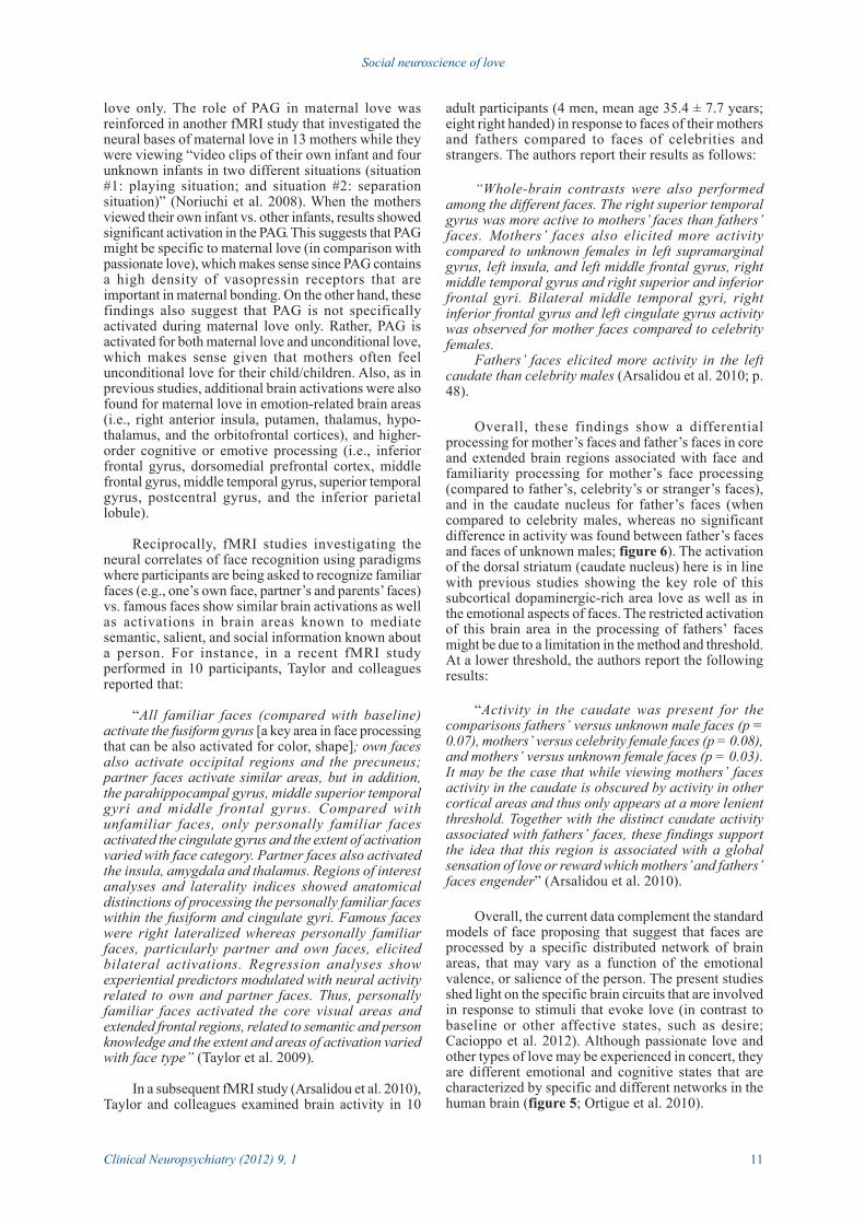
Figure 6. Rendered group activation maps of brain regions more active during processing of mother and fatherfaces (Arsalidou et al. 2010)
11
Social neuroscience of love
Clinical Neuropsychiatry (2012) 9, 1
love only. The role of PAG in maternal love wasreinforced in another fMRI study that investigated theneural bases of maternal love in 13 mothers while theywere viewing “video clips of their own infant and fourunknown infants in two different situations (situation#1: playing situation; and situation #2: separationsituation)” (Noriuchi et al. 2008). When the mothersviewed their own infant vs. other infants, results showedsignificant activation in the PAG. This suggests that PAGmight be specific to maternal love (in comparison withpassionate love), which makes sense since PAG containsa high density of vasopressin receptors that areimportant in maternal bonding. On the other hand, thesefindings also suggest that PAG is not specificallyactivated during maternal love only. Rather, PAG isactivated for both maternal love and unconditional love,which makes sense given that mothers often feelunconditional love for their child/children. Also, as inprevious studies, additional brain activations were alsofound for maternal love in emotion-related brain areas(i.e., right anterior insula, putamen, thalamus, hypo-thalamus, and the orbitofrontal cortices), and higher-order cognitive or emotive processing (i.e., inferiorfrontal gyrus, dorsomedial prefrontal cortex, middlefrontal gyrus, middle temporal gyrus, superior temporalgyrus, postcentral gyrus, and the inferior parietallobule).
Reciprocally, fMRI studies investigating theneural correlates of face recognition using paradigmswhere participants are being asked to recognize familiarfaces (e.g., one’s own face, partner’s and parents’ faces)vs. famous faces show similar brain activations as wellas activations in brain areas known to mediatesemantic, salient, and social information known abouta person. For instance, in a recent fMRI studyperformed in 10 participants, Taylor and colleaguesreported that:
“All familiar faces (compared with baseline)activate the fusiform gyrus [a key area in face processingthat can be also activated for color, shape]; own facesalso activate occipital regions and the precuneus;partner faces activate similar areas, but in addition,the parahippocampal gyrus, middle superior temporalgyri and middle frontal gyrus. Compared withunfamiliar faces, only personally familiar facesactivated the cingulate gyrus and the extent of activationvaried with face category. Partner faces also activatedthe insula, amygdala and thalamus. Regions of interestanalyses and laterality indices showed anatomicaldistinctions of processing the personally familiar faceswithin the fusiform and cingulate gyri. Famous faceswere right lateralized whereas personally familiarfaces, particularly partner and own faces, elicitedbilateral activations. Regression analyses showexperiential predictors modulated with neural activityrelated to own and partner faces. Thus, personallyfamiliar faces activated the core visual areas andextended frontal regions, related to semantic and personknowledge and the extent and areas of activation variedwith face type” (Taylor et al. 2009).
In a subsequent fMRI study (Arsalidou et al. 2010),Taylor and colleagues examined brain activity in 10
adult participants (4 men, mean age 35.4 ± 7.7 years;eight right handed) in response to faces of their mothersand fathers compared to faces of celebrities andstrangers. The authors report their results as follows:
“Whole-brain contrasts were also performedamong the different faces. The right superior temporalgyrus was more active to mothers’ faces than fathers’faces. Mothers’ faces also elicited more activitycompared to unknown females in left supramarginalgyrus, left insula, and left middle frontal gyrus, rightmiddle temporal gyrus and right superior and inferiorfrontal gyri. Bilateral middle temporal gyri, rightinferior frontal gyrus and left cingulate gyrus activitywas observed for mother faces compared to celebrityfemales.
Fathers’ faces elicited more activity in the leftcaudate than celebrity males (Arsalidou et al. 2010; p.48).
Overall, these findings show a differentialprocessing for mother’s faces and father’s faces in coreand extended brain regions associated with face andfamiliarity processing for mother’s face processing(compared to father’s, celebrity’s or stranger’s faces),and in the caudate nucleus for father’s faces (whencompared to celebrity males, whereas no significantdifference in activity was found between father’s facesand faces of unknown males; figure 6). The activationof the dorsal striatum (caudate nucleus) here is in linewith previous studies showing the key role of thissubcortical dopaminergic-rich area love as well as inthe emotional aspects of faces. The restricted activationof this brain area in the processing of fathers’ facesmight be due to a limitation in the method and threshold.At a lower threshold, the authors report the followingresults:
“Activity in the caudate was present for thecomparisons fathers’ versus unknown male faces (p =0.07), mothers’ versus celebrity female faces (p = 0.08),and mothers’ versus unknown female faces (p = 0.03).It may be the case that while viewing mothers’ facesactivity in the caudate is obscured by activity in othercortical areas and thus only appears at a more lenientthreshold. Together with the distinct caudate activityassociated with fathers’ faces, these findings supportthe idea that this region is associated with a globalsensation of love or reward which mothers’ and fathers’faces engender” (Arsalidou et al. 2010).
Overall, the current data complement the standardmodels of face proposing that suggest that faces areprocessed by a specific distributed network of brainareas, that may vary as a function of the emotionalvalence, or salience of the person. The present studiesshed light on the specific brain circuits that are involvedin response to stimuli that evoke love (in contrast tobaseline or other affective states, such as desire;Cacioppo et al. 2012). Although passionate love andother types of love may be experienced in concert, theyare different emotional and cognitive states that arecharacterized by specific and different networks in thehuman brain (figure 5; Ortigue et al. 2010).

12
Stephanie Cacioppo et al.
Clinical Neuropsychiatry (2012) 9, 1
Conclusion
Social neuroscience of love is a growing field ofresearch, which only recently has become the topic ofintensive and rigorous scientific empirical investi-gations. By identifying the specific cortico-subcorticalneural network as well as the central and peripheralelectrophysiological indices of love, we hope to providean interdisciplinary approach to better understand thecomplexity of love and its disorders. Althoughcombining knowledge from neuroimaging (fMRI andEEG) studies with standard approach in relationshipscience still doesn’t solve the hard problem of loveregarding its nature and origin, an integrative approachcombining neuroimaging techniques with otherdisciplines such as social psychology, animal studies,and genetics has the potential to answer age-old questionsas to the function of love, which can have usefulapplications in mental health and couple therapies.
Acknowledgments
The authors thank James W. Lewis, Chris Frum,and Elsa Juan. This work was supported by the SwissNational Science Foundation (Grant #PP00_1_128599/1 to SC), the University Funds Maurice Chalumeau ofthe University of Geneva in Switzerland (to SC andFBD), and the Mind Science Foundation (Grant #TSA2010-2 to SC, & FBD).
ReferencesAcevedo BP, Aron A, Fisher HE, Brown LL (2012). Neural
correlates of long-term intense romantic love. Socialcognitive and affective neuroscience 7, 145-159.
Aron A, Fisher H, Mashek DJ, Strong G, Li H, Brown LL (2005).Reward, motivation, and emotion systems associated withearly-stage intense romantic love. J Neurophysiol 94, 327–337.
Arsalidou M, Barbeau EJ, Bayless SJ, Taylor MJ (2010). Brainresponses differ to faces of mothers and fathers. Brain andCognition 74, 47-51.
Arzy S, Seeck M, Ortigue S, Spinelli L, Blanke O (2006).Induction of an illusory shadow person. Nature 443, 287.
Bartels A, Zeki S (2000). The neural basis of romantic love.Neuroreport 11, 3829-3834.
Bartels A, Zeki, S (2004). The neural correlates of maternal andromantic love. NeuroImage 21, 1155-1166.
Basar E, Schmiedt-Fehr C, Oniz A, Baþar-Eroðlu C (2008). Brainoscillations evoked by the face of a loved person. BrainResearch, 1214, 105-115.
Beauregard M, Courtemanche J, Paquette V, St-Pierre EL (2009).The neural basis of unconditional love. Psychiatry Res 172,93-98.
Berscheid E, Hatfield E (1969). Interpersonal attraction.Addison-Wesley, New York.
Birbaumer N, Lutzenberger W, Elbert T, Flor H, Rockstroh B(1993) Imagery and brain processes. In N. Birbaumer &A. Öhmann (Eds) The structure of emotion, pp. 298-321.Hogrefe and Huber, Toronto.
Blanke O, Ortigue S, Landis T, Seeck M (2002). Stimulatingillusory own-body perceptions. Nature 419, 269-270.
Cacioppo JT, Berntson GG (1992). Social psychologicalcontributions to the decade of the brain. Doctrine ofmultilevel analysis. Am Psychol 47, 1019-1028.
Cacioppo JT, Berntson GG (2005). Social Neuroscience.Psychology Press, New York.
Cacioppo JT, Berntson GG, Sheridan JF, McClintock MK (2000).Multilevel integrative analyses of human behavior: socialneuroscience and the complementing nature of social andbiological approaches. Psychological Bulletin 126, 829-843.
Cacioppo JT, Ortigue S (2011). Social Neuroscience: how amultidisciplinary field is uncovering the biology of humaninteractions. Cerebrum 12, 1-7.
Cacioppo S, Bianchi-Demicheli F, Frum C, Pfaus JG, Lewis JW(2012). The common neural bases between sexual desireand love: a multilevel kernel density fMRI analysis.Journal of Sexual Medicine, in press.
Darwin C (1872). The Expression of the Emotions in Man andAnimals. New York.
Diamond LM (2004). Emerging perspectives on distinctionsbetween romantic love and sexual desire. CurrentDirections in Psychological Science 13, 116-119.
Diamond LM (2003). What does sexual orientation orient? Abiobehavioral model distinguishing romantic love andsexual desire. Psychological Review 110, 173-192.
Ekman P (1992). Are there basic emotions? PsychologicalReview 99, 550-553.
Ekman P, Cordaro D (2011). What is Meant by Calling EmotionsBasic. Emotion Review 3, 364-370.
Ekman P (1999). Basic emotions. In T Dalgleish & T Power(Eds) The handbook of cognition and emotion, pp. 45-60.John Wiley & Sons, Ltd, Sussex, UK.
Guerra P, Vico C, Campagnoli R, Sánchez A, Anllo-Vento L, Vila J(2011). Affective processing of loved familiar faces:Integrating central and peripheral electrophysiologicalmeasures. International Journal of Psychophysiology in press.
Hatfield E, Rapson RL (1993). Love, sex and intimacy. Theirpsychology, biology and history. Harper & Collins, NewYork.
Hatfield E, Rapson RL (1996). Love and sex. Cross-culturalperspectives. Allyn and Bacon, Boston.
Hatfield E, Rapson RL (2002). Passionate love and sexual desire.Cross-cultural and historical perspectives. In A Vangelisti,HT Reis & MA Fitzpatrick (Eds) Stabillity and changes inrelationships, pp. 306-324. Cambridge University Press,Cambridge, England.
Hatfield E, Sprecher S (1986). Measuring passionate love inintimate relationships. J Adolesc 9, 383-410.
Hatfield E, Cacioppo JT, Rapson RL (1994). Emotionalcontagion. Cambridge University Press, New York.
Hatfield E, Rapson RL (1987). Passionate love/sexual desire:Can the same paradigm explain both? Archives of SexualBehavior 16, 259-278.
Hatfield E, Rapson RL (2009). The Neuropsychology ofpassionate love. In E Cuyler & M Ackhart (Eds)Psychology of Relationships. Nova Science Publishers.
Komisaruk BR, Whipple B (1998). Love as sensory stimulation:physiological consequences of its deprivation andexpression. Psychoneuroendocrinology 23, 927-944.
Noriuchi M, Kikuchi Y, Senoo A (2008). The functionalneuroanatomy of maternal love: mother’s response toinfant’s attachment behaviors. Biol Psychiatry 63, 415-423.
Ortigue S, Bianchi-Demicheli F, Patel N, Frum C, Lewis J (2010).Neuroimaging of Love: fMRI meta-analysis evidencetowards new perspectives in sexual medicine. Journal ofSexual Medicine 7, 3541-3552.
Russell JA, Rosenberg EL, Lewis MD (2011). Introduction to aSpecial Section on Basic Emotion Theory. Emotion Review3, 363-363.
Sabini J, Silver M (2005). Ekman’s basic emotions: Why notlove and jealousy? Cognition & Emotion 19, 693-712.
Shaver PR, Murdaya U, Fraley RC (2001). Structure of theIndonesian Emotion Lexicon. Asian Journal of SocialPsychology 4, 201-224.
Shaver P, Schwartz J, Kirson D, O’Connor G (1987). EmotionKnowledge: Further exploration of a prototype approach.Journal of Personality and Social Psychology 52, 1061-1086.

13
Social neuroscience of love
Clinical Neuropsychiatry (2012) 9, 1
Taylor MJ, Arsalidou M, Bayless SJ, Morris D, Evans JW,Barbeau EJ (2009). Neural correlates of personally familiarfaces: parents, partner and own faces. Human BrainMapping 30, 2008-2020.
Vico C, Guerra P, Robles H, Vila J, Anllo-Vento L (2010).Affective processing of loved faces: contributions fromperipheral and central electrophysiology. Neuro-psychologia 48, 2894-2902.