acc glands gi tract lect1 27oct15 - duke university
TRANSCRIPT

NORMAL BODY Microscopic Anatomy ���Accessory Glands of the GI Tract���
lecture 1
• Salivary glands • Pancreas
John Klingensmith [email protected]

Objectives���By the end of this lecture, students will be able to: ���
• describe the functional organization of the salivary glands and pancreas at the cellular level
• distinguish parenchymal tissue in the pancreas and salivary glands
• understand the structural relationships of exocrine and endocrine functions of the pancreas
• contrast the structure of the three major salivary glands relative to each other and the pancreas
(Lecture plan: overview of structure and function, then increasing resolution of microanatomy and cellular function)

Salivary Glands

Saliva functions to • Begin chemical digestion (salivary amylase) • Solubilize/suspend “flavor” compounds (water) • Lubricate food for swallowing (mucous, water) • Clean teeth and membranes (water) • Inhibit bacterial growth (lysozyme, sIgA) • Expel undesired material (water)
Contribution to saliva (~1 liter/day): 65% submandibular; 25% parotid; 5% sublingual; 5% minor glands

Secretory cells of the salivary glands • Mucous – triggered by
sympathetic stimuli (e.g. fright)…thick and viscous
• Serous – triggered by parasympathetic stimuli (e.g. food odors)…watery and protein-rich
• Striated ducts modify the exudate • Plasma cells outside secretory
acini produce IgA

Serous ���secretory cell
• Amino acids from the capillary blood
• Synthesis into proteins in rER, requires ATP
• Proteins move apically via Golgi
• Secretion vesicles/granules formed
• Granules extruded into lumen

Functional unit of salivary parenchyma
(Intralobular ducts)

Structure of salivary glands
• Lobular parenchyma • Connective tissue septa • Excretory (interlobular)
ducts – cuboidal to stratified columnar as diameter increases
• Intralobular ducts • Acini

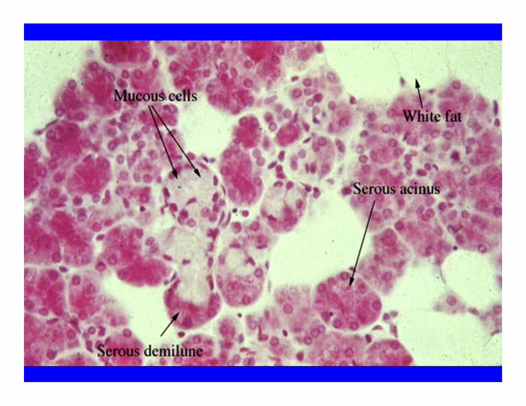
Histology of saliva secretion
• Serous acini • Mucous acini • “Serous demilunes” (mixed acini; demilune is fixation artifact)
• Myoepithelial cells • Intercalated ducts • Striated ducts

���serous mucous
• Nucleus round, near base • Basal cytoplasm has RER,
ribosomes (stains w/ H) • Apical cytoplasm has
zymogen secretion granules (stains w/ E)
• Typically in rounded acinus
• May cap mucous tubule as “demilune”
• Nucleus flat, near base • Organelles mostly near
base • Mucinogen granules in
apical cytoplasm (lost in paraffin/H&E)
• Cyclical activity • Typically tubular
arrangement

Myoepithelial cells
• Contractile cells • Occur between
secretory cells and basal lamina
• Stellate around serous acini
• Longitudinal along mucous glands and intercalated ducts

Intralobular salivary ducts • Intercalated
ducts: low cuboidal epithelium
• Striated ducts: cuboidal to columnar epithelium, big lumen

Cell types of the salivary gland

Striated ducts • Link intercalated to interlobular ducts • Cuboidal to columnar epithelial cells • Basal striations with stacks of elongated
mitochondria • Reabsorb Na, add K (makes secretion
hypotonic)

Parotid Gland • Serous acini,
no mucous • Long
intercalated ducts
• Striated ducts • Plasma cells • Adipose
tissue • Excretory ducts
pseudostratified to stratified

Blockage of the parotid duct is…

…a serous pain in the acinus!

Sublingual gland • Mucous
acini • Serous
demilunes (mixed acini)
• Plasma cells
• Intercalated ducts but no striated
• Many ducts to mouth

Submandibular Gland
• Serous acini • Mucous acini • Serous demilunes • Few adipose cells • Striated ducts • Short intercalated
ducts

Distinguishing the ���salivary glands
• Parotid has serous acini, lots of striated ducts, fibrous capsule, adipocytes in septa
• Sublingual has mostly mucous acini with some mixed (demilunes), loose CT septa, no striated ducts or fibrous capsule, many ducts to mouth
• Submandibular has serous and mucous acini, many mixed acini (demilunes), some striated ducts, few adipocytes, fibrous capsule

Pancreas

Pancreas has exocrine and endocrine functions���
exocrine endocrine • Serous acinar cells
secrete digestive juice • Secretion to
duodenum (pH 8, 1L/day)
• Digestive enzymes which hydrolyze proteins, carbs and fats
• Enzymes released largely as zymogens
• Islets of Langerhans produce hormones
• Hormones primarily involved in glucose metabolism and levels
• Released into efferent capillaries leaving islets

Pancreas

Pancreas has both exocrine and endocrine
functions • Exocrine -
serous acini • Endocrine -
islets of Langerhans

Pancreatic exocrine tissue

Pancreatic acini • Tightly packed • All serous • Acinar cells have
eosinophilic, granular apical cytoplasm
• AC have basophilic basal cytoplasm
• Round, basal nuclei • Drained by
intercalated ducts • surround centroacinar
cells

Pancreatic ���acinar cells
• Pyramidal in shape • Basal rough
endoplasmic reticulum • Basal, round nucleus
w/ large nucleolus • Prominent Golgi • Apical zymogen
granules • Apices face lumen

Centroacinar cells
• Line acinar lumen • Low cuboidal or
squamous epithelial cells
• Continuous with cells of intralobular duct
• Unique to pancreatic acini
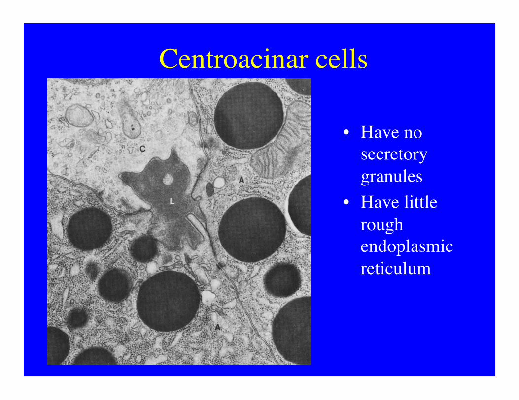
Centroacinar cells
• Have no secretory granules
• Have little rough endoplasmic reticulum

Intercalated ducts drain ���pancreatic acini
Pancreatic ductal system • Intercalated ducts - includes squamous
centroacinar cells, becomes cuboidal • Intralobular ducts - not striated as in SG. • Interlobular ducts - low columnar
epithelium, dense CT rim • Main pancreatic duct - runs length of
pancreas, wall contains SM, drains into duodenum at duodenal papilla
• Accessory pancreatic duct - similar duct from head of pancreas
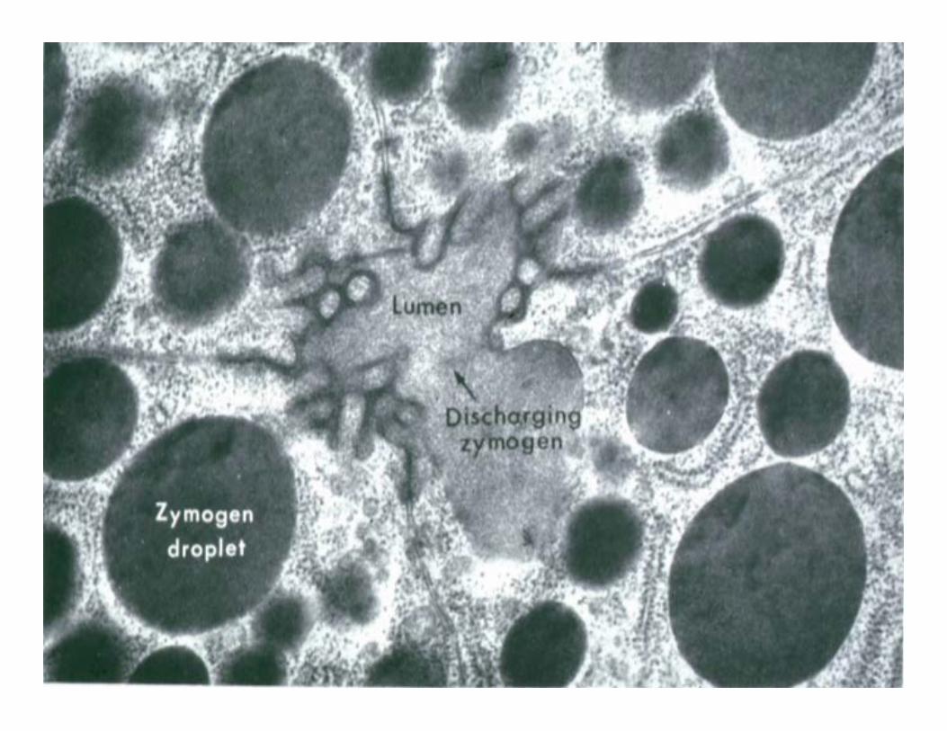

Zymogen excretion (e.g. trypsin)

Exocrine secretion by the pancreas • Zymogen granules released into lumen of
intercalated ducts, ultimately to duodenum • Release stimulated by hormones (CCK-
acini, secretin-IC ducts) • Secretion includes proenzymes activated in
small intestine • Enzymes digest various foodstuffs: protein
(endopeptidases); carbs (amylase); lipids (lipase); nucleic acids (nucleases)

Endocrine pancreas: ���Islets of Langerhans

Islet of Langerhans is ���well vascularized


Endocrine Cell types of the Islet ���- A, B, D ���
• Special staining • blue stains Type B • pink stains Type A • Immuno-
histochemistry • Function
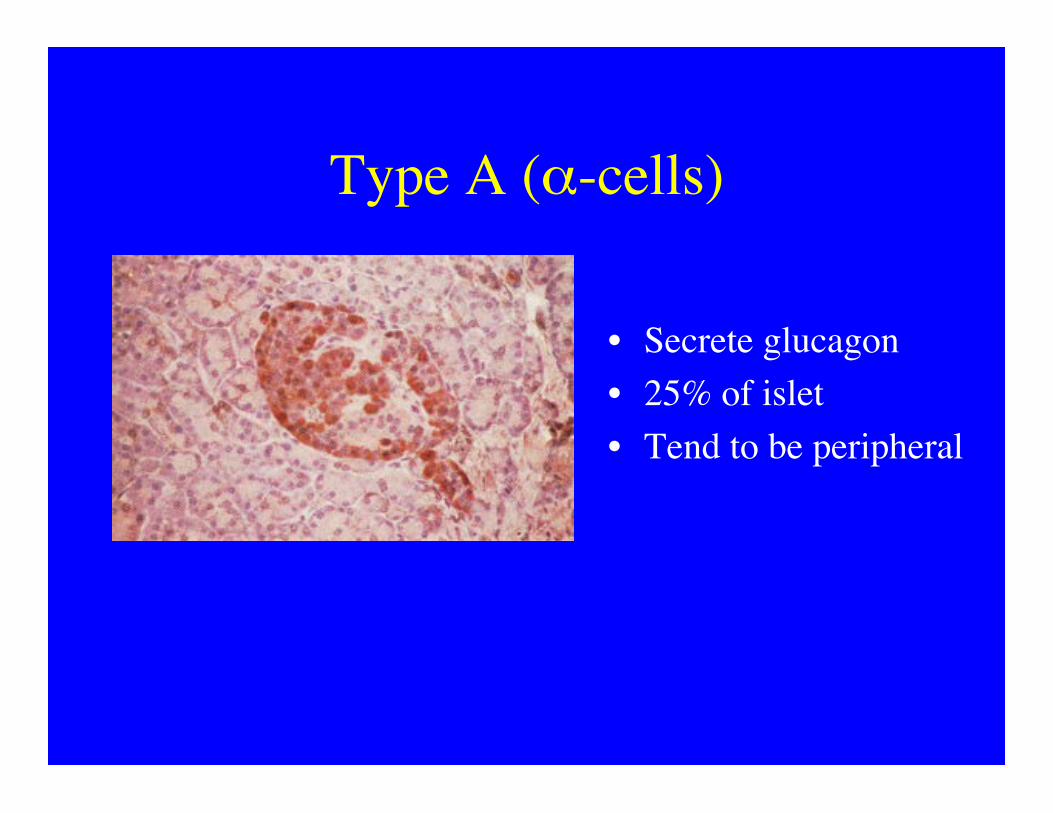
Type A (α-cells)
• Secrete glucagon • 25% of islet • Tend to be peripheral

Type B (β-cells)
• Secrete insulin • 65% of islet • Tend to be most dense
centrally • Polyhedral core in
granules (crystalline insulin?)

Type D (δ-cells) and others
• D cells secrete somatostatin • 5-10% of islet • Peripheral location • minor cell types comprise remaining 5%

Functions of insulin and glucagon are largely reciprocal
• Insulin lowers blood glucose
• Stimulates uptake from circulation
• Promotes storage, utilization and phos.
• Promotes synthesis of glycogen from phos. Glucose
• Secretion inhibited by somatistatin
• Glucagon raises blood glucose
• Stimulate release into blood
• Promotes synthesis of new glucose and breakdown of glycogen
• Secretion inhibited by somatistatin

Distinguishing pancreatic and salivary exocrine tissue
• Pancreas acini are all serous
• Long intercalated ducts
• No striated ducts • Centroacinar cells • Larger ducts simple
columnar • Islets of Langerhans
• Parotid acini are all serous
• Long intercalated ducts
• Lots of striated ducts • Large ducts
pseudostratified