acc/aha clinical data standardscirc.ahajournals.org/content/circulationaha/132/4/302.full.pdf ·...
TRANSCRIPT

ACC/AHA Clinical Data Standards
302
(Circulation. 2015;132:302-361. DOI: 10.1161/CIR.0000000000000156.)© 2015 by the American College of Cardiology Foundation and the American Heart Association, Inc.
Circulation is available at http://circ.ahajournals.org DOI: 10.1161/CIR.0000000000000156
*The findings and conclusions in this report are those of the authors and do not necessarily represent the official positions of the US Food and Drug Administration.†Former Task Force Chair; current chair during the writing effort.‡Former Task Force member; current member during the writing effort.This document was approved by the American Heart Association Science Advisory and Coordinating Committee on June 13, 2014, and by the American
College of Cardiology Board of Trustees on November 12, 2014.The online-only Comprehensive RWI Data Supplement table is available with this article at http://circ.ahajournals.org/lookup/suppl/
doi:10.1161/CIR.0000000000000156/-/DC1.Two online-only Data Supplements are available with this article at http://circ.ahajournals.org/lookup/suppl/doi:10.1161/CIR.0000000000000156/-/
DC2 and http://circ.ahajournals.org/lookup/suppl/doi:10.1161/CIR.0000000000000156/-/DC3.The American Heart Association requests that this document be cited as follows: Hicks KA, Tcheng JE, Bozkurt B, Chaitman BR, Cutlip DE,
Farb A, Fonarow GC, Jacobs JP, Jaff MR, Lichtman JH, Limacher MC, Mahaffey KW, Mehran R, Nissen SE, Smith EE, Targum SL. 2014 ACC/AHA key data elements and definitions for cardiovascular endpoint events in clinical trials: a report of the American College of Cardiology/American Heart Association Task Force on Clinical Data Standards (Writing Committee to Develop Cardiovascular Endpoints Data Standards). Circulation. 2015;132:302–361.
This article has been copublished in the Journal of the American College of Cardiology.Copies: This document is available on the World Wide Web sites of the American College of Cardiology (www.cardiosource.org) and the
American Heart Association (my.americanheart.org). A copy of the document is available at http://my.americanheart.org/statements by selecting either the “By Topic” link or the “By Publication Date” link. To purchase additional reprints, call 843-216-2533 or e-mail [email protected].
Expert peer review of AHA Scientific Statements is conducted by the AHA Office of Science Operations. For more on AHA statements and guidelines development, visit http://my.americanheart.org/statements and select the “Policies and Development” link.
Permissions: Multiple copies, modification, alteration, enhancement, and/or distribution of this document are not permitted without the express permission of the American Heart Association. Instructions for obtaining permission are located at http://www.heart.org/HEARTORG/General/Copyright-Permission-Guidelines_UCM_300404_Article.jsp. A link to the “Copyright Permissions Request Form” appears on the right side of the page.
2014 ACC/AHA Key Data Elements and Definitions for Cardiovascular Endpoint Events in Clinical Trials
A Report of the American College of Cardiology/American Heart Association Task Force on Clinical Data Standards (Writing Committee
to Develop Cardiovascular Endpoints Data Standards)WRITING COMMITTEE MEMBERS*
Karen A. Hicks, MD, FACC, Chair*; James E. Tcheng, MD, FACC, Vice-Chair; Biykem Bozkurt, MD, PhD, FACC, FAHA; Bernard R. Chaitman, MD, FACC;
Donald E. Cutlip, MD, FACC; Andrew Farb, MD, FACC*; Gregg C. Fonarow, MD, FACC, FAHA; Jeffrey P. Jacobs, MD, FACC;
Michael R. Jaff, DO, FACC; Judith H. Lichtman, MPH, PhD; Marian C. Limacher, MD, FACC, FAHA; Kenneth W. Mahaffey, MD, FACC; Roxana Mehran, MD, FACC, FAHA; Steven E. Nissen, MD, MACC, FAHA;
Eric E. Smith, MD, MPH, FAHA; Shari L. Targum, MD, FACC*
ACC/AHA TASK FORCE ON CLINICAL DATA STANDARDS MEMBERS
William S. Weintraub, MD, MACC, FAHA, Chair; Biykem Bozkurt, MD, PhD, FACC, FAHA; Gregg C. Fonarow, MD, FACC, FAHA; Robert C. Hendel, MD, FACC, FAHA†;
Jeffrey P. Jacobs, MD, FACC; Hani H. Jneid, MD, FACC, FAHA; Michael A. Kutcher, MD, FACC; Judith H. Lichtman, MPH, PhD; Eric E. Smith, MD, MPH, FAHA‡; James E. Tcheng, MD, FACC;
Tracy Y. Wang, MD, FACC, FAHA
by guest on June 24, 2018http://circ.ahajournals.org/
Dow
nloaded from
by guest on June 24, 2018http://circ.ahajournals.org/
Dow
nloaded from
by guest on June 24, 2018http://circ.ahajournals.org/
Dow
nloaded from
by guest on June 24, 2018http://circ.ahajournals.org/
Dow
nloaded from
by guest on June 24, 2018http://circ.ahajournals.org/
Dow
nloaded from
by guest on June 24, 2018http://circ.ahajournals.org/
Dow
nloaded from
by guest on June 24, 2018http://circ.ahajournals.org/
Dow
nloaded from
by guest on June 24, 2018http://circ.ahajournals.org/
Dow
nloaded from
by guest on June 24, 2018http://circ.ahajournals.org/
Dow
nloaded from

Hicks et al Cardiovascular Endpoints Data Standards 303
Table of Contents
Preamble . . . . . . . . . . . . . . . . . . . . . . . . . . . . . . . . . . . . . . 303Foreword . . . . . . . . . . . . . . . . . . . . . . . . . . . . . . . . . . . . . . 3041. Introduction. . . . . . . . . . . . . . . . . . . . . . . . . . . . . . . . . . 3042. Methodology . . . . . . . . . . . . . . . . . . . . . . . . . . . . . . . . . 305 2.1. Writing Committee Composition . . . . . . . . . . . . . 305 2.2. Relationships With Industry and
Other Entities . . . . . . . . . . . . . . . . . . . . . . . . . . . . 305 2.3. Review of Literature and Existing
Data Definitions . . . . . . . . . . . . . . . . . . . . . . . . . . 305 2.4. Development of Terminology Concepts . . . . . . . . 305 2.5. Consensus Development . . . . . . . . . . . . . . . . . . . . 306 2.6. Relation to Other Standards . . . . . . . . . . . . . . . . . 306 2.7. Peer Review, Public Review, and
Board Approval . . . . . . . . . . . . . . . . . . . . . . . . . . . 3063. Data Elements and Definitions . . . . . . . . . . . . . . . . . . . 306 3.1. Death Attribution . . . . . . . . . . . . . . . . . . . . . . . . . 306 3.1.1. Cardiovascular Cause of Death . . . . . . . . . 307 3.1.2. Noncardiovascular Cause of Death . . . . . . 307 3.1.3. Undetermined Cause of Death. . . . . . . . . . 307 3.2. Myocardial Infarction . . . . . . . . . . . . . . . . . . . . . . 307 3.3. Hospitalization for Unstable Angina. . . . . . . . . . . 308 3.4. Transient Ischemic Attack and Stroke. . . . . . . . . . 308 3.5. Heart Failure Event . . . . . . . . . . . . . . . . . . . . . . . . 308 3.6. Percutaneous Coronary Intervention. . . . . . . . . . . 309 3.7. Peripheral Vascular Intervention . . . . . . . . . . . . . . 309 3.8. Stent Thrombosis . . . . . . . . . . . . . . . . . . . . . . . . . 3094. Informatics of Controlled Vocabularies . . . . . . . . . . . . 310References . . . . . . . . . . . . . . . . . . . . . . . . . . . . . . . . . . . . . 310Appendix 1. Author Relationships With Industry and
Other Entities (Relevant)—2014 ACC/AHA Key Data Elements and Definitions for Cardiovascular Endpoint Events in Clinical Trials. . . . . . . . . . . . . . . . . . . . . . . . . . . . . . . 313
Appendix 2. Reviewer Relationships With Industry and Other Entities—2014 ACC/AHA Key Data Elements and Definitions for Cardiovascular Endpoint Events in Clinical Trials . . . . . . . . 314
Appendix 3. Death Attribution . . . . . . . . . . . . . . . . . . . . . 315Appendix 4. Myocardial Infarction. . . . . . . . . . . . . . . . . . 318Appendix 5. Hospitalization for Unstable Angina . . . . . . 324Appendix 6. Transient Ischemic Attack and Stroke . . . . . 327Appendix 7. Heart Failure Event . . . . . . . . . . . . . . . . . . . 329Appendix 8. Heart Failure Event: Additional Details . . . . 334Appendix 9. Percutaneous Coronary Intervention . . . . . . 336Appendix 10. Cardiovascular Anatomy . . . . . . . . . . . . . . 350Appendix 11. Peripheral Vascular Intervention . . . . . . . . 356
PreambleThe American College of Cardiology (ACC) and the American Heart Association (AHA) support their members’ goal to improve the care of patients with cardiovascular dis-ease through professional education, research, and develop-ment of guidelines and standards and by fostering policies that support optimal patient outcomes. The ACC and AHA recognize the importance of the use of clinical data standards for patient management, assessment of outcomes, and conduct of research, and the importance of defining the processes and outcomes of clinical care, whether in randomized trials, obser-vational studies, registries, or quality-improvement initiatives.
Clinical data standards strive to define and standardize data relevant to clinical concepts, with the primary goal of facilitat-ing uniform data collection by providing a platform of clinical terms with corresponding definitions and data elements. Broad agreement on a common vocabulary with reliable definitions used by all is vital to pool and/or compare data across clini-cal trials to promote interoperability with electronic health records (EHRs) and to assess the applicability of research to clinical practice. The ultimate purpose of clinical data stan-dards is to contribute to the infrastructure necessary to accom-plish the ACC’s mission of fostering optimal cardiovascular care and disease prevention and the AHA’s mission of build-ing healthier lives, free of cardiovascular diseases and stroke.
The specific goals of clinical data standards are:
1. To establish a consistent, interoperable, and universal clinical vocabulary as a foundation for both clinical care and clinical research, including clinical trials
2. To promote the ubiquitous use of EHRs and facilitate the exchange of data across systems through harmonized, standardized definitions of key data elements
3. To facilitate the further development of clinical registries, quality- and performance-improvement programs, out-comes evaluations, and clinical research, including the comparison of results within and across these initiatives
The key elements and definitions are intended to facilitate the consistent, accurate, and reproducible capture of clinical concepts; standardize the terminology used to describe car-diovascular diseases and procedures; create a data environ-ment conducive to the assessment of patient management and outcomes for quality and performance improvement and clin-ical and translational research; and increase opportunities for sharing data across disparate data sources. The ACC/AHA Task Force on Clinical Data Standards selects cardiovascu-lar conditions and procedures that will benefit from creation of a standard dataset. Subject matter experts are selected to examine/consider existing standards and develop a compre-hensive, yet not exhaustive, standard dataset. When a data collection effort is undertaken, only a subset of the elements contained in a clinical data standards listing may be needed, or conversely, users may want to consider whether it may be necessary to collect some elements not listed. For example, in the setting of a randomized clinical trial of a new drug, addi-tional information would likely be required regarding study procedures and drug therapies.
The ACC and AHA recognize that there are other national efforts to establish clinical data standards, and every attempt is made to harmonize newly published standards with exist-ing standards. Writing Committees are instructed to consider adopting or adapting existing nationally and internationally recognized data standards if the definitions and characteris-tics are useful and applicable to the set under development. In addition, the ACC and AHA are committed to continually expanding their portfolio of data standards and will create new standards and update existing standards as needed to maintain their currency and promote harmonization with other standards as health information technology and clinical practice evolve.
The Writing Committee for the current effort was intention-ally expanded to include a regulatory perspective. This reflects
by guest on June 24, 2018http://circ.ahajournals.org/
Dow
nloaded from

304 Circulation July 28, 2015
the key role of clinical event concepts in evaluating therapeu-tic safety and effectiveness in clinical trials. This unique col-laboration between the regulatory sector and the ACC and AHA acknowledges the need to align key clinical concepts for regulatory reporting and clinical care.
The Health Insurance Portability and Accountability Act privacy regulations, which went into effect in April 2003, have heightened all practitioners’ awareness of our profes-sional commitment to safeguard our patients’ privacy. The Health Insurance Portability and Accountability Act privacy regulations1 specify which information elements are consid-ered “protected health information.” These elements may not be disclosed to third parties (including registries and research studies) without the patient’s written permission. Protected health information may be included in databases used for healthcare operations under a data use agreement. Research studies using protected health information must be reviewed by an institutional review board or a privacy board.
We have included identifying information in all clinical data standards to facilitate uniform collection of these elements when appropriate. For example, a longitudinal clinic database may contain these elements because access is restricted to the patient’s caregivers. Conversely, registries may not con-tain protected health information unless specific permission is granted by each patient. These fields are indicated as protected health information in the data standards.
In clinical care, caregivers communicate with each other through a common vocabulary. In an analogous fashion, the integrity of clinical research depends on firm adherence to prespecified procedures for patient enrollment and follow-up; these procedures are guaranteed through careful attention to definitions enumerated in the study protocol, case report forms, and clinical event committee charters. When data ele-ments and definitions are standardized across studies, com-parison, pooled analysis, and meta-analysis are enabled, thus deepening our understanding of individual studies.
The recent development of quality-performance mea-surement initiatives, particularly those for which the com-parison of providers is an implicit or explicit aim, has further raised awareness about the importance of data standards. Indeed, a wide audience, including nonmedical professionals such as payers, regulators, and consumers, may draw conclu-sions about care and outcomes. To understand and compare care patterns and outcomes, the data elements that charac-terize them must be clearly defined, consistently used, and properly interpreted.
William S. Weintraub, MD, MACC, FAHAChair, ACC/AHA Task Force on Clinical Data Standards
ForewordThis publication, commissioned by the ACC/AHA, is the product of a novel collaboration between the ACC and AHA, the US Food and Drug Administration (FDA), and the Standardized Data Collection for Cardiovascular Trials Initiative (SCTI). The aim of the collaboration is to promul-gate, for regulatory submissions, clinical data standards for key cardiovascular and stroke endpoint events in clinical tri-als. The Writing Committee for this particular data standard is unique in that it was convened to achieve the following goals:
to address issues particular to regulatory submissions and to provide a framework of data standards that would simplify the design and conduct of clinical trials for those considering regulatory submissions. The intent of this Writing Committee is not to be overly prescriptive. For example, the stroke data elements are minimal by design to allow for flexibility needed to conduct global clinical trials for drugs and devices. In a trial focused on the treatment of stroke, investigators may define additional outcomes and data elements for a particular stroke treatment or indication. The Writing Committee recognizes that these standards may be used for other types of clinical tri-als and clinical care processes where appropriate. These data standards are a first step in developing a universal language for clinical trials and other types of health-related research.
1. IntroductionEffective communication is a cornerstone of the health-care enterprise. A prerequisite for providing seamless care is the universal and consistent use of medical vocabularies.2 Cardiovascular endpoints such as death, myocardial infarc-tion, stroke, and revascularization are critical in assessing diagnostic and therapeutic approaches in the clinical care, research, and regulatory domains. With the adoption of EHR solutions has come the opportunity to manage health-related information as discrete data.3 Therefore, the ACC/AHA Task Force on Clinical Data Standards established this Writing Committee to identify and harmonize the common data ele-ments involved in key cardiovascular endpoint events. Doing so will allow this vocabulary to be used to improve the assess-ment of process, performance, and outcomes across multiple dimensions of health care.
In this work, the term “vocabulary” includes the terminol-ogy concept, the concept definition, a suggested label for the corresponding data element, permissible values of the data element, and definitions for the permissible values.
The Writing Committee identified the ongoing work of the SCTI as the foundation for the development of this vocabu-lary. The Writing Committee’s task was to review, refine, and advance as a clinical standard the cardiovascular endpoint terminology set developed by the SCTI. This terminology set largely reflects endpoints related to the symptoms, manifesta-tions, treatment, and consequences of coronary artery disease in both cardiovascular and noncardiovascular drug and device trials. Endpoint concepts related to carotid/cerebral revascu-larization, peripheral surgical revascularization, and treatment of diseases of the aorta are beyond the scope of this document.
First convened in 2009 by the FDA, the SCTI is a working group formed to improve the quality and efficiency of clinical trials. It includes representatives from academia, professional societies, the Clinical Data Interchange Standards Consortium, Health Level 7, the Clinical Trials Transformation Initiative, pharmaceutical and cardiovascular device manufacturers, and the FDA (which includes the Center for Drug Evaluation and Research and the Center for Devices and Radiological Health). The original goal of the working group was to improve the quality and efficiency of cardiovascular clinical trials (eg, acute coronary syndrome trials, percutaneous coronary inter-vention [PCI] trials). Subsequently, in recognition of the growing interest in cardiovascular events in noncardiovascular
by guest on June 24, 2018http://circ.ahajournals.org/
Dow
nloaded from

Hicks et al Cardiovascular Endpoints Data Standards 305
trials (eg, diabetes control trials, weight loss trials), this focus expanded to include noncardiovascular trials. To achieve its goal, the SCTI acknowledged the need for a consistent car-diovascular and stroke endpoint vocabulary comprising terms defined by objective criteria and reported uniformly.4 This framework of standardized key data elements in clinical trials (and potentially the clinical care domain), could also facilitate the conduct of meta-analyses to assess cardiovascular safety and compare the effectiveness of drug and device products.
The SCTI-developed working draft “Standardized Definitions for Cardiovascular and Stroke Endpoint Events in Clinical Trials” is the source document for the data standards in this publication.4 This source document identifies the cardio-vascular and stroke endpoint terms relevant to clinical trials and regulatory submissions. The Writing Committee evaluated and harmonized these endpoint terms, categorized the attributes of each data element, developed permissible (ie, allowed) values, and tabulated the content to facilitate computational interoper-ability and the use of the terminology regulatory domains.
The objective is to use this controlled terminology, initially developed for clinical trials and regulatory submissions, to standardize event reporting and data collection when deter-minations must be made about cardiovascular and stroke endpoints in the context of clinical care processes, registries, EHRs, and longitudinal drug or device surveillance.
Nonetheless, the Writing Committee recognizes that this terminology is not applicable to all dimensions of health care and that some of these definitions are not applicable to clinical care processes. For example, the “Hospitalization for Unstable Angina” definition may not be optimal for the Centers for Medicare and Medicaid Services because other clinical scenarios could be consistent with unstable angina but not fulfill all the criteria needed to define this endpoint in a clinical trial. Similar scenarios could be relevant to other nonfatal endpoint definitions.
To avoid ambiguity, we recommend maintaining the car-diovascular and stroke endpoint vocabulary described in this document as “regulatory specific” distinct from other vocabularies. Although endpoint definitions may evolve over time, a period in which definitions remain static is needed for terms to be used successfully to conduct a meta-analysis. Furthermore, the ACC/AHA Task Force on Clinical Data Standards should include regulatory data standards experts—in addition to members of this Writing Committee, SCTI rep-resentatives, and other clinical trial experts—as an integral part of any necessary definition revision process, because such experts understand the requirements for evaluating drug and device products.
2. Methodology2.1. Writing Committee CompositionThe ACC/AHA Task Force on Clinical Data Standards selected the members of the Writing Committee. The committee con-sisted of 16 people with domain expertise in internal medi-cine, cardiovascular medicine, neurology, clinical research, epidemiology, invasive and interventional therapies, outcomes assessment, medical informatics, health information manage-ment, and healthcare services research and delivery.
2.2. Relationships With Industry and Other EntitiesThe ACC/AHA Task Force on Clinical Data Standards makes every effort to avoid actual or potential conflicts of inter-est that might arise as a result of an outside relationship or a personal, professional, or business interest of any mem-ber of the Writing Committee. Specifically, all members of the Writing Committee are required to complete and submit a disclosure form showing all such relationships that could be perceived as real or potential conflicts of interest. These statements are reviewed by the ACC/AHA Task Force on Clinical Data Standards and updated when changes occur. Authors’ and peer reviewers’ relationships with industry and other entities pertinent to this data standards document are disclosed in Appendixes 1 and 2, respectively. In addition, for complete transparency, the disclosure information of each Writing Committee member—including relationships not pertinent to this document—is available as an online supple-ment at http://circ.ahajournals.org/lookup/suppl/doi:10.1161/CIR.0000000000000156/-/DC1. The work of the Writing Committee was supported exclusively by the ACC and AHA without commercial support. Writing Committee members volunteered their time for this effort. Meetings of the Writing Committee were confidential and attended only by committee members and staff.
2.3. Review of Literature and Existing Data DefinitionsCardiovascular endpoint concepts have long been used in clinical care and research to ascertain and assess outcomes of diagnostic and therapeutic approaches. A series of refer-ence publications provided the foundation for the cardiovas-cular endpoint concepts identified by the SCTI and developed by the Writing Committee.5–14 What makes this work unique is that it reviews and refines the terms as developed by the SCTI explicitly for use in reporting clinical trial results and in regulatory submissions, and it delineates where these concepts could or should not be used as the foundational vocabulary in routine clinical care.
2.4. Development of Terminology ConceptsThe terminology set addressed in this body of work includes cardiovascular endpoints of universal interest in clinical care, research, and regulatory review: death (specifically attribution of the cause of death), myocardial infarction, stroke, transient ischemic attack (TIA), coronary intervention (including stent thrombosis), peripheral vascular intervention, hospitalization for unstable angina, and acute heart failure (HF) events.
The Writing Committee aggregated, reviewed, harmonized, and extended these terms to develop a controlled, semanti-cally interoperable, machine-computable terminology set that would be usable, as appropriate, in as many contexts as possible. As necessary, the Writing Committee identified the contexts where individual terms required differentiation depending on their proposed use (ie, research/regulatory ver-sus clinical care contexts).
The Writing Committee tabulated the content and provided sufficient structure to build and model the informatics formal-isms to achieve computational interoperability. The resulting
by guest on June 24, 2018http://circ.ahajournals.org/
Dow
nloaded from

306 Circulation July 28, 2015
appendices (Appendixes 3–7 and 9–11) list the “terminology concept” in the first column, followed by a clinical definition (“concept definition”) of the terminology concept in the sec-ond column. A data element label is suggested for forms-based approaches to data capture. The allowed responses (“permissi-ble values”) for each terminology concept in the next column are the acceptable “answers” for capturing the information. For terminology concepts with multiple permissible values, a bulleted list of the permissible values is provided in the same row as the terminology concept, with successive rows listing each permissible value and corresponding permissible value definition. The process of converting the prose description of an endpoint into this tabular format can be seen by compar-ing the source text for a HF endpoint event (Appendix 8, an excerpt from the SCTI draft document) and the tabular repre-sentation of the same concept in Appendix 7. Where possible, clinical definitions of endpoints (and the corresponding per-missible values) are repeated verbatim as defined by the SCTI or as previously published in reference documents.
2.5. Consensus DevelopmentThe ACC/AHA Task Force on Clinical Data Standards estab-lished the Writing Committee in November 2012, according to the processes described in the Task Force on Clinical Data Standards’ methodology statement.15 As described previously, the responsibility of the Writing Committee was to review and refine the list of candidate terms identified by the SCTI and to harmonize the attributes and other informatics formal-isms required to attain interoperability of the terms. During the first 6 months of 2013, the work of the Writing Committee was accomplished through a series of teleconference and Web conference meetings, along with extensive correspondence by e-mail. The review work was distributed among subgroups of the Writing Committee on the basis of their interest and exper-tise in the components of the terminology set. The proceed-ings of the work groups were then assembled, resulting in the vocabulary and associated descriptive prose in Section 3. All members reviewed and approved the final vocabulary.
2.6. Relation to Other StandardsThe Writing Committee reviewed the work of the SCTI along with available published data standards, specifically those developed for death, acute myocardial infarction, stroke, TIA, unstable angina/non–ST-elevation myocardial infarc-tion, HF, PCI, and peripheral vascular intervention.4–13,16–21 Existing published definitions were adjusted to eliminate verbiage not relevant to an actual definition (eg, instructions such as the phrase “indicate whether the patient has . . .” have been eliminated).
Through the affirmation and refinement of existing data stan-dards, the Writing Committee anticipates that the vocabulary will facilitate the uniform adoption of these terms, where appro-priate, by the clinical care, clinical and translational research, regulatory, quality and outcomes, and EHR communities.
2.7. Peer Review, Public Review, and Board ApprovalThe “2014 ACC/AHA Key Data Elements and Definitions for Cardiovascular Endpoint Events in Clinical Trials” statement
was reviewed by official reviewers nominated by the ACC and AHA. To increase its applicability further, the document was posted on the ACC Web site for a 30-day public comment period. This document was approved for publication by the ACC Board of Trustees on November 12, 2014, and by the AHA Science Advisory and Coordinating Committee on June 13, 2014. The Writing Committee anticipates that these data standards will require review and updating in the same man-ner as other published guidelines, performance measures, and appropriate use criteria. The Writing Committee will there-fore review the set of data elements periodically, starting with the anniversary of publication of the standards, to ascertain whether modifications should be considered.
3. Data Elements and DefinitionsAs described above, the SCTI identified candidate cardio-vascular and stroke endpoint event terms in the draft docu-ment “Standardized Definitions for Cardiovascular and Stroke Endpoint Events in Clinical Trials.” The document delineated the following cardiovascular and neurological endpoint events in the context of clinical trials and regulatory reporting:
1. Cardiovascular death2. Noncardiovascular death3. Undetermined cause of death4. Myocardial infarction5. Hospitalization for unstable angina6. Transient ischemic attack and stroke7. Heart failure event8. Percutaneous coronary intervention9. Peripheral vascular intervention
10. Stent thrombosis
The SCTI envisioned that data collection and exchange standards for these endpoint events would allow individual investigators and clinical research organizations to collect and exchange research data consistently and efficiently. It also envisioned that adoption of the controlled terminology within the research and regulatory sectors could facilitate consistency across the clinical care domain.
3.1. Death AttributionDeath is classified into 1 of 3 categories: 1) cardiovascular death; 2) noncardiovascular death; and 3) undetermined cause of death. The intent of the classification schema is to identify one, and only one, of the categories as the underlying cause of death. The key priority is differentiating between cardiovascu-lar and noncardiovascular causes of death (Appendix 3).
Collection of appropriate source documentation is criti-cal for rigorous adjudication of the cause of death. Although death certificates establish that the patient died, reliance on information included in death certificates may be problem-atic; several studies have demonstrated inaccurate coding in the death certificate when death certificates were compared with adjudicated outcomes.22,23 In contrast, autopsy reports are often valuable in assessing the cause of death and should be used whenever possible.
For sudden deaths, even when witnessed, death attribu-tion may be difficult if only limited information is available. Frequently, these deaths are attributed to either sudden cardiac
by guest on June 24, 2018http://circ.ahajournals.org/
Dow
nloaded from

Hicks et al Cardiovascular Endpoints Data Standards 307
death (cardiovascular death) or death due to an undetermined cause. Sensitivity analyses may be helpful in determining the effect of these events on the primary and major secondary end-points in a particular clinical trial or development program.
3.1.1. Cardiovascular Cause of DeathFrequently, the cardiovascular death category is not divided further into subcategories such as death resulting from an acute myocardial infarction, sudden cardiac death, or HF, because the cause of death is so often unknown or ambigu-ous (eg, Does a death after a myocardial infarction count as a myocardial infarction, sudden death, arrhythmic death, or HF death?). Moreover, the underlying cause of death and the mode of death (ie, most proximate event associated with death) may overlap substantially. In contrast, precision is more achievable with respect to nonfatal events.
However, in cases where subclassification is desired, the Writing Committee recommends a uniform approach for cat-egorizing the attributable cause (and not just the proximate event) for cardiovascular death. The suggested subcategories for attribution of death to a cardiovascular etiology are acute myocardial infarction, sudden cardiac death, HF, stroke, car-diovascular procedure, cardiovascular hemorrhage, and other cardiovascular causes. “Death due to other cardiovascular causes” refers to a cardiovascular death not included in the above categories but with a specific known cause, such as a pulmonary embolism or peripheral arterial disease. In addi-tion, “death due to cardiovascular hemorrhage” refers to a death related to hemorrhage such as a nonstroke intracranial hemorrhage, nonprocedural or nontraumatic vascular rupture (eg, aortic aneurysm), or pulmonary hemorrhage from a pul-monary embolism. In contrast, if a pulmonary hemorrhage were a result of a contusion from a motor vehicle accident, the cause of death would be noncardiovascular (death due to trauma). Although these subcategories may not be appli-cable to all study populations, therapeutic areas, or drug and device development programs, the events in these subcatego-ries occur relatively frequently in the general population and probably contribute to mortality in any observation. In some trials, subclassification of cardiovascular causes of death may prove helpful in understanding pathophysiology in the context of drug or device programs.
3.1.2. Noncardiovascular Cause of DeathIdentifying noncardiovascular causes of death is important when assessing competing mortality risks in both cardiovas-cular and noncardiovascular trials. The proposed schema for noncardiovascular causes of death is more general than that for cardiovascular causes of death. This noncardiovascular schema could be expanded to capture other causes for spe-cific trials in particular therapeutic areas or for specific drug or device development programs if a specific toxicity has been identified in nonclinical work or early clinical trials. When death is clearly due to a noncardiovascular cause, a cardiovas-cular cause of death is excluded. The proposed values repre-sent commonly used noncardiovascular categories.
3.1.3. Undetermined Cause of DeathIn general, this category of death should apply to few patients in well-run clinical trials. Attribution of causality may be
limited or impossible if information available at the time of death is minimal or nonexistent. In such cases, the date of death may be the only data element captured.
The key priority is to prespecify how these deaths will be classified and to implement a uniform approach throughout the conduct of the trial. Occasionally, it may not be possible to determine exact causality when 2 lethal conditions contrib-ute to death equally. In this circumstance, 1 condition should be chosen, with consideration of the issue being studied. For example, if cardiac safety is under consideration and the com-peting causes of death are cardiovascular and noncardiovascu-lar, cardiovascular death should take precedence.
3.2. Myocardial InfarctionThe categorization and definitions of the types of myo-cardial infarction are derived from the “Third Universal Definition of Myocardial Infarction,”13,14 the “2012 ACCF/AHA Guideline for the Management of Unstable Angina/Non-ST-Elevation Myocardial Infarction,”12 and the “2013 ACCF/AHA Guideline for the Management of ST-Elevation Myocardial Infarction.”18
The key recommendation is to base thresholds for biomarker detection of myocardial infarction on 99th percentile values (ie, the upper reference limit) rather than on “upper limit of normal” values. Multiple assays exist for cardiac troponin and the MB fraction of creatine kinase (CK-MB), and assay characteristics vary by manufacturer. Some assays reported by local laboratories provide the 99th percentile and a higher “decision limit” or upper limit of normal above which myo-cardial infarction should be considered. The “Third Universal Definition of Myocardial Infarction”13,14 recommends the use of the 99th percentile upper reference limit as the reference standard. The data elements developed in Appendix 4 allow both the 99th percentile and the upper limit of normal (or both) to be captured, depending on the reporting approach used in the central or local laboratory. Instead of listing every cardiac biomarker assay in Appendix 4, we have elected to represent all assays with the generic term [cardiac biomarker]. The actual biomarker assay used should replace the generic term [cardiac biomarker]. Collection of serial biomarker val-ues to capture all measurements (and to reflect rise or fall of the biomarker) would recursively use the same data element construct as the approach for capturing a single value. Cardiac troponin is the preferred biomarker. If troponin values are not available, then CK-MB mass is used as an alternative.
The terminology set includes data elements for stent reste-nosis without occlusion as a type of acute myocardial infarc-tion (type 4c) and asymptomatic postbaseline myocardial infarction detected during follow-up. The data elements that reflect old or prior myocardial infarction at baseline are not included here but can be found in the “2013 ACCF/AHA Key Data Elements and Definitions for Measuring the Clinical Management and Outcomes of Patients With Acute Coronary Syndromes and Coronary Artery Disease.”16
The Writing Committee acknowledges that there is disagree-ment about how to define a “clinically relevant myocardial infarction” after coronary revascularization (PCI or coronary artery bypass graft). An expert consensus group from the Society for Cardiovascular Angiography and Interventions24
by guest on June 24, 2018http://circ.ahajournals.org/
Dow
nloaded from

308 Circulation July 28, 2015
proposes the use of CK-MB instead of troponin and different cut points from those included in the “Third Universal Definition of Myocardial Infarction.”13,14 A detailed discussion of these differences is beyond the scope of this publication but is pro-vided by White.25 At this time, the Writing Committee contin-ues to support the “Third Universal Definition of Myocardial Infarction”13 for harmonization purposes but recognizes that this matter requires further study. As long as cardiac biomarker values (both cardiac troponin and CK-MB) and 99th percentile upper reference limit values are recorded, virtually any defini-tion of periprocedural myocardial infarction can be applied and examined with respect to outcome.
3.3. Hospitalization for Unstable AnginaHospitalization for unstable angina is a commonly used endpoint in clinical trials evaluating the efficacy or safety of cardiovascular therapies such as lipid-modifying agents, antihypertensive drugs, antithrombotic therapies, and coro-nary interventions. Unlike traditional endpoints such as death, myocardial infarction, or stroke, hospitalization for unstable angina, by necessity, involves some degree of subjective assessment of the most likely etiology of symptoms result-ing in hospital admission. The terminology set for unstable angina (Appendix 5) focuses on data elements needed for determining whether symptoms truly represent cardiovascular ischemia, including the character and duration of the present-ing symptoms, the proximity of symptom onset to hospitaliza-tion, and the duration of hospitalization. Electrocardiographic abnormalities are pivotal to the diagnosis. Such abnormali-ties include the presence or absence of deviations in the ST segment, morphology of ST-segment changes (horizon-tal or downsloping versus upsloping), and the magnitude of the deviation. Many patients without high-risk features (ie, patients with low TIMI [Thrombolysis in Myocardial Infarction] or GRACE [Global Registry of Acute Coronary Events] risk scores) undergo provocative testing for inducible myocardial ischemia, requiring measurement of ST eleva-tion or depression during electrocardiographic monitoring. Alternatively, exercise or pharmacological stress testing may involve assessment of wall motion abnormalities on echo-cardiography and/or reversible perfusion defects by nuclear scintigraphy or magnetic resonance imaging. Other important data elements include angiographic evidence of the severity of coronary stenosis or presence of coronary thrombus in a vessel believed to be responsible for the ischemic signs and symptoms. Additional data elements include the need for cor-onary revascularization by PCI or coronary bypass surgery of lesion(s) believed responsible for the hospitalization.
The need for escalation of pharmacological therapy (nitrates, beta-blockers, or other antianginal therapy) may pro-vide supportive evidence for a diagnosis of unstable angina. Last, to fulfill the criteria for unstable angina, cardiac bio-markers must be negative and there can be no evidence of acute myocardial infarction.
3.4. Transient Ischemic Attack and StrokeTIA and stroke endpoints (Appendix 6) are designed to cap-ture the incidence of new TIA and stroke, type of stroke (ie, ischemic, hemorrhagic, or undetermined), and severity of
stroke (ie, mortality and level of functional disability). The modified Rankin Scale26 is recommended as the measure of disability. Hemorrhagic stroke may be further subcategorized as intracerebral hemorrhage or subarachnoid hemorrhage if there is sufficient information to make this determination.
The Writing Committee proposes the following definition of TIA: “a transient episode of focal neurological dysfunc-tion caused by brain, spinal cord, or retinal ischemia, without acute infarction.” This definition is identical to that adopted by the SCTI (FDA Stroke Team) and is based on one previously proposed in an AHA/American Stroke Association (ASA) sci-entific statement9 with one subtle but important difference: as defined by the scientific statement, TIA is “a transient episode of neurological dysfunction caused by focal brain, spinal cord, or retinal ischemia, without acute infarction.” The SCTI defi-nition that the Writing Committee has adopted emphasizes the clinical presentation rather than the anatomic location of the TIA and may be more appropriate for clinical trial use because the availability of imaging modalities may vary greatly from one study center to the next.
In contrast to TIA, stroke is defined on the basis of the pres-ence of acute infarction as demonstrated by imaging or based on the persistence of symptoms. The Writing Committee acknowledges that the categorization of TIA versus ischemic stroke depends partly on the sensitivity of the diagnostic assessments for brain infarction. For example, patients with symptoms of short duration (eg, <24 hours) but evidence of infarction on magnetic resonance imaging could be categorized as having had an ischemic stroke. In contrast, in the absence of highly sensitive magnetic resonance imaging evidence, the same patient could be categorized as having had a TIA. The presence of persisting symptoms should be considered suf-ficient evidence for stroke rather than TIA. Primarily on the basis of consensual practice (rather than objective evidence), the AHA/ASA has recommended the existence of symptoms for at least 24 hours as an operational definition of persist-ing symptoms to indicate stroke rather than TIA.19 However, the time cut point that best discriminates between infarc-tion and the absence of infarction remains largely undefined. Accordingly, for any clinical trial that plans to use the duration of symptom persistence to operationally discriminate between TIA and stroke, the Writing Committee and SCTI recommend prespecifying the particular duration in the protocol, although both acknowledge that a duration ≥24 hours is frequently used.
Depending on the trial objectives, additional optional infor-mation could be recorded for analysis, including physical examination findings (eg, using the AHA/ASA guideline–recommended National Institutes of Health Stroke Scale), presumed mechanism of ischemic stroke, and impact on addi-tional patient-centered outcomes such as basic or instrumental activities of daily living. Investigators interested in collecting more detailed information on stroke outcomes should con-sider using the National Institute of Neurological Disorders and Stroke Common Data Elements, available online at www.commondataelements.ninds.nih.gov.
3.5. Heart Failure EventHF is a common outcome of many different etiologies and may be associated with cardiovascular and noncardiovascular
by guest on June 24, 2018http://circ.ahajournals.org/
Dow
nloaded from

Hicks et al Cardiovascular Endpoints Data Standards 309
treatment modalities. Accurate recognition of HF events is important because of the poor outcomes associated with them and because of their increasing prevalence and societal bur-den. In clinical trials, when a specific uniform definition is lacking, the concurrence between the initial and adjudicated assessment of HF is lower than is the case with adjudications of myocardial infarction and/or stroke.27 This lack of concur-rence illustrates the challenges investigators face in classify-ing HF events and underlines the importance of a standardized definition of event. A consistent definition will ensure that all HF events are accurately reported in all clinical trials and registries.
The proposed HF endpoint event (Appendix 7) has been constructed independent of whether the exacerbation of HF results in hospitalization, recognizing that exacerbation of HF can often be managed on an outpatient basis such as with an urgent or unscheduled outpatient office/practice or emergency room visit. Instead, the key characteristic of a HF event is the need for a resource-intensive response to failure of the primary therapeutic management strategy. The terminology set for a HF event requires both subjective and objective findings, including worsening symptoms and signs, as well as laboratory evidence supporting the diagnosis of worsening HF. Also incorporated into the definition is the requirement for a substantive intensifi-cation in HF therapies, whether pharmacological, mechanical, or both. For additional details, see Appendix 8.28–40
3.6. Percutaneous Coronary InterventionThe vast majority of catheter-based interventional cardiology procedures are performed to treat atherosclerotic coronary artery lesions.
For coronary revascularization procedures, it is important to determine whether the procedure was performed to treat symp-toms of myocardial ischemia or based solely on coronary ana-tomic characteristics. It is also important to document whether the Heart Team considered the patient to be inappropriate for surgical revascularization due to prohibitive comorbidities. Medical records that include a description of symptoms and objective assessments of ischemia should be reviewed to deter-mine whether the revascularization procedure was clinically indicated. Imaging reviews by independent core laboratories (eg, angiography, intravascular ultrasonography, optical coherence tomography) are particularly useful for reducing potential bias.
The terminology set for PCI (Appendix 9) concentrates on PCI status, procedural success, target lesion failure, target lesion revascularization, and both intraprocedural and vascu-lar complications. Of specific note, the Writing Committee identified limitations in the nomenclature of the coronary arteries as described for the Coronary Artery Surgery Study (CASS),41 which has subsequently been updated by the Bypass Angioplasty Revascularization Investigators (BARI)42 and is currently used by the ACC National Cardiovascular Data Registry. For example, the nomenclature does not address the concepts of ostial or bifurcation disease and does not fol-low a consistent convention for naming coronary segments. The Writing Committee therefore proposes an update to the CASS/BARI/National Cardiovascular Data Registry coronary artery nomenclature (Appendix 10) to better capture data eval-uating treatment approaches to ostial and bifurcation disease,
improve the consistency and completeness of coronary artery nomenclature, include the Medina Classification43,44 as a stan-dard, and reflect universal conventions and terminology cur-rently used by angiography core laboratories. Finally, as new classes of intracoronary therapy (eg, drug-coated balloons, bioresorbable drug-eluting stents/scaffolds) are developed, novel mechanisms of failure may be identified that will require modification and addition to this controlled vocabulary.
3.7. Peripheral Vascular InterventionPeripheral artery disease (PAD) is widespread. Of all the atherosclerotic syndromes, the clinical relevance of PAD is poorly appreciated by primary care physicians, cardio-vascular specialists, and patients alike. Not only does PAD reduce the physical functioning of affected patients, but it is associated with a marked increase in all-cause and cardio-vascular mortality.
Although vascular disease is defined as “all diseases of the arteries, veins, and lymphatic vessels,”10 for simplicity, this vocabulary for peripheral vascular intervention endpoints focuses on data elements that describe revascularization interventions involving the peripheral arterial circulation. These data standards concentrate on PAD involving the infra-renal aorta, iliac, and infrainguinal arteries and carotid, renal, mesenteric, and aortic interventions. Of note, upper extremity or intracranial vascular diseases are beyond the scope of this publication.
Appendix 11 lists the vocabulary to facilitate uniform reporting of endovascular and surgical interventions for patients with PAD, thereby allowing comparisons of drug, device, and surgical treatments for PAD. Included are har-monized definitions of success and failure that are derived from the coronary revascularization terminology, including concepts of target lesion and target vessel revascularization. Although somewhat arbitrary, the proposed construct includes the division of the lower extremity arterial circulation into the 3 “vessel” territories, or levels (aorto-iliac, femoral-popliteal, and tibioperoneal) analogous to the division of the 3 coronary vessel territories.
As new classes of endovascular therapy (eg, drug-coated bal-loons, bioresorbable drug-eluting stents/scaffolds) are devel-oped, novel mechanisms of failure may be identified that will require modification and addition to this controlled vocabulary.
3.8. Stent ThrombosisAccording to the classification proposed by the Academic Research Consortium, stent thrombosis is defined as definite, probable, or possible.7 Definite stent thrombosis is defined as occurring when clinical presentation is consistent with acute coronary syndrome and angiography or autopsy examination confirm stent occlusion or thrombus. Probable stent throm-bosis is defined as death occurring within 30 days that cannot be attributed to another cause or when myocardial infarction occurs at any time point and is attributable to the target ves-sel in the absence of angiography confirming another culprit lesion. Finally, possible stent thrombosis is defined as occur-ring when the patient dies after >30 days and death is not explained by another cause. The terminology set (Appendix 9) focuses on data elements required for confirmation of stent
by guest on June 24, 2018http://circ.ahajournals.org/
Dow
nloaded from

310 Circulation July 28, 2015
thrombosis. To classify these events accordingly, the follow-ing information is required: clinical details surrounding the acute event; dates and procedural information for all prior stent procedures; serial electrocardiograms at the time of the event and for appropriate duration of follow-up; serial cardiac biomarkers; results of coronary angiography with review by an independent angiographic core laboratory or independent clinical events committee; and clinical details surrounding all deaths, including death certificate and autopsy report if appli-cable. When available data support >1 classification, the high-est level of certainty should be reported.
4. Informatics of Controlled VocabulariesVariability in the definitions, formatting, and encoding of clinical concepts hinders the use, exchange, and analysis of information in health care. Efficient use of healthcare infor-mation requires both syntactic interoperability (ie, standards and protocols for formatting, packaging, and transmission required for computer-to-computer data transfer) and seman-tic interoperability (ie, the capacity of computer systems to transmit data with unambiguous, shared meaning, enabling machine-computable logic, data federation, inferential pro-cessing, and knowledge discovery).45,46 To achieve these forms of interoperability, the Writing Committee specified the attri-butes of the endpoint concepts relevant to the informatics of controlled vocabularies. These attributes (terminology con-cept, concept definition, permissible values, permissible value definitions) are only a subset of those needed to characterize data elements. Other attributes are still needed to fully qualify a terminology set as a controlled vocabulary; these include preferred abbreviation, concept unique identifier, data type, data format, relationships to other terms, use of case context describing where and when a concept is assessed, and con-cept steward. The need to be explicit is particularly relevant because the class of endpoint events represents summative concepts more useful for assessing responses and outcomes to therapeutic approaches and treatments. The use of sum-mative concepts contrasts with the emphasis in EHR solu-tions on diagnoses as classified by taxonomies such as the International Classification of Disease and the Systemized Nomenclature of Medicine—Clinical Terms.
Under FDA grant 1R24FD004411-01, this terminology set has been developed as a controlled vocabulary in the cardiovas-cular domain of the Clinical Data Acquisition and Standards Harmonization, and the tabular representation of this work is available for download at http://circ.ahajournals.org/lookup/suppl/doi:10.1161/CIR.0000000000000156/-/DC2 and http://circ.ahajournals.org/lookup/suppl/doi:10.1161/CIR.0000000000000156/-/DC3. The terminology set will also be developed in an International Organization for Standardization/International Electrotechnical Commission 11179 standard metadata repository; this specification pro-vides a standardized grammar and syntax for describing data elements and associated metadata, resulting in unambiguous representation and interpretation of data.47,48 Specifically, the endpoint concepts will be represented in the National Institutes of Health/National Cancer Institute Data Standards Registry and Repository to facilitate the use of the terminology set across the clinical care, research, and regulatory domains.
Finally, it is intended for this terminology set to be developed and balloted through the HL7 EHR System Functional Model process to further foster adoption in EHR systems.
The Writing Committee acknowledges that cardiovascular and stroke endpoint event concepts are a subset of a larger set of cardiovascular endpoints. In particular, additional con-cepts such as those describing carotid/cerebral revasculariza-tion, peripheral surgical revascularization, aortic dissection, abdominal aortic aneurysm, aortic surgery, and valvular heart disease remain to be developed.
Staff
American College of CardiologyPatrick T. O’Gara, MD, FACC, PresidentShalom Jacobovitz, Chief Executive OfficerLara E. Slattery, MHS, Team Leader, ACC Scientific
ReportingAmelia Scholtz, PhD, Publications Manager, Clinical Policy
and Pathways
American College of Cardiology/American Heart AssociationMaria Lizza D. Isler, BSMT, Specialist, Clinical Data
Standards
American Heart AssociationElliott Antman, MD, FAHA, PresidentNancy Brown, Chief Executive OfficerRose Marie Robertson, MD, FACC, FAHA, Chief Science
OfficerGayle R. Whitman, PhD, RN, FAHA, FAAN, Senior Vice
President, Office of Science OperationsMelanie B. Turner, MPH, Science and Medicine Advisor,
Office of Science OperationsJody Hundley, Production Manager, Scientific Publications,
Office of Science Operations
References 1. Health Insurance Portability and Accountability Act of 1996. Public Law
104-191. 1996. 2. Hammond WE. Seamless care: what is it; what is its value; what
does it require; when might we get it? Stud Health Technol Inform. 2010;155:3–13.
3. Cimino JJ. Desiderata for controlled medical vocabularies in the twenty-first century. Methods Inf Med. 1998;37:394–403.
4. Hicks KA, Hung HM, Mahaffey KW, et al. Standardized Definitions for Cardiovascular and Stroke End Point Events in Clinical Trials. Available at: http://www.cdisc.org/therapeutic. Accessed August 20, 2014.
5. Anderson JL, Adams CD, Antman EM, et al. ACC/AHA 2007 guidelines for the management of patients with unstable angina/non ST-elevation myocardial infarction: a report of the American College of Cardiology/American Heart Association Task Force on Practice Guidelines (Writing Committee to Revise the 2002 Guidelines for the Management of Patients With Unstable Angina/Non ST-Elevation Myocardial Infarction): Developed in collaboration with the American College of Emergency Physicians, the Society for Cardiovascular Angiography and Interventions, and the Society of Thoracic Surgeons. Circulation. 2007;116:e148–e304.
6. Campeau L. Letter: grading of angina pectoris. Circulation. 1976;54:522–3. 7. Cutlip DE, Windecker S, Mehran R, et al. Clinical end points in coro-
nary stent trials: a case for standardized definitions. Circulation. 2007;115:2344–51.
by guest on June 24, 2018http://circ.ahajournals.org/
Dow
nloaded from

Hicks et al Cardiovascular Endpoints Data Standards 311
8. Dickstein K, Cohen-Solal A, Filippatos G, et al. ESC guidelines for the diagnosis and treatment of acute and chronic heart failure 2008: the Task Force for the Diagnosis and Treatment of Acute and Chronic Heart Failure 2008 of the European Society of Cardiology. Developed in collaboration with the Heart Failure Association of the ESC (HFA). Eur J Heart Fail. 2008;10:933–89.
9. Easton JD, Saver JL, Albers GW, et al. Definition and evaluation of tran-sient ischemic attack: a scientific statement for healthcare professionals from the American Heart Association/American Stroke Association Stroke Council; Council on Cardiovascular Surgery and Anesthesia; Council on Cardiovascular Radiology and Intervention; Council on Cardiovascular Nursing; and the Interdisciplinary Council on Peripheral Vascular Disease. Stroke. 2009;40:2276–93.
10. Hiatt WR, Goldstone J, Smith SC Jr, et al. Atherosclerotic Peripheral Vascular Disease Symposium II: nomenclature for vascular diseases. Circulation. 2008;118:2826–9.
11. Hunt SA, Abraham WT, Chin MH, et al. 2009 Focused update incorporated into the ACC/AHA 2005 guidelines for the diagnosis and management of heart failure in adults: a report of the American College of Cardiology Foundation/American Heart Association Task Force on Practice Guidelines. Developed in collaboration with the International Society for Heart and Lung Transplantation. Circulation. 2009;119:e391–479.
12. Jneid H, Anderson JL, Wright RS, et al. 2012 ACCF/AHA focused update of the guideline for the management of patients with unstable angina/non-ST-elevation myocardial infarction (updating the 2007 guideline and replacing the 2011 focused update): a report of the American College of Cardiology Foundation/American Heart Association Task Force on Practice Guidelines. Circulation. 2012;126:875–910.
13. Thygesen K, Alpert JS, Jaffe AS, et al. Third universal definition of myo-cardial infarction. Circulation. 2012;126:2020–35.
14. Thygesen K, Alpert JS, Jaffe AS, et al. Third universal definition of myo-cardial infarction. J Am Coll Cardiol. 2012;60:1581–98.
15. Hendel RC, Bozkurt B, Fonarow GC, et al. ACC/AHA 2013 methodology for developing clinical data standards: a report of the American College of Cardiology/American Heart Association Task Force on Clinical Data Standards. Circulation. 2014;129:2346–57.
16. Cannon CP, Brindis RG, Chaitman BR, et al. 2013 ACCF/AHA key data elements and definitions for measuring the clinical management and out-comes of patients with acute coronary syndromes and coronary artery disease: a report of the American College of Cardiology Foundation/American Heart Association Task Force on Clinical Data Standards (Writing Committee to Develop Acute Coronary Syndromes and Coronary Artery Disease Clinical Data Standards). Circulation. 2013;127:1052–89.
17. Creager MA, Belkin M, Bluth EI, et al. 2012 ACCF/AHA/ACR/SCAI/SIR/STS/SVM/SVN/SVS key data elements and definitions for periph-eral atherosclerotic vascular disease: a report of the American College of Cardiology Foundation/American Heart Association Task Force on Clinical Data Standards (Writing Committee to develop Clinical Data Standards for Peripheral Atherosclerotic Vascular Disease). Circulation. 2012;125:395–467.
18. O’Gara PT, Kushner FG, Ascheim DD, et al. 2013 ACCF/AHA guideline for the management of ST-elevation myocardial infarction: a report of the American College of Cardiology Foundation/American Heart Association Task Force on Practice Guidelines. Circulation. 2013;127:529–55.
19. Sacco RL, Kasner SE, Broderick JP, et al. An updated definition of stroke for the 21st century: a statement for healthcare professionals from the American Heart Association/American Stroke Association. Stroke. 2013;44:2064–89.
20. Weintraub WS, Karlsberg RP, Tcheng JE, et al. ACCF/AHA 2011 key data elements and definitions of a base cardiovascular vocabulary for electronic health records: a report of the American College of Cardiology Foundation/American Heart Association Task Force on Clinical Data Standards. Circulation. 2011;124:103–23.
21. Zannad F, Garcia AA, Anker SD, et al. Clinical outcome endpoints in heart failure trials: a European Society of Cardiology Heart Failure Association consensus document. Eur J Heart Fail. 2013;15:1082–94.
22. Lloyd-Jones DM, Martin DO, Larson MG, et al. Accuracy of death certifi-cates for coding coronary heart disease as the cause of death. Ann Intern Med. 1998;129:1020–6.
23. Every NR, Parsons L, Hlatky MA, et al. Use and accuracy of state death certificates for classification of sudden cardiac deaths in high-risk popula-tions. Am Heart J. 1997;134:1129–32.
24. Moussa ID, Klein LW, Shah B, et al. Consideration of a new defi-nition of clinically relevant myocardial infarction after coronary revascularization: an expert consensus document from the Society
for Cardiovascular Angiography and Interventions (SCAI). J Am Coll Cardiol. 2013;62:1563–70.
25. White H. Avatar of the universal definition of periprocedural myocardial infarction. J Am Coll Cardiol. 2013;62:1571–4.
26. Rankin J. Cerebral vascular accidents in patients over the age of 60. II. Prognosis. Scott Med J. 1957;2:200–15.
27. Ives DG, Fitzpatrick AL, Bild DE, et al. Surveillance and ascertainment of cardiovascular events. The Cardiovascular Health Study. Ann Epidemiol. 1995;5:278–85.
28. Binanay C, Califf RM, Hasselblad V, et al. Evaluation study of conges-tive heart failure and pulmonary artery catheterization effectiveness: the ESCAPE trial. JAMA. 2005;294:1625–33.
29. Butman SM, Ewy GA, Standen JR, et al. Bedside cardiovascular examination in patients with severe chronic heart failure: importance of rest or inducible jugular venous distension. J Am Coll Cardiol. 1993;22:968–74.
30. Dao Q, Krishnaswamy P, Kazanegra R, et al. Utility of B-type natriuretic peptide in the diagnosis of congestive heart failure in an urgent-care set-ting. J Am Coll Cardiol. 2001;37:379–85.
31. Davis M, Espiner E, Richards G, et al. Plasma brain natriuretic peptide in assessment of acute dyspnoea. Lancet. 1994;343:440–4.
32. Drazner MH, Rame JE, Stevenson LW, et al. Prognostic importance of elevated jugular venous pressure and a third heart sound in patients with heart failure. N Engl J Med. 2001;345:574–81.
33. Drazner MH, Hellkamp AS, Leier CV, et al. Value of clinician assessment of hemodynamics in advanced heart failure: the ESCAPE trial. Circ Heart Fail. 2008;1:170–7.
34. Kirkpatrick JN, Vannan MA, Narula J, et al. Echocardiography in heart failure: applications, utility, and new horizons. J Am Coll Cardiol. 2007;50:381–96.
35. Maisel AS, Krishnaswamy P, Nowak RM, et al. Rapid measurement of B-type natriuretic peptide in the emergency diagnosis of heart failure. N Engl J Med. 2002;347:161–7.
36. Moe GW, Howlett J, Januzzi JL, et al. N-terminal pro-B-type natriuretic peptide testing improves the management of patients with suspected acute heart failure: primary results of the Canadian prospective randomized multicenter IMPROVE-CHF study. Circulation. 2007;115:3103–10.
37. Mueller C, Scholer A, Laule-Kilian K, et al. Use of B-type natriuretic pep-tide in the evaluation and management of acute dyspnea. N Engl J Med. 2004;350:647–54.
38. Nagueh SF, Appleton CP, Gillebert TC, et al. Recommendations for the evaluation of left ventricular diastolic function by echocardiography. Eur J Echocardiogr. 2009;10:165–93.
39. Nagueh SF, Bhatt R, Vivo RP, et al. Echocardiographic evaluation of hemodynamics in patients with decompensated systolic heart failure. Circ Cardiovasc Imaging. 2011;4:220–7.
40. van Kimmenade RR, Pinto YM, Bayes-Genis A, et al. Usefulness of intermediate amino-terminal pro-brain natriuretic peptide concentra-tions for diagnosis and prognosis of acute heart failure. Am J Cardiol. 2006;98:386–90.
41. Austen WG, Edwards JE, Frye RL, et al. A reporting system on patients evaluated for coronary artery disease. Report of the Ad Hoc Committee for Grading of Coronary Artery Disease, Council on Cardiovascular Surgery, American Heart Association. Circulation. 1975;51:5–40.
42. Alderman EL, Stadius M. The angiographic definitions of the bypass angioplasty revascularization investigation. Coronary Artery Disease. 1992;3:1189–207.
43. Louvard Y, Thomas M, Dzavik V, et al. Classification of coronary artery bifurcation lesions and treatments: time for a consensus! Catheter Cardiovasc Interv. 2008;71:175–83.
44. Medina A, Suarez de Lezo J., Pan M. [A new classification of coronary bifurcation lesions]. Rev Esp Cardiol. 2006;59:183.
45. Hammond WE. eHealth interoperability. Stud Health Technol Inform. 2008;134:245–53.
46. Mead CN. Data interchange standards in healthcare IT–computable semantic interoperability: now possible but still difficult, do we really need a better mousetrap? J Healthc Inf Manag. 2006;20:71–8.
47. Fragoso G, de Coronado S., Haber M, et al. Overview and utilization of the NCI thesaurus. Comp Funct Genomics. 2004;5:648–54.
48. Komatsoulis GA, Warzel DB, Hartel FW, et al. caCORE version 3: imple-mentation of a model driven, service-oriented architecture for semantic interoperability. J Biomed Inform. 2008;41:106–23.
49. CDISC Glossary. Available at: http://cdisc.org/therapeutic. Accessed December 12, 2014.
by guest on June 24, 2018http://circ.ahajournals.org/
Dow
nloaded from
312 Circulation July 28, 2015
50. LOINC Test. Available at: http://www.loinc.org. Accessed December 12, 2014.
51. van Swieten JC, Koudstaal PJ, Visser MC, et al. Interobserver agreement for the assessment of handicap in stroke patients. Stroke. 1988;19:604–7.
52. Radford MJ, Arnold JM, Bennett SJ, et al. ACC/AHA key data elements and definitions for measuring the clinical management and outcomes of patients with chronic heart failure: a report of the American College of Cardiology/American Heart Association Task Force on Clinical Data Standards (Writing Committee to Develop Heart Failure Clinical Data Standards). Developed in collaboration with the American College of Chest Physicians and the International Society for Heart and Lung Transplantation. Circulation. 2005;112:1888–916.
53. NCDR CathPCI Registry v4.4 Coder’s Data Dictionary. Available at: https://www.ncdr.com/WebNCDR/docs/public-data-collection-documents/ cathpci_v4_codersdictionary_4-4.pdf?sfvrsn=2. Accessed December 12, 2014.
54. Levine GN, Bates ER, Blankenship JC, et al. 2011 ACCF/AHA/SCAI guideline for percutaneous coronary intervention. A report of the American College of Cardiology Foundation/American Heart Association Task Force on Practice Guidelines and the Society for Cardiovascular Angiography and Interventions. Circulation. 2011; 124:e574–651.
55. Butman SM, ed. Complications of Percutaneous Coronary Interventions. New York, NY: Springer; 2010.
56. National Heart, Lung, and Blood Institute. Coronary artery angiographic changes after percutaneous transluminal coronary angioplasty. In: Manual of Operations: NHLBI PTCA Registry. Bethesda, MD: National Heart, Lung, and Blood Institute; 1985:6–9.
57. Capone G, Wolf NM, Meyer B, et al. Frequency of intracoronary filling defects by angiography in angina pectoris at rest. Am J Cardiol. 1985;56:403–6.
58. Cannon CP, Braunwald E, McCabe CH, et al. The Thrombolysis in Myocardial Infarction (TIMI) trials: the first decade. J Interv Cardiol. 1995;8:117–35.
59. STS/ACC TVT Registry v2.0 Coder’s Data Dictionary. Available at: https://www.ncdr.com/TVT/Libraries/TVT_Library/2_0_CoderDataDictionary.sflb.ashx. Accessed December 12, 2014.
60. Diehm N, Pattynama PM, Jaff MR, et al. Clinical endpoints in peripheral endovascular revascularization trials: a case for standardized definitions. Eur J Vasc Endovasc Surg. 2008;36:409–19.
KEY WORDS: AHA Scientific Statements ◼ clinical trials ◼ cardiovascular endpoints ◼ clinical events ◼ death ◼ myocardial infarction ◼ stroke ◼ percutaneous coronary intervention ◼ peripheral vascular intervention ◼ heart failure ◼ unstable angina
by guest on June 24, 2018http://circ.ahajournals.org/
Dow
nloaded from

Hicks et al Cardiovascular Endpoints Data Standards 313
Appendix 1. Author Relationships With Industry and Other Entities (Relevant)—2014 ACC/AHA Key Data Elements and Definitions for Cardiovascular Endpoint Events in Clinical Trials
Name Employment Consultant Speakers Bureau
Ownership/Partnership/
Principal Research
Institutional,Organizational, orOther Financial
BenefitExpert
Witness
Karen A. Hicks, Chair
FDA None None None None None None
James E. Tcheng, Vice-Chair
Duke University Medical Center
None None None NIH • Duke University Medical Center— Philips Medical Systems
None
Judith H. Lichtman, TFDS Liaison
Yale University School of Medicine, Department of Epidemiology and Public Health
None None None None None None
Marian C. Limacher, AHA Representative
University of Florida, Division of Cardiovascular Medicine
None None None None None None
Biykem Bozkurt Michael E. DeBakey VA Medical Center
None None None None None None
Bernard R. Chaitman St. Louis University School of Medicine, Core ECG Laboratory
None None None None None None
Donald E. Cutlip Beth Israel Deaconess Medical Center, Interventional Cardiology
None None None None None None
Andrew Farb FDA None None None None None None
Gregg C. Fonarow Ahmanson-UCLA Cardiomyopathy Center, Division of Cardiology
None None None None None None
Jeffrey P. Jacobs Cardiac Surgical Associates None None None None None None
Michael R. Jaff Massachusetts General Hospital
None None None None None None
Kenneth W. Mahaffey
Stanford University School of Medicine
None None None None None None
Roxana Mehran Mount Sinai Medical Center None None None None None None
Steven E. Nissen Cleveland Clinic Foundation, Department of Cardiovascular Medicine
None None None None None None
Eric E. Smith Calgary Stroke Program, Department of Clinical Neurosciences
None None None None None None
Shari L. Targum FDA None None None None None None
This table represents the relationships of committee members with industry and other entities that were determined to be relevant to this document. These relationships were reviewed and updated in conjunction with all meetings and/or conference calls of the Writing Committee during the document development process. The table does not necessarily reflect relationships with industry at the time of publication. A person is deemed to have a significant interest in a business if the interest represents ownership of 5% or more of the voting stock or share of the business entity, or ownership of $10 000 or more of the fair market value of the business entity; or if funds received by the person from the business entity exceed 5% of the person’s gross income for the previous year. Relationships that exist with no financial benefit are also included for the purpose of transparency. Relationships in this table are modest unless otherwise noted.
ACC indicates American College of Cardiology; AHA, American Heart Association; ECG, electrocardiography; FDA, US Food and Drug Administration; TFDS, Task Force on Data Standards; UCLA, University of California Los Angeles; and VA, Veterans Affairs.
by guest on June 24, 2018http://circ.ahajournals.org/
Dow
nloaded from

314 Circulation July 28, 2015
Appendix 2. Reviewer Relationships With Industry and Other Entities—2014 ACC/AHA Key Data Elements and Definitions for Cardiovascular Endpoint Events in Clinical Trials
Name Representation Employment ConsultantSpeakers Bureau
Ownership/Partnership/
Principal Research
Institutional, Organizational,
or Other Financial Benefit
Expert Witness
Athena Poppas
ACC—Board of Trustees
Rhode Island Hospital, Division of Cardiology, and Brown Medical School—Associate Professor of Medicine
None None None None None None
Dhanunjaya Lakkireddy
ACC—Board of Governors
University of Kansas Hospital—Professor of Medicine
• St. Jude • Boehringer Ingelheim
• Jansen
None None None None
Jean-Pierre Bassand
ACC— Assembly of International Governors
University Hospital Jean-Minjoz Pole Coeur Poumon
• Bayer • AstraZeneca• GlaxoSmithKline
None None None None
Harold Adams
AHA—Official Reviewer
University of Iowa Hospitals & Clinics— Professor, Neurology
None None None None None None
Steven J. Kittner
AHA—Official Reviewer
University of Maryland School of Medicine— Professor of Neurology
None None None None None None
Tracy Y. Wang
ACC/AHA Task Force on Data Standards Lead Reviewer
Duke University School of Medicine—Associate Professor of Medicine
• ACC• AstraZeneca
None None • ASNC• Eli Lilly/Daiichi
Sankyo Alliance• Gilead• GlaxoSmithKline
None None
Kelly P. Anderson
Content Reviewer
Marshfield Clinic None None None None None None
Alfred A. Bove
Content Reviewer
Temple University Hospital—Professor of Medicine
• Insight Telehealth, LLC
• World Health Networks, Inc
None • Insight Telehealth, LLC
• Merck Schering Plough
None None
Virginia Howard
Content Reviewer
University of Alabama at Birmingham— Professor
• Bayer Healthcare
None None • NIH Principal Investigator
None • Chantix and Risk of Adverse Events, 2012— Defendant
Ileana L. Pina
Content Reviewer
Montefiore Medical Center—Associate Chief for Academic Affairs
• GE Healthcare• Novartis
None None None None None
Diane Reeves
Content Reviewer
National Cancer Institute—Associate Director, Biomedical Data Standards
None None None None None None
Peter Smith Content Reviewer
Marshfield Clinic None None None None None None
This table represents the relationships of committee members with industry and other entities that were determined to be relevant to this document. These relationships were reviewed and updated in conjunction with all meetings and/or conference calls of the Writing Committee during the document development process. The table does not necessarily reflect relationships with industry at the time of publication. A person is deemed to have a significant interest in a business if the interest represents ownership of 5% or more of the voting stock or share of the business entity, or ownership of $10 000 or more of the fair market value of the business entity; or if funds received by the person from the business entity exceed 5% of the person’s gross income for the previous year. Relationships that exist with no financial benefit are also included for the purpose of transparency. Relationships in this table are modest unless otherwise noted.
ACC indicates American College of Cardiology; AHA, American Heart Association; ASNC, American Society of Nuclear Cardiology; and NIH, National Institutes of Health.
by guest on June 24, 2018http://circ.ahajournals.org/
Dow
nloaded from

Hicks et al Cardiovascular Endpoints Data Standards 315
Appendix 3. Death Attribution
Terminology Concept Concept Definition
Suggested Data Element Label Permissible Values Permissible Value Definitions
Citations and References
Death—System Attribution
Classification of the cause of death by physiological system.
Note: Classification may be difficult because this classification schema encompasses both underlying cause (eg, acute MI) and mode of death concepts (sudden/arrhythmic, progression of HF), and they overlap substantially.
Primary Cause of Death
• CV: acute MI• CV: sudden cardiac
death• CV: HF• CV: stroke• CV: CV procedure• CV: CV hemorrhage• CV: other• Pulmonary• Renal• Gastrointestinal• Hepatobiliary• Pancreatic• Infection• Inflammatory/ immune
(including autoimmune)• Hemorrhage• Non-CV procedure
or surgery• Trauma• Suicide• Nonprescription drug
reaction or overdose• Prescription drug
reaction or overdose• Neurological• Malignancy• Other non-CV
http://www. cdisc.org/ therapeutic49
CV: acute MI Death by any cardiovascular mechanism (arrhythmia, sudden death, HF, stroke, pulmonary embolus, PAD) within 30 d after an acute MI, related to the immediate consequences of the MI, such as progressive HF or recalcitrant arrhythmia. There may be assessable (attributable) mechanisms of cardiovascular death during this time period, but for simplicity, if the cardiovascular death occurs within 30 d of an acute MI, it will be considered a death due to MI.
Note: Acute MI should be verified to the extent possible by the diagnostic criteria outlined for acute MI or by autopsy findings showing recent MI or recent coronary thrombosis. Death resulting from a procedure to treat an MI (PCI or CABG), or to treat a complication resulting from MI, should also be considered death due to acute MI. Death resulting from an elective coronary procedure to treat myocardial ischemia (ie, chronic stable angina) or death due to an MI that occurs as a direct consequence of a cardiovascular investigation/procedure/operation should be considered as a death due to a cardiovascular procedure.
(Continued)
by guest on June 24, 2018http://circ.ahajournals.org/
Dow
nloaded from

316 Circulation July 28, 2015
CV: sudden cardiac death Death that occurs unexpectedly and not within 30 d of an acute MI.
Note: Sudden cardiac death includes the following scenarios:• Death witnessed and occurring without new or worsening
symptoms• Death witnessed within 60 min of the onset of new or
worsening cardiac symptoms unless the symptoms suggest acute MI
• Death witnessed and attributed to an identified arrhythmia (eg, captured on an electrocardiographic recording, witnessed on a monitor, or unwitnessed but found on ICD review)
• Death after unsuccessful resuscitation from cardiac arrest (eg, ICD unresponsive sudden cardiac death, pulseless electrical activity arrest)
• Death after successful resuscitation from cardiac arrest and without identification of a specific cardiac or noncardiac etiology
• Unwitnessed death in a subject seen alive and clinically stable ≤24 h before being found dead without any evidence supporting a specific noncardiovascular cause of death (information about the patient’s clinical status preceding death should be provided if available)
Unless additional information suggests an alternate specific cause of death (eg, Death due to Other Cardiovascular Causes), if a patient is seen alive ≤24 h before being found dead, sudden cardiac death (criterion 2f) should be recorded. For patients who were not observed alive within 24 h of death, undetermined cause of death should be recorded (eg, a subject found dead in bed but who had not been seen by family members for >24 h).
CV: HF Death associated with clinically worsening symptoms and/or signs of HF, regardless of HF etiology
Note: Deaths due to HF can have various etiologies, including single or recurrent MIs, ischemic or nonischemic cardiomyopathy, hypertension, or valvular disease.
CV: stroke Death after a stroke that is either a direct consequence of the stroke or a complication of the stroke.
Note: Acute stroke should be verified to the extent possible by the diagnostic criteria outlined for stroke.
CV: CV procedure Death caused by the immediate complication(s) of a Cardiovascular procedure
CV: CV hemorrhage Death related to hemorrhage such as a nonstroke intracranial hemorrhage, (eg, subdural hematoma) nonprocedural or nontraumatic vascular rupture (eg, aortic aneurysm), or hemorrhage causing cardiac tamponade
CV: other Cardiovascular death not included in the above categories but with specific, known cause (eg, PE, PAD)
Pulmonary Noncardiovascular death attributable to disease of the lungs (excludes malignancy)
Renal Noncardiovascular death attributable to renal failure
Gastrointestinal Noncardiovascular death attributable to disease of the esophagus, stomach, or intestines (excludes malignancy)
Appendix 3. Continued
Terminology Concept Concept Definition
Suggested Data Element Label Permissible Values Permissible Value Definitions
Citations and References
(Continued)
by guest on June 24, 2018http://circ.ahajournals.org/
Dow
nloaded from
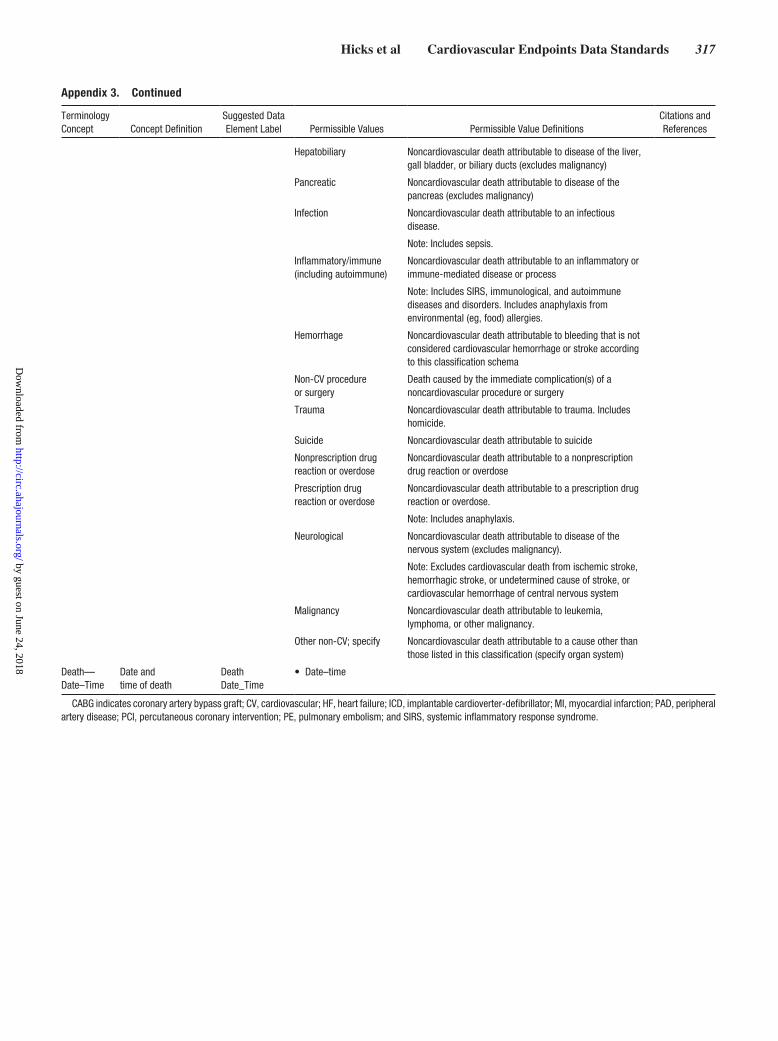
Hicks et al Cardiovascular Endpoints Data Standards 317
Hepatobiliary Noncardiovascular death attributable to disease of the liver, gall bladder, or biliary ducts (excludes malignancy)
Pancreatic Noncardiovascular death attributable to disease of the pancreas (excludes malignancy)
Infection Noncardiovascular death attributable to an infectious disease.
Note: Includes sepsis.
Inflammatory/immune (including autoimmune)
Noncardiovascular death attributable to an inflammatory or immune-mediated disease or process
Note: Includes SIRS, immunological, and autoimmune diseases and disorders. Includes anaphylaxis from environmental (eg, food) allergies.
Hemorrhage Noncardiovascular death attributable to bleeding that is not considered cardiovascular hemorrhage or stroke according to this classification schema
Non-CV procedure or surgery
Death caused by the immediate complication(s) of a noncardiovascular procedure or surgery
Trauma Noncardiovascular death attributable to trauma. Includes homicide.
Suicide Noncardiovascular death attributable to suicide
Nonprescription drug reaction or overdose
Noncardiovascular death attributable to a nonprescription drug reaction or overdose
Prescription drug reaction or overdose
Noncardiovascular death attributable to a prescription drug reaction or overdose.
Note: Includes anaphylaxis.
Neurological Noncardiovascular death attributable to disease of the nervous system (excludes malignancy).
Note: Excludes cardiovascular death from ischemic stroke, hemorrhagic stroke, or undetermined cause of stroke, or cardiovascular hemorrhage of central nervous system
Malignancy Noncardiovascular death attributable to leukemia, lymphoma, or other malignancy.
Other non-CV; specify Noncardiovascular death attributable to a cause other than those listed in this classification (specify organ system)
Death— Date–Time
Date and time of death
Death Date_Time
• Date–time
CABG indicates coronary artery bypass graft; CV, cardiovascular; HF, heart failure; ICD, implantable cardioverter-defibrillator; MI, myocardial infarction; PAD, peripheral artery disease; PCI, percutaneous coronary intervention; PE, pulmonary embolism; and SIRS, systemic inflammatory response syndrome.
Appendix 3. Continued
Terminology Concept Concept Definition
Suggested Data Element Label Permissible Values Permissible Value Definitions
Citations and References
by guest on June 24, 2018http://circ.ahajournals.org/
Dow
nloaded from

318 Circulation July 28, 2015
Appendix 4. Myocardial Infarction
Terminology Concept Concept Definition
Suggested Data Element Label
Permissible Values
Permissible Value Definitions
Citations and References
Acute MI Clinical syndrome where there is evidence of myocardial necrosis in a clinical setting consistent with acute myocardial ischemia.
Acute Myocardial Infarction Indicator
• Yes• No
Thygesen K, Alpert JS, Jaffe AS, et al. Third universal definition of myocardial infarction. J Am Coll Cardiol. 2012;60:1581–98.14
Thygesen K, Alpert JS, Jaffe AS, et al. Third universal definition of myocardial infarction. Circulation. 2012;126:2020–35.13
Acute MI—Date–Time
Date and time of onset of acute MI.
Acute Myocardial Infarction Date_Time
• Date–time
Acute MI—Type Type of acute MI, classified according to the Joint ESC/ACCF/AHA/WHF Joint Task Force for the Universal Definition of Myocardial Infarction.
Note: cTn-I or -T is the preferred biomarker. If a cTn assay is not available, the best alternative is CK-MB (measured by mass assay).
Acute Myocardial Infarction Type
• Type 1: spontaneous
• Type 2: ischemic imbalance
• Type 3: death, no biomarkers
• Type 4a: PCI related
• Type 4b: stent thrombosis
• Type 4c: stent restenosis
• Type 5: CABG related
Thygesen K, Alpert JS, Jaffe AS, et al. Third universal definition of myocardial infarction. J Am Coll Cardiol. 2012;60:1581–98.14
Thygesen K, Alpert JS, Jaffe AS, et al. Third universal definition of myocardial infarction. Circulation. 2012;126:2020–35.13
Type 1: spontaneous Spontaneous clinical syndrome related to atherosclerotic plaque rupture, ulceration, fissuring, erosion, or dissection, with resulting intraluminal thrombus, and leading to decreased myocardial blood flow or distal platelet emboli with ensuing myocyte necrosis. This classification requires a) Detection of a rise and/or fall of [cardiac biomarker] values (preferably cTn) with at least 1 value >99th percentile of the URL and b) At least 1 of the following:
1) Symptoms of myocardial ischemia 2) New or presumed new significant
ST-segment–T wave (ST–T) changes or new LBBB on the ECG
3) Development of pathological Q waves on the ECG
4) Imaging evidence of new loss of viable myocardium or new regional wall motion abnormality
5) Identification of an intracoronary thrombus by angiography or autopsy.
Notes: One or more coronary arteries may be involved. The patient may have underlying severe CAD but on occasion may have nonobstructive CAD.
(Continued)
by guest on June 24, 2018http://circ.ahajournals.org/
Dow
nloaded from
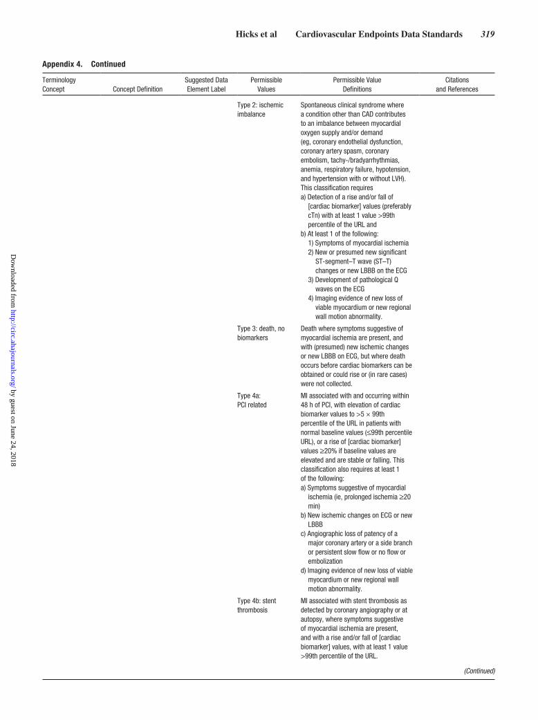
Hicks et al Cardiovascular Endpoints Data Standards 319
Type 2: ischemic imbalance
Spontaneous clinical syndrome where a condition other than CAD contributes to an imbalance between myocardial oxygen supply and/or demand (eg, coronary endothelial dysfunction, coronary artery spasm, coronary embolism, tachy-/bradyarrhythmias, anemia, respiratory failure, hypotension, and hypertension with or without LVH). This classification requiresa) Detection of a rise and/or fall of
[cardiac biomarker] values (preferably cTn) with at least 1 value >99th percentile of the URL and
b) At least 1 of the following: 1) Symptoms of myocardial ischemia 2) New or presumed new significant
ST-segment–T wave (ST–T) changes or new LBBB on the ECG
3) Development of pathological Q waves on the ECG
4) Imaging evidence of new loss of viable myocardium or new regional wall motion abnormality.
Type 3: death, no biomarkers
Death where symptoms suggestive of myocardial ischemia are present, and with (presumed) new ischemic changes or new LBBB on ECG, but where death occurs before cardiac biomarkers can be obtained or could rise or (in rare cases) were not collected.
Type 4a: PCI related
MI associated with and occurring within 48 h of PCI, with elevation of cardiac biomarker values to >5 × 99th percentile of the URL in patients with normal baseline values (≤99th percentile URL), or a rise of [cardiac biomarker] values ≥20% if baseline values are elevated and are stable or falling. This classification also requires at least 1 of the following: a) Symptoms suggestive of myocardial
ischemia (ie, prolonged ischemia ≥20 min)
b) New ischemic changes on ECG or new LBBB
c) Angiographic loss of patency of a major coronary artery or a side branch or persistent slow flow or no flow or embolization
d) Imaging evidence of new loss of viable myocardium or new regional wall motion abnormality.
Type 4b: stent thrombosis
MI associated with stent thrombosis as detected by coronary angiography or at autopsy, where symptoms suggestive of myocardial ischemia are present, and with a rise and/or fall of [cardiac biomarker] values, with at least 1 value >99th percentile of the URL.
Appendix 4. Continued
Terminology Concept Concept Definition
Suggested Data Element Label
Permissible Values
Permissible Value Definitions
Citations and References
(Continued)
by guest on June 24, 2018http://circ.ahajournals.org/
Dow
nloaded from
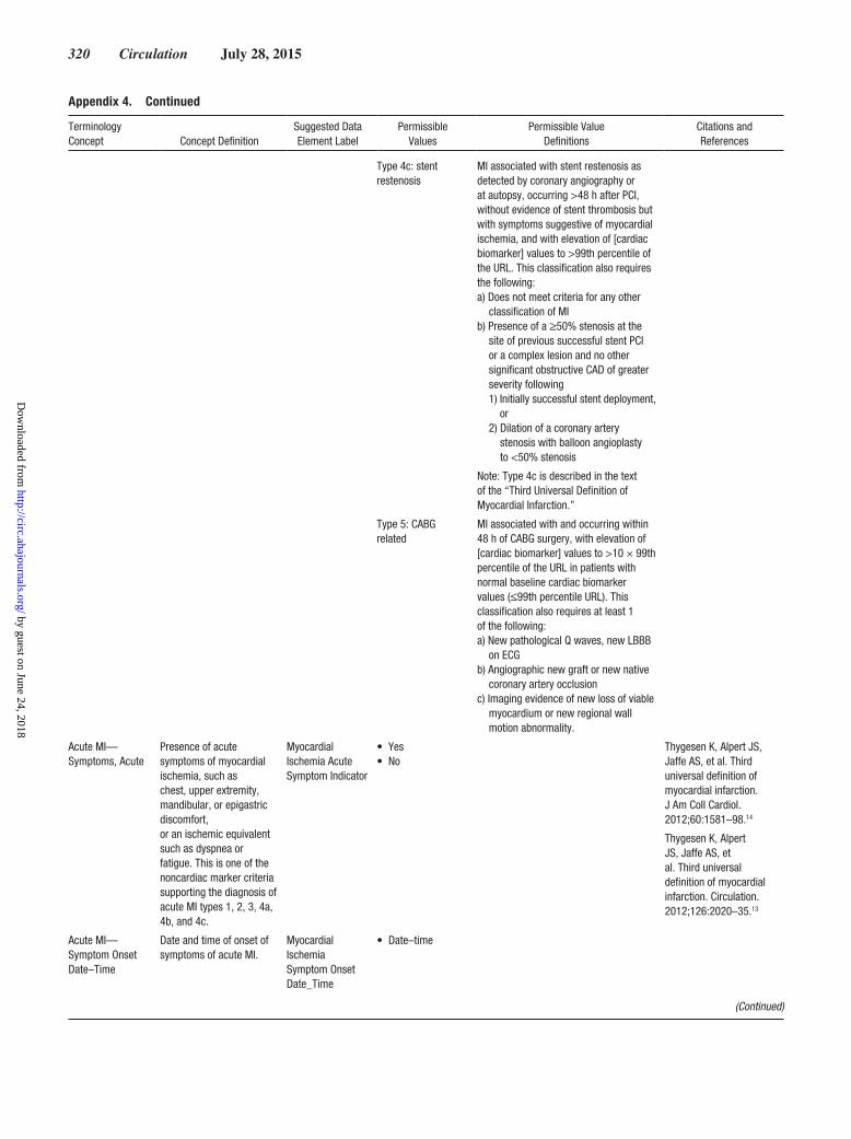
320 Circulation July 28, 2015
Type 4c: stent restenosis
MI associated with stent restenosis as detected by coronary angiography or at autopsy, occurring >48 h after PCI, without evidence of stent thrombosis but with symptoms suggestive of myocardial ischemia, and with elevation of [cardiac biomarker] values to >99th percentile of the URL. This classification also requires the following: a) Does not meet criteria for any other
classification of MI b) Presence of a ≥50% stenosis at the
site of previous successful stent PCI or a complex lesion and no other significant obstructive CAD of greater severity following1) Initially successful stent deployment,
or2) Dilation of a coronary artery
stenosis with balloon angioplasty to <50% stenosis
Note: Type 4c is described in the text of the “Third Universal Definition of Myocardial Infarction.”
Type 5: CABG related
MI associated with and occurring within 48 h of CABG surgery, with elevation of [cardiac biomarker] values to >10 × 99th percentile of the URL in patients with normal baseline cardiac biomarker values (≤99th percentile URL). This classification also requires at least 1 of the following: a) New pathological Q waves, new LBBB
on ECG b) Angiographic new graft or new native
coronary artery occlusion c) Imaging evidence of new loss of viable
myocardium or new regional wall motion abnormality.
Acute MI—Symptoms, Acute
Presence of acute symptoms of myocardial ischemia, such as chest, upper extremity, mandibular, or epigastric discomfort, or an ischemic equivalent such as dyspnea or fatigue. This is one of the noncardiac marker criteria supporting the diagnosis of acute MI types 1, 2, 3, 4a, 4b, and 4c.
Myocardial Ischemia Acute Symptom Indicator
• Yes• No
Thygesen K, Alpert JS, Jaffe AS, et al. Third universal definition of myocardial infarction. J Am Coll Cardiol. 2012;60:1581–98.14
Thygesen K, Alpert JS, Jaffe AS, et al. Third universal definition of myocardial infarction. Circulation. 2012;126:2020–35.13
Acute MI— Symptom Onset Date–Time
Date and time of onset of symptoms of acute MI.
Myocardial Ischemia Symptom Onset Date_Time
• Date–time
(Continued)
Appendix 4. Continued
Terminology Concept Concept Definition
Suggested Data Element Label
Permissible Values
Permissible Value Definitions
Citations and References
by guest on June 24, 2018http://circ.ahajournals.org/
Dow
nloaded from

Hicks et al Cardiovascular Endpoints Data Standards 321
Acute MI—Acute Ischemic Changes on ECG
Presence of new or presumed new significant ST-segment–T wave (ST-T) changes or new LBBB consistent with acute myocardial ischemia. This is one of the noncardiac marker criteria supporting the diagnosis of acute MI types 1, 2, 3, and 4.
Acute Ischemic ECG Change Type
• Ischemic changes on ECG
• LBBB
Thygesen K, Alpert JS, Jaffe AS, et al. Third universal definition of myocardial infarction. J Am Coll Cardiol. 2012;60:1581–98.14
Thygesen K, Alpert JS, Jaffe AS, et al. Third universal definition of myocardial infarction. Circulation. 2012;126:2020–35.13
Ischemic changes on ECG
In the absence of LVH and LBBB pattern (or other confounder such as a paced rhythm) on ECG, either a) new (or presumed new) ST elevation at the J point in 2 contiguous leads with the following cut points: ≥0.1 mV in all leads other than leads V2 to V3 where the following cut points apply: ≥0.2 mV in men ≥40 y of age; ≥0.25 mV in men <40 y of age, or ≥0.15 mV in women; or b) new (or presumed new) horizontal or downsloping ST-segment depression ≥0.05 mV in 2 contiguous leads and/or T inversion ≥0.1 mV in 2 contiguous leads with prominent R wave or R/S ratio >1.
LBBB New (or presumed new) LBBB pattern on ECG.
Acute MI—New Q Waves on ECG
Presence of new or presumed new pathological Q waves consistent with MI. This is one of the noncardiac marker criteria supporting the diagnosis of acute MI types 1, 2, 4, and 5.
ECG Change, New Q-Wave Indicator
• New Q waves New (or presumed new) a) Q wave in leads V
2 to V3 ≥0.02 s or QS complex in leads V2 and V3; b) Q wave ≥0.03 s and ≥0.1 mV deep or QS complex in leads I, II, aVL, aVF, or V4 to V6 in any 2 leads of a contiguous lead grouping (I, aVL; V1 to V6; II, III, aVF; V7 to V9); or c) R wave ≥0.04 s in V1 to V2 and R/S ≥1 with a concordant positive T wave in the absence of a conduction defect.
Thygesen K, Alpert JS, Jaffe AS, et al. Third universal definition of myocardial infarction. J Am Coll Cardiol. 2012;60:1581–98.14
Thygesen K, Alpert JS, Jaffe AS, et al. Third universal definition of myocardial infarction. Circulation. 2012;126:2020–35.13
Acute MI—Coronary Thrombus Present
Presence of thrombus in a major epicardial vessel consistent with an acute MI. This is one of the noncardiac marker criteria supporting the diagnosis of acute MI type 1.
Identification of Coronary Artery Thrombus With Acute Myocardial Infarction
• Thrombus on angiography
• Thrombus at autopsy
Thygesen K, Alpert JS, Jaffe AS, et al. Third universal definition of myocardial infarction. J Am Coll Cardiol. 2012;60:1581–98.14
Thygesen K, Alpert JS, Jaffe AS, et al. Third universal definition of myocardial infarction. Circulation. 2012;126:2020–35.13
Thrombus on angiography
In the patient with a presumed acute STEMI, the angiographic appearance of thrombus (typically a filling defect) on angiography. This includes the aspiration of thrombus from the infarct vessel before coronary intervention during primary PCI for acute STEMI.
(Continued)
Appendix 4. Continued
Terminology Concept Concept Definition
Suggested Data Element Label
Permissible Values
Permissible Value Definitions
Citations and References
by guest on June 24, 2018http://circ.ahajournals.org/
Dow
nloaded from

322 Circulation July 28, 2015
Thrombus at autopsy
Identification of thrombus in a major epicardial vessel at autopsy.
Acute MI—Change in Noninvasive Imaging
Demonstration of a new change in myocardial viability or function consistent with MI. This is one of the noncardiac marker criteria supporting the diagnosis of acute MI types 1, 2, 4a, and 5.
Change in Non-Invasive Imaging Type
• New loss of viable myocardium
• New regional wall motion abnormality
Thygesen K, Alpert JS, Jaffe AS, et al. Third universal definition of myocardial infarction. J Am Coll Cardiol. 2012;60:1581–98.14
Thygesen K, Alpert JS, Jaffe AS, et al. Third universal definition of myocardial infarction. Circulation. 2012;126:2020–35.13
New loss of viable myocardium
Noninvasive imaging evidence of a loss of viable myocardium when compared with the most recent previous noninvasive imaging study.
New regional wall motion abnormality
Noninvasive imaging evidence of a decrease in regional wall motion contractility compared with the most recent previous noninvasive imaging study.
Acute MI—PCI Angiographic Complication
Occurrence of an adverse angiographic finding during PCI consistent with acute myocardial ischemia. This is one of the noncardiac marker criteria supporting the diagnosis of acute MI type 4a.
PCI Acute Angiographic Complication
• Loss of major coronary
• Loss of side branch
• Slow flow/no flow/embolization
Thygesen K, Alpert JS, Jaffe AS, et al. Third universal definition of myocardial infarction. J Am Coll Cardiol. 2012;60:1581–98.14
Thygesen K, Alpert JS, Jaffe AS, et al. Third universal definition of myocardial infarction. Circulation. 2012;126:2020–35.13
Loss of major coronary
Angiographic loss of patency of a major epicardial vessel.
Loss of side branch
Angiographic loss of patency of a side branch.
Slow flow/no flow/embolization
Angiographic reduction of flow into the coronary microcirculation.
Acute MI—Acute Vessel Occlusion After CABG
Angiographic documentation of a new CABG or new native coronary artery occlusion within 48 h of CABG surgery. This is one of the noncardiac marker criteria supporting the diagnosis of acute MI type 5.
Vessel Occlusion After Coronary Artery Bypass Graft Surgery Indicator
• Yes• No
Thygesen K, Alpert JS, Jaffe AS, et al. Third universal definition of myocardial infarction. J Am Coll Cardiol. 2012;60:1581–98.14
Thygesen K, Alpert JS, Jaffe AS, et al. Third universal definition of myocardial infarction. Circulation. 2012;126:2020–35.13
(Continued)
Appendix 4. Continued
Terminology Concept Concept Definition
Suggested Data Element Label
Permissible Values
Permissible Value Definitions
Citations and References
by guest on June 24, 2018http://circ.ahajournals.org/
Dow
nloaded from

Hicks et al Cardiovascular Endpoints Data Standards 323
Prior MI Presence of any one of the following criteria meets the diagnosis for prior MI (before study initiation): a) Pathological Q
waves with or without symptoms in the absence of nonischemic causes
b) Imaging evidence of a region of loss of viable myocardium that is thinned and/or fails to contract, in the absence of a nonischemic cause
c) Pathological findings of a prior MI.
Prior Myocardial Infarction Indicator
• Yes• No
Thygesen K, Alpert JS, Jaffe AS, et al. Third universal definition of myocardial infarction. J Am Coll Cardiol. 2012;60:1581–98.14
Thygesen K, Alpert JS, Jaffe AS, et al. Third universal definition of myocardial infarction. Circulation. 2012;126:2020–35.13
Cardiac Biomarker—Name
Name of cardiac biomarker test, per LOINC nomenclature.
Cardiac Biomarker Test
• LOINC Test Name
LOINC®. Available at www.loinc.org.50
Cardiac Biomarker—Value
Serum concentration result of a cardiac biomarker test.
Cardiac Biomarker Value
• Value
Cardiac Biomarker—Unit
Unit of measure of the result of a cardiac biomarker test.
Cardiac Biomarker Unit
• nanogram per liter
• nanogram per milliliter
• pictogram per milliliter
Cardiac Biomarker—Date–Time
Date and time a specimen was obtained for the assay of a cardiac biomarker.
Cardiac Biomarker Date_Time
• Date–time
Cardiac Biomarker—99% URL
The 99% URL of a cardiac biomarker.
Cardiac Biomarker 99% URL Value
• Value
Cardiac Biomarker—99% URL Unit
Unit of measure of the 99% URL of a cardiac biomarker.
Cardiac Biomarker 99% URL Unit
• nanogram per liter
• nanogram per milliliter
• pictogram per milliliter
Cardiac Biomarker—ULN
The ULN of a cardiac biomarker.
Cardiac Biomarker ULN Value
• Value
Cardiac Biomarker—ULN Unit
Unit of measure of the ULN of a cardiac biomarker.
Cardiac Biomarker ULN Unit
• nanogram per liter
• nanogram per milliliter
• pictogram per milliliter
ACC indicates American College of Cardiology; ACCF, American College of Cardiology Foundation; AHA, American Heart Association; CABG, coronary artery bypass graft; CAD, coronary artery disease; CK-MB, creatinine kinase MB; cTn, cardiac troponin; ECG, electrocardiogram; ESC, European Society of Cardiology; HF, heart failure; ICD, implantable cardioverter-defibrillator; LBBB, left bundle-branch block; LOINC, Logical Observation Identifiers Names and Codes; LVH, left ventricular hypertrophy; MI, myocardial infarction; PCI, percutaneous coronary intervention; STEMI, ST-segment elevation myocardial infarction; ULN, upper limit of normal; URL, upper reference limit; and WHF, World Heart Federation.
Appendix 4. Continued
Terminology Concept Concept Definition
Suggested Data Element Label
Permissible Values
Permissible Value Definitions
Citations and References
by guest on June 24, 2018http://circ.ahajournals.org/
Dow
nloaded from

324 Circulation July 28, 2015
Appendix 5. Hospitalization for Unstable Angina
Terminology Concept Concept DefinitionSuggested Data Element Label
Permissible Values
Permissible Value Definitions
Citations and References
UA Hospitalization Unscheduled hospitalization for the management of UA, occurring within 24 h of the most recent symptoms.
Hospitalization is defined as an admission to an inpatient unit or a visit to an ED that results in at least a 24-h stay (or a change in calendar date if the hospital admission or discharge times are not available).
This classification requires that 4 separate criteria be met: a) Worsening ischemic
discomfort b) Unscheduled hospitalizationc) Objective evidence of
myocardial ischemia d) Negative cardiac
biomarkers
Note: Escalation of pharmacotherapy for myocardial ischemia, while considered supportive evidence, is not sufficient to qualify as UA hospitalization without objective evidence of ischemia.
Admission for suspected UA does not qualify as a UA hospitalization if a noncardiac or nonischemic etiology is subsequently identified.
An ischemic event meeting the criteria for acute MI is not a UA hospitalization.
Planned hospitalization or rehospitalization for performance of an elective revascularization procedure (eg, positive stress test, staged revascularization) is not a UA hospitalization.
Hospitalization with revascularization of CAD identified during elective cardiac catheterization does not qualify as a UA hospitalization.
Unstable Angina Event Indicator
• Yes• No
Adapted from Cannon CP, Brindis RG, Chaitman BR, et al. 2013 ACCF/AHA key data elements and definitions for measuring the clinical management and outcomes of patients with acute coronary syndromes and coronary artery disease. A report of the American College of Cardiology Foundation/American Heart Association Task Force on Clinical Data Standards (Writing Committee to Develop Acute Coronary Syndromes and Coronary Artery Disease Clinical Data Standards). Circulation. 2013;127:1052–89.16
UA Hospitalization— Date–Time
Date and time of the presentation of the patient to the hospital for management of UA.
Date and time of presentation is defined as the date and time of registration in the ED (or hospital, if the patient is not seen in the ED).
Unstable Angina Hospitalization Date_Time
• Date–time
(Continued)
by guest on June 24, 2018http://circ.ahajournals.org/
Dow
nloaded from

Hicks et al Cardiovascular Endpoints Data Standards 325
UA Hospitalization—Symptoms of Ischemia
Documentation of ischemic discomfort (angina, or symptoms thought to be equivalent) ≥10 min in duration occurring eithera) At rest, orb) In an accelerating pattern
with frequent episodes associated with progressively decreasing exercise capacity.
Unstable Angina Ischemic Discomfort Indicator
• Yes• No
Adapted from Cannon CP, Brindis RG, Chaitman BR, et al. 2013 ACCF/AHA key data elements and definitions for measuring the clinical management and outcomes of patients with acute coronary syndromes and coronary artery disease. A report of the American College of Cardiology Foundation/American Heart Association Task Force on Clinical Data Standards (Writing Committee to Develop Acute Coronary Syndromes and Coronary Artery Disease Clinical Data Standards). Circulation. 2013;127:1052–89.16
UA Hospitalization—Evidence of Ischemia
Documentation of objective evidence of new or worsening coronary ischemia.
Unstable Angina Ischemic Evidence Type
• Changes on resting ECG
• Inducible myocardial ischemia
• Coronary lesion on angiography
• Coronary revascularization
Changes on resting ECG
New or worsening ST or T-wave changes on resting ECG (in the absence of confounders, such as LBBB or LVH), defined as eithera) Transient ST elevation
(duration <20 min): new ST elevation at the J point ≥0.1 mV in any 2 contiguous leads (other than leads V
2 to V3); in leads V2 to V3, the following cut points apply: ≥0.2 mV in men ≥40 y of age, ≥0.25 mV in men <40 y of age, and ≥0.15 mV in women.
b) ST depression and T-wave changes: new horizontal or downsloping ST depression ≥0.05 mV in 2 contiguous leads and/or new T inversion ≥0.3 mV in 2 contiguous leads with prominent R wave or R/S ratio >1.
Appendix 5. Continued
Terminology Concept Concept DefinitionSuggested Data Element Label
Permissible Values
Permissible Value Definitions
Citations and References
(Continued)
by guest on June 24, 2018http://circ.ahajournals.org/
Dow
nloaded from

326 Circulation July 28, 2015
Inducible myocardial ischemia
Definite evidence of inducible myocardial ischemia believed to be responsible for the myocardial ischemic symptoms/signs, demonstrated by any of the following:a) An early positive exercise
stress test result, defined as ST elevation or ≥2 mm ST depression before 5 METs
b) Stress echocardiography (reversible wall motion abnormality)
c) Myocardial scintigraphy (reversible perfusion defect)
d) MRI (myocardial perfusion deficit under pharmacological stress)
Coronary lesion on angiography
Angiographic evidence of new or worsening lesion ≥70% (≥50% for left main coronary artery lesion) and/or thrombus in an epicardial coronary artery believed to be responsible for the myocardial ischemic symptoms/signs.
Coronary revascularization
Need for coronary revascularization procedure (PCI or CABG) for the presumed culprit lesion(s). This criterion would be fulfilled if revascularization was undertaken during the unscheduled hospitalization or subsequent to transfer to another institution without interceding home discharge.
UA Hospitalization—MI Excluded
Exclusion of the diagnosis of MI as the reason for hospitalization, including negative cardiac biomarkers and no other evidence of acute MI.
Myocardial Infarction Exclusion Indicator
• Yes• No
ACCF indicates American College of Cardiology Foundation; AHA, American Heart Association; CABG, coronary artery bypass graft; CAD, coronary artery disease; ECG, electrocardiogram; ED, emergency department; LBBB, left bundle-branch block; LVH, left ventricular hypertrophy; METs, metabolic equivalents; MI, myocardial infarction, MRI, magnetic resonance imaging; PCI, percutaneous coronary intervention; and UA, unstable angina.
Appendix 5. Continued
Terminology Concept Concept DefinitionSuggested Data Element Label
Permissible Values
Permissible Value Definitions
Citations and References
by guest on June 24, 2018http://circ.ahajournals.org/
Dow
nloaded from

Hicks et al Cardiovascular Endpoints Data Standards 327
Appendix 6. Transient Ischemic Attack and Stroke
Terminology Concept Concept Definition
Suggested Data Element Name
Permissible Values
Permissible Value Definitions
Citations and References
Stroke An acute episode of focal or global neurological dysfunction caused by brain, spinal cord, or retinal vascular injury as a result of hemorrhage or infarction.
Stroke Indicator • Yes• No
Adapted from Sacco RL, Kasner SE, Broderick JP, et al. An updated definition of stroke for the 21st century: a statement for healthcare professionals from the American Heart Association/American Stroke Association. Stroke. 2013;44:2064–89.19
Stroke— Date–Time
Date and time of the onset of a stroke.
Stroke Date_Time • Date–time
Stroke—Type Categorical description of stroke type, classified into 1 of 3 mutually exclusive categories (ischemic, hemorrhagic, undetermined).
Stroke Type • Ischemic• Hemorrhagic• Undetermined
Adapted from Sacco RL, Kasner SE, Broderick JP, et al. An updated definition of stroke for the 21st century: a statement for healthcare professionals from the American Heart Association/American Stroke Association. Stroke. 2013;44:2064–89.19
Ischemic An acute episode of focal cerebral, spinal, or retinal dysfunction caused by infarction of central nervous system tissue.
Note: Hemorrhage may be a consequence of ischemic stroke. In this situation, the stroke is an ischemic stroke with hemorrhagic transformation and not a hemorrhagic stroke.
Hemorrhagic Hemorrhagic: An acute episode of focal or global cerebral or spinal dysfunction caused by intraparenchymal, intraventricular, or subarachnoid hemorrhage.
Note: Subdural hematomas are intracranial hemorrhagic events and not strokes.
Undetermined Undetermined: An acute episode of focal or global neurological dysfunction caused by presumed brain, spinal cord, or retinal vascular injury as a result of hemorrhage or infarction but with insufficient information to allow categorization as either ischemic or hemorrhagic.
Modified Rankin Scale
Validated scale for assessment of disability following a stroke.
Note: Measure disability with the modified Rankin Scale at each visit and 90 d after the event.
Modified Rankin Scale Value
• 0• 1• 2• 3• 4• 5• 6
Rankin J. Cerebral vascular accidents in patients over the age of 60. Scott Med J. 1957;2:200–15.26
van Swieten J, Koudstaal P, Visser M, et al. Interobserver agreement for the assessment of handicap in stroke patients. Stroke. 1988;19:604–7.51
(Continued)
by guest on June 24, 2018http://circ.ahajournals.org/
Dow
nloaded from

328 Circulation July 28, 2015
0 No symptoms at all.
1 No significant disability despite symptoms; able to carry out all usual duties and activities.
2 Slight disability; unable to perform all previous activities but able to look after own affairs without assistance.
3 Moderate disability; requiring some help but able to walk without assistance.
4 Moderately severe disability; unable to walk without assistance and unable to attend to own bodily needs without assistance.
5 Severe disability; bedridden, incontinent, and requiring constant nursing care and attention.
6 Dead.
Modified Rankin Scale—Date–Time
Date and time of neurological assessment to determine modified Rankin scale grade.
Rankin Scale Date_Time
Date–time
TIA Transient episode of focal neurological dysfunction caused by brain, spinal cord, or retinal ischemia without acute infarction.
Note: Persistence of symptoms is an acceptable indicator of acute infarction. If it is used, duration of symptom persistence that will be used to distinguish between transient ischemia and acute infarction should be defined for any clinical trial in which it is used.
Note: AHA/ASA recommends duration ≥24 h as an operational definition of persisting symptoms of stroke rather than TIA, based mostly on consensual practice rather than objective evidence.
Transient Ischemic Attack Indicator
• Yes• No
Easton JD, Saver JL, Albers GW, et al. Definition and evaluation of transient ischemic attack: a scientific statement for healthcare professionals from the American Heart Association/American Stroke Association Stroke Council; Council on Cardiovascular Surgery and Anesthesia; Council on Cardiovascular Radiology and Intervention; Council on Cardiovascular Nursing; and the Interdisciplinary Council on Peripheral Vascular Disease. Stroke. 2009;40:2276–93.9
Sacco RL, Kasner SE, Broderick JP, et al. An updated definition of stroke for the 21st century: a statement for healthcare professionals from the American Heart Association/American Stroke Association. Stroke. 2013;44:2064–89.19
TIA—Date–Time Date and time of the onset of a TIA.
Transient Ischemic Attack Date_Time
Date–time
AHA indicates American Heart Association; ASA, American Stroke Association; and TIA, transient ischemic attack.
Appendix 6. Continued
Terminology Concept Concept Definition
Suggested Data Element Name
Permissible Values
Permissible Value Definitions
Citations and References
by guest on June 24, 2018http://circ.ahajournals.org/
Dow
nloaded from

Hicks et al Cardiovascular Endpoints Data Standards 329
Appendix 7. Heart Failure Event
Terminology Concept Concept Definition
Suggested Data Element Name
Permissible Values
Permissible Value Definitions
Citations and References
HF Event Presentation of the patient for an urgent, unscheduled clinic/office/ED visit or hospital admission, with a primary diagnosis of HF, where the patient exhibits new or worsening symptoms of HF on presentation, has objective evidence of new or worsening HF, and receives initiation or intensification of treatment specifically for HF.
Objective evidence consists of at least 2 physical examination findings OR at least 1 physical examination finding and at least 1 laboratory criterion of new or worsening HF on presentation.
Heart Failure Endpoint Event
• Yes• No
http://www.cdisc.org/therapeutic49
HF Event—Date–Time
Date and time of the presentation of a patient for management of a HF event.
Heart Failure Endpoint Event Date_Time
• Date–time
HF Event—Encounter Type
The type of healthcare encounter of a patient who presents for the management of new-onset or worsening HF.
Heart Failure Endpoint Event Healthcare Encounter Type
• HF hospitalization• Urgent HF visit
http://www.cdisc.org/therapeutic49
HF hospitalization An event where the patient is admitted to the hospital with a primary diagnosis of HF where the length of stay is at least 24 h (or extends over a calendar date if the hospital admission and discharge times are unavailable), where the patient exhibits new or worsening symptoms of HF on presentation, has objective evidence of new or worsening HF, and receives initiation or intensification of treatment specifically for HF.
Urgent HF visit An event where the patient has an urgent, unscheduled office/practice/ED visit for a primary diagnosis of HF but is not admitted to the hospital, where the patient exhibits new or worsening symptoms of HF on presentation, has objective evidence of new or worsening HF, and receives initiation or intensification of treatment specifically for HF, with the exception that changes to oral diuretic therapy do not qualify as initiation or intensification of treatment.
(Continued)
by guest on June 24, 2018http://circ.ahajournals.org/
Dow
nloaded from

330 Circulation July 28, 2015
HF Event—Symptoms, New or Worsening
Documentation of new or worsening symptoms of HF on patient presentation.
Criterion for new or worsening symptoms due to HF is to have at least 1 of the following on presentation: a) Dyspnea b) Decreased exercise tolerance c) Fatigue d) Worsened end-organ
perfusion e) Volume overload
Heart Failure Symptoms, New or Worsening
• Dyspnea• Decreased
exercise tolerance• Fatigue• Worsened end-
organ perfusion• Volume overload
Adapted from Radford MJ, Arnold JM, Bennett SJ, et al. ACC/AHA key data elements and definitions for measuring the clinical management and outcomes of patients with chronic heart failure: a report of the American College of Cardiology/American Heart Association Task Force on Clinical Data Standards (Writing Committee to Develop Heart Failure Clinical Data Standards). Developed in collaboration with the American College of Chest Physicians and the International Society for Heart and Lung Transplantation. Circulation. 2005;112:1888–916.52
Dyspnea Includes dyspnea on exertion, dyspnea at rest, orthopnea, and paroxysmal nocturnal dyspnea.
Decreased exercise tolerance
Decreased exercise tolerance: reduced ability to withstand or participate in activities that induce physical or mental exertion.
Fatigue Unusual tiredness and inability to perform usual activities.
Worsened end-organ perfusion
Decreased blood supply to the vital organs (kidney, liver, lungs, heart, and brain).
Note: Parameters must be specified and described by the study protocol.
Volume overload Excessive accumulation of intravascular fluid resulting from compromised regulatory mechanisms.
Note: Parameters must be specified and described by the study protocol.
Appendix 7. Continued
Terminology Concept Concept Definition
Suggested Data Element Name
Permissible Values
Permissible Value Definitions
Citations and References
(Continued) by guest on June 24, 2018
http://circ.ahajournals.org/D
ownloaded from
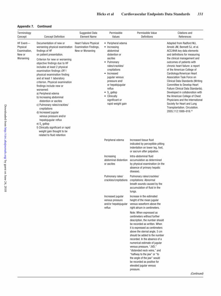
Hicks et al Cardiovascular Endpoints Data Standards 331
HF Event—Physical Examination, New or Worsening
Documentation of new or worsening physical examination findings of HF on patient presentation.
Criterion for new or worsening objective findings due to HF includes at least 2 physical examination findings OR 1 physical examination finding and at least 1 laboratory criterion. Physical examination findings include new or worsened: a) Peripheral edema b) Increasing abdominal
distention or ascites c) Pulmonary rales/crackles/
crepitations d) Increased jugular
venous pressure and/or hepatojugular reflux
e) S3 gallop f) Clinically significant or rapid
weight gain thought to be related to fluid retention
Heart Failure Physical Examination Findings, New or Worsening
• Peripheral edema• Increasing
abdominal distention or ascites
• Pulmonary rales/crackles/crepitations
• Increased jugular venous pressure and/or hepatojugular reflux
• S3 gallop• Clinically
significant or rapid weight gain
Adapted from Radford MJ, Arnold JM, Bennett SJ, et al. ACC/AHA key data elements and definitions for measuring the clinical management and outcomes of patients with chronic heart failure: a report of the American College of Cardiology/American Heart Association Task Force on Clinical Data Standards (Writing Committee to Develop Heart Failure Clinical Data Standards). Developed in collaboration with the American College of Chest Physicians and the International Society for Heart and Lung Transplantation. Circulation. 2005;112:1888–916.52
Peripheral edema Increased tissue fluid indicated by perceptible pitting indentation on lower leg, foot, or sacrum after palpation.
Increasing abdominal distention or ascites
Intra-abdominal fluid accumulation as determined by physical examination (in the absence of primary hepatic disease).
Pulmonary rales/crackles/crepitations
Pulmonary rales/crackles/crepitations: Abnormal breath sounds caused by the accumulation of fluid in the lungs.
Increased jugular venous pressure and/or hepatojugular reflux
Increase in the estimated height of the mean jugular venous waveform above the right atrium in centimeters.
Note: When expressed as centimeters without further description, the number should be recorded as written. When it is expressed as centimeters above the sternal angle, 5 cm should be added to the number recorded. In the absence of a numerical estimate of jugular venous pressure, “JVD,” “distended neck veins,” and “halfway to the jaw” or “to the angle of the jaw” would be recorded as positive for elevated jugular venous pressure.
Appendix 7. Continued
Terminology Concept Concept Definition
Suggested Data Element Name
Permissible Values
Permissible Value Definitions
Citations and References
(Continued)
by guest on June 24, 2018http://circ.ahajournals.org/
Dow
nloaded from

332 Circulation July 28, 2015
S3 gallop Presence of an S3 mid-diastolic heart sound.
Clinically significant or rapid weight gain
Weight gain thought to be related to fluid retention.
HF Event—Laboratory Data, New or Worsening
Documentation of new or worsening laboratory evidence of HF obtained within 24 h of patient presentation. Criterion for new or worsening objective findings due to HF includes at least 2 physical examination findings OR 1 physical examination finding and at least 1 laboratory criterion. Laboratory criteria include new or worsened: a) Increased BNP/NT-proBNP b) Radiological evidence of
pulmonary congestion c) Noninvasive diagnostic
evidence of HF d) Invasive diagnostic
evidence of HF
Heart Failure Laboratory Findings, New or Worsening
• Increase in HF biomarker
• Radiological evidence of pulmonary congestion
• Noninvasive diagnostic evidence of HF
• Invasive diagnostic evidence of HF
Adapted from: Radford MJ, Arnold JM, Bennett SJ, et al. ACC/AHA key data elements and definitions for measuring the clinical management and outcomes of patients with chronic heart failure: a report of the American College of Cardiology/American Heart Association Task Force on Clinical Data Standards (Writing Committee to Develop Heart Failure Clinical Data Standards). Developed in collaboration with the American College of Chest Physicians and the International Society for Heart and Lung Transplantation. Circulation. 2005;112:1888–916.52
Increase in HF biomarker
Biomarker increase BNP/NT-pro BNP with decompensation of HF (such as BNP >500 pg/mL or NT-proBNP >2000 pg/mL). In patients with chronically elevated natriuretic peptides, a significant increase above baseline is required.
Radiological evidence of pulmonary congestion
Radiological evidence of pulmonary congestion: imaging findings consistent with increased intravascular blood volume in the lungs.
Noninvasive diagnostic evidence of HF
Noninvasive diagnostic evidence of HF: Noninvasive diagnostic evidence of clinically significant elevated left- or right-sided ventricular filling pressure or low cardiac output. For example, echocardiographic criteria could include E/e’ >15 or D-dominant pulmonary venous inflow pattern, plethoric inferior vena cava with minimal collapse on inspiration, or decreased LVOT minute stroke distance (TVI).
Invasive diagnostic evidence of HF
Invasive diagnostic evidence with right-sided catheterization of heart showing a PCWP (pulmonary artery occlusion pressure) ≥18 mm Hg, central venous pressure ≥12 mm Hg, or a cardiac index <2.2 L/min/m2.
Appendix 7. Continued
Terminology Concept Concept Definition
Suggested Data Element Name
Permissible Values
Permissible Value Definitions
Citations and References
(Continued)
by guest on June 24, 2018http://circ.ahajournals.org/
Dow
nloaded from

Hicks et al Cardiovascular Endpoints Data Standards 333
HF Event—Treatment Intensification
Initiation or intensification of treatment specifically for HF. The criterion is that the patient receives initiation or intensification of treatment specifically for HF, including at least 1 of the following: a) Augmentation in oral diuretic
therapy b) Intravenous diuretic or
vasoactive agent (eg, inotrope, vasopressor, or vasodilator)
c) Mechanical or surgical intervention
Heart Failure Therapy, Intensification
• Augmentation of oral diuretic therapy
• Intravenous diuretic, inotrope, or vasodilator therapy
• Mechanical or surgical intervention
Adapted from Radford MJ, Arnold JM, Bennett SJ, et al. ACC/AHA key data elements and definitions for measuring the clinical management and outcomes of patients with chronic heart failure: a report of the American College of Cardiology/American Heart Association Task Force on Clinical Data Standards (Writing Committee to Develop Heart Failure Clinical Data Standards). Developed in collaboration with the American College of Chest Physicians and the International Society for Heart and Lung Transplantation. Circulation. 2005;112:1888–916.52
Augmentation of oral diuretic therapy
Initiation or intensification of orally administered medication(s) that promote diuresis to treat HF.
Intravenous diuretic, inotrope, vasopressor, or vasodilator therapy
Initiation or intensification of medication(s) administered by vein to treat HF, increase production of urine, increase cardiac performance, and/or reduce cardiac preload or afterload.
Mechanical or surgical intervention
Mechanical circulatory support (eg, intra-aortic balloon pump, ventricular assist device, extracorporeal membrane oxygenation, total artificial heart) or mechanical fluid removal (eg, ultrafiltration, hemofiltration, dialysis).
Cardiogenic Shock
Sustained (>30 min) episode of systolic BP <90 mm Hg and/or cardiac index <2.2 L/min/m2 determined to be secondary to cardiac dysfunction, and/or the requirement for parenteral inotropic or vasopressor agents or mechanical support (eg, intra-aortic balloon pump, extracorporeal circulatory support, ventricular assist device) to maintain BP and cardiac index above those specified levels.
Cardiogenic Shock Indicator
• Yes• No
Cannon CP, Brindis RG, Chaitman BR, et al. 2013 ACCF/AHA key data elements and definitions for measuring the clinical management and outcomes of patients with acute coronary syndromes and coronary artery disease: a report of the American College of Cardiology Foundation/American Heart Association Task Force on Clinical Data Standards (Writing Committee to Develop Acute Coronary Syndromes and Coronary Artery Disease Clinical Data Standards). Circulation. 2013;127:1052–89.16
ACCF indicates American College of Cardiology Foundation; AHA, American Heart Association; BNP, B-type natriuretic peptide; BP, blood pressure; cm, centimeter; ED, emergency department; HF, heart failure; JVD, jugular venous distention; L/min/m2, liter per minute per square millimeter; LVOT, left ventricular outflow tract; mm Hg, millimeters of mercury; NT-pro BNP, N-terminal pro B-type natriuretic peptide; PCWP, pulmonary capillary wedge pressure; and TVI, time velocity integral.
Appendix 7. Continued
Terminology Concept Concept Definition
Suggested Data Element Name
Permissible Values
Permissible Value Definitions
Citations and References
by guest on June 24, 2018http://circ.ahajournals.org/
Dow
nloaded from

334 Circulation July 28, 2015
Appendix 8. Heart Failure Event: Additional Details
• A Heart Failure Event includes hospitalization for HF and may include urgent, unscheduled outpatient office/practice or ED visits.
– A Heart Failure Hospitalization is defined as an event in which the patient is admitted to the hospital with a primary diagnosis of HF, the length of stay is at least 24 h (or extends over a calendar date), the patient exhibits new or worsening symptoms of HF on presentation, has objective evidence of new or worsening HF, and receives initiation or intensification of treatment specifically for HF.
– An Urgent Heart Failure Visit is defined as an event in which the patient has an urgent, unscheduled office/practice or ED visit for a primary diagnosis of HF but is not admitted to the hospital and exhibits new or worsening symptoms of HF on presentation, has objective evidence of new or worsening HF, and receives initiation or intensification of treatment specifically for HF. Note that changes to oral diuretic therapy do not qualify as initiation or intensification of treatment.
• Heart Failure Symptoms: The patient may present with new or worsening symptoms due to HF on presentation. To be defined as having new or worsening symptoms due to HF, the patient should have at least ONE of the following on presentation:
–Dyspnea (dyspnea with exertion, dyspnea at rest, orthopnea, paroxysmal nocturnal dyspnea, nocturnal cough in supine position, tachypnea);
–Decreased exercise tolerance (reduced ability to perform activities that involve dynamic movement of large skeletal muscles because of symptoms of dyspnea or fatigue);
– Fatigue (usually described as feeling a lack of energy and motivation in both mental and physical activities, easily tiring and not being able to complete usual activities, and sometimes accompanied by dizziness, lightheadedness);
– Worsened end-organ perfusion (worsening cerebral, renal, liver, abdominal or gastrointestinal, peripheral circulatory function manifested by symptoms such as dizziness, lightheadedness, syncope, confusion, altered mental status, restlessness, decline in cognitive state, nausea, vomiting, abdominal pain, abdominal fullness, abdominal discomfort or abdominal tenderness, cold clammy extremities, discoloration of extremities or lips, jaundice, pain in extremities, reduced urine output, darkening of urine color, chest pain, and/or palpitations); or
– Other symptoms of volume overload (swelling of lower extremities; swelling or indentation of pressure marks in areas of fluid accumulation such as the legs, ankles, or lower back; an increase in abdominal girth, right-sided abdominal fullness, discomfort, or tenderness; an increase in body weight; oozing and development of skin breakdown in lower extremities).
• Heart Failure Signs: The patient may present with objective evidence of new or worsening HF. The objective evidence or signs of new or worsening HF should consist of at least 2 physical examination findings OR 1 physical examination finding and at least 1 laboratory criterion. Physical examination findings considered to be due to HF include new or worsened:
–Peripheral edema (swelling or pitting indentation when pressed in feet, ankles, legs, thighs, upper extremities, scrotum, presacral area, or abdominal wall).
–Increasing abdominal distention or ascites (in the absence of primary hepatic disease)
–Pulmonary rales/crackles/crepitations
–Increased jugular venous pressure and/or hepatojugular reflux
–S3 gallop
–Clinically significant or rapid weight gain thought to be related to fluid retention (usually >3–4 lb in 3–4 d).
Other physical findings such as the following may also support the diagnosis: a decline in systolic or diastolic blood pressure; orthostatic hypotension; cool, mottled or clammy skin; lip discoloration and cyanosis; tachypnea; irregular or periodic breathing pattern (eg, Cheyne-Stokes respirations); tachycardia or bradycardia; arrhythmia; irregular pulse; pulsus alternans; displaced point of maximum impulse of cardiac apex; RV heave; loud S
2; diminished S1; S4; valvular murmurs; reduced urine output; liver enlargement; physical findings compatible with pleural effusion such as decreased breath sounds and or decreased egophony; narrow pulse pressure; abnormal arterial pulse pressure response during the strain phase of the Valsalva maneuver; weight loss; and/or wheezing.29,32,33 Because these physical findings are not as common, sensitive, specific, or easily reproducible for HF as those listed in the bullet points above, they are not included in the required physical findings criteria.
• Laboratory Evidence of HF: Laboratory evidence of new or worsening HF should be obtained within 24 h of presentation.
Laboratory criteria include new or worsened:
– Increased BNP or NT-proBNP concentrations. In patients with suspected new onset of HF, or in patients with dyspnea but uncertainty of HF, measurement of BNP or NT-proBNP is useful to support the clinical diagnosis of HF.30,31,35,39,40 In acute decompensated HF, natriuretic peptide levels are usually significantly elevated (eg, BNP >500 pg/mL or NT-proBNP >2000 pg/mL). The exclusion threshold differs for patients presenting with acute onset or worsening of symptoms and for those presenting with more gradual onset of symptoms. For patients presenting with acute onset or worsening of symptoms, the optimal exclusion cut-off points are usually quoted as 300 pg/mL for NT-proBNP and 100 pg/mL for BNP levels. Lower values of natriuretic peptides usually make the diagnosis of HF less likely, and higher values have reasonably high positive predictive value in diagnosing HF, especially in dyspneic patients presenting to urgent care or ED settings. That being said, elevated plasma levels of natriuretic peptides can be seen with a variety of other cardiac and noncardiac causes. Other causes of elevated natriuretic peptide levels in the acute setting are acute coronary syndrome, atrial or ventricular arrhythmias, pulmonary embolism, cor pulmonale, renal failure, and sepsis. Other causes of an elevated natriuretic level in the nonacute setting are old age (>75 y), atrial arrhythmias, LV hypertrophy, chronic obstructive pulmonary disease, and chronic kidney disease. Conversely, natriuretic peptide levels can be lower in obese patients. In patients with chronically elevated natriuretic peptides, levels usually correlate with HF severity and prognosis, and an increase above the baseline is usually noted during acute decompensated HF. Current evidence suggests there is no specific cut-off point for an absolute or relative rise above the baseline level to diagnose a HF event in patients with chronically elevated natriuretic peptide levels.
– Radiological evidence of pulmonary congestion (CXR or other imaging modality such as CT or MRI with evidence of pulmonary venous or alveolar congestion, interstitial or pulmonary edema, pleural effusion, or cephalization of venous flow. It is important to note that CXR may also reflect evidence of cardiomegaly.)
(Continued)
by guest on June 24, 2018http://circ.ahajournals.org/
Dow
nloaded from

Hicks et al Cardiovascular Endpoints Data Standards 335
– Noninvasive diagnostic evidence of HF (echocardiography, cardiac MRI, cardiac PET scan, and nuclear imaging) can provide information about cardiac anatomy such as LV or RV function, volumes, geometry, wall motion, or valvular function, and other findings such as elevated filling pressures by Doppler echocardiography) supporting the diagnosis and etiology of HF. In systolic HF, LV ejection fraction (<50%) and LV fractional shortening are reduced (<25%). There may be regional LV wall motion abnormalities, especially in patients with ischemic HF. In patients with chronic systolic HF, LV end-diastolic size is usually increased (diameter ≥60 mm, >32 mm/m2, LV volume >97 mL/m2), as is LV end-systolic size (diameter >45 mm/>25 mm/m2, volume >43 mL/m2). Cardiac output is usually reduced, and LV outflow tract velocity time integral is reduced (<15 cm). LV diastolic dysfunction parameters may reflect abnormalities of the mitral inflow pattern, with tissue velocities (e) or the E/e’ ratio indicating the presence and degree of LV diastolic dysfunction. In cases of diastolic dysfunction, e’ is decreased (<8 cm/s septal, <10 cm/s lateral, or <9 cm/s average), suggesting delayed LV relaxation. High E/e’ ratios (>15) usually indicate high LV filling pressures, whereas low E/e’ ratios (<8) usually indicate normal LV filling pressures. A mitral inflow E/A ratio >2 suggests restrictive, high LV filling pressures. In contrast, a mitral inflow E/A ratio of <1 suggests delayed LV relaxation with normal LV filling pressures. An E/A ratio of 1–2 may be inconclusive or pseudonormal. Mitral inflow during the Valsalva maneuver may reflect a change from the pseudonormal to the impaired relaxation pattern (with a decrease in E/A ratio >0.5), suggesting high LV filling pressures (unmasked through Valsalva). An A-wave duration >30 ms usually suggests high LV filling pressures. In patients with elevated filling pressures, left atrial volume index is usually increased (volume >34 mL/m2)34,38,39 In HF, LV mass index is usually increased to >95 g/m2 in women and >115 g/m2 in men. Valvular structure and function may reveal valvular stenosis or regurgitation, especially aortic stenosis and mitral regurgitation, and may be the cause, result, or a complicating factor of HF (secondary mitral regurgitation). RV function (eg, TAPSE) may be reduced (TAPSE <16 mm). Tricuspid regurgitation peak velocity may be increased (>3.4 m/s), suggesting increased RV systolic pressures. Systolic pulmonary artery pressure may be increased (>50 mm Hg) with pulmonary hypertension. The inferior vena cava may be dilated, with no respiratory collapse, suggesting increased right atrial pressures, RV dysfunction, volume overload, or pulmonary hypertension.
– Invasive diagnostic evidence of HF (right-sided heart catheterization) may reveal elevated PCWP, reduced cardiac output or cardiac index, elevated right atrial pressure, or reduced mixed venous oxygen saturation. Left-sided heart catheterization may reveal elevated LVEDP. In patients with LV failure, PCWP or LVEDP is usually elevated over 18 mm Hg. In patients with decompensated HF, right atrial pressure or central venous pressure is usually >12 mm Hg, and cardiac index is usually <2.2 L/min/m2.28
All results from diagnostic tests should be reported if available, even if they do not meet the above criteria, because they provide important information for the adjudication of these events.
• Initiation or Intensification of HF Treatment: The criterion is that the patient receives initiation or intensification of treatment specifically for HF, including at least 1 of the following:
1) Augmentation in oral diuretic therapy (increase in oral diuretic dose or addition of another oral diuretic)
2) Intravenous diuretic or intravenous vasoactive therapy. Vasoactive therapy may include an intravenous inotrope, vasodilator, or vasopressor.
3) Mechanical or surgical intervention, including:
• Mechanical circulatory support (eg, intra-aortic balloon pump, ventricular assist device, extracorporeal membrane oxygenation, total artificial heart)
• Mechanical fluid removal (eg, ultrafiltration, hemofiltration, dialysis)
BNP indicates B-type natriuretic peptide; CT, computed tomography; CXR, chest X-ray; ED, emergency department; HF, heart failure; LV, left ventricular; LVEDP, left ventricular end-diastolic pressure; MRI, magnetic resonance imaging; NT-proBNP, N-terminal pro-B-type natriuretic peptide; PCWP, pulmonary capillary wedge pressure; PET, positron emission tomography; RV, right ventricular; and TAPSE, Tricuspid Annular Plane Systolic Excursion.
Appendix 8. Continued
by guest on June 24, 2018http://circ.ahajournals.org/
Dow
nloaded from
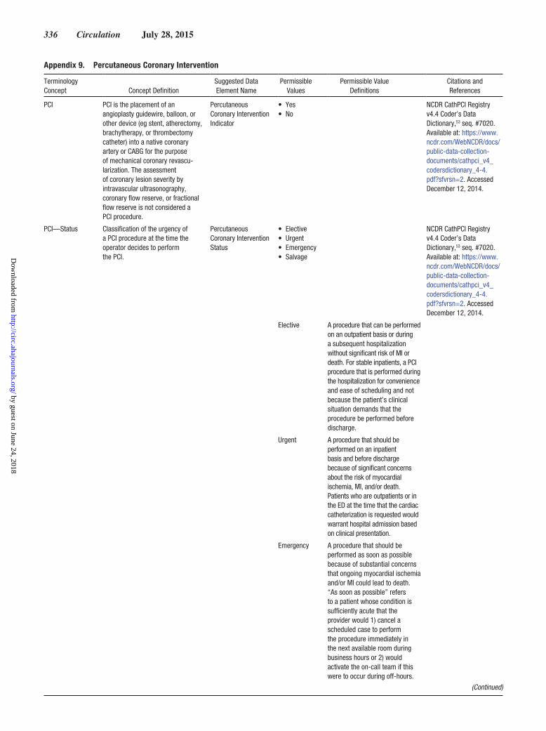
336 Circulation July 28, 2015
Appendix 9. Percutaneous Coronary Intervention
Terminology Concept Concept Definition
Suggested Data Element Name
Permissible Values
Permissible Value Definitions
Citations and References
PCI PCI is the placement of an angioplasty guidewire, balloon, or other device (eg stent, atherectomy, brachytherapy, or thrombectomy catheter) into a native coronary artery or CABG for the purpose of mechanical coronary revascu-larization. The assessment of coronary lesion severity by intravascular ultrasonography, coronary flow reserve, or fractional flow reserve is not considered a PCI procedure.
Percutaneous Coronary Intervention Indicator
• Yes• No
NCDR CathPCI Registry v4.4 Coder’s Data Dictionary,53 seq. #7020.Available at: https://www.ncdr.com/WebNCDR/docs/public-data-collection-documents/cathpci_v4_codersdictionary_4-4.pdf?sfvrsn=2. Accessed December 12, 2014.
PCI—Status Classification of the urgency of a PCI procedure at the time the operator decides to perform the PCI.
Percutaneous Coronary Intervention Status
• Elective• Urgent• Emergency• Salvage
NCDR CathPCI Registry v4.4 Coder’s Data Dictionary,53 seq. #7020. Available at: https://www.ncdr.com/WebNCDR/docs/public-data-collection-documents/cathpci_v4_codersdictionary_4-4.pdf?sfvrsn=2. Accessed December 12, 2014.
Elective A procedure that can be performed on an outpatient basis or during a subsequent hospitalization without significant risk of MI or death. For stable inpatients, a PCI procedure that is performed during the hospitalization for convenience and ease of scheduling and not because the patient’s clinical situation demands that the procedure be performed before discharge.
Urgent A procedure that should be performed on an inpatient basis and before discharge because of significant concerns about the risk of myocardial ischemia, MI, and/or death. Patients who are outpatients or in the ED at the time that the cardiac catheterization is requested would warrant hospital admission based on clinical presentation.
Emergency A procedure that should be performed as soon as possible because of substantial concerns that ongoing myocardial ischemia and/or MI could lead to death. “As soon as possible” refers to a patient whose condition is sufficiently acute that the provider would 1) cancel a scheduled case to perform the procedure immediately in the next available room during business hours or 2) would activate the on-call team if this were to occur during off-hours.
(Continued)
by guest on June 24, 2018http://circ.ahajournals.org/
Dow
nloaded from
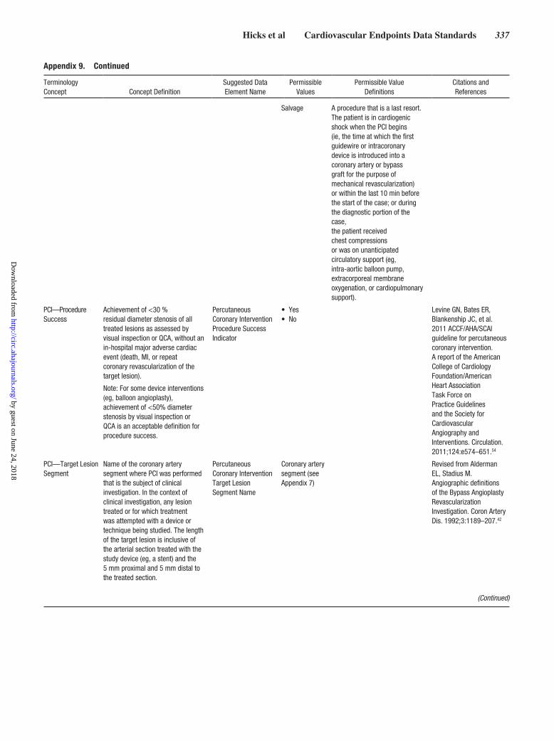
Hicks et al Cardiovascular Endpoints Data Standards 337
Salvage A procedure that is a last resort. The patient is in cardiogenic shock when the PCI begins (ie, the time at which the first guidewire or intracoronary device is introduced into a coronary artery or bypass graft for the purpose of mechanical revascularization) or within the last 10 min before the start of the case; or during the diagnostic portion of the case, the patient received chest compressions or was on unanticipated circulatory support (eg, intra-aortic balloon pump, extracorporeal membrane oxygenation, or cardiopulmonary support).
PCI—Procedure Success
Achievement of <30 % residual diameter stenosis of all treated lesions as assessed by visual inspection or QCA, without an in-hospital major adverse cardiac event (death, MI, or repeat coronary revascularization of the target lesion).
Note: For some device interventions (eg, balloon angioplasty), achievement of <50% diameter stenosis by visual inspection or QCA is an acceptable definition for procedure success.
Percutaneous Coronary Intervention Procedure Success Indicator
• Yes• No
Levine GN, Bates ER, Blankenship JC, et al. 2011 ACCF/AHA/SCAI guideline for percutaneous coronary intervention. A report of the American College of Cardiology Foundation/American Heart Association Task Force on Practice Guidelines and the Society for Cardiovascular Angiography and Interventions. Circulation. 2011;124:e574–651.54
PCI—Target Lesion Segment
Name of the coronary artery segment where PCI was performed that is the subject of clinical investigation. In the context of clinical investigation, any lesion treated or for which treatment was attempted with a device or technique being studied. The length of the target lesion is inclusive of the arterial section treated with the study device (eg, a stent) and the 5 mm proximal and 5 mm distal to the treated section.
Percutaneous Coronary Intervention Target Lesion Segment Name
Coronary artery segment (see Appendix 7)
Revised from Alderman EL, Stadius M. Angiographic definitions of the Bypass Angioplasty Revascularization Investigation. Coron Artery Dis. 1992;3:1189–207.42
Appendix 9. Continued
Terminology Concept Concept Definition
Suggested Data Element Name
Permissible Values
Permissible Value Definitions
Citations and References
(Continued)
by guest on June 24, 2018http://circ.ahajournals.org/
Dow
nloaded from

338 Circulation July 28, 2015
PCI—Target Lesion Procedure Success
Achievement of <30 % residual diameter stenosis of the target lesion as assessed by visual inspection or QCA, without an in-hospital major adverse cardiac event (death, MI, or repeat coronary revascularization of the target lesion). For some device interventions (eg, balloon angioplasty), achievement of <50% diameter stenosis by visual inspection or QCA is an acceptable definition for technical (angiographic) success.
In the context of clinical investigation, ideally the assessment of the residual stenosis at the end of the procedure should be performed by an angiographic core laboratory.
Percutaneous Coronary Intervention Target Lesion Procedure Success Indicator
• Yes• No
Adapted from Levine GN, Bates ER, Blankenship JC, et al. 2011 ACCF/AHA/SCAI guideline for percutaneous coronary intervention. A report of the American College of Cardiology Foundation/American Heart Association Task Force on Practice Guidelines and the Society for Cardiovascular Angiography and Interventions. Circulation. 2011;124:e574–651.54
PCI—Abrupt Closure
New intraprocedural severely reduced flow (TIMI grade 0–1) within the target vessel that persists and requires intervention by stenting or other treatment or results in MI or death. Abrupt closure requires an association with a vascular dissection, thrombus, or severe spasm at the treatment site or within the instrumented vessel.
Coronary Artery Abrupt Closure
• Yes• No
Adapted from Butman SM, ed. Complications of Percutaneous Coronary Interventions. New York, NY: Springer; 2010.55
PCI—Dissection New appearance of contrast and/or radiolucencies during PCI inconsistent with the expected luminal dimensions of a lesion and/or vessel.
Coronary Artery Dissection Grade
• Grade A• Grade B• Grade C• Grade D• Grade E• Grade F
Adapted from National Heart, Lung, and Blood Institute. Coronary artery angiographic changes after percutaneous transluminal coronary angioplasty. In: Manual of Operations: NHLBI PTCA Registry. Bethesda, MD: National Heart, Lung, and Blood Institute; 1985:6–9.56
Grade A Minor radiolucencies within the lumen during contrast injection with no persistence after dye clearance.
Grade B Parallel tracts or double lumen separated by a radiolucent area during contrast injection with no persistence after dye clearance.
Grade C Extraluminal cap with persistence of contrast after dye clearance from the lumen.
Grade D Spiral luminal filling defect with delayed but complete distal flow.
Grade E New persistent filling defect with delayed antegrade flow. May represent thrombus.
Grade F Non–A-E types with total coronary occlusion and no distal antegrade flow. May represent thrombus.
Appendix 9. Continued
Terminology Concept Concept Definition
Suggested Data Element Name
Permissible Values
Permissible Value Definitions
Citations and References
(Continued)
by guest on June 24, 2018http://circ.ahajournals.org/
Dow
nloaded from

Hicks et al Cardiovascular Endpoints Data Standards 339
PCI—No Reflow New acute reduction in coronary flow (TIMI grade 0–1) in the absence of dissection, thrombus, spasm, or high-grade residual stenosis at the original PCI lesion site.
Coronary Artery No Reflow Indicator
• Yes• No
Adapted from Butman SM, ed. Complications of Percutaneous Coronary Interventions. New York, NY: Springer; 2010.55
PCI—Coronary Thrombosis
New acute development of compromise of a PCI vessel by coronary artery thrombus (blood clot) occurring at any time during a procedure and independent of evidence of stenosis or dissection.
Criteria for an intraprocedural thrombosis event include ≥1 of the following: a) Development of new or increasing
coronary thrombusb) Abrupt vessel closure c) No reflow (from TIMI flow 3/2
to 1/0) or slow reflow (from TIMI 3 to 2)
d) Distal embolization
Coronary Artery Thrombosis, Intra-Procedural Indicator
• Yes• No
Adapted from Butman SM, ed. Complications of Percutaneous Coronary Interventions. New York, NY: Springer; 2010.55
PCI—Intraprocedural Stent Thrombosis
Occlusion of the lumen of a newly implanted stent by thrombus (blood clot) during the index stent implantation procedure.
Stent Thrombosis Intra-Procedural Indicator
• Yes• No
Adapted from Butman SM, ed. Complications of Percutaneous Coronary Interventions. New York, NY: Springer; 2010.55
PCI—Stent Thrombosis
Compromise of the lumen of a coronary stent by thrombus (blood clot) and not as a result of restenosis or atherosclerosis after completion of the stent implantation procedure.
Note: Angiographic nonocclusive intracoronary thrombus is defined as a (spheric, ovoid, or irregular) noncalcified filling defect or lucency surrounded by contrast material (on 3 sides or within a coronary stenosis) seen in multiple projections, or persistence of contrast material within the lumen, or a visible embolization of intraluminal material downstream.
Angiographic occlusive thrombus is defined as the presence of TIMI 0 or TIMI 1 flow intrastent or proximal to a stent up to the most adjacent proximal side branch or main branch (if it originates from the side branch).
Pathological confirmation of stent thrombosis is evidence of recent thrombus within the stent determined at autopsy or by examination of tissue retrieved following thrombectomy.
Stent Thrombosis, Coronary, Indicator
• Yes• No
Cutlip DE, Windecker S, Mehran R, et al. Clinical endpoints in coronary stent trials: a case for standardized definitions. Circulation. 2007;115:2344–51.7
PCI—Stent Thrombosis Date–Time
Date and time of the onset of stent thrombosis.
Stent Thrombosis, Coronary, Date_Time
• Date–time
Appendix 9. Continued
Terminology Concept Concept Definition
Suggested Data Element Name
Permissible Values
Permissible Value Definitions
Citations and References
(Continued)
by guest on June 24, 2018http://circ.ahajournals.org/
Dow
nloaded from

340 Circulation July 28, 2015
PCI—Stent Thrombosis ARC Grade
Probability that coronary artery stent thrombosis has occurred, according to ARC grading criteria.
Note: The incidental angiographic documentation of stent occlusion in the absence of clinical signs or symptoms is not considered a confirmed stent thrombosis (silent occlusion).
Stent Thrombosis, Coronary, ARC grade
• Definite• Probable• Possible
Cutlip DE, Windecker S, Mehran R, et al. Clinical endpoints in coronary stent trials: a case for standardized definitions. Circulation. 2007;115:2344–51.7
Definite Definite stent thrombosis is considered to have occurred when there is either angiographic or pathological confirmation. Angiographic confirmation is the presence of a thrombus at coronary angiography that originates in the stent or in the segment 5 mm proximal or distal to the stent, with the presence of at least 1 of 3 clinical criteria within a 48-h time window:a) Acute onset of ischemic
symptoms at restb) New ischemic changes on ECG
that suggest acute ischemiac) Typical rise and fall in cardiac
biomarkers. Pathological confirmation is evidence of recent thrombus within the stent determined at autopsy or by examination of tissue retrieved following thrombectomy.
Probable Probable stent thrombosis is considered to have occurred after intracoronary stenting in the following situations:a) Any unexplained death within
the first 30 d (for studies of patients with an ST-segment elevation MI, exclusion of unexplained death within 30d as evidence of probable stent thrombosis)
b) irrespective of the time after the index procedure, any MI that is related to documented acute ischemia in the territory of the implanted stent without angiographic confirmation of stent thrombosis and in the absence of any other obvious cause.
Possible Possible: Possible stent thrombosis is considered to have occurred with any unexplained death >30 d after intracoronary stenting.
Appendix 9. Continued
Terminology Concept Concept Definition
Suggested Data Element Name
Permissible Values
Permissible Value Definitions
Citations and References
(Continued)
by guest on June 24, 2018http://circ.ahajournals.org/
Dow
nloaded from

Hicks et al Cardiovascular Endpoints Data Standards 341
PCI—Stent Thrombosis ARC Timing
Timing of the detection of stent thrombosis according to the ARC grading criteria, with time 0 defined as the time when the patient departs the catheterization laboratory after a stent implantation procedure.
Note: The combination of acute or subacute stent thrombosis can be replaced by the term early stent thrombosis (0–30 d).
Stent Thrombosis, Coronary, ARC timing
• Acute• Subacute• Late• Very late
Cutlip DE, Windecker S, Mehran R, et al. Clinical endpoints in coronary stent trials: a case for standardized definitions. Circulation. 2007;115:2344–51.7
Acute 0–24 h after stent implantation
Subacute >24 h to 30 d after stent implantation
Late >30 d to 1 y after stent implantation
Very late >1 y after stent implantation
PCI—Stent Restenosis
Renarrowing of a stent implanted at a lesion site to treat a prior stenosis, to a diameter stenosis of >50% within the stent, inclusive of the original treated site plus the adjacent vascular segments 5 mm proximal and 5 mm distal to the stent.
Percutaneous Coronary Intervention Restenosis, In-Stent, Angiographic Binary Indicator
• Yes• No
Adapted from Levine GN, Bates ER, Blankenship JC, et al. 2011 ACCF/AHA/SCAI guideline for percutaneous coronary intervention. A report of the American College of Cardiology Foundation/American Heart Association Task Force on Practice Guidelines and the Society for Cardiovascular Angiography and Interventions. Circulation. 2011;124:e574–651.54
PCI—Lesion Restenosis
Renarrowing of a lesion site following treatment of a prior stenosis, to a diameter stenosis of >50% at the previously treated lesion site, inclusive of the original treated site plus the adjacent vascular segments 5 mm proximal and 5 mm distal to the treated segment.
Percutaneous Coronary Intervention, Restenosis, Lesion Angiographic Binary Indicator
• Yes• No
Adapted from Levine GN, Bates ER, Blankenship JC, et al. 2011 ACCF/AHA/SCAI guideline for percutaneous coronary intervention. A report of the American College of Cardiology Foundation/American Heart Association Task Force on Practice Guidelines and the Society for Cardiovascular Angiography and Interventions. Circulation. 2011;124:e574–651.54
Appendix 9. Continued
Terminology Concept Concept Definition
Suggested Data Element Name
Permissible Values
Permissible Value Definitions
Citations and References
(Continued)
by guest on June 24, 2018http://circ.ahajournals.org/
Dow
nloaded from
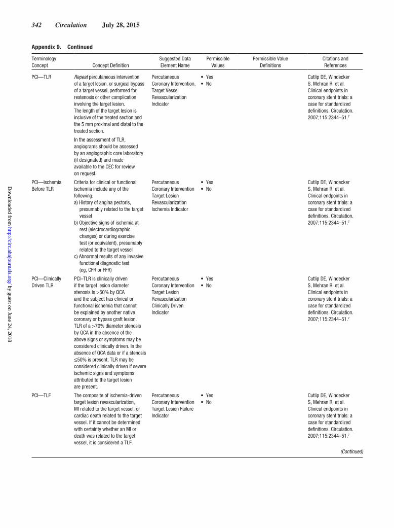
342 Circulation July 28, 2015
PCI—TLR Repeat percutaneous intervention of a target lesion, or surgical bypass of a target vessel, performed for restenosis or other complication involving the target lesion. The length of the target lesion is inclusive of the treated section and the 5 mm proximal and distal to the treated section.
In the assessment of TLR, angiograms should be assessed by an angiographic core laboratory (if designated) and made available to the CEC for review on request.
Percutaneous Coronary Intervention, Target Vessel Revascularization Indicator
• Yes• No
Cutlip DE, Windecker S, Mehran R, et al. Clinical endpoints in coronary stent trials: a case for standardized definitions. Circulation. 2007;115:2344–51.7
PCI—Ischemia Before TLR
Criteria for clinical or functional ischemia include any of the following: a) History of angina pectoris,
presumably related to the target vessel
b) Objective signs of ischemia at rest (electrocardiographic changes) or during exercise test (or equivalent), presumably related to the target vessel
c) Abnormal results of any invasive functional diagnostic test (eg, CFR or FFR)
Percutaneous Coronary Intervention Target Lesion Revascularization Ischemia Indicator
• Yes• No
Cutlip DE, Windecker S, Mehran R, et al. Clinical endpoints in coronary stent trials: a case for standardized definitions. Circulation. 2007;115:2344–51.7
PCI—Clinically Driven TLR
PCI–TLR is clinically driven if the target lesion diameter stenosis is >50% by QCA and the subject has clinical or functional ischemia that cannot be explained by another native coronary or bypass graft lesion. TLR of a >70% diameter stenosis by QCA in the absence of the above signs or symptoms may be considered clinically driven. In the absence of QCA data or if a stenosis ≤50% is present, TLR may be considered clinically driven if severe ischemic signs and symptoms attributed to the target lesion are present.
Percutaneous Coronary Intervention Target Lesion Revascularization Clinically Driven Indicator
• Yes• No
Cutlip DE, Windecker S, Mehran R, et al. Clinical endpoints in coronary stent trials: a case for standardized definitions. Circulation. 2007;115:2344–51.7
PCI—TLF The composite of ischemia-driven target lesion revascularization, MI related to the target vessel, or cardiac death related to the target vessel. If it cannot be determined with certainty whether an MI or death was related to the target vessel, it is considered a TLF.
Percutaneous Coronary Intervention Target Lesion Failure Indicator
• Yes• No
Cutlip DE, Windecker S, Mehran R, et al. Clinical endpoints in coronary stent trials: a case for standardized definitions. Circulation. 2007;115:2344–51.7
Appendix 9. Continued
Terminology Concept Concept Definition
Suggested Data Element Name
Permissible Values
Permissible Value Definitions
Citations and References
(Continued)
by guest on June 24, 2018http://circ.ahajournals.org/
Dow
nloaded from

Hicks et al Cardiovascular Endpoints Data Standards 343
PCI—Target Vessel
In the context of clinical investigation, the major native coronary artery (eg, left main coronary artery, LAD coronary artery, left circumflex coronary artery, or right coronary artery) or bypass graft containing the target lesion. A native coronary artery target vessel includes the arterial segments upstream and downstream from the target lesion plus major side branches.
Percutaneous Coronary Intervention Target Vessel Name
• Left main artery
• LAD artery• Left
circumflex artery
• Ramus intermedius artery
• Right coronary artery
Adapted from Alderman EL, Stadius M. Angiographic definitions of the Bypass Angioplasty Revascularization Investigation. Coronary Artery Disease. 1992;3:1189–207.42
Left main artery Left main coronary artery. SNOMED-CT: 3227004
LAD artery LAD coronary artery, including septal and diagonal branches.
SNOMED-CT: 59438005
Left circumflex artery
Left circumflex coronary artery, including marginal branches; if mixed dominance, also including left posterolateral branches; if left dominant, also including posterolateral and posterior descending branches.
SNOMED-CT: 57396003
Ramus intermedius artery
Ramus intermedius coronary branch.
SNOMED-CT: 244252004
Right coronary artery
Right coronary artery and its branches; if mixed dominance, also including posterior descending branch; if right dominant, also including right posterolateral and posterior descending branches.
SNOMED-CT: 13647002
PCI—TVR Repeat PCI or surgical bypass of any segment of a coronary artery containing a target lesion. A target vessel is defined as the entire major coronary vessel proximal and distal to a target lesion, including upstream and downstream branches and the target lesion itself.
In the assessment of TVR, angiograms should be assessed by an angiographic core laboratory (if designated) and made available to the CEC for review on request.
Percutaneous Coronary Intervention Target Vessel Revascularization Indicator
• Yes• No
Cutlip DE, Windecker S, Mehran R, et al. Clinical endpoints in coronary stent trials: a case for standardized definitions. Circulation. 2007;115:2344–51.7
PCI—TVF The composite of ischemia-driven TVR, MI related to the target vessel, or cardiac death related to the target vessel. If it cannot be determined with certainty whether the MI or death was related to the target vessel, it is considered a TVF.
Percutaneous Coronary Intervention Target Vessel Failure Indicator
• Yes• No
Cutlip DE, Windecker S, Mehran R, et al. Clinical endpoints in coronary stent trials: a case for standardized definitions. Circulation. 2007;115:2344–51.7
Appendix 9. Continued
Terminology Concept Concept Definition
Suggested Data Element Name
Permissible Values
Permissible Value Definitions
Citations and References
(Continued)
by guest on June 24, 2018http://circ.ahajournals.org/
Dow
nloaded from
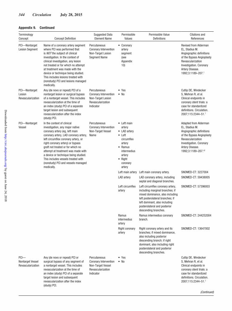
344 Circulation July 28, 2015
PCI—Nontarget Lesion Segment
Name of a coronary artery segment where PCI was performed that is NOT the subject of clinical investigation. In the context of clinical investigation, any lesion not treated or for which no attempt at treatment was made with the device or technique being studied. This includes lesions treated with (nonstudy) PCI and lesions managed medically.
Percutaneous Coronary Intervention Non-Target Lesion Segment Name
• Coronary artery segment (see Appendix 10)
Revised from Alderman EL, Stadius M. Angiographic definitions of the Bypass Angioplasty Revascularization Investigation. Coronary Artery Disease. 1992;3:1189–207.7
PCI—Nontarget Lesion Revascularization
Any (de novo or repeat) PCI of a nontarget lesion or surgical bypass of a nontarget vessel. This includes revascularization at the time of an index (study) PCI of a separate target lesion and subsequent revascularization after the index (study) PCI.
Percutaneous Coronary Intervention Non-Target Lesion Revascularization Indicator
• Yes• No
Cutlip DE, Windecker S, Mehran R, et al. Clinical endpoints in coronary stent trials: a case for standardized definitions. Circulation. 2007;115:2344–51.7
PCI—Nontarget Vessel
In the context of clinical investigation, any major native coronary artery (eg, left main coronary artery, LAD coronary artery, left circumflex coronary artery, or right coronary artery) or bypass graft not treated or for which no attempt at treatment was made with a device or technique being studied. This includes vessels treated with (nonstudy) PCI and vessels managed medically.
Percutaneous Coronary Intervention Non-Target Vessel Name
• Left main artery
• LAD artery• Left
circumflex artery
• Ramus intermedius artery
• Right coronary artery
Adapted from Alderman EL, Stadius M. Angiographic definitions of the Bypass Angioplasty Revascularization Investigation. Coronary Artery Disease. 1992;3:1189–207.42
Left main artery Left main coronary artery. SNOMED-CT: 3227004
LAD artery LAD coronary artery, including septal and diagonal branches.
SNOMED-CT: 59438005
Left circumflex artery
Left circumflex coronary artery, including marginal branches; if mixed dominance, also including left posterolateral branches; if left dominant, also including posterolateral and posterior descending branches.
SNOMED-CT: 57396003
Ramus intermedius artery
Ramus intermedius coronary branch.
SNOMED-CT: 244252004
Right coronary artery
Right coronary artery and its branches; if mixed dominance, also including posterior descending branch; if right dominant, also including right posterolateral and posterior descending branches.
SNOMED-CT: 13647002
PCI— Nontarget Vessel Revascularization
Any (de novo or repeat) PCI or surgical bypass of any segment of a nontarget vessel. This includes revascularization at the time of an index (study) PCI of a separate target lesion and subsequent revascularization after the index (study) PCI.
Percutaneous Coronary Intervention Non-Target Vessel Revascularization Indicator
• Yes• No
Cutlip DE, Windecker S, Mehran R, et al. Clinical endpoints in coronary stent trials: a case for standardized definitions. Circulation. 2007;115:2344–51.7
Appendix 9. Continued
Terminology Concept Concept Definition
Suggested Data Element Name
Permissible Values
Permissible Value Definitions
Citations and References
(Continued)
by guest on June 24, 2018http://circ.ahajournals.org/
Dow
nloaded from
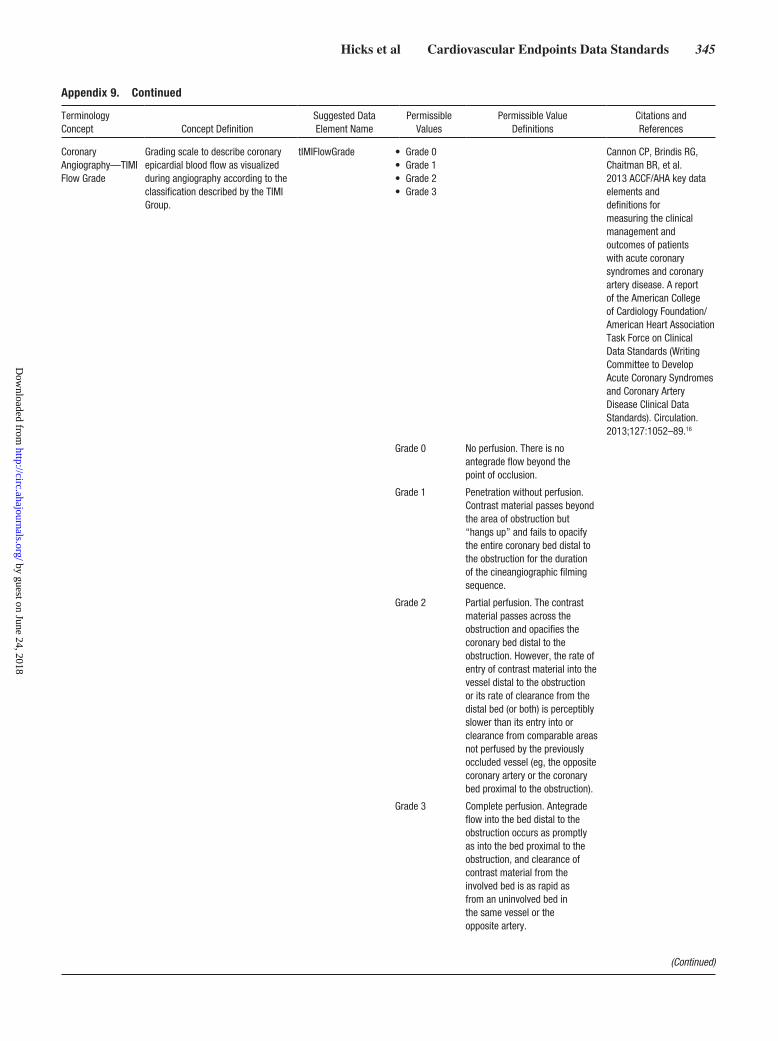
Hicks et al Cardiovascular Endpoints Data Standards 345
Coronary Angiography—TIMI Flow Grade
Grading scale to describe coronary epicardial blood flow as visualized during angiography according to the classification described by the TIMI Group.
tIMIFlowGrade • Grade 0• Grade 1• Grade 2• Grade 3
Cannon CP, Brindis RG, Chaitman BR, et al. 2013 ACCF/AHA key data elements and definitions for measuring the clinical management and outcomes of patients with acute coronary syndromes and coronary artery disease. A report of the American College of Cardiology Foundation/American Heart Association Task Force on Clinical Data Standards (Writing Committee to Develop Acute Coronary Syndromes and Coronary Artery Disease Clinical Data Standards). Circulation. 2013;127:1052–89.16
Grade 0 No perfusion. There is no antegrade flow beyond the point of occlusion.
Grade 1 Penetration without perfusion. Contrast material passes beyond the area of obstruction but “hangs up” and fails to opacify the entire coronary bed distal to the obstruction for the duration of the cineangiographic filming sequence.
Grade 2 Partial perfusion. The contrast material passes across the obstruction and opacifies the coronary bed distal to the obstruction. However, the rate of entry of contrast material into the vessel distal to the obstruction or its rate of clearance from the distal bed (or both) is perceptibly slower than its entry into or clearance from comparable areas not perfused by the previously occluded vessel (eg, the opposite coronary artery or the coronary bed proximal to the obstruction).
Grade 3 Complete perfusion. Antegrade flow into the bed distal to the obstruction occurs as promptly as into the bed proximal to the obstruction, and clearance of contrast material from the involved bed is as rapid as from an uninvolved bed in the same vessel or the opposite artery.
Appendix 9. Continued
Terminology Concept Concept Definition
Suggested Data Element Name
Permissible Values
Permissible Value Definitions
Citations and References
(Continued)
by guest on June 24, 2018http://circ.ahajournals.org/
Dow
nloaded from

346 Circulation July 28, 2015
Coronary Angiography—Coronary Artery Thrombus
A discrete, mobile, intraluminal filling defect with defined borders with or without associated contrast staining.
Coronary Artery Thrombus Indicator
• Yes• No
Adapted from Capone G, Wolf NM, Meyer B, Meister SG. Frequency of intracoronary filling defects by angiography in angina pectoris at rest. Am J Cardiol. 1985;56:403–6.57
Coronary Angiography—TIMI Thrombus Grade
Grading scale to describe coronary thrombus as visualized during angiography per the classification described by the TIMI Group.
TIMI Coronary Thrombus Grade
• Grade 1• Grade 2• Grade 3• Grade 4• Grade 5
Cannon CP, Braunwald E, McCabe CH, et al. The Thrombolysis in Myocardial Infarction trials: the first decade. J Interv Cardiol. 1995; 8:117–35.58
Grade 1 Possible thrombus present: angiography demonstrates characteristics such as reduced contrast density, haziness, irregular lesion contour, or a smooth convex “meniscus” at the site of total occlusion suggestive, but not diagnostic, of thrombus.
Grade 2 Small definite thrombus, with the greatest dimension less than or equal to one half of the vessel diameter.
Grade 3 Moderate definite thrombus, with the greatest linear dimension greater than one half but <2 vessel diameters.
Grade 4 Large definite thrombus, with the greatest dimension ≥2 vessel diameters.
Grade 5 Total vessel occlusion.
Cardiovascular Catheterization—Access Site Hematoma
Development of a new, localized collection of blood at a vascular access site sufficient to produce a palpable mass within 72 h of a procedure.
Access Site Hematoma Indicator
• Yes• No
Adapted from Butman SM, ed. Complications of Percutaneous Coronary Interventions. New York, NY: Springer; 2010.55
Cardiovascular Catheterization—Arteriovenous Fistula
Development of a new, unintended communication between an artery and a vein occurring at a vascular access site within 72 h of a procedure.
Arteriovenous Fistula Indicator
• Yes• No
Adapted from Butman SM, ed. Complications of Percutaneous Coronary Interventions. New York, NY: Springer; 2010.55
Cardiovascular Catheterization—Peripheral Ischemia
Development of new arterial insufficiency sufficient to produce clinical signs or symptoms of ischemia (pallor, pain, paresthesia) distal to a vascular access site within 72 h of a procedure.
Peripheral Ischemia Indicator
• Yes• No
Adapted from Butman SM, ed. Complications of Percutaneous Coronary Interventions. New York, NY: Springer; 2010.55
Cardiovascular Catheterization—Peripheral Nerve Injury
Development of new sensory or motor loss of peripheral nerve function from external nerve compression (eg, as a result of positioning during a procedure), or internal compression or direct nerve damage from the procedure, occurring within 72 h of a procedure.
Peripheral Nerve Injury Indicator
•Yes•No
Adapted from Butman SM, ed. Complications of Percutaneous Coronary Interventions. New York, NY: Springer; 2010.55
Appendix 9. Continued
Terminology Concept Concept Definition
Suggested Data Element Name
Permissible Values
Permissible Value Definitions
Citations and References
(Continued)
by guest on June 24, 2018http://circ.ahajournals.org/
Dow
nloaded from

Hicks et al Cardiovascular Endpoints Data Standards 347
Cardiovascular Catheterization—Pseudoaneurysm
Development of a new localized collection of blood with a persistent communication (neck) originating at a vascular access site and occurring within 72 h of a procedure.
Pseudoaneurysm Indicator
• Yes• No
Adapted from Butman SM, ed. Complications of Percutaneous Coronary Interventions. New York, NY: Springer; 2010.55
Cardiovascular Catheterization—Retroperitoneal Hemorrhage
Development of new bleeding into the retroperitoneal space originating at a vascular access site and occurring within 72 h of a procedure.
Retroperitoneal Hemorrhage Indicator
• Yes• No
Adapted from Butman SM, ed. Complications of Percutaneous Coronary Interventions. New York, NY: Springer; 2010.55
CABG CABG surgery is a procedure performed to bypass partially or completely occluded coronary arteries with veins and/or arteries harvested from elsewhere in the body, thereby improving the blood supply to the coronary circulation supplying the myocardium (heart muscle).
Coronary Artery Bypass Graft Surgery Indicator
• Yes• No
CABG—Status Classification of the urgency of a CABG surgical procedure, based on the patient’s clinical status before entering the operating room
Coronary Artery Bypass Graft Surgery Status
• Elective• Urgent• Emergency• Salvage
Elective Patient cardiac status has been stable in the days or weeks before the operation. The procedure can be deferred without increased risk of compromised cardiac outcome.
Urgent Procedure required during the same hospitalization to minimize chances of clinical deterioration or adverse outcome. Clinical conditions include (but are not limited to) acute or worsening chest pain, acute or worsening HF, acute MI, critical coronary stenosis, IABP support, UA with intravenous nitroglycerin, and rest angina.
Appendix 9. Continued
Terminology Concept Concept Definition
Suggested Data Element Name
Permissible Values
Permissible Value Definitions
Citations and References
(Continued)
by guest on June 24, 2018http://circ.ahajournals.org/
Dow
nloaded from

348 Circulation July 28, 2015
Emergency Procedure required because of ongoing, refractory (difficult, complicated, and/or unmanageable), unrelenting cardiac compromise, with or without hemodynamic instability, and not responsive to any form of therapy except cardiac surgery. An emergency operation is one in which there should be no delay in providing operative intervention. The clinical status of the patient can include any of the following:a) Ischemic dysfunction (any
of the following): 1) ongoing ischemia, including rest angina despite maximal medical therapy (medical and/or IABP); 2) acute evolving MI within 24 h before surgery; or 3) pulmonary edema requiring intubation.
b) Mechanical dysfunction (either of the following): 1) shock with circulatory support or 2) shock without circulatory support.
Salvage The patient is undergoing CPR or is being managed with extracorporeal membrane oxygenation en route to the operating room or before induction of anesthesia. The clinical acuity of the patient is a dying state.
Cardiothoracic Surgery—Inoperable/Extreme Risk
Heart Team assessment that a patient is inappropriate for cardiothoracic surgery, based on predicted operative mortality, comorbidities, frailty/debilitation, previous procedures, technical inoperability, and/or other extenuating circumstances.
Cardiothoracic Surgery Inoperable-Extreme Risk Indicator
• Yes• No
Adapted from STS/ACC TVT Registry v2.0 Coder’s Data Dictionary. Available at: https://www.ncdr.com/TVT/Libraries/TVT_Library/2_0_CoderDataDictionary.sflb.ashx. Accessed December 12, 2014.59
CABG—Type Type of CABG conduit. Coronary Artery Graft Type
• Saphenous vein graft
• Arterial graft, in situ
• Arterial graft, free
Saphenous vein graft
CABG composed of saphenous vein.
Arterial graft, in situ
CABG composed of arterial conduit, where the origin of the graft remains intact.
Arterial graft, free
CABG composed of an artery that has been completely freed from its original location.
Appendix 9. Continued
Terminology Concept Concept Definition
Suggested Data Element Name
Permissible Values
Permissible Value Definitions
Citations and References
(Continued)
by guest on June 24, 2018http://circ.ahajournals.org/
Dow
nloaded from

Hicks et al Cardiovascular Endpoints Data Standards 349
PCI—Bypass Graft Lesion Location
Location of a lesion within a CABG where PCI was performed.
Coronary Artery Graft Lesion Location
• Graft origin• Graft body• Graft
anastomosis
http://www.cdisc.org/therapeutic49
Graft origin The section of a graft from the connection (anastomosis) of the graft with the aorta (or from the origin if in situ), inclusive of the first 3 mm of the graft.
Graft body The section of a graft between the origin and the anastomosis.
Graft anastomosis
The section of a graft from the connection (anastomosis) of the graft with the coronary artery, inclusive of the retrograde 3 mm of the graft.
CABG— Anastomosis
The coronary artery segment to which a CABG is connected.
Coronary Artery Graft Anastomosis
• Coronary artery segment (see Appendix 10)
ACCF indicates American College of Cardiology Foundation; AHA, American Heart Association; ARC, Academic Research Consortium; CABG, coronary artery bypass graft; CEC, Clinical Events Committee; CFR, coronary flow reserve; CPR, cardiopulmonary resuscitation; ECG, electrocardiogram; ED, emergency department; FFR, fractional flow reserve; IABP, intra-aortic balloon bump; LAD, left anterior descending; MI, myocardial infarction; mm, millimeter; NCDR, National Cardiovascular Data Registry; NHLBI, National Heart, Lung, and Blood Institute; PCI, percutaneous coronary intervention; PTCA, percutaneous transluminal coronary angioplasty; QCA, quantitative coronary angiography; SCAI, Society for Cardiac Angiography and Interventions; SNOMED-CT, Systemized Nomenclature of Medicine—Clinical Terms; TIMI, Thrombolysis in Myocardial Infarction; TLF, target lesion failure; TLR, target lesion revascularization; TVF, target vessel failure; TVR, target lesion revascularization; and UA, unstable angina.
Appendix 9. Continued
Terminology Concept Concept Definition
Suggested Data Element Name
Permissible Values
Permissible Value Definitions
Citations and References
by guest on June 24, 2018http://circ.ahajournals.org/
Dow
nloaded from

350 Circulation July 28, 2015
Appendix 10. Cardiovascular AnatomyThis table describes coronary artery dominance, coronary artery segments, and lower abdominal, pelvic, and lower extremity artery segment concepts. The bulleted lists are the permissible values for the terminology concept; the following rows provide more detail about the individual permissible values. The table includes a draft simple numbering code for the coronary and peripheral artery segments, as well as references to existing coding systems describing the specific segments where available. The numbering code schema uses the letter “a” to designate the ostium, “b” to designate the body, “c” to designate the bifurcation terminus, and “d” to identify a lateral branch of a branch.
Terminology Concept Concept Definition
Suggested Data Element
NamePermissible
ValuesPermissible Value
DefinitionsCitations and References
Coronary Artery Dominance
Pattern of the coronary arteries that supply blood to the inferior wall of the left ventricle, classified into 1 of 3 mutually exclusive categories (right, left, co-dominant).
Coronary Artery Dominance
• Left• Right• Co-dominant
Revised from Austen WG, Edwards JE, Frye RL, et al. A reporting system on patients evaluated for coronary artery disease. Report of the Ad Hoc Committee for Grading of Coronary Artery Disease, Council on Cardiovascular Surgery, American Heart Association. Circulation. 1975;51:5–40.41
Alderman EL, Stadius M. Angiographic definitions of the Bypass Angioplasty Revascularization Investigation. Coronary Artery Disease. 1992;3:1189–207.42
Medina A, Suarez de Lezo J, Pan M. [A new classification of coronary bifurcation lesions]. Rev Esp Cardiol. 2006;59:183.44
Left The PDA and PLA arise from the left circumflex artery.
Right The PDA and PLA arise from the right coronary artery.
Co-dominant The right coronary artery supplies the PDA, and the circumflex artery supplies the PLA. Thus, both the right coronary and left circumflex arteries contribute to the blood supply of the inferior wall of the left ventricle.
(Continued)
by guest on June 24, 2018http://circ.ahajournals.org/
Dow
nloaded from

Hicks et al Cardiovascular Endpoints Data Standards 351
Coronary Artery Segments
[value set] • Right coronary artery ostium• Proximal right coronary artery• Mid right coronary artery• Distal right coronary artery• Right PDA• Posterolateral segmental artery• First right posterolateral branch• Second right posterolateral branch• Third right posterolateral branch• Posterior descending septal perforator• Right ventricular branch• Left main coronary artery ostium• Left main coronary artery body• Left main coronary artery bifurcation• LAD artery ostium• Proximal LAD• Mid LAD• Distal LAD• First diagonal branch• First diagonal lateral branch• Second diagonal branch• Second diagonal lateral branch• Third diagonal branch• Third diagonal lateral branch• Anterior descending septal perforator• Left circumflex artery ostium• Proximal left circumflex artery• Mid left circumflex artery• Distal left circumflex artery• First obtuse marginal branch• First obtuse marginal lateral branch• Second obtuse marginal branch• Second obtuse marginal lateral
branch• Third obtuse marginal branch• Third obtuse marginal lateral branch• Left atrioventricular artery• Left PDA• First left posterolateral branch• Second left posterolateral branch• Third left posterolateral branch• Ramus intermedius branch• Ramus intermedius lateral branch
Revised from Austen WG, Edwards JE, Frye RL, et al. A reporting system on patients evaluated for coronary artery disease. Report of the Ad Hoc Committee for Grading of Coronary Artery Disease, Council on Cardiovascular Surgery, American Heart Association. Circulation. 1975;51:5–40.41
Alderman EL, Stadius M. Angiographic definitions of the Bypass Angioplasty Revascularization Investigation. Coronary Artery Disease. 1992;3:1189–207.42
Medina A, Suarez de Lezo J, Pan M. [A new classification of coronary bifurcation lesions]. Rev Esp Cardiol. 2006;59:183.44
Right coronary artery ostium Origin of the right coronary artery, including the first 3 mm of the artery
Draft code: 1aSNOMED-CT: 56789007Draft code: 1a
Proximal right coronary artery Proximal portion of the right coronary artery, from the ostium of the right coronary artery to the origin of the first right ventricular branch
Draft code: 1BARI: 1NCI: C102337SNOMED-CT: 91083009
Mid right coronary artery Middle portion of the right coronary artery, from the origin of the first right ventricular branch to the acute margin
Draft code: 2BARI: 2NCI: C102329SNOMED-CT: 13647002+255562008
Distal right coronary artery Distal portion of the right coronary artery, from the acute margin to the origin of the posterior descending artery
Draft code: 3BARI: 3NCI: C102296SNOMED-CT: 41879009
Appendix 10. Continued
Terminology Concept
Concept Definition
Suggested Data Element
Name Permissible ValuesPermissible Value
DefinitionsCitations and References
(Continued)
by guest on June 24, 2018http://circ.ahajournals.org/
Dow
nloaded from

352 Circulation July 28, 2015
Right PDA In right dominant and mixed circulations, the vessel that runs in the posterior interventricular groove and supplies septal perforator branches
Draft code: 4BARI: 4NCI: C102342SNOMED-CT: 53655008
Posterolateral segmental artery In right-dominant circulations, the distal continuation of the right coronary artery in the posterior atrioventricular groove after the origin of the right PDA
Draft code: 5BARI: 5NCI: C102341SNOMED-CT: 12800002
First right posterolateral branch In right-dominant circulations, the first posterolateral branch originating from the right posterior atrioventricular artery
Draft code: 6BARI: 6SNOMED-CT: 91761002
Second right posterolateral branch In right-dominant circulations, the second posterolateral branch originating from the right posterior atrioventricular artery
Draft code: 7BARI: 7SNOMED-CT: 91762009
Third right posterolateral branch In right-dominant circulations, the third posterolateral branch originating from the right posterior atrioventricular artery
Draft code: 8BARI: 8SNOMED-CT: 91763004
Posterior descending septal perforator
Septal perforator vessel originating from the PDA
Draft code: 9BARI: 9
Right ventricular branch Branch arising from the right coronary artery to supply the right ventricular wall
Draft code: 10BARI: 10SNOMED-CT: 22765000
Left main coronary artery ostium Origin of the left coronary artery, including the first 3 mm of the artery
Draft code: 11aSNOMED-CT: 76862008
Left main coronary artery body Body of the left main coronary artery, from the ostium to the bifurcation
Draft code: 11bBARI: 11SNOMED-CT: 3227004
Left main coronary artery bifurcation
Distal end of the left main coronary artery, including the terminal 3 mm through the bifurcation of the left main into the LAD and left circumflex arteries
Draft code: 11c
LAD artery ostium Origin of the LAD coronary artery, including the first 3 mm of the artery
Draft code: 12a
Proximal LAD artery Proximal portion of the LAD coronary artery, from the ostium to the origin of the first septal
Draft code: 12BARI: 12SNOMED-CT: 68787002
Mid LAD artery Middle portion of the LAD coronary artery, from the origin of the first septal artery to the origin of the third septal artery
Draft code: 13BARI: 13SNOMED-CT: 91748002
Distal LAD artery Distal portion of the LAD coronary artery, from the origin of the third septal artery to the terminus
Draft code: 14BARI: 14SNOMED-CT: 36672000
First diagonal branch First of the 3 longest branches originating from the LAD artery to supply the anterolateral wall of the left ventricle
Draft code: 15BARI: 15SNOMED-CT: 91750005
First diagonal lateral branch Branch of the first diagonal branch Draft code: 15dBARI: 15a
(Continued)
Appendix 10. Continued
Terminology Concept
Concept Definition
Suggested Data Element
Name Permissible ValuesPermissible Value
DefinitionsCitations and References
by guest on June 24, 2018http://circ.ahajournals.org/
Dow
nloaded from

Hicks et al Cardiovascular Endpoints Data Standards 353
Second diagonal branch Second of the 3 longest branches originating from the LAD artery to supply the anterolateral wall of the left ventricle
Draft code: 16BARI: 16SNOMED-CT: 91751009
Second diagonal lateral branch Branch of the second diagonal branch Draft code: 16dBARI: 16a
Third diagonal branch Third of the 3 longest branches originating from the LAD artery to supply the anterolateral wall of the left ventricle
Draft code: 17BARI: 29SNOMED-CT: 91752002
Third diagonal lateral branch Branch of the third diagonal branch Draft code: 17dBARI: 29a
Anterior descending septal perforator Septal perforator vessel originating from the LAD artery to supply the interventricular septum
Draft code: 18BARI: 17SNOMED-CT: 244251006
Left circumflex artery ostium Origin of the left circumflex coronary artery, including the first 3 mm of the artery
Draft code: 19a
Proximal left circumflex artery Proximal portion of the left circumflex coronary artery, from the ostium to the origin (or the nominal location of) the first marginal branch
Draft code: 19BARI: 18SNOMED-CT: 52433000
Mid left circumflex artery Middle portion of the left circumflex coronary artery, from the origins of (or nominal locations of) the first marginal to the second marginal
Draft code: 20BARI: 19SNOMED-CT: 91753007
Distal left circumflex artery Distal portion of the left circumflex coronary artery, from the origin of (or the nominal location of) the second marginal to the terminus (in right-dominant systems), or to the origin of the first left posterolateral in all other dominant systems
Draft code: 21BARI: 19aSNOMED-CT: 6511003
First obtuse marginal branch First of the 3 longest branches originating from the left circumflex artery to supply the lateral wall of the left ventricle
Draft code: 22BARI: 20NCI: C102299SNOMED-CT: 91754001
First obtuse marginal lateral branch Branch of the first marginal branch Draft code: 22dBARI: 20a
Second obtuse marginal branch Second of the 3 longest branches originating from the left circumflex artery to supply the lateral wall of the left ventricle
Draft code: 23BARI: 21SNOMED-CT: 91755000
Second obtuse marginal lateral branch Branch of the second marginal branch Draft code: 23dBARI: 21a
Third obtuse marginal branch Third of the 3 longest branches originating from the left circumflex artery to supply the lateral wall of the left ventricle
Draft code: 24BARI: 22SNOMED-CT: 91756004
Third obtuse marginal lateral branch Branch of the third marginal branch Draft code: 24dBARI: 22a
Left atrioventricular artery In left-dominant and mixed circulations, the distal continuation of the left circumflex coronary artery in the posterior atrioventricular groove
Draft code: 25BARI: 23NCI: C102287SNOMED-CT: 75902001
(Continued)
Appendix 10. Continued
Terminology Concept
Concept Definition
Suggested Data Element
Name Permissible ValuesPermissible Value
DefinitionsCitations and References
by guest on June 24, 2018http://circ.ahajournals.org/
Dow
nloaded from

354 Circulation July 28, 2015
Left posterior descending artery In left-dominant circulations, the vessel that arises from the distal continuation of the left atrioventricular artery, travels in the posterior interventricular groove and supplies septal perforator branches
Draft code: 26BARI: 27SNOMED-CT: 56322004
First left posterolateral branch In left-dominant and mixed circulations, the first posterolateral branch originating from the posterior atrioventricular left circumflex artery
Draft code: 27BARI: 24SNOMED-CT: 91757008
Second left posterolateral branch In left-dominant and mixed circulations, the second posterolateral branch originating from the posterior atrioventricular left circumflex artery
Draft code: 28BARI: 25SNOMED-CT: 91758003
Third left posterolateral branch In left-dominant and mixed circulations, the third posterolateral branch originating from the posterior atrioventricular left circumflex artery
Draft code: 29BARI: 26SNOMED-CT: 91759006
Ramus intermedius branch Branch vessel whose origin bisects the origins of the LAD and circumflex arteries
Draft code: 30BARI: 28SNOMED-CT: 244252004
Ramus intermedius lateral branch Branch of the ramus intermedius branch
Draft code: 30dBARI: 28a
Peripheral Artery Segments
[value set] • Infrarenal aorta• Left renal artery• Right renal artery• Left common iliac artery• Right common iliac artery• Left external iliac artery• Right external iliac artery• Left internal iliac artery• Right internal iliac artery• Left common femoral artery• Right common femoral artery• Left superficial femoral artery• Right superficial femoral artery• Left profunda femoris artery• Right profunda femoris artery• Left popliteal artery—above knee• Right popliteal artery—above knee• Left popliteal artery—below knee• Right popliteal artery—below knee• Left anterior tibial artery• Right anterior tibial artery• Left tibio-peroneal trunk• Right tibio-peroneal trunk• Left peroneal artery• Right peroneal artery• Left posterior tibial artery• Right posterior tibial artery• Left accessory renal artery• Right accessory renal artery
Diehm N, Pattynama PM, Jaff MR, et al. Clinical endpoints in peripheral endovascular revascularization trials: a case for standardized definitions. Eur J Vasc Endovasc Surg. 2008;36:409–19.60
Infrarenal aorta Draft code: 400
Left renal artery Draft code: 501
Right renal artery Draft code: 401
Left common iliac artery Draft code: 502
Right common iliac artery Draft code: 402
(Continued)
Appendix 10. Continued
Terminology Concept
Concept Definition
Suggested Data Element
Name Permissible ValuesPermissible Value
DefinitionsCitations and References
by guest on June 24, 2018http://circ.ahajournals.org/
Dow
nloaded from

Hicks et al Cardiovascular Endpoints Data Standards 355
Left external iliac artery Draft code: 503
Right external iliac artery Draft code: 403
Left internal iliac artery Draft code: 504
Right internal iliac artery Draft code: 404
Left common femoral artery Draft code: 505
Right common femoral artery Draft code: 405
Left superficial femoral artery Draft code: 506
Right superficial femoral artery Draft code: 406
Left profunda femoris artery Draft code: 507
Right profunda femoris artery Draft code: 407
Left popliteal artery—above knee Draft code: 508
Right popliteal artery—above knee Draft code: 408
Left popliteal artery—below knee Draft code: 509
Right popliteal artery—below knee Draft code: 409
Left anterior tibial artery Draft code: 510
Right anterior tibial artery Draft code: 410
Left tibio-peroneal trunk Draft code: 511
Right tibio-peroneal trunk Draft code: 411
Left peroneal artery Draft code: 512
Right peroneal artery Draft code: 412
Left posterior tibial artery Draft code: 513
Right posterior tibial artery Draft code: 413
Left accessory renal artery Draft code: 514
Right accessory renal artery Draft code: 414
BARI indicates Bypass Angioplasty Revascularization Investigators; LAD, left anterior descending; NCI, National Cancer Institute; PDA, posterior descending artery; PLA, posterolateral artery; and SNOMED-CT, Systemized Nomenclature of Medicine—Clinical Terms.
Appendix 10. Continued
Terminology Concept
Concept Definition
Suggested Data Element
Name Permissible ValuesPermissible Value
DefinitionsCitations and References
by guest on June 24, 2018http://circ.ahajournals.org/
Dow
nloaded from

356 Circulation July 28, 2015
Appendix 11. Peripheral Vascular Intervention
Terminology Concept Concept Definition
Suggested Data Element Name
Permissible Values
Permissible Value Definitions
Citations and References
PVI A PVI is a catheter-based or open surgical procedure designed to improve arterial or venous blood flow or otherwise modify or revise vascular conduits. Procedures may include, but are not limited to, percutaneous transluminal balloon angioplasty, stent placement, thrombectomy, embolectomy, atherectomy, dissection repair, aneurysm exclusion, treatment of dialysis conduits, placement of various devices, intravascular thrombolysis or other pharmacotherapies, and open surgical bypass or revision. In general, the intention to perform percutaneous vascular intervention is denoted by the insertion of a guidewire into an artery or vein. For the sake of simplicity, this definition applies to the extracranial carotid artery and other noncardiac arteries and excludes the intracranial vessels, veins, and lymphatics.
Peripheral Vascular Intervention Indicator
• Yes http://www.cdisc.org/ therapeutic49
PVI—Status Classification of the urgency of a PVI procedure at the time the operator decides to perform the PVI.
Nonelective procedures include urgent and emergency procedures.
Peripheral Vascular Intervention Status
• Elective• Urgent• Emergency
http://www.cdisc.org/ therapeutic,49 modeled on the similar coronary PCI concept.
Elective A procedure that is scheduled and performed on a patient with stable disease or in whom there is no urgency and/or increased morbidity or mortality associated with a planned procedure.
Urgent A procedure that is not an emergency but is required to be performed on a timely basis (≤24 h) (eg, a patient who has been stabilized following initial treatment of acute limb ischemia and there is clinical consensus that a definitive procedure should occur within the next 24 h).
Emergency A procedure that is required to be performed immediately because of the acute nature of the medical condition (eg, acute limb ischemia, acute aortic dissection) and the increased morbidity or mortality associated with a temporal delay in treatment.
(Continued)
by guest on June 24, 2018http://circ.ahajournals.org/
Dow
nloaded from

Hicks et al Cardiovascular Endpoints Data Standards 357
PVI—Target Lesion Segment
Name of the peripheral artery segment where PVI was performed that is the subject of clinical investigation. In the context of clinical investigation, any lesion treated or for which treatment was attempted with a device or technique being studied. The length of the target lesion is inclusive of the section of vessel treated with the study device (eg, a stent) and the 10 mm proximal and 10 mm distal to the treated section.
Peripheral Vascular Intervention Target Lesion Segment Name
• Peripheral artery segment (see Appendix 10)
PVI—Procedure Success
Achievement of a satisfactory final residual diameter stenosis by angiography at the end of the procedure (and without flow-limiting dissection or hemodynamically significant translesional pressure gradient). The specific parameter for final percent residual stenosis is typically between <30% and <50%; selection of the appropriate percentage may vary depending on the specific intervention applied, the vascular territory, and anticipated or desired therapeutic response. Procedural success also implies absence of in-hospital major adverse events (eg, death, stroke, MI, acute onset of limb ischemia, need for urgent/emergency vascular surgery, and other procedure-specific major adverse events).
In the context of clinical investigation, ideally the assessment of the residual stenosis at the end of the procedure should be performed by an angiographic core laboratory.
The balloon inflation, stent placement, or other therapeutic intervention may be preceded by use of adjunctive devices (eg, percutaneous mechanical thrombectomy, directional or rotational atherectomy, laser, and chronic total occlusion crossing device), as predefined in the protocol.
Peripheral Vascular Intervention Procedure Success Indicator
• Yes• No
Adapted from Diehm N, Pattynama PM, Jaff MR, et al. Clinical endpoints in peripheral endovascular revascularization trials: a case for standardized definitions. Eur J Vasc Endovasc Surg. 2008;36:409–19.60
PVI—Stent Restenosis
Renarrowing of a stent implanted at a lesion site to treat a prior stenosis, to a diameter stenosis of >50% within the stent, inclusive of the original treated site plus the adjacent vascular segments 10 mm proximal and 10 mm distal to the stent.
Peripheral Vascular Intervention Restenosis, In-Stent— Angiographic Binary Indicator
• Yes• No
Adapted from Diehm N, Pattynama PM, Jaff MR, et al. Clinical endpoints in peripheral endovascular revascularization trials: a case for standardized definitions. Eur J Vasc Endovasc Surg. 2008;36:409–19.60
PVI—Lesion Restenosis
Renarrowing of a lesion site following treatment of a prior stenosis, to a diameter stenosis of >50% at the previously treated lesion site, inclusive of the original treated site plus the adjacent vascular segments 10 mm proximal and 10 mm distal to the treated segment.
Peripheral Vascular Intervention Restenosis, Lesion— Angiographic Binary Indicator
• Yes• No
Adapted from Diehm N, Pattynama PM, Jaff MR, et al. Clinical endpoints in peripheral endovascular revascularization trials: a case for standardized definitions. Eur J Vasc Endovasc Surg. 2008;36:409–19.60
Appendix 11. Continued
Terminology Concept Concept Definition
Suggested Data Element Name
Permissible Values
Permissible Value Definitions
Citations and References
(Continued)
by guest on June 24, 2018http://circ.ahajournals.org/
Dow
nloaded from

358 Circulation July 28, 2015
PVI—TLR Repeat PVI of a target lesion (including 10 mm proximal and 10 mm distal to the index device) or surgical intervention/bypass of a target vessel, performed for restenosis or other complication involving the target lesion.
In the assessment of TLR, angiograms should be assessed by an angiographic core laboratory (if designated). Angiograms (and core laboratory assessment thereof) and other source documentation should be made available to the CEC for review on request.
Peripheral Vascular Intervention Target Lesion Revascularization Indicator
• Yes• No
Adapted from Diehm N, Pattynama PM, Jaff MR, et al. Clinical endpoints in peripheral endovascular revascularization trials: a case for standardized definitions. Eur J Vasc Endovasc Surg. 2008;36:409–19.60
PVI—Ischemia Before TLR
Criteria for clinical or functional ischemia include any of the following: a) Recurrent/progressive intermittent
claudication b) Critical limb ischemia c) Recurrence of the clinical syndrome for
which the initial procedure was performed.
Percutaneous Coronary Intervention Target Lesion Revascularization Ischemia Indicator
• Yes• No
http://www.cdisc.org/ therapeutic,49 modeled on the similar coronary PCI concept.
PVI—Clinically Driven TLR
TLR is clinically driven if the target lesion diameter stenosis is >50% AND the subject has either clinical or functional ischemia (eg, recurrent/progressive intermittent claudication, critical limb ischemia) OR recurrence of the clinical syndrome for which the initial procedure was performed. Clinically driven TLR occurs in the absence of protocol-directed surveillance ultrasonography or angiography.
Percutaneous Coronary Intervention Target Lesion Revascularization Clinically Driven Indicator
• Yes• No
Adapted from Diehm N, Pattynama PM, Jaff MR, et al. Clinical endpoints in peripheral endovascular revascularization trials: a case for standardized definitions. Eur J Vasc Endovasc Surg. 2008;36:409–19.60
PVI—TLF The composite of ischemia-driven TLR AND either evidence of clinical or functional ischemia (eg, recurrent/progressive intermittent claudication, critical limb ischemia) OR recurrence of the clinical syndrome for which the initial procedure was performed. If it cannot be determined with certainty whether clinical/functional ischemia or the clinical syndrome was related to the target vessel, it is considered a TLF.
Peripheral Vascular Intervention Target Lesion Failure Indicator
• Yes• No
Adapted from Diehm N, Pattynama PM, Jaff MR, et al. Clinical endpoints in peripheral endovascular revascularization trials: a case for standardized definitions. Eur J Vasc Endovasc Surg. 2008;36:409–19.60
PVI—Target Vessel
Name of the peripheral artery level containing a PVI target lesion that is the subject of clinical investigation. In the context of clinical investigation, a target vessel is any noncardiac or nonintracranial blood vessel that contains the target lesion treated with the study device. The target vessel includes the target lesion as well as the entire length of native vessel upstream and downstream from the target lesion, including side branches.
For the arteries of the leg, the vasculature is divided into 3 vessel “levels”: aorto-iliac, femoral-popliteal, and tibial-crural.
Peripheral Vascular Intervention Target Vessel Name
• Aorto-iliac• Femoro-
popliteal• Tibial-crural
Adapted from Diehm N, Pattynama PM, Jaff MR, et al. Clinical endpoints in peripheral endovascular revascularization trials: a case for standardized definitions. Eur J Vasc Endovasc Surg. 2008;36:409–19.60
Aorto-iliac Infrarenal aorta and iliac arteries (to the bottom of the pelvic rim/inguinal ligament), including branches of these vessels.
Appendix 11. Continued
Terminology Concept Concept Definition
Suggested Data Element Name
Permissible Values
Permissible Value Definitions
Citations and References
(Continued)
by guest on June 24, 2018http://circ.ahajournals.org/
Dow
nloaded from

Hicks et al Cardiovascular Endpoints Data Standards 359
Femoro-popliteal Femoral and popliteal arteries (to the origin of the anterior tibial artery), including branches of these vessels.
Tibial-crural Anterior tibial and below, including the arteries of the foot.
PVI—TVR Repeat intervention or surgical bypass of any segment of a target vessel. In the assessment of TVR, angiograms should be assessed by an angiographic core laboratory (if designated). Angiograms (and core laboratory assessment thereof) and other source documentation should be made available to the CEC for review on request.
Peripheral Vascular Intervention Target Vessel Revascularization Indicator
• Yes• No
http://www.cdisc.org/ therapeutic,49 modeled on the similar coronary PCI concept.
PVI—Vessel Patency
The absence of clinically driven TLR and/ or absence of recurrent target lesion diameter stenosis >50% by imaging (eg, invasive angiography or, most commonly, duplex ultrasonography). If patency data are incorporated within the primary endpoint of a clinical trial, the angiographic images or duplex ultrasonographic images should be assessed by appropriate core laboratories and made available to the CEC for review on request.
Vessel Patency Indicator
• Yes• No
Adapted from Diehm N, Pattynama PM, Jaff MR, et al. Clinical endpoints in peripheral endovascular revascularization trials: a case for standardized definitions. Eur J Vasc Endovasc Surg. 2008;36:409–19.60
PVI—Target Limb
The target limb is the extremity that contains the target lesion and all the native vessels upstream and downstream from it, including side branches.
Peripheral Vascular Intervention Target Limb Name
• Right leg• Left leg
Adapted from Diehm N, Pattynama PM, Jaff MR, et al. Clinical endpoints in peripheral endovascular revascularization trials: a case for standardized definitions. Eur J Vasc Endovasc Surg. 2008;36:409–19.60
PVI—Target Limb Failure
TLF is the composite of ischemia-driven revascularization of the target limb and major adverse ischemic events affecting the target limb. If it cannot be determined with certainty whether an event was related to the target limb, it is considered a TLF.
Peripheral Vascular Intervention Target Limb Failure Indicator
• Yes• No
Adapted from Diehm N, Pattynama PM, Jaff MR, et al. Clinical endpoints in peripheral endovascular revascularization trials: a case for standardized definitions. Eur J Vasc Endovasc Surg. 2008;36:409–19.60
PVI—Nontarget Lesion Segment
Name of a peripheral artery segment where PVI was performed that is NOT the subject of clinical investigation. In the context of clinical investigation, any lesion not treated or for which treatment was not attempted with the device or technique being studied. This includes lesions treated with (nonstudy) PVI and lesions managed medically.
Peripheral Vascular Intervention Non- Target Lesion Segment Name
• Peripheral artery segment (see Appendix 10)
Appendix 11. Continued
Terminology Concept Concept Definition
Suggested Data Element Name
Permissible Values
Permissible Value Definitions
Citations and References
(Continued)
by guest on June 24, 2018http://circ.ahajournals.org/
Dow
nloaded from
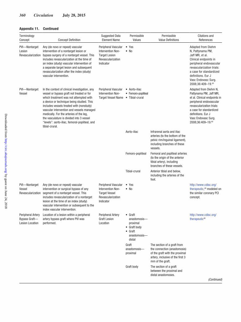
360 Circulation July 28, 2015
PVI—Nontarget Lesion Revascularization
Any (de novo or repeat) vascular intervention of a nontarget lesion or bypass surgery of a nontarget vessel. This includes revascularization at the time of an index (study) vascular intervention of a separate target lesion and subsequent revascularization after the index (study) vascular intervention.
Peripheral Vascular Intervention Non- Target Lesion Revascularization Indicator
• Yes• No
Adapted from Diehm N, Pattynama PM, Jaff MR, et al. Clinical endpoints in peripheral endovascular revascularization trials: a case for standardized definitions. Eur J Vasc Endovasc Surg. 2008;36:409–19.60
PVI—Nontarget Vessel
In the context of clinical investigation, any vessel or bypass graft not treated or for which treatment was not attempted with a device or technique being studied. This includes vessels treated with (nonstudy) vascular intervention and vessels managed medically. For the arteries of the leg, the vasculature is divided into 3 vessel “levels”: aorto-iliac, femoral-popliteal, and tibial-crural.
Peripheral Vascular Intervention Non- Target Vessel Name
• Aorto-iliac• Femoro-popliteal• Tibial-crural
Adapted from Diehm N, Pattynama PM, Jaff MR, et al. Clinical endpoints in peripheral endovascular revascularization trials: a case for standardized definitions. Eur J Vasc Endovasc Surg. 2008;36:409–19.60
Aorto-iliac Infrarenal aorta and iliac arteries (to the bottom of the pelvic rim/inguinal ligament), including branches of these vessels.
Femoro-popliteal Femoral and popliteal arteries (to the origin of the anterior tibial artery), including branches of these vessels.
Tibial-crural Anterior tibial and below, including the arteries of the foot.
PVI—Nontarget Vessel Revascularization
Any (de novo or repeat) vascular intervention or surgical bypass of any segment of a nontarget vessel. This includes revascularization of a nontarget lesion at the time of an index (study) vascular intervention or subsequent to the index vascular intervention.
Peripheral Vascular Intervention Non- Target Vessel Revascularization Indicator
• Yes• No
http://www.cdisc.org/ therapeutic,49 modeled on the similar coronary PCI concept.
Peripheral Artery Bypass Graft— Lesion Location
Location of a lesion within a peripheral artery bypass graft where PVI was performed.
Peripheral Artery Graft Lesion Location
• Graft anastomosis— proximal
• Graft body• Graft
anastomosis— distal
http://www.cdisc.org/ therapeutic49
Graft anastomosis— proximal
The section of a graft from the connection (anastomosis) of the graft with the proximal artery, inclusive of the first 3 mm of the graft.
Graft body The section of a graft between the proximal and distal anastomoses.
Appendix 11. Continued
Terminology Concept Concept Definition
Suggested Data Element Name
Permissible Values
Permissible Value Definitions
Citations and References
(Continued)
by guest on June 24, 2018http://circ.ahajournals.org/
Dow
nloaded from

Hicks et al Cardiovascular Endpoints Data Standards 361
Graft anastomosis— distal
The section of a graft from the connection (anastomosis) of the graft with the distal artery, inclusive of the retrograde 3 mm of the graft.
Peripheral Artery Bypass Graft—Type
Type of peripheral artery bypass graft conduit.
Peripheral Artery Graft Type
• Autologous vein graft
• Synthetic graft• Composite
graft• Cadaveric
graft—arterial• Cadaveric
graft—venous• Other graft
Autologous vein graft
Bypass graft composed of autologous vein (harvested from the patient).
Synthetic graft Bypass graft composed of nonbiological conduit material.
Composite graft Bypass graft composed of a composite of materials.
Cadaveric graft—arterial
Bypass graft composed of artery harvested from a cadaver.
Cadaveric graft—venous
Bypass graft composed of vein harvested from a cadaver.
Other graft Bypass graft not composed of autologous vein, synthetic conduit, composite construction, or of cadaveric origin.
Peripheral Artery Bypass Graft— Synthetic Material
Type of material used in a synthetic peripheral artery bypass graft conduit.
Peripheral Artery Graft, Synthetic Material
• Gortex• PTFE• Polyester• Dacron• Polyurethane
Peripheral Artery Bypass Graft— Anastomosis
The peripheral artery segment to which a peripheral artery bypass graft is connected.
Peripheral Artery Graft Anastomosis
• Peripheral artery segment (see Appendix 10)
Adapted from Diehm N, Pattynama PM, Jaff MR, et al. Clinical endpoints in peripheral endovascular revascularization trials: a case for standardized definitions. Eur J Vasc Endovasc Surg. 2008;36:409–19.60
CEC indicates Clinical Events Committee; PTFE, polytetrafluoroethylene; PVI, peripheral vascular intervention; TLF, target lesion failure; TLR, target lesion revascularization; and TVR, target vessel revascularization.
Appendix 11. Continued
Terminology Concept Concept Definition
Suggested Data Element Name
Permissible Values
Permissible Value Definitions
Citations and References
by guest on June 24, 2018http://circ.ahajournals.org/
Dow
nloaded from

and Shari L. TargumMarian C. Limacher, Kenneth W. Mahaffey, Roxana Mehran, Steven E. Nissen, Eric E. Smith
Andrew Farb, Gregg C. Fonarow, Jeffrey P. Jacobs, Michael R. Jaff, Judith H. Lichtman, Karen A. Hicks, James E. Tcheng, Biykem Bozkurt, Bernard R. Chaitman, Donald E. Cutlip,
Cardiovascular Endpoints Data Standards)Association Task Force on Clinical Data Standards (Writing Committee to Developin Clinical Trials: A Report of the American College of Cardiology/American Heart
2014 ACC/AHA Key Data Elements and Definitions for Cardiovascular Endpoint Events
Print ISSN: 0009-7322. Online ISSN: 1524-4539 Copyright © 2014 American Heart Association, Inc. All rights reserved.
is published by the American Heart Association, 7272 Greenville Avenue, Dallas, TX 75231Circulation doi: 10.1161/CIR.0000000000000156
2015;132:302-361; originally published online December 29, 2014;Circulation.
http://circ.ahajournals.org/content/132/4/302World Wide Web at:
The online version of this article, along with updated information and services, is located on the
/content/132/8/e129.full.pdfAn erratum has been published regarding this article. Please see the attached page for:
http://circ.ahajournals.org/content/suppl/2014/12/23/CIR.0000000000000156.DC3 http://circ.ahajournals.org/content/suppl/2014/12/23/CIR.0000000000000156.DC2 http://circ.ahajournals.org/content/suppl/2014/12/23/CIR.0000000000000156.DC1
Data Supplement (unedited) at:
http://circ.ahajournals.org//subscriptions/
is online at: Circulation Information about subscribing to Subscriptions:
http://www.lww.com/reprints Information about reprints can be found online at: Reprints:
document. Permissions and Rights Question and Answer this process is available in the
click Request Permissions in the middle column of the Web page under Services. Further information aboutOffice. Once the online version of the published article for which permission is being requested is located,
can be obtained via RightsLink, a service of the Copyright Clearance Center, not the EditorialCirculationin Requests for permissions to reproduce figures, tables, or portions of articles originally publishedPermissions:
by guest on June 24, 2018http://circ.ahajournals.org/
Dow
nloaded from

Correction
e129
In the article by Hicks et al, “2014 ACC/AHA Key Data Elements and Definitions for Cardiovascular Endpoint Events in Clinical Trials: A Report of the American College of Cardiology/American Heart Association Task Force on Clinical Data Standards (Writing Committee to Develop Cardiovascular Endpoints Data Standards),” which published online December 29, 2014, and appeared in the July 28, 2015, issue of the journal (Circulation. 2015;132:302–361. DOI: 10.1161/CIR.0000000000000156), several corrections were needed.
1. The following amendments were made to the reference list of the accepted manuscript:• reference 16 was deleted because it was a duplicate of reference 5• reference 17 was deleted because it was a duplicate of reference 6• reference 20 was deleted because it was a duplicate of reference 8• reference 21 was deleted because it was a duplicate of reference 9• reference 22 was deleted because it was a duplicate of reference 11
2. All references have been renumbered accordingly, as illustrated in the table below.
(Circulation. 2015;132:e129. DOI: 10.1161/CIR.0000000000000294.)© 2015 American Heart Association, Inc.
Circulation is available at http://circ.ahajournals.org DOI: 10.1161/CIR.0000000000000294
These corrections have been made to print version and to the current online version of the article, which is available at http://circ.ahajournals.org/content/132/4/302.full.pdf+html.
Accepted Manuscript Reference Number
Typeset Document Reference Number
Accepted Manuscript Reference Number
Typeset Document Reference Number
Accepted Manuscript Reference Number
Typeset Document Reference Number
1 1 23 18 45 40
2 2 24 19 46 41
3 3 25 20 47 42
4 4 26 21 48 43
5 5 27 22 49 44
6 6 28 23 50 45
7 7 29 24 51 46
8 8 30 25 52 47
9 9 31 26 53 48
10 10 32 27 54 49
11 11 33 28 55 50
12 12 34 29 56 51
13 13 35 30 57 52
14 14 36 31 58 53
15 15 37 32 59 54
16 5 38 33 60 55
17 6 39 34 61 56
18 16 40 35 62 57
19 17 41 36 63 58
20 8 42 37 64 59
21 9 43 38 65 60
22 11 44 39

Hicks KA, et al. Cardiovascular Endpoints Data Standards © 2014 by the American College of Cardiology Foundation and the American Heart Association, Inc.
Appendix 1. Comprehensive Author Relationships Wit h Industry and Other Entities — 2014 ACC/AHA Key Data Elements and Definitions for Cardiovascular En dpoint Events in Clinical Trials
Name Employment Consultant Speaker Ownership/ Partnership/
Principal
Research Institutional, Organizational or Other Financial
Benefit
Expert Witness
Karen A. Hicks (Chair)
FDA None None None None None None
James E. Tcheng (Vice-Chair)
Duke University Medical Center
• American Board of Internal Medicine
None
None • NIH • Duke University Medical Center – Philips Medical Systems
None
Judith H. Lichtman (TFDS Liaison)
Yale University School of Medicine, Department of Epidemiology and Public Health
None None None None
None None
Marian C. Limacher (AHA Rep)
University of Florida, Division of Cardiovascular Medicine
None None None • NIH None None
Biykem Bozkurt Michael E. DeBakey VA Medical Center
None None None None None None
Bernard R. Chaitman St. Louis University School of Medicine, Core ECG Laboratory
• Gilead • Relypsa • Takeda
None None None • Forest • Johnson &
Johnson • Lilly • Merck • NovoNordisc • Pfizer • Roche • Sanofi
None
Donald E. Cutlip Beth Israel • STENTYS None None • Boston None None

Hicks KA, et al. Cardiovascular Endpoints Data Standards © 2014 by the American College of Cardiology Foundation and the American Heart Association, Inc.
Name Employment Consultant Speaker Ownership/
Partnership/ Principal
Research Institutional, Organizational or Other Financial
Benefit
Expert Witness
Deaconess Medical Center, Interventional Cardiology
Scientific • Celonova • Medtronic
Andrew Farb FDA None None None None None None Gregg C. Fonarow
Ahmanson-UCLA Cardiomyopathy Center Division of Cardiology
• Amgen • Bayer • Boston
Scientific • J&J • Medicines
Company • Medtronic • Novartis • Takeda
None None • Gambro • Medtronic • NHLBI • NIH/NIAID • Novartis
None None
Jeffrey P. Jacobs Cardiac Surgical Associates
None None None • COAST DSMB • NIH Challenge
Grant
None None
Michael R. Jaff Massachusetts General Hospital
• Abbott Vascular
• Access Closure
• American Genomics
• Cardinal Health
• Cordis • Covidien • Medtronic
Vascular • Micell • Vascular
Therapies • WebMD
None • Icon Interventional
• Janacare • MC10 • Northwind • PQ Bypass • Primacea • Sadra Medical • Sano V
• Ekos • IC Sciences • VIVA
Physicians Group
None
Kenneth W. Mahaffey Stanford • Amgen None None • Amgen • Duke Clinical None

Hicks KA, et al. Cardiovascular Endpoints Data Standards © 2014 by the American College of Cardiology Foundation and the American Heart Association, Inc.
Name Employment Consultant Speaker Ownership/
Partnership/ Principal
Research Institutional, Organizational or Other Financial
Benefit
Expert Witness
University School of Medicine
• AstraZeneca • Bayer • Boehringer
Ingelheim • Bristol
Myers Squibb
• Cubist • Daiichi
Sankyo • Dialouges • Duke Center
for Educational Excellence
• Elsevier • Glaxo Smith
Kline • Haemonetics • Johns
Hopkins University
• Purdue Pharma
• Sanofi-Aventis
• South East Area Health Education Center
• Springer Publishing
• Sun Pharma • University of
British Columbia
• WebMD
• Amylin • AstraZeneca • Baxter • BMS • Boehringer
Ingleheim • Cubist
Pharmaceuticals • Edwards
Lifesciences • Eli Lilly • Forest • Glaxo Smith
Kline • Ikaria • Johnson &
Johnson • Merck • NHLBI • NIAID • NIH • Novartis
Pharmaceutical • Portola • Regado • Roche
Diagnostic • Sanofi
Research Institute
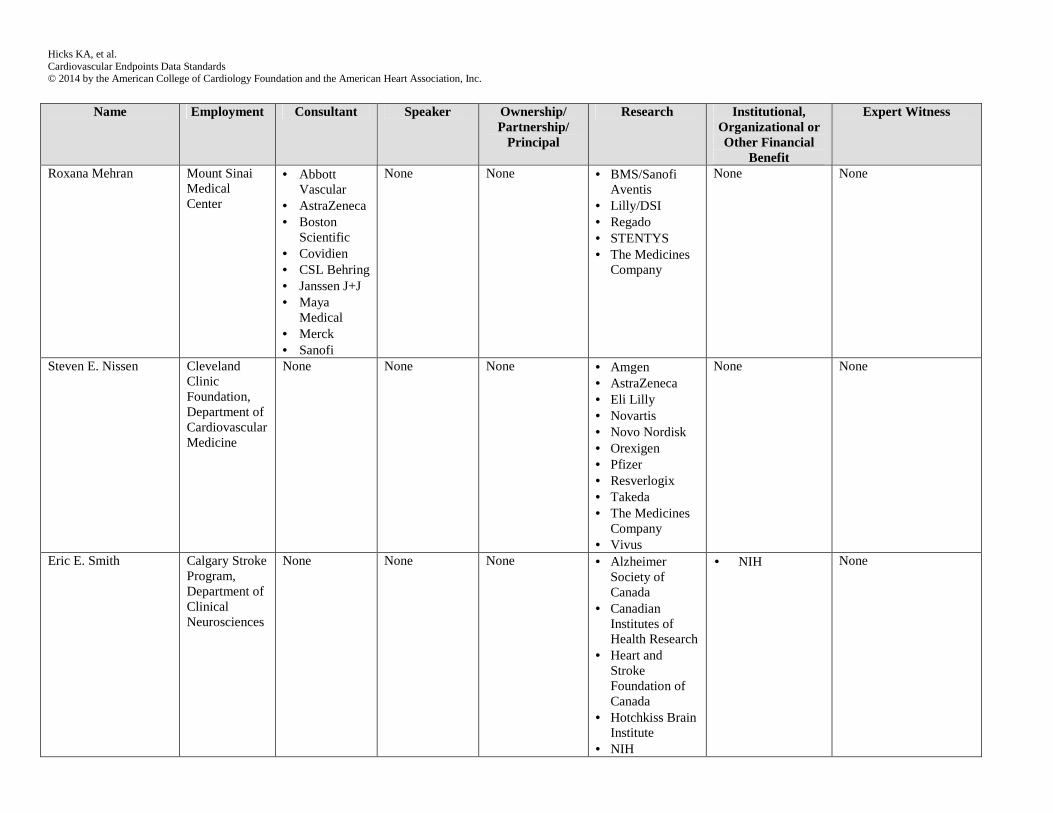
Hicks KA, et al. Cardiovascular Endpoints Data Standards © 2014 by the American College of Cardiology Foundation and the American Heart Association, Inc.
Name Employment Consultant Speaker Ownership/
Partnership/ Principal
Research Institutional, Organizational or Other Financial
Benefit
Expert Witness
Roxana Mehran Mount Sinai Medical Center
• Abbott Vascular
• AstraZeneca • Boston
Scientific • Covidien • CSL Behring • Janssen J+J • Maya
Medical • Merck • Sanofi
None None • BMS/Sanofi Aventis
• Lilly/DSI • Regado • STENTYS • The Medicines
Company
None None
Steven E. Nissen Cleveland Clinic Foundation, Department of Cardiovascular Medicine
None
None None • Amgen • AstraZeneca • Eli Lilly • Novartis • Novo Nordisk • Orexigen • Pfizer • Resverlogix • Takeda • The Medicines
Company • Vivus
None None
Eric E. Smith Calgary Stroke Program, Department of Clinical Neurosciences
None None None • Alzheimer Society of Canada
• Canadian Institutes of Health Research
• Heart and Stroke Foundation of Canada
• Hotchkiss Brain Institute
• NIH
• NIH None

Hicks KA, et al. Cardiovascular Endpoints Data Standards © 2014 by the American College of Cardiology Foundation and the American Heart Association, Inc.
Name Employment Consultant Speaker Ownership/
Partnership/ Principal
Research Institutional, Organizational or Other Financial
Benefit
Expert Witness
Shari L. Targum FDA None None None None None None This table represents the relationships of committee members with industry and other entities that were determined to be relevant to this document. These relationships were reviewed and updated in conjunction with all meetings and/or conference calls of the Writing Committee during the document development process. The table does not necessarily reflect relationships with industry at the time of publication. A person is deemed to have a significant interest in a business if the interest represents ownership of 5% or more of the voting stock or share of the business entity, or ownership of $10,000 or more of the fair market value of the business entity; or if funds received by the person from the business entity exceed 5% of the person’s gross income for the previous year. Relationships that exist with no financial benefit are also included for the purpose of transparency. Relationships in this table are modest unless otherwise noted. BMS indicates Bristol Myers Squibb; NHLBI, National Heart, Lung and Blood Institute; NIAID, National Institute of Allergy and Infectious Diseases; NIH, National Institute of Health.