acdis day2-6 track5-5 pres 0517-bridgeman-f · 2017-05-23 · shunt (modified blalock‐taussig)...
TRANSCRIPT

1
Karen Bridgeman, MSN, RN, CCDSCDI Educator
Medical University of South CarolinaCharleston, SC
Out of the Sandbox: Congenital Anomalies and Syndromes
2
Learning Objectives
• At the completion of this educational activity, the learner will be able to:
– Discuss the intricacies of various congenital anomalies for accurate code assignment in ICD‐10
– Discuss documentation needs related to congenital anomalies
– Identify query opportunities in congenital anomalies
– Identify root operations for repair of various congenital anomalies
3
2017 Copyright, HCPro, an H3.Group division of Simplify Compliance LLC. All rights reserved. These materials may not be copied without written permission.
1
4
Polling Question #1
• What is your biggest challenge in pediatric patients with congenital anomalies and syndromes?
– Understanding congenital heart defects
– Coding the procedures
– Understanding the different syndromes
– Finding resources
– I do not have any challenges in this patient population
5
“The only true wisdom is in
knowingyou know nothing”
Socrates
https://commons.wikimedia.org/wiki/File%3AHead_of_Socrates_in_Palazzo_Massimo_alle_Terme_(Rome).JPG
6
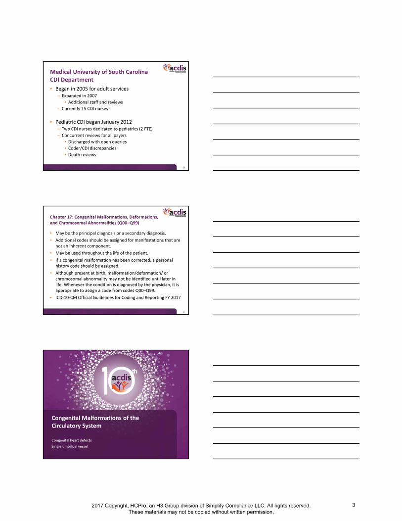
Medical University of South Carolina Children's Hospital
Medical University of South Carolina
• Major academic and tertiary patient referral center for all of South Carolina
• Three hospitals
– University
– Institute of Psychiatry
– Children’s
• 709 beds
• Level I trauma center
Children’s Hospital
• 186 licensed beds
• Service lines
– Pediatric ICU
– Pediatric cardiology
– Pediatric hospitalists
– Pediatric hem‐onc
– Pediatric pulmonary
– Pediatric neurology
– Pediatric emergency dept
– Neonatal services
• Level III nursery
2017 Copyright, HCPro, an H3.Group division of Simplify Compliance LLC. All rights reserved. These materials may not be copied without written permission.
2
7
Medical University of South Carolina CDI Department
• Began in 2005 for adult services– Expanded in 2007
• Additional staff and reviews
– Currently 15 CDI nurses
• Pediatric CDI began January 2012– Two CDI nurses dedicated to pediatrics (2 FTE)
– Concurrent reviews for all payers
• Discharged with open queries
• Coder/CDI discrepancies
• Death reviews
8
Chapter 17: Congenital Malformations, Deformations, and Chromosomal Abnormalities (Q00–Q99)
• May be the principal diagnosis or a secondary diagnosis.
• Additional codes should be assigned for manifestations that are not an inherent component.
• May be used throughout the life of the patient.
• If a congenital malformation has been corrected, a personal history code should be assigned.
• Although present at birth, malformation/deformation/ or chromosomal abnormality may not be identified until later in life. Whenever the condition is diagnosed by the physician, it is appropriate to assign a code from codes Q00–Q99.
• ICD‐10‐CM Official Guidelines for Coding and Reporting FY 2017
9
Congenital Malformations of the Circulatory System
Congenital heart defects
Single umbilical vessel
2017 Copyright, HCPro, an H3.Group division of Simplify Compliance LLC. All rights reserved. These materials may not be copied without written permission.
3
10
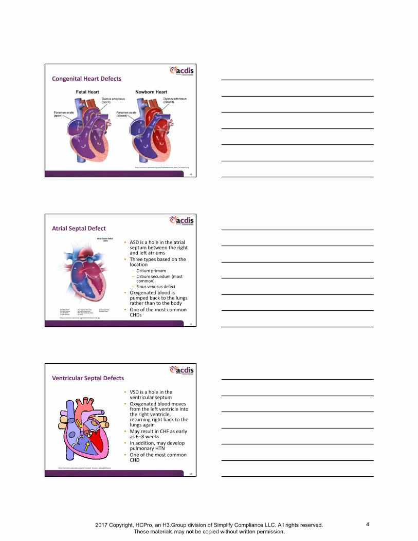
Congenital Heart Defects
https://commons.wikimedia.org/wiki/File%3ANeonatal_Heart_Circulation.png
11
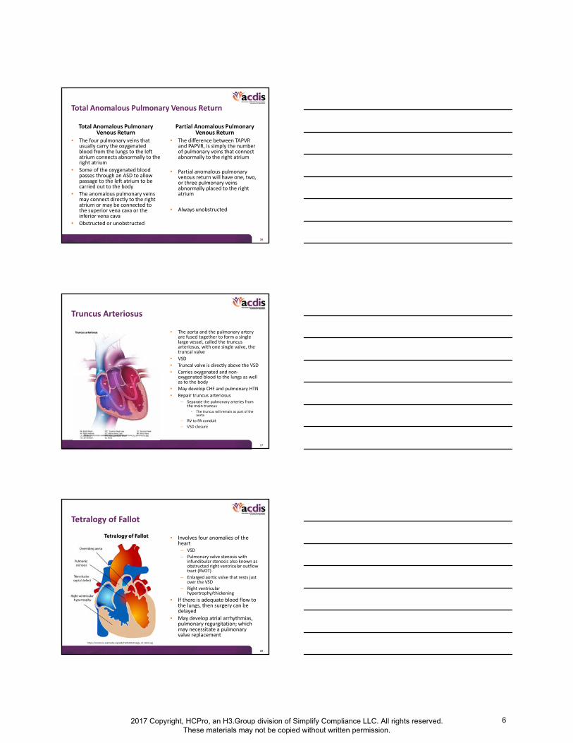
Atrial Septal Defect
• ASD is a hole in the atrial septum between the right and left atriums
• Three types based on the location– Ostium primum– Ostium secundum (most
common)– Sinus venosus defect
• Oxygenated blood is pumped back to the lungs rather than to the body
• One of the most common CHDs
https://commons.wikimedia.org/wiki/File%3AAsd‐web.jpg
12
Ventricular Septal Defects
• VSD is a hole in the ventricular septum
• Oxygenated blood moves from the left ventricle into the right ventricle, returning right back to the lungs again
• May result in CHF as early as 6–8 weeks
• In addition, may develop pulmonary HTN
• One of the most common CHD
https://commons.wikimedia.org/wiki/File:Heart_stenosis_vsd.svg#filehistory
2017 Copyright, HCPro, an H3.Group division of Simplify Compliance LLC. All rights reserved. These materials may not be copied without written permission.
4

13
Atrioventricular Septal Defect
• AVSD is a combination of closely associated defects with an ASD, VSD, and anomalies of the mitral and tricuspid valves
– Partial AVSD includes only the atrial septal defect and a common AV valve (usually a cleft left mitral valve)
– Complete AVSD includes defects of both an atrial septal defect and a ventricular septal defect and a common AV valve
– The right AV valve is the tricuspid valve
– The left AV valve is the mitral valve
– Rastelli types A‐B‐C describe the structure of the AV valve leaflets
• Root operation
– Supplement – patch closure
– Repair – direct closure
– Creation
• Mitral and tricuspid valve from common AV valve
Q21.2 ‐ AV canal defect AV septal defectEndocardial cushion defectOstium primum ASD type 1
https://commons.wikimedia.org/wiki/File%3AAvsd‐with‐normal.jpg
14
Coarctation of the Aorta
• Coarctation of the aorta is a stricture usually noted in the descending aorta
• Prostaglandins to keep the PDA open
• Root operation– Excision
• End‐to‐end anastomosis
– Dilation
• Balloon angioplasty
– Supplement
• Aortoplasty patch
• Subclavian artery flap
• Body part– Ascending aorta
• Aortic valve to innominate artery
– Aorta arch
• Innominate artery, left common carotid artery, and left subclavian artery
– Descending aorta
• Left subclavian artery to level of diaphragm
https://commons.wikimedia.org/wiki/File%3ABlausen_0243_CoarctationofAorta_CloseUp.png
15
Transposition of Great Vessels
• Dextro‐TGA – Q20.3– Aorta and pulmonary
artery are connected to the wrong chambers of the heart
– Patent foramen ovale
– Patent ductus arteriosus
• Levo‐TGA – Q20.5– Corrected
– Abnormal ventricle position, aorta, and pulmonary artery
– VSDs
– Pulmonary stenosis
• Root operation
• Reposition– Arterial switch
• One or two coronary arteries
• Replacement– Aortic root
replacement
• Occlusion– PDA ligation
• Supplement– Aorta root patch graft
• Excision– Pulmonary trunk
excision
https://commons.wikimedia.org/wiki/File%3AD‐tga‐575px.jpg
2017 Copyright, HCPro, an H3.Group division of Simplify Compliance LLC. All rights reserved. These materials may not be copied without written permission.
5
16
Total Anomalous Pulmonary Venous Return
Total Anomalous Pulmonary Venous Return
• The four pulmonary veins that usually carry the oxygenated blood from the lungs to the left atrium connects abnormally to the right atrium
• Some of the oxygenated blood passes through an ASD to allow passage to the left atrium to be carried out to the body
• The anomalous pulmonary veins may connect directly to the right atrium or may be connected to the superior vena cava or the inferior vena cava
• Obstructed or unobstructed
Partial Anomalous Pulmonary Venous Return
• The difference between TAPVR and PAPVR, is simply the number of pulmonary veins that connect abnormally to the right atrium
• Partial anomalous pulmonary venous return will have one, two, or three pulmonary veins abnormally placed to the right atrium
• Always unobstructed
17
Truncus Arteriosus
• The aorta and the pulmonary artery are fused together to form a single large vessel, called the truncus arteriosus, with one single valve, the truncal valve
• VSD
• Truncal valve is directly above the VSD
• Carries oxygenated and non‐oxygenated blood to the lungs as well as to the body
• May develop CHF and pulmonary HTN
• Repair truncus arteriosus – Separate the pulmonary arteries from
the main truncus
• The truncus will remain as part of the aorta
– RV to PA conduit
– VSD closure
https://commons.wikimedia.org/wiki/File%3ATruncus_arteriosus.jpg
18
Tetralogy of Fallot
• Involves four anomalies of the heart– VSD
– Pulmonary valve stenosis with infundibular stenosis also known as obstructed right ventricular outflow tract (RVOT)
– Enlarged aortic valve that rests just over the VSD
– Right ventricular hypertrophy/thickening
• If there is adequate blood flow to the lungs, then surgery can be delayed
• May develop atrial arrhythmias, pulmonary regurgitation; which may necessitate a pulmonary valve replacement
https://commons.wikimedia.org/wiki/File%3ATetralogy_of_Fallot.svg
2017 Copyright, HCPro, an H3.Group division of Simplify Compliance LLC. All rights reserved. These materials may not be copied without written permission.
6

19
Hypoplastic Left Heart Syndrome
• Small and inadequate functionality of the left ventricle of the heart
• Mitral valve atresia• Aortic valve atresia• Hypoplasia of ascending aorta • Blood flow to the body is
severely restricted• When the patent ductus
arteriosus (PDA) closes, acute cardiogenic shock will develop
• Due to palliative surgery, patients are now living into adulthood
https://commons.wikimedia.org/wiki/File%3AHypoplastic_left_heart_syndrome.svg
20
Pulmonary Atresia
Pulmonary Atresia
• No pulmonary valve to allow blood to enter the pulmonary artery and be carried to the lungs
• Pulmonary atresia with intact ventricular septum– Blocks flow of blood to lungs
– Hypoplastic right ventricle with defective tricuspid valve
• Pulmonary atresia with ventricular septal defect– Provides blood flow out through
right ventricle
Treatment
• Prostaglandin to keep the patent ductus arteriosus open
• Balloon valvuloplasty– Root operation – Dilation
• Pulmonary valve patch– Root operation – Supplement
• Aorta to pulmonary artery shunt (modified Blalock‐Taussig)
– Root operation – Bypass
• Based on the physiology, the pulmonary atresia may be treated as a single ventricle
21
Tricuspid Atresia
Tricuspid Atresia
• No tricuspid valve
• An atrial septal defect (ASD) is needed to allow blood flow out of the right atrium which allows mixing of oxygenation and non‐oxygenated blood
• May have a hypoplastic right ventricle and pulmonary valve stenosis
• Usually will have a ventricular septal defect (VSD)
• Usually will have pulmonary valvar stenosis
Treatment
• Treated as single ventricle because the heart has only one functioning ventricle (the left ventricle)
• Surgical interventions:
– Norwood
– Bidirectional Glenn
– Fontan
2017 Copyright, HCPro, an H3.Group division of Simplify Compliance LLC. All rights reserved. These materials may not be copied without written permission.
7

22
Three‐Stage Palliation Surgery
Norwood
• Within the first two weeks of life
Root operation
• Bypass– Modified Blalock‐
Taussig shunt
– Aortopulmonaryshunt
– Sano modification
• Excision– Atrial septectomy
• Occlusion– PDA ligation
Bidirectional Glenn
• Between 3 to 6 months of age
Root operation
• Bypass– Cavo‐pulmonary shunt
– (Right pulmonary artery to the superior vena cava)
• Removal of the previous shunt (not coded)
– Modified Blalock‐Taussig shunt
– Aortopulmonary shunt
– Sano modified shunt
Fontan
• Between 18 months to 4 years
Root operation
• Bypass– Right pulmonary
artery to the inferior vena cava
23
Rastelli Procedure
Conditions Treated
• Double outlet right ventricle
• Dextro‐transposition of the great vessels (d‐TGA)
• Truncus arteriosus
• Combinations of congenital heart defects with– VSD
– Right ventricular outflow tract obstruction (RVOTO)
• Pulmonary atresia
• Pulmonary stenosis
• Subpulmonary stenosis
– Aortic atresia
Rastelli Procedure• Root operation:
– Right ventricle to pulmonary artery (RV‐PA) conduit
• Bypass
• Root operation:– Closure of VSD
• Supplement
• Repair
– Infundibulectomy• Excision of right ventricle
– Division of RVOT obstruction• Release
• Root operation:– Pulmonary valve closed
• Occlusion
24
Secondary Diagnoses
• Acidosis/alkalosis
• Acquired coagulation factor deficiency– Administration of Factor VII
• Awaiting transplant status
• Cardiomyopathy
• Cyanosis
• Developmental delays
• Down syndrome
• Electrolyte imbalances
• Exposure to smoke
• Failure to thrive
• Feeding difficulties
• Hypoxia or hypercapnia
• Heart arrhythmias– Complete heart block
– Junctional ectopic tachycardia (JET)
• Heart failure– Congestive heart failure
– Neonatal heart failure
• Long‐term use of aspirin or anticoagulants
• Presence of prosthetic heart valve
• Pulmonary artery HTN/secondary pulmonary HTN
• Respiratory distress
• Respiratory failure
• 22q11.2 deletion syndrome
2017 Copyright, HCPro, an H3.Group division of Simplify Compliance LLC. All rights reserved. These materials may not be copied without written permission.
8

25
Query for Factor VII
• Patient is a 10‐day‐old neonate with d‐transposition of great vessels, subpulmonary ventricular septal defect, and patent ductus arteriosus. S/P arterial switch, VSD patch, and division of PDA. OP report notes “Obtaining hemostasis required administration of platelets, cryoprecipitate, and two doses of recombinant factor 7.” Can these clinical findings be further specified? E.g.,
– Acquired coagulation deficiency
– Acquired coagulation disorder
– Other __________________
– Clinically undetermined
26
Complications of Congenital Heart Surgery
• Abnormal shaped heart valve may develop– Endocarditis
– Regurgitation
• Arrhythmias– Complete heart block
– Junctional ectopic tachycardia (JET)
• Cardiomegaly
• Chylothorax
• Developmental delays
• Frequent respiratory infections
• Heart failure– Congestive heart failure (CHF)
– Neonatal heart failure
• Horner’s syndrome
• Hypocalcemia (DiGeorge syndrome)
• Learning disability
• Plastic bronchitis
• Pleural effusions
• Post‐pericardiotomy syndrome
• Protein‐losing enteropathy (PLE)
• Pulmonary hypertension
• Residual defects
• Shunt stenosis
• Stroke
• Shunt thrombosis
• Wound infections
27
Congenital Heart Defects—Palliative
• Partially repaired congenital heart conditions:– Common ventricle
– Double inlet left ventricle
– Double outlet right ventricle
– Hypoplastic left heart syndrome
– Tricuspid atresia
• History of Norwood, Bidirectional Glenn (BDG) (Glenn), and/or Fontan procedures– These procedures are performed for common ventricle conditions
– Patients with a common ventricle condition will always have the condition unless they receive a heart transplant
2017 Copyright, HCPro, an H3.Group division of Simplify Compliance LLC. All rights reserved. These materials may not be copied without written permission.
9

28
Stenosis: Complication vs. End of Life
• Many congenital heart repair surgeries require replacement of valves and/or placement of conduits to redirect blood flow
• Most surgically implanted valves or conduits have a life span of 10–20 years before they wear out, become stenotic or calcified, or develop impaired function
• When their function becomes impaired, the valves or conduits need to be replaced
• Both mechanical and bioprosthetic implanted valves and conduits will eventually need to be repaired or replaced
• Principal diagnosis for these patients:– Z45.09 Encounter for adjustment and management of other
cardiac device
• Do not code removal of the device
29
Coding Clinic—End of Life
Prosthetic valve stenosis due to end of valve life ICD‐9‐CM Coding Clinic, Second Quarter 2008 Page: 9 to 10 Effective with discharges: July 7, 2008
Question:The patient was admitted due to end of life of the heart valve prosthesis. The cardiothoracic surgeon documents "prosthetic valve stenosis due to end of life." In ICD‐9‐CM, the index directs to code 996.71, Other complications of internal (biological) (synthetic) prosthetic device, implant, and graft, due to heart valve prosthesis, for prosthetic valve stenosis. However, the surgeon does not agree with this code assignment. He stated that the patient's heart valve prosthesis was placed 19 years ago and has reached its end of life, which is an expected outcome, not a complication. How should end of life of the heart valve prosthesis be coded?
30
Coding Clinic—End of Life: Response
Prosthetic valve stenosis due to end of valve life ICD‐9‐CM Coding Clinic, Second Quarter 2008 Page: 9 to 10 Effective with discharges: July 7, 2008
Answer:Assign code V53.39, Fitting and adjustment of other device, other cardiac device, as the principal diagnosis. This situation would not be classified as a complication of the device since the device is eventually expected to wear out and the patient was not experiencing any problems due to the device. When the device is causing problems or complications, such as mechanical breakdown, a code from subcategory 996.6, Mechanical complications of cardiac device, implant and graft, is assigned.
2017 Copyright, HCPro, an H3.Group division of Simplify Compliance LLC. All rights reserved. These materials may not be copied without written permission.
10

31
Root Operations for Congenital Heart Defects
• Bypass– RV‐PA conduit
– Blalock‐Taussig Shunt• Thoracic aorta – right pulmonary
artery
– Fontan• Right atrium – right pulmonary artery
• Creation– Right and left AV valve from common
atrioventricular valve
– Truncus arteriosus repair
• Dilation– Balloon valvuloplasty
– Enlargement of existing ASD
– Creation of new ASD
• Excision– Coarctation repair end‐to‐end
anastomosis
– Infundibulectomy – excision of right ventricle
• Occlusion– PDA
– Rastelli – Occlusion of pulmonary valve
• Release– Vascular ring
– Division of right ventricular outflow tract muscle bundles
– Commissurotomy of cardiac valves
• Repair– ASD/VSD with direct closure
• Replacement – Heart valves
• Reposition– Arterial switch
• Pulmonary trunk
• Thoracic aorta
• Supplement– Annuloplasty ring
– ASD/VSD with patch repair
32
Valvuloplasty
Dilation
• A 18‐day‐old neonate with congenital pulmonary valve stenosis
• S/P percutaneous balloon pulmonary valvuloplasty
Replacement
• A 6‐year‐old with congenital aortic valve stenosis
• S/P valvuloplasty with an Edwards SAPIEN heart valve implanted
Supplement
• A 18‐month‐old with history of HLHS post Fontan presents with tricuspid regurgitation
• S/P valvuloplasty with annuloplasty ring sutured into place
Valvuloplasty is the surgical reconstruction of a deformed cardiac valve, for the relief of stenosis or incompetence.
33
Common Congenital Heart Devices
• Zooplastic
– Admedus cardiocel scaffold collagen
– Edwards SAPIEN valve
– Mosaic bioprosthesis aortic or mitral valve
– Contegra conduits
– Baxter PCO404N (bovine)
– Bioprosthesis
• Synthetic
– Goretex
– Silicone
– Carpentier‐Edwards annuloplasty ring
– CryoLife On‐X Valves
• Allograft
– CryoLife
2017 Copyright, HCPro, an H3.Group division of Simplify Compliance LLC. All rights reserved. These materials may not be copied without written permission.
11

34
Single Umbilical Artery
• Q27.0 Congenital absence and hypoplasia of umbilical artery– Single umbilical artery
• P02.6 Newborn (suspected to be) affected by other and unspecified conditions of umbilical cord– Short umbilical cord
– Vasa previa
• Excludes1 Newborn affected by single umbilical artery (Q27.0)
• Most umbilical cords will have two arteries and one vein • The umbilical vein carries oxygenated blood from the placenta to
the baby while the umbilical arteries carry non‐oxygenated blood from the baby to the placenta
• May indicate a congenital heart abnormality, or skeletal, intestinal, or renal problems
35
Cleft Lip and Cleft Palate
36
Cleft Lip and Cleft Palate
• Cleft lip
– Unilateral
– Bilateral
– Median
• Cleft palate
– Soft palate
– Hard palate
– Hard palate and soft palate
– Uvula
• Cleft lip & palate
– Unilateral
– Bilateral
– Median
• Cleft lip may be partial or complete– Partial cleft lip is a small groove or opening of the lip – Complete cleft lip is a small groove or opening that continues upward to the nose
• Cleft palate may be incomplete or complete– Incomplete cleft palate is a groove or opening usually in the soft palate– Complete cleft palate is a groove or opening in the soft palate and the hard palate
2017 Copyright, HCPro, an H3.Group division of Simplify Compliance LLC. All rights reserved. These materials may not be copied without written permission.
12

37
Congenital Malformation of the Digestive System
Biliary atresia
38
Biliary Atresia
• Biliary atresia is the blockage of the biliary ducts, which leads to liver damage and cirrhosis of the liver
• Usually become symptomatic around 4–8 weeks of age
• The Kasai procedure is a palliative treatment
– Code the biliary atresia as the condition still exists
• A liver transplant is the only cure for biliary atresia
– A personal history code should be used to capture the history of biliary atresia
39
Congenital Malformation of the Genital Organs
Ambiguous genitalia
2017 Copyright, HCPro, an H3.Group division of Simplify Compliance LLC. All rights reserved. These materials may not be copied without written permission.
13

40
Ambiguous Genitalia
• Ambiguous genitalia is a disorder in which an infant’s external genitals do not appear to be clearly male or female
• The genitals may be underdeveloped or may have characteristics of both sexes
• Q56.4, Ambiguous genitalia
• Condition code 45, Ambiguous gender category
– This code will allow gender‐specific coding
– Reduces denials based on gender‐specific edits
41
Congenital Malformation of the Urinary System
Congenital hydronephrosis
42
Congenital Hydronephrosis
• Congenital hydronephrosis (Q62.0) is a condition associated with distention and dilation of the renal pelvis that may be due to various congenital obstructive anomalies of the kidneys and urinary tract that may develop into renal atrophy
– Ureteropelvic junction obstruction (UPJO)
– Posterior urethral valve (PUV)
– Vesicoureteral reflux (VUR)
– No significant abnormality
• Pelviectasis or pyelectasis (Q62.0) is a mild enlargement of the kidney pelvis, whereas hydronephrosis is an extreme ballooning of the kidney
• A query for congenital hydronephrosis will be needed if only noted on x‐ray report
• Many newborns with a UTI will have a urinary tract abnormality
2017 Copyright, HCPro, an H3.Group division of Simplify Compliance LLC. All rights reserved. These materials may not be copied without written permission.
14

43
Congenital Malformations of the Musculoskeletal System
Craniosynostosis
Congenital diaphragmatic hernia
Congenital kyphosis and scoliosis
Pectus excavatum
44
Craniosynostosis
• Sagittal synostosis most common
• Premature fusion of the fibrous sutures in an infant’s skull
• Develops a misshapen head
• May increase intracranial pressure with neuro deficits, developmental delays, cognitive impairment, blindness, seizures, or death
• Root operation: Reposition of skull
45
Congenital Diaphragmatic Hernia
• Congenital diaphragmatic hernia– Q79.0 Congenital diaphragmatic hernia
– Boldek hernia
• Posterior‐lateral diaphragmatic hernia
– Morgagni hernia
• Parasternal diaphragmatic hernia
• Diaphragm eventration– Q79.1 Other congenital malformation of diaphragm
– Abnormal elevation of diaphragm
– Intact diaphragm
• Congenital hiatal hernia vs. acquired hiatal hernia– Congenital hiatal hernia (Q40.1)
– Acquired hiatal hernia
• K44.9 Diaphragmatic hernia without obstruction or gangrene– Includes hiatal hernia (sliding)
2017 Copyright, HCPro, an H3.Group division of Simplify Compliance LLC. All rights reserved. These materials may not be copied without written permission.
15

46
Scoliosis: Congenital and Idiopathic
• Scoliosis is a spinal deformity characterized by a curvature and rotation of the spine – Birth defect
– Accident
– Neuromuscular disease
– Idiopathic • Early‐onset scoliosis (infantile)
– Birth to 3 years
• Juvenile
– Age 4 to onset of puberty
• Adolescence
– Between age 10 and skeletal maturity
– Past the adolescent growth spurt
• Adolescent scoliosis – Bracing – does not correct
scoliosis, but may prevent progression of the spinal curve
– A spinal fusion with instrumentation may be done once the child has reached skeletal maturity
• Juvenile scoliosis– For children who are still
growing and have not reached skeletal maturity, growing rod implantation is a treatment to correct spinal curve and allow continued growth of the spine
47
Vertical Expandable Prosthetic Titanium Rib (VEPTR) Growing Rods
• Thoracic insufficiency syndrome is a condition that involves chest wall deformities that affect normal breathing and lung growth. It is closely associated with scoliosis and other congenital spinal disorders.
• The Vertical Expandable Prosthetic Titanium Rib (VEPTR) is a device that expands the chest, stabilizes the spine curvature, and is lengthened periodically as the child grows.
• Initial placement– Principal diagnosis – scoliosis (most common)
– Root operation – Reposition
• Adjustment of VEPTR– Principal diagnosis – Z45.89, Encounter for adjustment and management of other
implanted devices
– Root operation – Revision
• Removal and replacement of VEPTR– Principal diagnosis – T84.328x, Displacement of other bone devices, implants, and
grafts
– Root operations – Insertion and Removal
48
Early‐Onset Scoliosis: MAGEC
• Growing rods require multiple surgeries that increase the risk of complications and increased hospitalizations
• MAGnetic Expansion Control (MAGEC) is a non‐invasive treatment for children with early‐onset scoliosis (EOS)
– After the initial implantation of the rod, doctors use an external remote control outside of the body to lengthen the magnetically controlled rod as the child grows
– Non‐invasive adjustments done in clinics
• CMS new technology add‐on payment
2017 Copyright, HCPro, an H3.Group division of Simplify Compliance LLC. All rights reserved. These materials may not be copied without written permission.
16

49
Pectus Excavatum
• Pectus excavatum is a congenital deformity characterized by a sunken chest, which can affect heart and lung function
• The heart is often displaced to the left side of the chest, and there is often compression of the heart and lungs
• Pectus excavatum may be associated with Marfan syndrome, Ehlers‐Danlos syndrome, or other skeletal abnormalities including scoliosis
• The deformity usually becomes more pronounced during the teen years, requiring correction
50
Pectus Excavatum—NUSS Procedure
• The Nuss procedure is often referred to as minimally invasive repair of pectus excavatum (MIRPE). This procedure places a curved steel bar under the sternum, which expands the pectus deformity. The approach is usually percutaneous endoscopic. The bar corrects the depression and can be removed in a minimum of 2 years.
• Submitted to Coding Clinic for guidance on appropriate root operation.
51
AHA Central Office Response
• AHA Central Office response:
– The intent of the NUSS procedure is to reposition the sternum, which then corrects the deformity (pectus excavatum). Therefore, assign the appropriate code with the root operation “reposition” with body part “sternum” for the correction of pectus excavatum using a Nuss bar.
• ICD‐10‐PCS: 0PS044Z• Section: 0 ‐ Med‐surg
• Body system: P ‐ Upper bones
• Operation: S ‐ Reposition
• Body part: 0 ‐ Sternum
• Approach: 4 ‐ Percutaneous endoscopic
• Device: 4 ‐ Internal fixation device
• Qualifier: Z ‐ None
2017 Copyright, HCPro, an H3.Group division of Simplify Compliance LLC. All rights reserved. These materials may not be copied without written permission.
17
52
Pectus Excavatum—Ravitch Procedure
• The Ravitch procedure involves removing the ends of the ribs and costal cartilage while preserving the lining of the cartilage. The sternum is broken and the stabilizing bar is attached to reposition the sternum.
• Root operation for Ravitch procedure: Reposition.
ICD‐10‐CM/PCS Coding Clinic, Fourth Quarter 2015 Pages: 33‐34 Effective with discharges: November 13, 2015
53
ICD‐10 Mapping of Pectus Excavatum Repair
• Using the root operation of Reposition for the NUSS or Ravitch procedure maps out to:– MS‐DRG 983, Extensive OR Procedure Unrelated to Principal
Diagnosis
or
– APR‐DRG 951, Moderate Extensive Procedure‐Unrelated Principal Diagnosis
• In ICD‐9, the repair of a pectus excavatum is mapped to:– MS‐DRG 165‐164‐163, Major Chest Procedures
– APR‐DRG 120, Major Respiratory & Chest Procedures
54
Congenital Malformation of the Nervous System
Congenital hydrocephalus
2017 Copyright, HCPro, an H3.Group division of Simplify Compliance LLC. All rights reserved. These materials may not be copied without written permission.
18

55
Hydrocephalus—ETV
• Endoscopic third ventriculostomy (ETV)
– Root operation: Bypass
– Normalized pressure on the brain without use of a shunt
• Endoscopic third ventriculostomy/choroid plexus cauterization (ETV/CPC)
– ETV/CPC is usually performed on infants
– Choroid plexus cauterization reduces the production of CSF fluid
– Root operation:
• Bypass
• Destruction
56
Ventriculoperitoneal Shunt
Insertion
Root operation: Bypass
00160J6
0 = Med‐surg
0 = Central nervous system
1 = Bypass
6 = Cerebral ventricle
0 = Open*
J = Synthetic
6 = Peritoneal cavity
Replacement of Entire Shunt System
Root operation: Bypass 00160J6
and Removal 00P60JZ
0 = Med‐surg
0 = Central nervous system
P = Removal
6 = Cerebral ventricle
0 = Open*
J = Synthetic
6 = Peritoneal cavity* Dependent on approach
57
Ventriculoperitoneal Shunt Revision
Root operation: Revision00W60JZ
0 = Med‐surg
0 = Central nervous system*
W = Revision
6 = Cerebral ventricle*
0 = Open*
J = Synthetic
Z = No qualifier
Root operation: Revision0WWG0JZ
0 = Med‐surg
W = Anatomical regions, general
W = Revision
G = Peritoneal cavity
0 = Open*
J = Synthetic
Z = No qualifier
Distal Revision Proximal Revision
* Dependent on documentation
2017 Copyright, HCPro, an H3.Group division of Simplify Compliance LLC. All rights reserved. These materials may not be copied without written permission.
19

58
Chromosomal Abnormalities
Down syndrome
DiGeorge syndrome
59
Syndromes vs. Association
SyndromeA syndrome is a group of symptoms that consistently occur together.
AssociationAn association is a group of symptoms that occurs together more frequently
than likely by chance alone.
60
ICD‐10‐CM Official Guidelines
Syndromes
Follow the Alphabetic Index guidance when coding syndromes. In the absence of Alphabetic Index guidance, assign codes for the documented manifestations of the syndrome. Additional codes for manifestations that are not an integral part of the disease process may also be assigned when the condition does not have a unique code.
ICD‐10‐CM Official Guidelines for Coding and Reporting FY 2017
2017 Copyright, HCPro, an H3.Group division of Simplify Compliance LLC. All rights reserved. These materials may not be copied without written permission.
20

61
Down Syndrome
Specificity
• Q90 Down syndrome
– Trisomy 21
– Trisomy 18
– Trisomy 13
– Whole‐chromosome trisomy
– Partial trisomy
Code
• Use additional code(s) to identify any associated physical conditions and degree of intellectual disability
– Mild intellectual disability
• IQ level 50–55 to 70
– Moderate intellectual disability
• IQ level 35–40 to 50–55
– Severe intellectual disability
• IQ level 20–25 to 35–40
– Profound intellectual disability
• IQ level below 20–25
62
DiGeorge Syndrome
Manifestations
• Heart defects– Tetralogy of Fallot– Interrupted aorta– Ventricular septal defects– Vascular rings
• Cleft lip and/or palate– Feeding difficulties
• Hypoparathyroidism– Hypocalcemia
• Hypoplastic thymus– Poor immune system function
• Developmental delay• Behavioral and emotional problems• Learning disability• Intellectual disability (borderline to mild)
Coding• 22q11.2 deletion
– “Q” Congenital malformations, deformations, and chromosomal abnormalities
– Q93 Monosomies and deletions from the autosomes, not elsewhere classified
– Q93.81 Velo‐cardio‐facial syndrome (MCC)• Inclusion – deletion 22q11.2
• DiGeorge syndrome– “D” Diseases of the blood and blood‐forming organs
and certain disorders involving the immune mechanism
– D82 Immunodeficiency associated with other major defects
– D82.1 DiGeorge syndrome (CC)
DiGeorge Syndrome is also called 22q11.2 deletion syndrome, a disorder caused by a defect in the short arm of chromosome 22.
63
Query for DiGeorge Syndrome
• Patient is a 2‐year‐old with tetralogy of Fallot. PMH notes history of DiGeorge syndrome. A review of this patient’s electronic health records notes a cytogenetics report of chromosome deletion 22q11.2. Please provide further specification of DiGeorge syndrome, if known. E.g., – Chromosome 22q11.2 deletion– Velo‐cardio‐facial syndrome – T cell deficiency without validated chromosomal deletion– Pharyngeal pouch syndrome– Thymic alymphoplasia– Thymic aplasia or hypoplasia with immunodeficiency– Other ________________________– Clinically undetermined
2017 Copyright, HCPro, an H3.Group division of Simplify Compliance LLC. All rights reserved. These materials may not be copied without written permission.
21
64
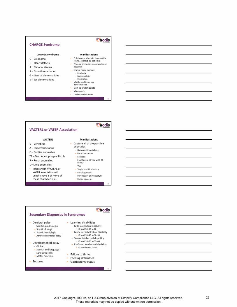
CHARGE Syndrome
CHARGE syndrome
C – Coloboma
H – Heart defects
A – Choanal atresia
R – Growth retardation
G – Genital abnormalities
E – Ear abnormalities
Manifestations• Coloboma – a hole in the eye (iris,
retina, choroid, or optic dis)
• Choanal stenosis – narrowed nasal passages
• Cranial nerve damage
– Dysphagia
– Facial paralysis
– Hearing loss
• Middle and inner ear abnormalities
• Cleft lip or cleft palate
• Micropenis
• Undescended testes
65
VACTERL or VATER Association
VACTERL
V – Vertebrae
A – Imperforate anus
C – Cardiac anomalies
TE – Tracheoesophageal fistula
R – Renal anomalies
L – Limb anomalies
• Infants with VACTERL or VATER association will usually have 3 or more of these characteristics
Manifestations
• Capture all of the possible anomalies– Hypoplastic vertebrae
– Fused vertebrae
– Scoliosis
– Esophageal atresia with TE fistula
– VSD
– Single umbilical artery
– Renal agenesis
– Polydactyly or syndactyly
– Radial agenesis
66
Secondary Diagnoses in Syndromes
• Cerebral palsy– Spastic quadriplegia– Spastic diplegic – Spastic hemiplegic– Athetoid cerebral palsy
• Developmental delay– Global– Speech and language– Scholastic skills– Motor function
• Seizures
• Learning disabilities– Mild intellectual disability
• IQ level 50–55 to 70
– Moderate intellectual disability• IQ level 35–40 to 50–55
– Severe intellectual disability• IQ level 20–25 to 35–40
– Profound intellectual disability• IQ level below 20–25
• Failure to thrive• Feeding difficulties• Gastrostomy status
2017 Copyright, HCPro, an H3.Group division of Simplify Compliance LLC. All rights reserved. These materials may not be copied without written permission.
22

67
Resources
• Cove Point Foundation – Congenital Heart Disease, Helen B. Taussig Children’s Heart Center, Johns Hopkins University
• ICD10data.com – a free reference website designed to look up all current ICD‐10‐CM and ICD‐10‐PCS codes
• NORD – National Organization for Rare Disorders
• Orphanet – a portal for rare diseases and orphan drugs
68
Thank you. Questions?
In order to receive your continuing education certificate(s) for this program, you must complete the online evaluation. The link can be found in the continuing education section at the front of the program guide.
2017 Copyright, HCPro, an H3.Group division of Simplify Compliance LLC. All rights reserved. These materials may not be copied without written permission.
23