acetylation of rb by pcaf is required for nuclear...
TRANSCRIPT
3718 Research Article
IntroductionThe retinoblastoma protein (Rb) belongs to a family of proteinscalled ‘pocket proteins’, which also includes p107 and p130. Theseproteins have common functionality including cell cycle controlvia interactions with E2F transcription factors (Dyson, 1998;Nevins, 1992). However, Rb differs from p107 and p130 in that ithas been shown to have a role in regulating cellular differentiation.Ablation of the Rb1 gene in mice results in embryonic lethalitywith defects in proliferation, cell death and differentiation in variousembryonic tissues (Clarke et al., 1992; Lee et al., 1992). p107- andp130-knockout mice, however, are viable (Cobrinik et al., 1996;Lee et al., 1996). Furthermore, numerous in vivo and in vitromodels of differentiation have shown that Rb is crucial incontrolling the differentiation process (for a review, see Khidr andChen, 2006).
During differentiation, Rb function is altered by post-translationalmodifications. In the switch between actively growing cells anddifferentiated cells, Rb becomes hypophosphorylated (Martinez etal., 1999) and acetylated (Chan et al., 2001; Nguyen et al., 2004).Hypophosphorylation of Rb is required for cell cycle arrest throughsequestration of E2F transcription factors; however, the ability ofRb to induce differentiation is independent of E2F binding (Sellerset al., 1998). Relatively little is known about Rb acetylation but itappears to be essential for differentiation of muscle cells (Nguyenet al., 2004). Acetylation is known to regulate various aspects ofprotein biology including: stability, function (including DNAbinding, transcriptional activity and enzymatic activity), protein-protein interactions and localization (Spange et al., 2009). It issuggested that acetylation regulates interaction with E2F1 in cyclingcells (Markham et al., 2006), yet the effects of acetylation on otheraspects of Rb functions have not been determined. The majoracetylation sites (lysines 873 and 874) reside within a nuclear
localization sequence (NLS) close to the C-terminus of the protein(Zacksenhaus et al., 1993). Rb mutants lacking the NLS cannotinhibit cell growth in Rb-null sarcoma ostogenic (Saos)-2 cellsand, importantly, are unable to induce differentiation (Zacksenhauset al., 1993). We report in this study that, during keratinocytedifferentiation, Rb is acetylated by the acetyltransferase PCAFand, through mutation of lysines 873 and 874, show that acetylationis required for differentiation and to retain Rb within the nucleus.Knockdown of endogenous PCAF levels or exogenous expressionof the deacetylase SIRT1 also resulted in the mislocalization of Rb,suggesting that acetylation is a mechanism for controlling Rblocalization during differentiation.
ResultsRb is acetylated during differentiationRb acetylation has been observed during differentiation of themonoblastoid cell line U937 (Chan et al., 2001) and the murinemuscle cell line C2C12 (Nguyen et al., 2004). Here, the acetylationstatus of Rb was assessed during calcium-mediated differentiationof primary human keratinocytes. Whole-cell lysates were obtainedfrom early pass keratinocytes that were cycling, grown toconfluence (0 hours) or differentiated for various times by serumwithdrawal and addition of 1.5 mM CaCl2. Acetylation was assessedby immunoprecipitation of Rb and western blot analysis to detectacetylated lysine residues (Fig. 1A). Rb was acetylated following72 hours differentiation, confirmed by expression of thedifferentiation marker keratin 1 (K1). Acetylation was observedduring differentiation in three independently isolated batches ofkeratinocytes, and the reciprocal immunoprecipitations using anti-acetylated lysine also confirmed acetylation of Rb duringdifferentiation (data not shown). Protein extracts obtained frommouse skin were also subjected to immunoprecipitation with Rb
Acetylation of Rb by PCAF is required for nuclearlocalization and keratinocyte differentiationAdam Pickard, Ping-Pui Wong and Dennis J. McCance*Centre for Cancer Research and Cell Biology, Queen’s University Belfast, Belfast, BT9 7BL, UK*Author for correspondence ([email protected])
Accepted 9 July 2010Journal of Cell Science 123, 3718-3726© 2010. Published by The Company of Biologists Ltddoi:10.1242/jcs.068924
SummaryAlthough the retinoblastoma protein (Rb) functions as a checkpoint in the cell cycle, it also regulates differentiation. It has recentlybeen shown that Rb is acetylated during differentiation; however, the role of this modification has not been identified. Depletion ofRb levels with short hairpin RNA resulted in inhibition of human keratinocyte differentiation, delayed cell cycle exit and allowed cellcycle re-entry. Restoration of Rb levels rescued defects in differentiation and cell cycle exit and re-entry; however, re-expression ofRb with the major acetylation sites mutated did not. During keratinocyte differentiation, acetylation of Rb is mediated by PCAF andit is further shown that PCAF acetyltransferase activity is also required for normal differentiation. The major acetylation sites in Rbare located within the nuclear localization sequence and, although mutation did not alter Rb localization in cycling cells, the mutantis mislocalized to the cytoplasm during differentiation. Studies indicate that acetylation is a mechanism for controlling Rb localizationin human keratinocytes, with either reduction of the PCAF or exogenous expression of the deacetylase SIRT1, resulting in mislocalizationof Rb. These findings identify PCAF-mediated acetylation of Rb as an event required to retain Rb within the nucleus duringkeratinocyte differentiation.
Key words: Differentiation, Retinoblastoma, Acetylation, PCAF, SIRT1
Jour
nal o
f Cel
l Sci
ence

and acetylated lysine antibodies and identified that Rb is acetylatedin vivo (Fig. 1B). As both p300 and PCAF have previously beenshown to acetylate Rb and both are associated with Rb in mouseskin (Fig. 1B), their expression was monitored during keratinocytedifferentiation. The expression of p300 did not alter significantlyduring differentiation, whereas PCAF levels increased during thetime-course (Fig. 1C). Both p300 and PCAF are known to trafficbetween the nucleus and cytoplasm (Chen et al., 2007; Santos-Rosa et al., 2003); therefore, their localization during keratinocytedifferentiation was investigated. In confluent cells, p300 and PCAFstaining was diffuse throughout the cell. Following initiation ofdifferentiation, p300 became almost exclusively nuclear and PCAFexclusively cytoplasmic (Fig. 2A). However, later in differentiation,both p300 and PCAF were localized in the nucleus.Immunohistochemical staining of human foreskin with antibodiesagainst PCAF also demonstrated that the localization of PCAFwas cytoplasmic in basal epithelial cells (undifferentiated cells)and nuclear in parabasal cells (differentiated; Fig. 2B).Immunohistochemical staining also showed that Rb and p300 arenuclear during skin differentiation (Fig. 2B). In keratinocytesdifferentiated for 72 hours, areas that had nuclear PCAF stainingwere also the regions that were positive for differentiation markers(Fig. 2C). As both p300 and PCAF were localized within thenucleus when Rb was acetylated, levels of the two acetyltransferaseswere individually reduced using short hairpin RNAs (shRNA) todetermine if they are required for keratinocyte differentiation.
p300 and PCAF are required for keratinocytedifferentiationPrimary human foreskin keratinocytes (HFKs) were transducedwith retroviruses that express shRNA targeted to either p300 orPCAF and western blots were used to determine the knockdown(Fig. 3A). Knockdown of either PCAF or p300 levels resulted ininhibition of differentiation, as determined by reduced keratin 1and transglutaminase 1 (TG1) expression (Fig. 3B). Knockdownof PCAF and p300 with alterative shRNA molecules gave similarresults (data not shown). The effects of p300 and PCAF knockdownon Rb acetylation were investigated by an in vitro acetylation
3719Rb acetylation during differentiation
assay. Cell lysates from control or knockdown cells at 0 and 72hours after differentiation were used to acetylate the large pocketof human Rb (GST-Rb-LP). Knockdown of PCAF levels resultedin a reduction of Rb acetylation following 72 hours differentiationcompared with control cells, whereas knockdown of p300 hadlittle effect on Rb acetylation (Fig. 3C). This suggests that althoughp300 and PCAF are required for keratinocyte differentiation, onlyPCAF acetylates Rb. To further investigate the role of PCAFduring differentiation, PCAF levels were depleted using siRNAtargeted to the 3� UTR and then rescued using adenovirus-encodingfull-length PCAF or an acetyltransferase-inactive mutant with adeletion in the active site, PCAF�HAT. The levels of PCAFexpression in keratinocytes were assessed by western blot prior tothe onset of differentiation in organotypic rafts (siPCAF-UTR/AdGFP; Fig. 3D). Knockdown of PCAF reduced the level ofdifferentiation with the expression of keratin 1, keratin 10 andinvolucrin all significantly reduced (Fig. 3E). Re-expression ofPCAF in PCAF-depleted keratinocytes restored the differentiationprogramme, as seen by the re-expression of differentiation markers(siPCAF-UTR/AdPCAF; Fig. 3E). However, expression ofPCAF�HAT was unable to restore differentiation in PCAF-depletedcells (siPCAF-UTR/AdPCAFDHAT; Fig. 3E), indicating that theacetylation function of PCAF is required during differentiation. Infact, the mutant PCAF appeared to act in a dominant-negativemanner and caused a reduction in the expression of differentiationmarkers in control-treated keratinocytes (si-scram/AdPCAFDHAT;Fig. 3E).
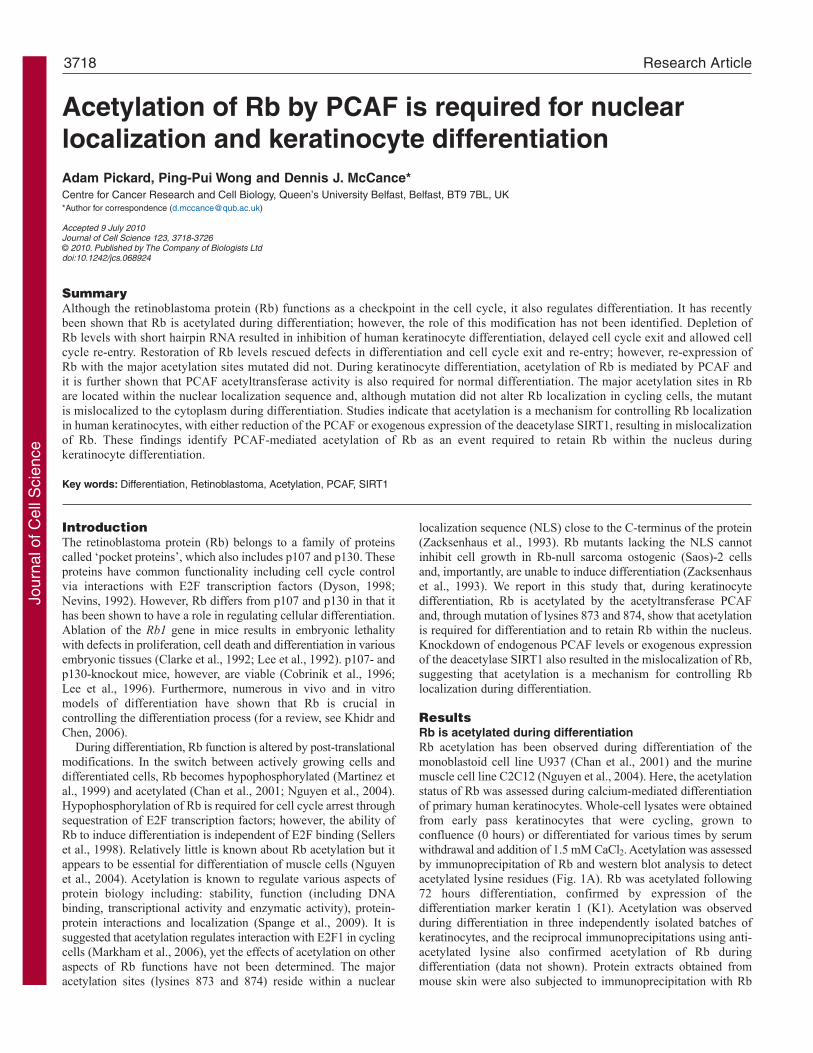
Rb acetylation is required for keratinocyte differentiationIn order to investigate the significance of Rb acetylation in primarykeratinocytes, cells were firstly infected with shRNA targeted tothe Rb1 transcript. HFKs depleted of Rb using two different shRNAmolecules did not differentiate as efficiently as control-infectedHFKs (supplementary material Fig. S1A,B) demonstrated by thereduced expression of keratin 1 and transglutaminase 1. Despitereduced expression of differentiation-specific proteins, Rb-depletedcells have the morphology of differentiated cells as there is anincrease in the formation of cell-cell contacts, as visualized by
Fig. 1. Rb is acetylated during keratinocytedifferentiation. (A)Rb immunoprecipitations from primaryhuman foreskin keratinocytes (HFKs) at different stagesthrough differentiation were analysed by western blotting.Acetylation was detected using an antibody raised againstacetylated lysine (anti-AcK). Differentiation was confirmedin these cells by assessing expression of keratin-1 (K1).(B)Immunoprecipitations from two samples of mouse skinusing either anti-Rb or anti-AcK antibodies. Bothprecipitations identified that pRb is acetylated in vivo andalso that pRb is associated with p300, PCAF and E2F1. * indicates non-specific bands in IgG control. (C) Westernblot analysis of p300 and PCAF during calcium-mediateddifferentiation shows that during differentiation the steady-state levels of PCAF increase whereas p300 remainsrelatively constant; differentiation was monitored by K1 andtransglutaminase-1 expression.
Jour
nal o
f Cel
l Sci
ence

staining for E-cadherin following 72 hours of differentiation. Thissuggests that not all aspects of calcium-mediated differentiationare affected by loss of Rb. Infection of Rb-knockdown HFKs(generated using shRNA targeted to the 3� UTR region of the Rbtranscript) with a recombinant adenovirus encoding either an HA-tagged wild-type Rb (RbWT) or an HA-tagged mutated Rb thatcannot be acetylated (RbKK873/874RR, Ad-RbRR) (Markham etal., 2006; Nguyen et al., 2004; Wong and Weber, 2007; Zhang etal., 1998) was used to assess the role of Rb acetylation duringdifferentiation. RbWT and RbRR expressed by recombinantadenoviruses were functional proteins, as measured by their abilityto arrest Saos-2 cells (supplementary material Fig. S2A). Expressionof RbWT protein was able to restore expression of differentiation-specific markers K1 and TG1 in response to calcium (Fig. 4A) andK1, K10 and loricrin in organotypic culture (supplementary materialFig. S3); however, expression of the acetylation mutant RbRRcould not. The exogenous expression of RbWT and RbRR wasequivalent, as observed by western blotting using an anti-HAantibody (Fig. 4A).
Because expression of RbWT was able to restore K1, TG1 andloricrin expression and RbRR was not, suggests that the acetylationsites are important even during the early stages of differentiation.Rb is known to play a key role in controlling proliferation and cellcycle exit (Buttitta and Edgar, 2007); therefore, we examined
3720 Journal of Cell Science 123 (21)
whether knockdown of Rb in primary keratinocytes would altercell cycle exit and if this could be reversed by expression of RbWTand RbRR. The proliferative capacity was monitored at varioustimes after addition of a differentiation stimulus, using BrdUincorporation to assess the number of cells in S-phase. shRb-UTR-knockdown cells had an increased proportion of cells incorporatingBrdU compared with controls in subconfluent (data not shown)and confluent cultures (Fig. 4B; time 0, shRb-UTR + AdGFP vssh-scram + AdGFP), suggesting that these cells are moreproliferative. Following 24 hours differentiation, control cellsrapidly exited the cell cycle, whereas shRb-UTR cells retainedtheir proliferative capacity, suggesting a delay in cell cycle exit(Fig. 4B). Restoration of Rb levels in shRb-UTR cycling cells withinfection of Ad-RbWT or Ad-RbRR reduced the number ofproliferating cells (Fig. 4B), suggesting that Rb acetylation doesnot have a role controlling the cell cycle. However, upondifferentiation, RbRR-rescued cells were unable to exit the cellcycle as efficiently as cells expressing RbWT and this mightexplain why these cells do not differentiate. This is also apparentin organotypic rafts where BrdU-positive cells were observed inthe suprabasal layers of the cultures (supplementary material Fig.S3). To further assess the differentiation status of these cells, cellswere returned to full growth medium 48 hours after addition of adifferentiation stimulus, shRb-UTR cells infected with Ad-GFP
Fig. 2. Localization of PCAF and p300 during differentiation.(A)Immunofluorescence analysis of differentiating keratinocytesfor expression and localization of p300 and PCAF shows that,shortly after addition of differentiation stimulus, p300 is localizedmainly within the nucleus, whereas PCAF is almost exclusivelycytoplasmic. Following 72 hours differentiation, both PCAF andp300 were identified in the nucleus. Scale bar: 200m.(B)Immunohistochemical staining of human foreskin identifiespRb and p300 localized within the nucleus throughout theepithelium, whereas PCAF stains positively in the cytoplasm of thebasal cells and then becomes nuclear as cells differentiate. Scalebar: 100m. (C)Immunofluorescence analysis of differentiatedkeratinocytes shows that cells with nuclear PCAF are also positivefor the differentiation marker K1. Scale bar: 200m.
Jour
nal o
f Cel
l Sci
ence

showed a significant increase in the percentage of cellsincorporating BrdU (P<0.05), suggesting that they can re-enter thecell cycle and are therefore not terminally differentiated (Fig. 4B;48 + 24 hours recovery). Re-expression of RbWT allowed cells toterminally differentiate whereas RbRR could not prevent cell cyclere-entry suggesting that these cells are not irreversibly arrested(Fig. 3B; 48 + 24 hours recovery). Together, this suggests thatacetylation at lysine residues 873 and 874 is required for thepermanent cell cycle exit of primary human keratinocytes.
Acetylation controls Rb localization during differentiationIn order to understand why RbRR could not rescue differentiationin Rb-knockdown cells, several aspects of Rb function wereinvestigated. RbRR functions identically to RbWT in its ability toreduce cellular proliferation (Fig. 3B; supplementary material Fig.S2A) and this has been attributed to its equivalent ability to bindand inhibit E2F1 transcriptional activity (Markham et al., 2006;Nguyen et al., 2004). It was confirmed that RbRR was able to bindand inhibit E2F1 in primary keratinocytes (data not shown) and,therefore, altered E2F1 activity could not explain the difference infunction during differentiation. Likewise, the mutation does notalter protein stability (data not shown). The acetylation sites in Rbare located within an NLS at the C-terminus of the protein and, asacetylation has been shown to modulate the cellular localization ofnon-histone proteins (Spange et al., 2009), it was postulated that
3721Rb acetylation during differentiation
acetylation might regulate the cellular localization of Rb. Toinvestigate the effects of mutating the acetylation sites on Rblocalization during keratinocyte differentiation, recombinantadenoviruses were generated encoding wild-type Rb and RbRRwith a C-terminal GFP tag. The GFP-tagged Rb proteins were bothfunctional as Ad-GFP-RbWT and Ad-GFP-RbRR were able toarrest Saos-2 cells, whereas Ad-GFP could not (supplementarymaterial Fig. S2B). Like endogenous Rb, both GFP-RbWT andGFP-RbRR were localized to the nucleus in live cycling cells (Fig.5A), whereas GFP alone was diffuse throughout the cells. Followingthe onset of differentiation, both RbWT and RbRR continued to belocalized in the nucleus; however, following 72 hoursdifferentiation, when the majority of GFP-RbWT is in the nucleus,GFP-RbRR was relocated to the cytoplasm (Fig. 5A). This wasconfirmed by fractionation of confluent (t0) and differentiatedHFKs (t72 hours) expressing GFP-RbWT and GFP-RbRR (Fig.5B). It is at this time that Rb acetylation was observed and whenPCAF translocated to the nucleus (Fig. 2), which suggests thatacetylation acts to retain Rb within the nucleus. The separation andpurity of the different fractions was assessed using beta-tubulinand p63 as cytoplasmic and nuclear markers, respectively.Immunofluorescence analysis also confirmed that although theRbRR-expressing cells appear to have a differentiated morphologywith flattened cells and an increased cytoplasm:nuclear ratio, theydo not express the differentiation marker K1 (Fig. 5C). This is
Fig. 3. Knockdown of PCAF inhibits Rb acetylation andkeratinocyte differentiation. (A)Stable knockdownkeratinocytes were generated by retroviral transduction ofshRNA targeted to PCAF and p300. Western blot analysis ofcell lysates demonstrates knockdown of the targetedproteins; levels of Rb and -actin show equal loading.(B)Stable knockdown cell lines, generated as described inA, were differentiated using calcium treatment. Neither sh-PCAF- nor sh-p300-knockdown cells differentiated asefficiently as control cells, as determined by keratin 1 (K1)and transglutaminase-1 (TG1) expression. (C)Lysates fromconfluent (t0) and cells differentiated for 72 hours wereused to acetylate GST-Rb-LP. Induction of acetylation wasobserved in differentiated control cell lysates, as detected byan anti-acetylated lysine (AcK) antibody. However, Rbacetylation was reduced in PCAF-knockdown cell lysates,whereas it was unaffected by reduction of p300 levels.(D)Expression of PCAF in PCAF-depleted cells (siPCAF)and when wild-type (AdPCAF) or an acetylase-deficientmutant (Ad-PCAFDHAT) PCAF is re-expressed usingadenoviruses. PCAF�HAT lacks 28 amino acids and resultsin a band running 3 kDa below full-length PCAF.(E)Expression of K1, K10 and involucrin in 5-dayorganotypic cultures of the keratinocytes generated in D,showing that re-expression of wild-type PCAF, but not theacetylase-deficient mutant, is able to rescue differentiation.Scale bar: 100m.Jo
urna
l of C
ell S
cien
ce
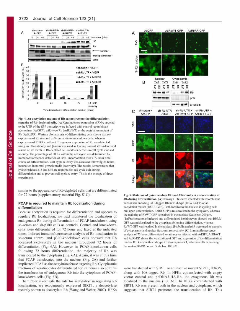
similar to the appearance of Rb-depleted cells that are differentiatedfor 72 hours (supplementary material Fig. S1C).
PCAF is required to maintain Rb localization duringdifferentiationBecause acetylation is required for differentiation and appears toregulate Rb localization, we next monitored the localization ofendogenous Rb during differentiation of PCAF knockdown usingsh-scram and sh-p300 cells as controls. Control and knockdowncells were differentiated for 72 hours and fixed at the indicatedtimes. Indirect immunofluorescence analysis of Rb localization insh-scram control and p300-knockdown cells showed that Rblocalized exclusively in the nucleus throughout 72 hours ofdifferentiation (Fig. 6A). However, in PCAF-knockdown cellsfollowing 72 hours differentiation, the majority of Rb wastranslocated to the cytoplasm (Fig. 6A). Again, it was at this timethat PCAF translocated into the nucleus (Fig. 2A) and furtherimplicated PCAF as the acetyltransferase targeting Rb. Cytoplasmicfractions of keratinocytes differentiated for 72 hours also confirmthe translocation of endogenous Rb into the cytoplasm of PCAF-knockdown cells (Fig. 6B).
To further investigate the role for acetylation in regulating Rblocalization, we exogenously expressed SIRT1, a deacetylaserecently shown to deacetylate Rb (Wong and Weber, 2007). HFKs
3722 Journal of Cell Science 123 (21)
were transfected with SIRT1 or an inactive mutant SIRT1, H363Y,along with HA-tagged Rb. In HFKs cotransfected with emptyvector control and pcDNA3-HA-Rb, the exogenous Rb waslocalized to the nucleus (Fig. 6C). In HFKs cotransfected withSIRT1, Rb was present both in the nucleus and cytoplasm, whichsuggests that SIRT1 promotes the translocation of Rb. This
Fig. 4. An acetylation mutant of Rb cannot restore the differentiationcapacity of Rb-depleted cells. (A)Keratinocytes expressing shRNA targetedto the UTR of the Rb1 transcript were infected with control recombinantadenovirus (AdGFP), wild-type Rb (AdRbWT) or the acetylation mutant ofRb (AdRbRR). Western blot analysis of differentiating cells shows that re-expression of Rb restored differentiation to knockdown cells, whereasexpression of RbRR could not. Exogenous expression of Rb was detectedusing an HA-antibody and -actin was used as loading control. (B)Adenoviralrescue of Rb levels in Rb-depleted cells restores defects in cell cycle exit andre-entry. The percentage of HFKs within the cell cycle was determined byimmunofluorescence detection of BrdU incorporation over a 72-hour time-course of differentiation. Cell cycle re-entry was assessed following 24 hoursincubation in normal growth media (recovery). The results demonstrated thatlysine residues 873 and 874 are required for cell cycle exit duringdifferentiation and to prevent cell cycle re-entry. This is the average of threeexperiments.
Fig. 5. Mutation of lysine residues 873 and 874 results in mislocalization ofRb during differentiation. (A)Primary HFKs were infected with recombinantadenovirus encoding GFP-tagged Rb in wild-type (RbWT-GFP) or anacetylation mutant (RbRR-GFP). Both localize to the nucleus in cycling cellsbut, upon differentiation, RbRR-GFP is mislocalized to the cytoplasm, whereasthe majority of RbWT-GFP is retained in the nucleus. Scale bar: 200m.(B)Fractionation of infected and differentiated keratinocytes showed that RbRR-GFP was mislocalized to the cytoplasm following differentiation, whereasRbWT-GFP was retained in the nucleus. -tubulin and p63 were used as markersof cytoplasmic and nuclear fractions, respectively. (C)Immunofluorescenceanalysis of 72-hour differentiated keratinocytes infected with AdGFP, AdRbWTand AdRbRR shows the localization of GFP and expression of the differentiationmarker K1. Cells with wild-type Rb also express K1, whereas cells expressingthe mutant RbRR do not. Scale bar: 100 M.
Jour
nal o
f Cel
l Sci
ence

translocation was dependent on SIRT1 deacetylase activity as, incells transfected with the mutant SIRT1-H363Y, Rb remained inthe nucleus. In addition, expression of SIRT2 (localized in thecytoplasm) and SIRT6 (localized in the nucleus) had no effect onRb localization (Fig. 6C). Quantification of the percentage of cellswhere Rb staining was evident in the cytoplasm of cotransfectedHFKs is shown in Fig. 6D. Similar effects of SIRT1 mislocalizationof Rb were also observed in Saos-2 cells cotransfected withpcDNA3-HA-Rb (Fig. 6E). Taken together, these results suggestthat acetylation is a mechanism for retaining Rb within the nucleusduring differentiation.
Rb maintains keratinocyte differentiationOur data suggests that acetylation is required to retain Rb withinthe nucleus during the later stages of differentiation. In order to testwhy cells might require Rb at this time, an Rb-specfic RNAimolecule was transfected into HFKs that had been differentiatedfor 72 hours. At this time, almost all cells had exited the cell cycleand control cells could not be induced to re-enter the cycle (Fig.4B). Following transfection, cells were grown for 24 hours in fullgrowth medium and pulsed with BrdU. The number of cells in S-phase was determined by immunofluorescence. The reduction ofRb levels resulted in a significant increase in the number of cells
3723Rb acetylation during differentiation
able to re-enter the cell cycle, suggesting that the continuedexpression of Rb during differentiation is important for terminaldifferentiation (Fig. 7A). Western blot analysis was used to confirmthe knockdown of Rb levels (Fig. 7B). Blotting also showed thatRb depletion resulted in a reduced expression of K1, TG1 andloricrin, suggesting that these cells have either dedifferentiated or,because they have not permanently exited the cell cycle, have notterminally differentiated and so exhibit low levels of differentiationmarkers (Fig. 7B). This can be more clearly observed where cellsthat are positive for BrdU have reduced K1 staining in si-Rb-treated cultures (Fig. 7C). Co-staining Rb-depleted cells for BrdUand E-cadherin shows that BrdU-positive cells have a differentiatedflattened morphology (supplementary material Fig. S1C). Three-dimensional visualisation of confocal images shows that BrdU-positive nuclei are in the same plane as the E-cadherin staining(supplementary material Fig. S4). Although the Rb depleted cellscan re-enter the cell cycle on growth stimulation, they do notcontinue to proliferate as cells replated after 72 hours ofdifferentiation do not grow out into colonies (data not shown). Insummary, our results indicate that Rb acetylation is an importantmodification that acts to retain Rb within the nucleus duringdifferentiation, an event required to maintain the differentiatedstate.
Fig. 6. Acetylation controls Rb localization. (A)Control, sh-PCAF and sh-p300 retroviral stable keratinocyte lines weredifferentiated with calcium for the indicated times and cells werefixed and stained with an anti-Rb antibody. In control and p300-knockdown cells, Rb localized to the nucleus throughoutdifferentiation, whereas, in sh-PCAF cells, Rb was mainlycytoplasmic following 72 hours differentiation. Scale bar:100m. (B)Following 72 hours differentiation, nuclear andcytoplasmic fractions were separated from the stable lines usedin A and western blotting showed that endogenous Rb levelswere significantly reduced in the nucleus of sh-PCAF cells butnot in the scrambled control (sh-scram). -tubulin and p63 wereused as cytoplasmic and nuclear markers.(C)Immunofluorescence analysis of primary humankeratinocytes cotransfected with pcDNA3-HA-Rb and Flag-tagged members of the family of SIRT deacetylases showed thatSIRT1 mislocalizes Rb to the cytoplasm. A mutant form ofSIRT1 (SIRT1-H363Y) that lacks deacetylase activity did notmislocalize Rb. Mislocalization of Rb is a targeted function ofSIRT1, as expression of SIRT2 or SIRT6 did not alter Rblocalization. Scale bar: 100m. (D,E)Keratinocytes and Saos-2cells transfected as in C were counted based on the localizationof Rb. The percentage of cells with cytoplasmic Rb staining isshown from three independent experiments.
Jour
nal o
f Cel
l Sci
ence

DiscussionThis study shows that Rb is a key regulator of differentiation andthat acetylation is an important modification during this process.We have investigated the role of Rb acetylation in keratinocytedifferentiation by mutating the major acetylation sites, lysines 873and 874, to arginine and then determining the ability of the mutantto restore differentiation in Rb-knockdown keratinocytes. Mutationof the acetylation sites did not affect the ability of Rb to inhibit theproliferation, probably because of the fact that RbRR can interactwith and inhibit E2F1 (Markham et al., 2006). This also suggeststhat inhibition of E2F family members is not sufficient to induceterminal differentiation, as has previously reported in the Saos-2cell line (Sellers et al., 1998). However, unlike wild-type Rb, theacetylation mutant is unable to induce either early or latedifferentiation markers and, because Rb is acetylated relatively lateduring differentiation, this suggests that either the early events areacetylation-independent or the mutated lysine residues can impactfunctions of Rb. The delay in cell cycle exit observed with theacetylation mutant might also be independent of acetylation,although again we show that lysine residues 873 and 874 appearto be important in controlling the exit. Overall, it appears that Rbis required for permanent cell cycle exit and differentiation but theearly functions might be acetylation-independent. Recently, it hasbeen identified that lysine 873 is methylated and mutation of this
3724 Journal of Cell Science 123 (21)
residue alone was shown to delay the onset of differentiation inSaos-2 and C2C12 cells (Munro et al., 2010), suggesting importantroles for these residues.
Although acetylation of Rb at lysines 873 and 874 has beenshown to be mediated by both p300 (Chan et al., 2001) and PCAF(Chan et al., 2001; Nguyen et al., 2004), we show here that onlyknockdown of PCAF reduces Rb acetylation, alters its localizationduring keratinocyte differentiation and inhibits expression ofdifferentiation markers, implying that PCAF might be the majoracetyltransferase targeting Rb in vivo. In addition, acetylation ofRb, nuclear localization and expression of differentiation markerscan be reinstated by exogenous expression of active PCAF but notan acetylase-dead mutant. These results also suggest that p300cannot compensate for the loss of PCAF and confirm previousreports that knockdown of p300 does not affect Rb acetylationduring cell cycle (Iyer et al., 2007). We have recently demonstratedthat p300 knockdown inhibits keratinocyte differentiation viaregulation of p21WAF1/CIP1 expression (Wong et al., 2010) and ourobservation that Rb is acetylated in p300-depleted cells suggeststhat although acetylation of Rb is required during the differentiationprocess, alone it is not sufficient to induce differentiation.
The identification of a low-penetrance mutation of Rb1, whereexons 24 and 25 are deleted, led us to investigate whetheracetylation would affect Rb localization. This mutation generatesa protein lacking amino acids 830-888, which removes the NLSand acetylation site of Rb, resulting in its mislocalization to thecytoplasm (Bremner et al., 1997). We have shown that a mutantlacking the acetylation sites is translocated to the cytoplasm upondifferentiation, suggesting that acetylation acts to retain Rb in thenucleus. Additionally, either knockdown of PCAF or overexpressionof SIRT1, a deacetylase, also led to mislocalization of Rb. AlteredRb localization has been described previously in an array of cancercell lines and primary cancer tissues, with cytoplasmic or nuclear-cytoplasmic staining of Rb observed in approximately 90% and60% of tested samples, respectively (Jiao et al., 2008). This wasattributed to aberrant phosphorylation of Rb in at least some of thecell lines (Jiao et al., 2006), although our data would suggest thatacetylation is another important modification that could control Rblocalization. Intriguingly, mislocalization of Rb also correlatedwith moderate and poorly differentiated primary cancer specimens(Jiao et al., 2008). It has also been proposed that acetylation caninhibit cyclin-E–CDK2-mediated phosphorylation of Rb in in vitrophosphorylation assays (Chan et al., 2001); however, duringdifferentiation, Rb becomes hypophosphorylated and, therefore,acetylation might be an independent method to control Rblocalization.
In addition, our results imply that Rb has an important role in thecontrol of differentiation even when the majority of cells have exitedthe cell cycle, suggesting that Rb is required to maintaindifferentiation. It is not clear at present if the role of Rb is tomaintain permanent cell cycle exit or if this plus other functions ofRb are required for successful differentiation. Rb-depleted cells dochange to a differentiation-like morphology, with a high cytoplasmto nuclear ratio, but do not express early or late differentiation-specific markers and a portion of these cells can re-enter the cellcycle. Our results are consistent with a number of recent reportswhere inhibition of Rb function allows differentiated retinal ormuscle cells to re-enter the cell cycle (Ajioka and Dyer, 2008; Blaiset al., 2007). Our findings are also supported by recent work that hasidentified that inactivation of Rb and Hippo in Drosophila inducesdedifferentiation in the retina (Nicolay et al., 2010).
Fig. 7. Rb is required throughout differentiation of primary keratinocytes.(A)Following 72 hours of differentiation, Rb levels were depleted using siRNAand BrdU incorporation was measured 24 hours later. Data is from threeindependent experiments and represents counts from at least 2000 cells in eachexperiment. (B)Western blots of cells treated as in A showed significantdepletion of Rb and a reduction in differentiation marker expression.(C)Immunofluorescence demonstrated that knockdown of Rb allowed cells tore-enter the cell cycle, with reduced K1 expression. Scale bar: 100 M.(D)E-cadherin and BrdU staining of cells treated as in A show that cellspositive for BrdU have a differentiated-like morphology. Scale bar: 200 M.Jo
urna
l of C
ell S
cien
ce

3725Rb acetylation during differentiation
In summary, our results show that acetylation functions to retainRb in the nucleus in the later stages of differentiation in order tomaintain Rb function and, in turn, maintain permanent cell cyclearrest.
Materials and MethodsKeratinocyte culture and generation of stable knockdown cell linesPrimary human foreskin keratinocytes (HFK) were isolated from neonatal foreskinsand grown in Epilife medium (Cascade) containing human keratinocyte growthsupplement (HKGS, Cascade). Keratinocytes were transduced with retrovirusproduced in fNYX-GP packaging cell line as described previously (Guess andMcCance, 2005; Rheinwald and Green, 1975), using the following plasmids: pSuper-retro constructs expressing short hairpin RNAs (shRNA) against no known annotatedgene (sh-scram) or targeting the Rb coding region (sh-Rb-CR) or 3� UTR (sh-Rb-UTR) were cloned as described previously (Incassati et al., 2006); p300- and PCAF-knockdown lines were generated using plasmids purchased from Origene. Todifferentiate HFKs, the cells were grown to confluence in the medium describedabove and then differentiation was induced by addition of 1.5 mM calcium chlorideand withdrawal of HKGS (Fang et al., 1998). Organotypic raft culture was asdescribed previously (Menges et al., 2006).
Generation of adenovirusRecombinant adenoviruses were cloned using ViraPower Adenoviral GatewayExpression Kit (Invitrogen). To generate AdGFP, pMAX-GFP (Amaxa) was digestedwith KpnI and XhoI and inserted into the same sites of pENTR11. AdRbWT andAdRbRR were generated by first subcloning the NcoI-XhoI fragment from pSG5-RbWT or pSG5-RbRR (Miyake et al., 2000; Nguyen et al., 2004) into the same sitesof pENTR11 (pENTR11-NcoI-RbWT-XhoI and pENTR11-NcoI-RbRR-XhoI). Thenthe NcoI fragment from the pSG5-RbWT was introduced into pENTR11-NcoI-RbWT-XhoI and pENTR11-NcoI-RbRR-XhoI and the directionality confirmed bysequencing.
To generate GFP-tagged Rb, EGFP was first excised from pEGFP-N1 by XhoIand NotI digestion and ligated into SalI and NotI in pENTR11. The HindIII-EcoRIand EcoRI-EcoRI fragments of pSG5-RbWT were ligated into the vector to generatepENTR-RbWT-stop-N1-GFP. Finally, PCR was conducted to remove the stop codonusing the following primers: forward, TcTTgAATTcgcTAgccTATcTccg; and reverse,AgAAggTAccTTcTcTTccTTgTT. pSG5-RbWT or pSG5-RbRR was used as template.The PCR product was digested with DraIII and KpnI and ligated into the same sitesof pENTR-RbWT-stop-N1-GFP. AdPCAF and AdPCAF�HAT were generated byligating the EcoRI and KpnI fragment of pCX-PCAF and pCX-PCAF�HAT, asdescribed previously (Santos-Rosa et al., 2003), into the same sites of pENTR11 andthe orientation was corrected by digesting the resulting vector with SalI and EcoRIand ligating into EcoRI and XhoI of pENTR11. All constructs were then recombinedinto pAd/CMV/V5-DEST. All recombinant adenoviruses were generated usingHEK293 cells and were purified and titrated as previously described (Enquist andLowenstein, 1996).
Transient transfectionFugene HD was used for transfection of siRNA; control and Rb1 siRNA werepurchased from Ambion. siRNA targeting the UTR of PCAF was as previouslydescribed (Linares et al., 2007): 24 hours after transfection, cells were infected withadenovirus encoding PCAF. Fugene 6 was used for transfection of keratinocyteswith plasmid DNA, whereas Fugene HD (Roche) was used for siRNA transfections.pECE-Flag-SIRT1 and pECE-Flag-SIRT1-H363Y, described in Brunet et al. (Brunetet al., 2004), were obtained from Addgene (plasmids 1791 and 1792). pcDNA3-Flag-SIRT2 and pcDNA3-Flag-SIRT6, described in North et al. (North et al., 2003),were also obtained from Addgene (plasmids 13813 and 13817).
ImmunofluorescenceTo determine proliferation in cell cultures, cells were pulsed for 15 minutes with 10M BrdU prior to fixing with 4% paraformaldehyde. Following antigen retrievalwith citrate buffer (DAKO), cells were stained with 1:200 dilution of anti-BrdU (BDBiosciences). The number of BrdU-positive cells and DAPI-counterstained cellswere counted to determine the percentage of proliferating cells. For organotypicrafts, sections were deparaffinised with xylene and then rehydrated with step-downconcentrations of ethanol. Sections were blocked using 10% bovine calf serum and0.2% Triton X-100. Sections were then stained with anti-keratin-1 (1:2000, Covance),anti-keratin-10 (1:100, Biodesign) or anti-loricrin (1:100; Neomarkers). Secondaryantibodies were goat anti-mouse and goat anti-rabbit conjugated to fluorophores 488or 594 (1:400, Invitrogen). Sections were mounted with ProLong Gold AntifadeReagent plus DAPI (Invitrogen).
Protein extracts, immunoprecipitation and in vitro acetylation assaysWhole-cell lysates for western blot were generated using urea buffer (8 M urea, 50mM Tris pH 7.5, 0.1% v/v beta-mercaptoethanol and protease inhibitor cocktail;Roche). Cell fractions were prepared as previously described (Sen et al., 2006). Forimmunoprecipitation and in vitro acetylation assays, cells were lysed in 50 mM Tris
pH 7.5, 150 mM NaCl, 0.5% NP40, 10 mM EDTA, 5 M trichostatin A and proteininhibitor cocktail. For Rb immunoprecipitation, 1 mg protein lysate was used with1 g anti-Rb (BD Biosciences) and 10 L Protein-G agarose (Santa Cruz). For invitro acetylation, 500 g protein lysate was incubated with 0.3 g purified GST-Rb-LP, as previously described (Nguyen et al., 2004), for 4 hours at 30°C. Additionalantibodies used in this study were: monoclonal IgG2A anti-acetylated lysine (CellSignaling), goat anti-mouse IgG2A (Santa Cruz), goat anti-mouse IgG1 (SantaCruz), mouse anti-p300 (Abnova), rabbit anti-p300 (Santa Cruz), anti-PCAF (SantaCruz), anti-HA (Santa Cruz), anti-beta actin (Sigma), anti-TBP (Abcam), anti--tubulin (Sigma) and anti-Flag 488 (Sigma).
The research was supported by a Wellcome Trust grant,WT082840AIA. For help in obtaining confocal images, we would liketo thank Karen McCloskey, Queens University Belfast. For generationof 3D images, we would like to thank Perkin Elmer for use of the 3Dvisualisation module of its Volocity software. Deposited in PMC forrelease after 6 months.
Supplementary material available online athttp://jcs.biologists.org/cgi/content/full/123/21/3718/DC1
ReferencesAjioka, I. and Dyer, M. A. (2008). A new model of tumor susceptibility following tumor
suppressor gene inactivation. Cell Cycle 7, 735-740.Blais, A., van Oevelen, C. J., Margueron, R., Acosta-Alvear, D. and Dynlacht, B. D.
(2007). Retinoblastoma tumor suppressor protein-dependent methylation of histone H3lysine 27 is associated with irreversible cell cycle exit. J. Cell Biol. 179, 1399-1412.
Bremner, R., Du, D. C., Connolly-Wilson, M. J., Bridge, P., Ahmad, K. F., Mostachfi,H., Rushlow, D., Dunn, J. M. and Gallie, B. L. (1997). Deletion of RB exons 24 and25 causes low-penetrance retinoblastoma. Am. J. Hum. Genet. 61, 556-570.
Brunet, A., Sweeney, L. B., Sturgill, J. F., Chua, K. F., Greer, P. L., Lin, Y., Tran, H.,Ross, S. E., Mostoslavsky, R., Cohen, H. Y. et al. (2004). Stress-dependent regulationof FOXO transcription factors by the SIRT1 deacetylase. Science 303, 2011-2015.
Buttitta, L. A. and Edgar, B. A. (2007). Mechanisms controlling cell cycle exit uponterminal differentiation. Curr. Opin. Cell Biol. 19, 697-704.
Chan, H. M., Krstic-Demonacos, M., Smith, L., Demonacos, C. and La Thangue, N.B. (2001). Acetylation control of the retinoblastoma tumour-suppressor protein. Nat.Cell Biol. 3, 667-674.
Chen, J., Halappanavar, S., Th’ng, J. P. and Li, Q. (2007). Ubiquitin-dependentdistribution of the transcriptional coactivator p300 in cytoplasmic inclusion bodies.Epigenetics 2, 92-99.
Clarke, A. R., Maandag, E. R., van Roon, M., van der Lugt, N. M., van der Valk, M.,Hooper, M. L., Berns, A. and te Riele, H. (1992). Requirement for a functional Rb-1gene in murine development. Nature 359, 328-330.
Cobrinik, D., Lee, M. H., Hannon, G., Mulligan, G., Bronson, R. T., Dyson, N.,Harlow, E., Beach, D., Weinberg, R. A. and Jacks, T. (1996). Shared role of the pRB-related p130 and p107 proteins in limb development. Genes Dev. 10, 1633-1644.
Dyson, N. (1998). The regulation of E2F by pRB-family proteins. Genes Dev. 12, 2245-2262.
Enquist, L. W. and Lowenstein, P. R. (1996). Protocols For Gene Transfer inNeuroscience: Towards Gene Therapy of Neurological Disorders. Chichester, NY:Wiley and Sons.
Fang, K. S., Farboud, B., Nuccitelli, R. and Isseroff, R. R. (1998). Migration of humankeratinocytes in electric fields requires growth factors and extracellular calcium. J.Invest. Dermatol. 111, 751-756.
Guess, J. C. and McCance, D. J. (2005). Decreased migration of Langerhans precursor-like cells in response to human keratinocytes expressing human papillomavirus type 16E6/E7 is related to reduced macrophage inflammatory protein-3alpha production. J.Virol. 79, 14852-14862.
Incassati, A., Patel, D. and McCance, D. J. (2006). Induction of tetraploidy through lossof p53 and upregulation of Plk1 by human papillomavirus type-16 E6. Oncogene 25,2444-2451.
Iyer, N. G., Xian, J., Chin, S. F., Bannister, A. J., Daigo, Y., Aparicio, S., Kouzarides,T. and Caldas, C. (2007). p300 is required for orderly G1/S transition in human cancercells. Oncogene 26, 21-29.
Jiao, W., Datta, J., Lin, H. M., Dundr, M. and Rane, S. G. (2006). Nucleocytoplasmicshuttling of the retinoblastoma tumor suppressor protein via Cdk phosphorylation-dependent nuclear export. J. Biol. Chem. 281, 38098-38108.
Jiao, W., Lin, H. M., Datta, J., Braunschweig, T., Chung, J. Y., Hewitt, S. M. andRane, S. G. (2008). Aberrant nucleocytoplasmic localization of the retinoblastomatumor suppressor protein in human cancer correlates with moderate/poor tumordifferentiation. Oncogene 27, 3156-3164.
Khidr, L. and Chen, P. L. (2006). RB, the conductor that orchestrates life, death anddifferentiation. Oncogene 25, 5210-5219.
Lee, E. Y., Chang, C. Y., Hu, N., Wang, Y. C., Lai, C. C., Herrup, K., Lee, W. H. andBradley, A. (1992). Mice deficient for Rb are nonviable and show defects in neurogenesisand haematopoiesis. Nature 359, 288-294.
Lee, M. H., Williams, B. O., Mulligan, G., Mukai, S., Bronson, R. T., Dyson, N.,Harlow, E. and Jacks, T. (1996). Targeted disruption of p107: functional overlapbetween p107 and Rb. Genes Dev. 10, 1621-1632.
Jour
nal o
f Cel
l Sci
ence

3726 Journal of Cell Science 123 (21)
Linares, L. K., Kiernan, R., Triboulet, R., Chable-Bessia, C., Latreille, D., Cuvier, O.,Lacroix, M., Le Cam, L., Coux, O. and Benkirane, M. (2007). Intrinsic ubiquitinationactivity of PCAF controls the stability of the oncoprotein Hdm2. Nat. Cell Biol. 9, 331-338.
Markham, D., Munro, S., Soloway, J., O’Connor, D. P. and La Thangue, N. B. (2006).DNA-damage-responsive acetylation of pRb regulates binding to E2F-1. EMBO Rep.7, 192-198.
Martinez, L. A., Chen, Y., Fischer, S. M. and Conti, C. J. (1999). Coordinated changesin cell cycle machinery occur during keratinocyte terminal differentiation. Oncogene18, 397-406.
Menges, C. W., Baglia, L. A., Lapoint, R. and McCance, D. J. (2006). Humanpapillomavirus type 16 E7 up-regulates AKT activity through the retinoblastoma protein.Cancer Res. 66, 5555-5559.
Miyake, S., Sellers, W. R., Safran, M., Li, X., Zhao, W., Grossman, S. R., Gan, J.,DeCaprio, J. A., Adams, P. D. and Kaelin, W. G., Jr (2000). Cells degrade a novelinhibitor of differentiation with E1A-like properties upon exiting the cell cycle. Mol.Cell. Biol. 20, 8889-8902.
Munro, S., Khaire, N., Inche, A., Carr, S. and La Thangue, N. B. (2010). Lysinemethylation regulates the pRb tumour suppressor protein. Oncogene 29, 2357-2367.
Nevins, J. R. (1992). E2F: a link between the Rb tumor suppressor protein and viraloncoproteins. Science 258, 424-429.
Nguyen, D. X., Baglia, L. A., Huang, S. M., Baker, C. M. and McCance, D. J. (2004).Acetylation regulates the differentiation-specific functions of the retinoblastoma protein.EMBO J. 23, 1609-1618.
Nicolay, B. N., Bayarmagnai, B., Moon, N. S., Benevolenskaya, E. V. and Frolov, M.V. (2010). Combined inactivation of pRB and hippo pathways induces dedifferentiationin the Drosophila retina. PLoS Genet. 6, e1000918.
North, B. J., Marshall, B. L., Borra, M. T., Denu, J. M. and Verdin, E. (2003). Thehuman Sir2 ortholog, SIRT2, is an NAD+-dependent tubulin deacetylase. Mol. Cell 11,437-444.
Rheinwald, J. G. and Green, H. (1975). Serial cultivation of strains of human epidermalkeratinocytes: the formation of keratinizing colonies from single cells. Cell 6, 331-343.
Santos-Rosa, H., Valls, E., Kouzarides, T. and Martinez-Balbas, M. (2003). Mechanismsof P/CAF auto-acetylation. Nucleic Acids Res. 31, 4285-4292.
Sellers, W. R., Novitch, B. G., Miyake, S., Heith, A., Otterson, G. A., Kaye, F. J.,Lassar, A. B. and Kaelin, W. G., Jr (1998). Stable binding to E2F is not required forthe retinoblastoma protein to activate transcription, promote differentiation, and suppresstumor cell growth. Genes Dev. 12, 95-106.
Sen, A., Agresti, D. and Mackow, E. R. (2006). Hyperphosphorylation of the rotavirusNSP5 protein is independent of serine 67, NSP2, or the intrinsic insolubility of NSP5and is regulated by cellular phosphatases. J. Virol. 80, 1807-1816.
Spange, S., Wagner, T., Heinzel, T. and Kramer, O. H. (2009). Acetylation of non-histone proteins modulates cellular signalling at multiple levels. Int. J. Biochem. CellBiol. 41, 185-198.
Wong, P. P., Pickard, A. and McCance, D. J. (2010). p300 alters keratinocyte cell growthand differentiation through regulation of p21(Waf1/CIP1). PLoS ONE 5, e8369.
Wong, S. and Weber, J. D. (2007). Deacetylation of the retinoblastoma tumour suppressorprotein by SIRT1. Biochem. J. 407, 451-460.
Zacksenhaus, E., Bremner, R., Phillips, R. A. and Gallie, B. L. (1993). A bipartitenuclear localization signal in the retinoblastoma gene product and its importance forbiological activity. Mol. Cell. Biol. 13, 4588-4599.
Zhang, W., Bone, J. R., Edmondson, D. G., Turner, B. M. and Roth, S. Y. (1998).Essential and redundant functions of histone acetylation revealed by mutation of targetlysines and loss of the Gcn5p acetyltransferase. EMBO J. 17, 3155-3167.
Jour
nal o
f Cel
l Sci
ence