acid-induced gelation of milk protein concentrates with added pectin-effect of casein micelle...
TRANSCRIPT
ARTICLE IN PRESS
FOODHYDROCOLLOIDS
0268-005X/$ - s
doi:10.1016/j.fo
�CorrespondE-mail addr
1Current add
Palmerston No
Food Hydrocolloids 21 (2007) 765–775
www.elsevier.com/locate/foodhyd
Acid-induced gelation of milk protein concentrates with added pectin:Effect of casein micelle dissociation
Lara Matia-Merino1, Harjinder Singh�
Riddet Centre, Massey University, Private Bag 11 222, Palmerston North, New Zealand
Received 16 May 2006; accepted 4 December 2006
Abstract
The dynamics of the formation of the acid gel network for mixtures of milk protein concentrate (MPC) and low methoxyl amidated
(LMA) pectin were studied using rheological measurements. The results as a function of pectin content and casein micelle integrity, from
neutral pH to approximately pH 4.2, together with the microstructural changes observed in some of these systems, are presented.
The gelation profiles of a mixture of 4% w/v MPC and LMA pectin (0–0.075% w/v) after the addition of 1.2% w/v glucono-d-lactoneshowed a gradual decrease in the shear modulus with the incorporation of pectin. The effects of casein micelle integrity on casein–pectin
interactions were studied, by preparing MPC dispersions containing various levels of micellar casein. A gradual change in the shear
modulus, from a disrupting effect of pectin added to MPC, in which the casein micelles are intact, to a clear synergistic effect of pectin
added to dissociated casein systems, was found in the acid-induced milk gels.
r 2007 Elsevier Ltd. All rights reserved.
Keywords: Acid milk gels; Pectin; Milk protein concentrates; Rheology; Calcium; CSLM
1. Introduction
The rheology and the structure of milk protein gels havebeen the subject of many studies during the last decade, asrecently reviewed by a number of authors (Lucey, 2002;van Vliet, Lakemond, & Visschers, 2004). Whereas theaggregation and gelation of casein micelles as a result ofacidification have been studied frequently (Lucey & Singh,2003), the presence of hydrocolloids during the acid-induced gelation presents another degree of complexity ofmilk protein systems that needs to be further explored. Thekinetics and the dynamics of the formation of casein gelscan be controlled by varying the amount of glucono-d-lactone (GDL) to produce different rates of acidification(Horne, 2001). The first stages of acidification are criticalfor the development of the network and the aggregationprocess appears to be more complicated than simply
ee front matter r 2007 Elsevier Ltd. All rights reserved.
odhyd.2006.12.007
ing author. Tel.: +646 350 4401; fax: +64 6 350 5655.
ess: [email protected] (H. Singh).
ress: Institute of Food, Nutrition and Human Health,
rth, Massey University, New Zealand.
reaching a critical pH for the collapse of the hairy k-caseinlayer on the micelles, as believed until recently (De Kruif,1997).Pectin is an anionic carboxylated polysaccharide that is
frequently used in the food industry. The proportion ofcarboxyl groups esterified with methanol and with acidamide units and the ester distribution along the polymerdetermine the functionality of pectin (May, 2000; Morris,1998; Voragen, Pilnik, Thibault, Axelos, & Renard, 1995).Pectin is widely used in many dairy products as a gelling/thickening agent (acid and non-acid milk desserts) and as astabilizer ingredient (acid milk drinks, milk/juice blends).In particular, the gelation of low methoxyl pectin, over awide range of pH and solids content, is mainly the result ofstrong interactions between calcium ions and blocks ofgalacturonic acid (Braccini & Perez, 2001).Previous rheological and microstructural studies have
shown that low methoxyl amidated (LMA) pectin is achallenging polysaccharide to investigate when it is presentduring the acidification of casein-based systems (Matia-Merino, Lau, & Dickinson, 2004). The fact that (1) ioniccalcium is released into the serum phase during the
ARTICLE IN PRESSL. Matia-Merino, H. Singh / Food Hydrocolloids 21 (2007) 765–775766
acidification of casein micelles (Law & Leaver, 1998; Singh,Roberts, Munro, & Teo, 1996 ), creating favourable gellingconditions for the pectin, and (2) adsorption of pectinaround the casein particles through electrostatic interactionseems to occur at or below pH 5.0 (Tuinier, Rolin, & deKruif, 2002) makes casein micelles and pectin an interestingbiopolymer mixture that could result in different gelnetworks upon acidification.
Through its role in the ionization of the protein andpectin molecules, pH is the most significant factor affectingthe electrostatic protein–pectin interactions. At neutral pH,where both casein micelles and pectin carry negativecharges, the interaction is minimal (Oakenfull & Scott,1998) and the non-adsorbing pectin induces depletionflocculation above a certain critical concentration. At alower pH, when the pectin is adsorbed onto the caseinmicelles, the stability of the system may vary. Dependingupon the amount of pectin, bridging flocculation anddepletion flocculation can also induce phase separation of acasein and pectin mixture (Maroziene & de Kruif, 2000;Syrbe, Bauer, & Klostermeyer, 1998).
In general, the relative concentration of a biopolymermixture is crucial for a subsequent gelation processbecause the relative kinetics of phase separation andgelation will determine the morphology of a mixed gel(Morris, 1990; Tolstoguzov, 1990). This can be seenspecifically for LMA pectin added to caseinate-stabilizedemulsion systems prior to acidification (Matia-Merino &Dickinson, 2004).
It has been established that casein micelles maydissociate to various degrees when the pH of milk isdecreased. The combined effect of low temperature andlow pH is more than additive in causing dissociation ofcasein and solubilization of the colloidal calcium phos-phate (CCP) from the casein micelles (Anema & Kloster-meyer, 1997; Dalgleish & Law, 1988; Law & Leaver, 1998;Le Graet & Gaucheron, 1999; Singh et al., 1996; Udabage,McKinnon, & Augustin, 2000). CCP can be removed frommilk in a controlled manner by using an acidification–dialysis technique (Pyne & McGann, 1960) that involvesthe acidification of cold milk (5 1C) to pH values in therange 4.8–6.7, followed by equilibrium dialysis againstexcess skim milk. This results in milks that are reduced inCCP content (and consequently that have different levels ofmicellar casein), but otherwise are identical in compositionto skim milk. The impact of these changes in casein micellestructure and CCP on the interactions of casein micelleswith LMA pectin is unknown.
In this paper, we report on the effect of casein micelleintegrity, prior to acidification, on casein–pectin interac-tions. The dynamics of the formation of the acid gelnetwork, along with the microstructural changes, werestudied through rheological measurements of mixtures ofmilk protein concentrate (MPC) and LMA pectin as afunction of pectin content and casein micelle integrity inMPC dispersions, from neutral pH to approximatelypH 4.2.
2. Materials and methods
2.1. Materials
A freshly made batch of MPC powder was obtainedfrom Fonterra Co-operative Group Ltd, New Zealand.The typical composition of the MPC (% w/w) was: 82%total protein (71.3% casein, 10.9% whey protein), 1.5%fat, 2.3% lactose, 6.6% moisture and 7.0% ash (21.7 gcalcium/kg, 13.4 g phosphorus/kg, 0.57 g sodium/kg and0.95 g magnesium/kg). The pectin, GRINDSTEDs PectinLA 410, was provided by Danisco; it is a low esteramidated pectin (approximately 31% degree of esterifica-tion and approximately 19% degree of amidation) withhigh calcium reactivity and is standardized with sugars.GDL and Rhodamine B were obtained from SigmaChemical Co. Ltd (St. Louis, MO, USA).
2.2. Preparation of reconstituted MPC and compositional
analysis
Reconstituted MPC samples were prepared by dissolvingMPC powder in Milli-Q water (Millipore Corp., Bedford,MA, USA) with continuous stirring at 60–62 1C for 2 h.Sodium azide (0.02%) was added to the milk dispersion asa preservative. The samples were stirred overnight beforeuse. Under these conditions, MPC was completely solublein water and no denaturation of whey proteins wasobserved.The calcium levels in the milk powder samples, milk
dispersions and serum phases were determined by induc-tively coupled plasma emission spectroscopy (Alkanani,Friel, Jackson, & Longerich, 1994). The total protein,casein and whey protein contents were calculated from thetotal nitrogen, non-casein nitrogen and non-protein nitro-gen contents using methods described previously (Hill,1993).
2.3. Adjustment of pH, dialysis and ultracentrifugation
MPC samples (10% w/v) were cooled to approximately8 1C and adjusted to pH 5.670.005 or pH 5.170.005 bythe slow addition of 1M/0.1M HCl under vigorousstirring. A stable pH reading was achieved after approxi-mately 2 h. Sub-samples (100ml) were then dialysed againstreconstituted MPC (20 times volume, three changes,36–48 h) to restore the soluble mineral components andthe pH to levels similar to those in the original MPCdispersion (Pyne & McGann, 1960). After dialysis, allsamples were allowed to equilibrate at room temperatureand the pH was adjusted to 7.0 by the slow addition of 2MNaOH. Particle size, protein and mineral content andrheological measurements were carried out on thesesamples.Dialysed MPC samples (or the original MPC disper-
sions) were also subjected to ultracentrifugation (100,000 g,for 1 h at 20 1C) in a Beckman L8-80M ultracentrifuge,
ARTICLE IN PRESSL. Matia-Merino, H. Singh / Food Hydrocolloids 21 (2007) 765–775 767
Ti-80 rotor (Beckman Instruments Inc., Palo Alto, CA,USA). The clear supernatants were carefully removed fromthe centrifuge tubes with a syringe and the protein andmineral contents were determined as described above.
To use concise and clear terms in this paper, the MPCssubjected to acidification to pH 5.6 or pH 5.1+dialysis+final pH adjustment are referred to as ‘MPC-treated-pH5.6’ and ‘MPC-treated-pH 5.1’.
2.4. Calcium ion activity
The ionic calcium levels in the reconstituted MPCsamples were measured using a Radiometer (Copenhagen,Denmark) F2112Ca calcium-specific electrode coupledwith an Orion (Beverly, Massachusetts, USA) 90-02 doublejunction reference electrode with 0.01M KCl in the outerchamber. Electrode potentials were measured by recordingthe mV response after dipping the electrodes in thesamples. A stable mV response was obtained after 1min.The ionic calcium levels were calculated from the recordedpotentials by using a calibration curve obtained fromstandard (0.25–3.0mM) CaCl2 � 2H2O in 0.01M KCl.
2.5. Particle size analysis
Casein micelle size was measured by photon correlationspectroscopy using a Malvern Zetasizer 4 instrument andthe associated ZET5110 particle sizing cell (MalvernInstruments Ltd, Malvern, Worcs., UK) at 2070.5 1C.MPC samples were dispersed in Ca-imidazole buffer(20mM imidazole, 5mM CaCl2, 30mM NaCl, pH 7.0) asdescribed previously (Anema, 1997).
2.6. Preparation of MPC and pectin mixtures
A 2% w/v LMA pectin stock solution was prepared at65 1C for 2 h. Different volumes of pectin were diluted intoMilli-Q water and enough volume of MPC solution wasthen added under continuous stirring to achieve final pectinconcentrations between 0.01 and 0.18% w/v (or up to 0.3%w/v for visual observations) and a constant concentrationof 4% w/v MPC.
2.7. Preparation of acid milk gels and rheological
measurements
Appropriate amounts of GDL (1.2–0.855% w/v) wereadded to the protein or protein+pectin mixtures (equili-brated at 25 1C) to reach approximately pH 4.2 after 8 h.After mixing thoroughly for 2min, 17ml of sample wasintroduced into a CC Z3 DIN measurement cell (concentriccylinder geometry) to study the development of viscoelas-ticity in situ by dynamic oscillatory rheometry using aPaar Physica rheometer UDS 200 (Ostfildern, Germany).A thin layer of low viscosity silicone oil was layered onthe surface of the sample to prevent evaporation. Thedevelopment of complex modulus (G*), storage modulus (G0),
loss modulus (G00) and tand was monitored in the linearregion (0.5% strain) at a constant frequency of 1Hz at 25 1Cfor a period of 7h. The decrease in pH was monitored inparallel samples using a pH meter (Jenway 3310, Essex, UK).All gels were characterized by a storage modulus that
was greater than the loss modulus and close in value to thecomplex modulus, which was the rheological parameterused throughout this study. The gelation point was definedas the time of sharp increase in the shear modulus G* fromthe baseline, this being signalled by the detection of amodulus above the instrument noise level (ca. 1 Pa).
2.8. Confocal laser scanning microscopy
A Leica DM RBE confocal laser scanning microscopeLS 510 (Leica Lasertechnik GmbH, Heidelberg, Germany)was used to examine the gel microstructures, with 100x and40x oil immersion objectives (numerical apertures 1.4 and1.3, respectively). MPC solution was stained with Rhoda-mine B (0.05% w/v) prior to acidification. After GDLaddition to the MPC (with or without the added pectin),approximately 80 ml of the dyed protein solution wasplaced into a laboratory-made welled slide. A cover slidewas then placed over the sample, which was observedunder the microscope at room temperature (similar to thetemperature used during the rheological measurements,25 1C). An Ar/Kr mixed gas laser was used at an excitationwavelength of 568 nm. Images were scanned at 10–15 mmbelow the coverslip. The fluorescence collected from thesample was an average of eight scans that were used tocreate each image (512 pixels� 512 pixels). Experimentswere done in duplicate.The gelation process of some samples was observed in
situ. In all gels the final structure was observed after 15 h ofgelation, in order to maximize the differences between thefinal networks. From previous experience, we chose 15 h ofgelation as a convenient representative period for compar-ison since the differences in the microstructure of gelsoccurring during the first hours of gelation always resultedin large differences within a 15 h period.
3. Results and discussion
3.1. MPC and pectin mixtures
Dispersions containing 4% w/v MPC and a range ofpectin concentrations (0–0.3% w/v) were examined visuallyprior to acidification, with the aim of identifying any phaseseparation or pre-gelation phenomenon. Mixtures contain-ing MPC were homogeneous without any signs of gelationat up to 0.09% w/v added pectin over the observationperiod of 5 days. At between 0.1% and 0.2% w/v pectin,the mixtures were uniform and liquid during the first day,but non-homogeneous mixtures (gelled material suspendedin the solution) were formed after 2 days in cold storage. AtX0.3% w/v pectin, the mixtures gradually gelled after
ARTICLE IN PRESSL. Matia-Merino, H. Singh / Food Hydrocolloids 21 (2007) 765–775768
mixing, going from a thick viscous fluid to a firm gel thatsupported its own weight after 2 days.
The interaction between casein micelles and pectin isexpected to be minimal at neutral pH, when both proteinand polysaccharide carry a negative charge (Oakenfull &Scott, 1998). The phase diagram established by Marozieneand de Kruif (2000) showed that about 0.2% w/v LMApectin is needed to induce phase separation by depletionflocculation effects in skim milk. Our observationsindicated that, at this concentration of LMA pectin, signsof gelation occurred before any sign of macrophaseseparation was detected at the fixed protein level used inthis study (approximately 3.2% total protein).
With respect to the observed gelation of the mixturesover time at X0.1% w/v pectin, previous studies onmixtures of casein micelles and LMA pectin haveattributed an increase in the shear modulus under quiescentaging to an increased availability of diffusible calcium ionsfrom within the casein micelles (Abbasi & Dickinson,2002). Calcium ion activity was measured after addingpectin to the MPC dispersions. A gradual slight reductionin ionic calcium with increasing pectin was detected for themixtures containing MPC. In general, no dramatic changesin ionic calcium with increasing amounts of pectin were
0
50
100
150
200
250
300
350
400
450
t (h)
G*
(Pa
)
0.2
0.3
0.4
0.5
0.6
0 1 2 3
0 1 2 3 4
t (
tan δ
a
b
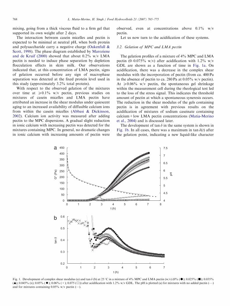
Fig. 1. Development of complex shear modulus (a) and tan d (b) at 25 1C in a m
(m); 0.045% (x); 0.05% (E); 0.06% (+); 0.075 (&)) after acidification with 1.2
and for mixtures containing 0.05% w/v pectin (—).
observed, even at concentrations above 0.1% w/vpectin.Let us now turn to the acidification of these systems.
3.2. Gelation of MPC and LMA pectin
The gelation profiles of a mixture of 4% MPC and LMApectin (0–0.075% w/v) after acidification with 1.2% w/vGDL are shown as a function of time in Fig. 1a. Onacidification, there was a decrease in the complex shearmodulus with the incorporation of pectin (from ca. 400 Pain the absence of pectin to ca. 280 Pa at 0.05% w/v pectin).At X0.06% w/v pectin, the spontaneous gel shrinkagewithin the measurement cell during the rheological test ledto the loss of the stress signal. This indicates the thresholdamount of pectin at which a spontaneous syneresis occurs.The reduction in the shear modulus of the gels containingpectin is in agreement with previous results on theacidification of mixtures of sodium caseinate containingcalcium+low LMA pectin concentrations (Matia-Merinoet al., 2004) and is discussed later.The development of tan d in the same system is shown in
Fig. 1b. In all cases, there was a maximum in tan d(t) afterthe gelation point, indicating a new liquid-like character
8
4
4.5
5
5.5
6
6.5
7
7.5
pH
4 5 6 7
5 6 7
h)
ixture of 4%MPC and LMA pectin (w/v) (0% (K); 0.025% (’); 0.035%
% w/v GDL. The pH is plotted (a) for mixtures with no added pectin (– –)
ARTICLE IN PRESSL. Matia-Merino, H. Singh / Food Hydrocolloids 21 (2007) 765–775 769
emerging as a result of further acidification. The morepectin added, the lower were the values of tan d at thegelation point and at the subsequent maximum. Amaximum in tan d has previously been observed duringthe formation of acid-set gels produced from heated skimmilk (Lucey, Tamehana, Singh, & Munro, 1998; Walsh-O’Grady, O’Kennedy, Fitzgerald, & Lane, 2001). Thismaximum has been related to a partial loosening of theweak initial gel network due to solubilization of CCP fromthe casein micelle in the pH range 6.0–5.3. A minimumfollowed by a maximum in tan d has also been observedduring the acidification of micellar casein systems within arange of milk salt concentrations (Auty, O’Kennedy,Allan-Wojtas, & Mulvihill, 2005), which has been attrib-uted to rearrangements of the casein protein. Consistentwith the above study, it appears that MPC acid-inducedgels undergo extensive rearrangements during the earlystages of gelation.
In order to discuss the observed effect of pectin on thegelation of MPCs, it is important to state the changes thatare likely to occur during the acidification of the mixturesused in this study. (1) There will be solubilization of theCCP plus dissociation of the casein from the casein micellesalong with a simultaneous decrease in the net negativecharge on the caseins. Consequently, casein particles beginto aggregate to form a gel around pH 5.1 where thesolubility of casein diminishes as previously observedduring the acidification of milk (Horne, 2001). (2) As thepH is reduced, an increased amount of the calcium ions willbe available for binding to the pectin, hence promoting itsaggregation/gelation. (3) The carboxylic groups of LMApectin become less ionized under acidification, promotingconformational ordering and intermolecular association(Gilsenan, Richardson, & Morris, 2000). It is also wellestablished that pectin will adsorb onto the casein micellesas a result of an electrostatic interaction at around andbelow pH 5.0 between the net positively charged casein andthe negatively charged pectin (Maroziene & de Kruif, 2000;Tuinier et al., 2002). Similarly, whey protein–pectinelectrostatic interactions will also take place underacidification as shown for pure protein systems (Girard,Turgeon, & Gauthier, 2002). Therefore, the final acid-
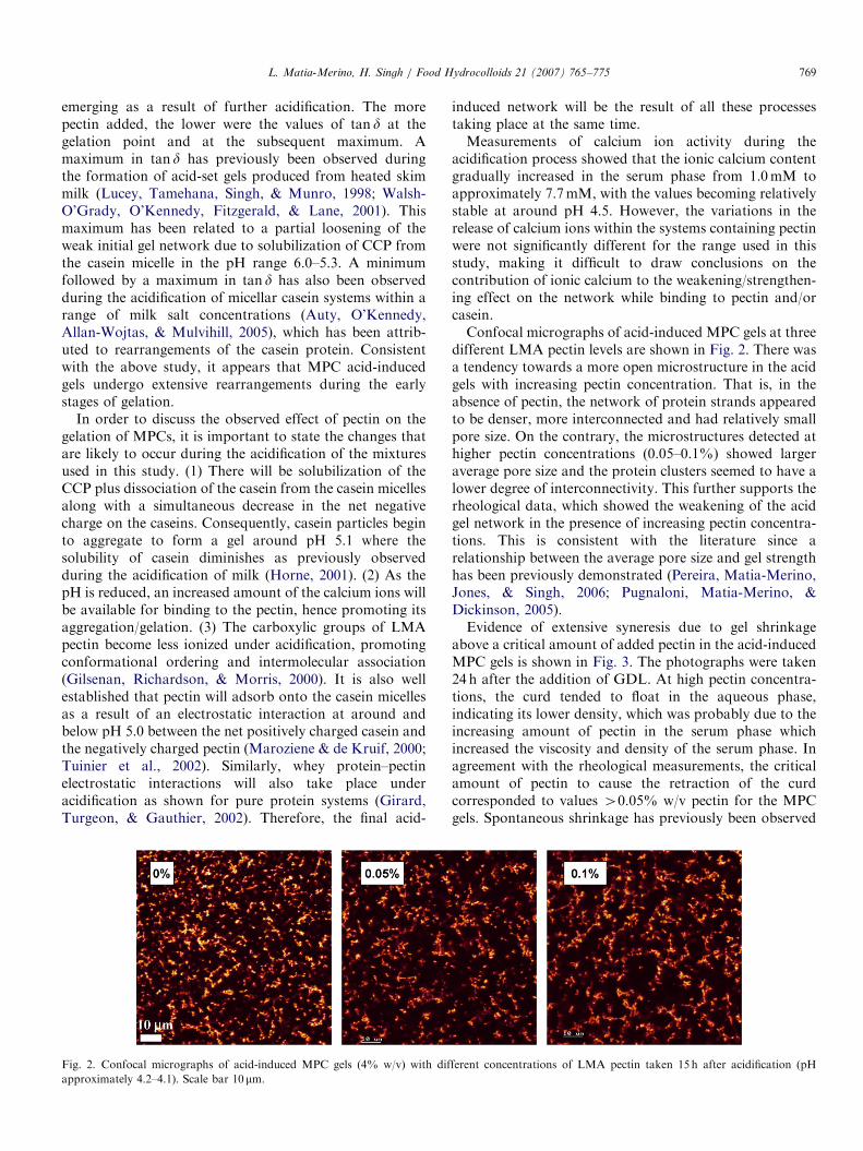
Fig. 2. Confocal micrographs of acid-induced MPC gels (4% w/v) with dif
approximately 4.2–4.1). Scale bar 10 mm.
induced network will be the result of all these processestaking place at the same time.Measurements of calcium ion activity during the
acidification process showed that the ionic calcium contentgradually increased in the serum phase from 1.0mM toapproximately 7.7mM, with the values becoming relativelystable at around pH 4.5. However, the variations in therelease of calcium ions within the systems containing pectinwere not significantly different for the range used in thisstudy, making it difficult to draw conclusions on thecontribution of ionic calcium to the weakening/strengthen-ing effect on the network while binding to pectin and/orcasein.Confocal micrographs of acid-induced MPC gels at three
different LMA pectin levels are shown in Fig. 2. There wasa tendency towards a more open microstructure in the acidgels with increasing pectin concentration. That is, in theabsence of pectin, the network of protein strands appearedto be denser, more interconnected and had relatively smallpore size. On the contrary, the microstructures detected athigher pectin concentrations (0.05–0.1%) showed largeraverage pore size and the protein clusters seemed to have alower degree of interconnectivity. This further supports therheological data, which showed the weakening of the acidgel network in the presence of increasing pectin concentra-tions. This is consistent with the literature since arelationship between the average pore size and gel strengthhas been previously demonstrated (Pereira, Matia-Merino,Jones, & Singh, 2006; Pugnaloni, Matia-Merino, &Dickinson, 2005).Evidence of extensive syneresis due to gel shrinkage
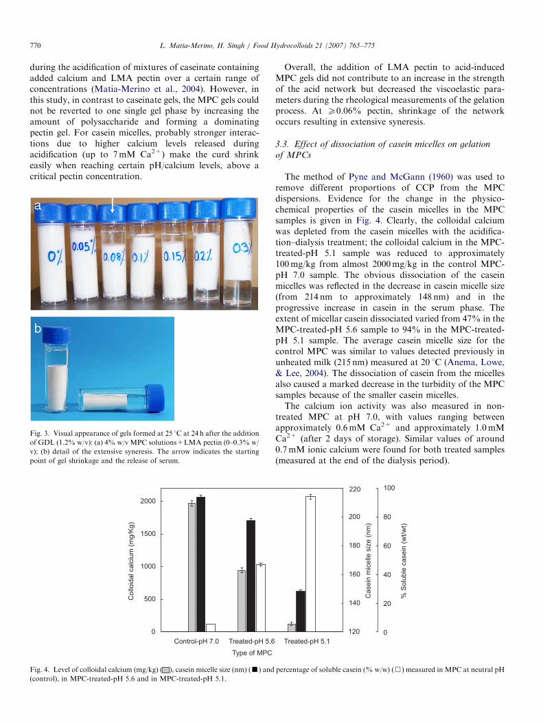
above a critical amount of added pectin in the acid-inducedMPC gels is shown in Fig. 3. The photographs were taken24 h after the addition of GDL. At high pectin concentra-tions, the curd tended to float in the aqueous phase,indicating its lower density, which was probably due to theincreasing amount of pectin in the serum phase whichincreased the viscosity and density of the serum phase. Inagreement with the rheological measurements, the criticalamount of pectin to cause the retraction of the curdcorresponded to values 40.05% w/v pectin for the MPCgels. Spontaneous shrinkage has previously been observed
ferent concentrations of LMA pectin taken 15 h after acidification (pH
ARTICLE IN PRESSL. Matia-Merino, H. Singh / Food Hydrocolloids 21 (2007) 765–775770
during the acidification of mixtures of caseinate containingadded calcium and LMA pectin over a certain range ofconcentrations (Matia-Merino et al., 2004). However, inthis study, in contrast to caseinate gels, the MPC gels couldnot be reverted to one single gel phase by increasing theamount of polysaccharide and forming a dominatingpectin gel. For casein micelles, probably stronger interac-tions due to higher calcium levels released duringacidification (up to 7mM Ca2+) make the curd shrinkeasily when reaching certain pH/calcium levels, above acritical pectin concentration.
Fig. 3. Visual appearance of gels formed at 25 1C at 24 h after the addition
of GDL (1.2% w/v): (a) 4% w/v MPC solutions+LMA pectin (0–0.3% w/
v); (b) detail of the extensive syneresis. The arrow indicates the starting
point of gel shrinkage and the release of serum.
Type of MPC
Co
lloid
al ca
lciu
m (
mg
/Kg
)
0
500
1000
1500
2000
Control-pH 7.0 Treated-pH 5.6
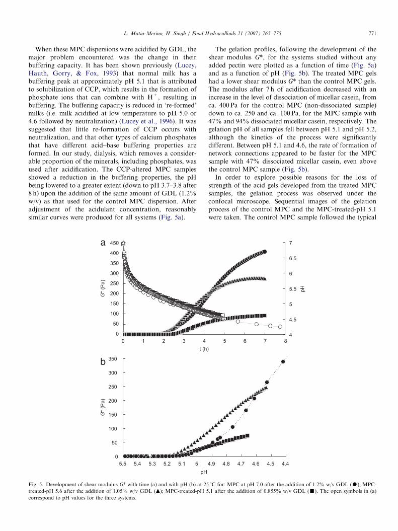
Fig. 4. Level of colloidal calcium (mg/kg) ( ), casein micelle size (nm) (’) and
(control), in MPC-treated-pH 5.6 and in MPC-treated-pH 5.1.
Overall, the addition of LMA pectin to acid-inducedMPC gels did not contribute to an increase in the strengthof the acid network but decreased the viscoelastic para-meters during the rheological measurements of the gelationprocess. At X0.06% pectin, shrinkage of the networkoccurs resulting in extensive syneresis.
3.3. Effect of dissociation of casein micelles on gelation
of MPCs
The method of Pyne and McGann (1960) was used toremove different proportions of CCP from the MPCdispersions. Evidence for the change in the physico-chemical properties of the casein micelles in the MPCsamples is given in Fig. 4. Clearly, the colloidal calciumwas depleted from the casein micelles with the acidifica-tion–dialysis treatment; the colloidal calcium in the MPC-treated-pH 5.1 sample was reduced to approximately100mg/kg from almost 2000mg/kg in the control MPC-pH 7.0 sample. The obvious dissociation of the caseinmicelles was reflected in the decrease in casein micelle size(from 214 nm to approximately 148 nm) and in theprogressive increase in casein in the serum phase. Theextent of micellar casein dissociated varied from 47% in theMPC-treated-pH 5.6 sample to 94% in the MPC-treated-pH 5.1 sample. The average casein micelle size for thecontrol MPC was similar to values detected previously inunheated milk (215 nm) measured at 20 1C (Anema, Lowe,& Lee, 2004). The dissociation of casein from the micellesalso caused a marked decrease in the turbidity of the MPCsamples because of the smaller casein micelles.The calcium ion activity was also measured in non-
treated MPC at pH 7.0, with values ranging betweenapproximately 0.6mM Ca2+ and approximately 1.0mMCa2+ (after 2 days of storage). Similar values of around0.7mM ionic calcium were found for both treated samples(measured at the end of the dialysis period).
% S
olu
ble
ca
se
in (
wt/
wt)
0
20
40
60
80
100
Ca
se
in m
ice
lle s
ize
(n
m)
140
160
180
200
Treated-pH 5.1
220
120
percentage of soluble casein (% w/w) (&) measured in MPC at neutral pH
ARTICLE IN PRESSL. Matia-Merino, H. Singh / Food Hydrocolloids 21 (2007) 765–775 771
When these MPC dispersions were acidified by GDL, themajor problem encountered was the change in theirbuffering capacity. It has been shown previously (Lucey,Hauth, Gorry, & Fox, 1993) that normal milk has abuffering peak at approximately pH 5.1 that is attributedto solubilization of CCP, which results in the formation ofphosphate ions that can combine with H+, resulting inbuffering. The buffering capacity is reduced in ‘re-formed’milks (i.e. milk acidified at low temperature to pH 5.0 or4.6 followed by neutralization) (Lucey et al., 1996). It wassuggested that little re-formation of CCP occurs withneutralization, and that other types of calcium phosphatesthat have different acid–base buffering properties areformed. In our study, dialysis, which removes a consider-able proportion of the minerals, including phosphates, wasused after acidification. The CCP-altered MPC samplesshowed a reduction in the buffering properties, the pHbeing lowered to a greater extent (down to pH 3.7–3.8 after8 h) upon the addition of the same amount of GDL (1.2%w/v) as that used for the control MPC dispersion. Afteradjustment of the acidulant concentration, reasonablysimilar curves were produced for all systems (Fig. 5a).
400
450
0 1 2 3 4
t (h
G*
(Pa
)G
* (P
a)
0
50
100
150
200
250
300
350
0
50
100
150
200
250
300
350
55.15.25.35.45.5
pH
b
a
Fig. 5. Development of shear modulus G* with time (a) and with pH (b) at 25
treated-pH 5.6 after the addition of 1.05% w/v GDL (m); MPC-treated-pH 5
correspond to pH values for the three systems.
The gelation profiles, following the development of theshear modulus G*, for the systems studied without anyadded pectin were plotted as a function of time (Fig. 5a)and as a function of pH (Fig. 5b). The treated MPC gelshad a lower shear modulus G* than the control MPC gels.The modulus after 7 h of acidification decreased with anincrease in the level of dissociation of micellar casein, fromca. 400 Pa for the control MPC (non-dissociated sample)down to ca. 250 and ca. 100 Pa, for the MPC sample with47% and 94% dissociated micellar casein, respectively. Thegelation pH of all samples fell between pH 5.1 and pH 5.2,although the kinetics of the process were significantlydifferent. Between pH 5.1 and 4.6, the rate of formation ofnetwork connections appeared to be faster for the MPCsample with 47% dissociated micellar casein, even abovethe control MPC sample (Fig. 5b).In order to explore possible reasons for the loss of
strength of the acid gels developed from the treated MPCsamples, the gelation process was observed under theconfocal microscope. Sequential images of the gelationprocess of the control MPC and the MPC-treated-pH 5.1were taken. The control MPC sample followed the typical
5 6 7 8
)
4
4.5
5
5.5
6
6.5
7
pH
4.44.54.64.74.84.9
1C for: MPC at pH 7.0 after the addition of 1.2% w/v GDL (K); MPC-
.1 after the addition of 0.855% w/v GDL (’). The open symbols in (a)

ARTICLE IN PRESS
Fig. 6. Confocal micrographs taken during the gelation process at 25 1C after addition of: (a) 1.2% w/v GDL to MPC (control) and (b) 0.855% w/v GDL
to MPC-treated-pH 5.1. Scale bar 10 mm. White arrow indicates the point at which it was necessary to refocus.
L. Matia-Merino, H. Singh / Food Hydrocolloids 21 (2007) 765–775772
aggregation process of GDL-induced gelation of skim milk(Auty, Fenelon, Guinee, Mullins, & Mulvihill, 1999). Atthe onset of gelation, a visible protein network was formed,at which point much of the stained protein becameimmobilized in the focus plane under study (Fig. 6a). Incontrast, after the onset of gelation in the MPC-treated-pH5.1 (with 94% dissociated micellar casein), the aggregationprocess was more difficult to follow because of thecontinuous loss of the focal plane. The connections of theaggregating milk protein exhibited a tendency for strandbreakage and rearrangements during the acidificationprocess until the protein network was fixed (Fig. 6b). Thiscould account for the weaker network formed from thetreated MPC samples. It seems reasonable to suggest thatthe difference in the dissociated casein micelle systemsduring the aggregation process can also be attributed to thedifferent building blocks forming the aggregating net-work—smaller remaining casein micelles and greateramounts of casein in the serum phase—which do notaggregate in the same way to form a strong network as theoriginal micelles.
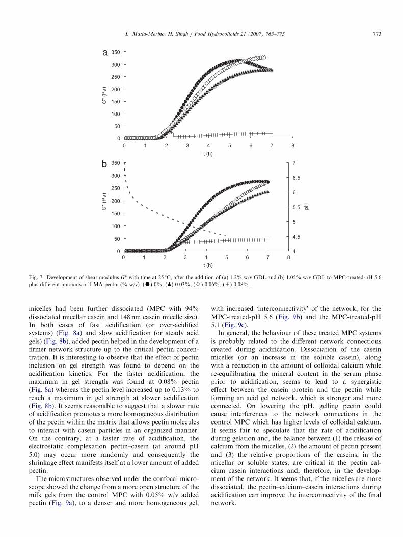
Finally, let us turn to the acidification of these treatedMPC systems in the presence of LMA pectin. Aspreviously demonstrated, the rate of acidification (or theamount of GDL) is one of the factors that influences thegel properties (Horne, 2001). In our study, acidificationwas carried out after the addition of (1) 1.2% w/v GDLand (2) a lower amount of GDL calculated to give a similarrate of acidification to that described above. The gelationcurves obtained are shown in Fig. 7 for the MPC with 47%dissociated micellar casein (or MPC-treated-pH 5.6) and inFig. 8 for the MPC with 94% dissociated micellar casein(or MPC-treated-pH 5.1).
The contribution of pectin to the development of thenetwork was clear in both treated MPC samples and at tworates of acidification. In Figs. 7a and 8a, the gelation ofMPC without added pectin showed the typical over-acidification effect—the pH being lowered to a valuebeyond the isoelectric point—which is detected rheologi-cally as a maximum in the storage modulus beyond thecritical gelation time (Horne, 2001; Koh, Matia-Merino, &Dickinson, 2002; Pugnaloni et al., 2005). The incorpora-tion of pectin changed the kinetics of gelation. In thecontrol MPC systems, addition of pectin caused aconsiderable decrease in G* (Fig. 1a), whereas the G*values increased markedly when the same amount of pectinwas incorporated into MPC-treated-pH 5.1 (Fig. 8b). Thistrend was observed until shrinkage of the network tookplace (i.e. up to 0.05% w/v pectin for the control MPC andup to 0.1% or 0.18% w/v pectin for the MPC-treated-pH5.1 depending on the rate of acidification, fast or slowrespectively).When the casein micelles were disaggregated to inter-
mediate values (MPC with 47% dissociated micellar caseinand 197 nm casein micelle size), the effect of pectin startedto reverse, showing the beginning of the synergistic effect.That is, during the experimental time scale the values of G*with added pectin were still lower than the G* valueswithout added pectin. However, the profiles indicated thatthe systems with added pectin may reach greater G* valuesbeyond the 7 h of the experiment, especially at a slower rateof acidification (Fig. 7b). Additionally, there were nosubstantial changes in gelation times with the incorpora-tion of pectin (up to 0.06% w/v).Independently of the rate of acidification, the synergistic
effect of pectin became more obvious when the casein
ARTICLE IN PRESS
1 5 6 7
G*
(Pa
)
0
50
100
150
200
250
300
350
0
50
100
150
200
250
300
350
0 2 3 4 8
1 5 6 70 2 3 4 8
t (h)
t (h)
G*
(Pa
)
4
4.5
5
5.5
6
6.5
7
pH
a
b
Fig. 7. Development of shear modulus G* with time at 25 1C, after the addition of (a) 1.2% w/v GDL and (b) 1.05% w/v GDL to MPC-treated-pH 5.6
plus different amounts of LMA pectin (% w/v): (K) 0%; (m) 0.03%; (B) 0.06%; (+) 0.08%.
L. Matia-Merino, H. Singh / Food Hydrocolloids 21 (2007) 765–775 773
micelles had been further dissociated (MPC with 94%dissociated micellar casein and 148 nm casein micelle size).In both cases of fast acidification (or over-acidifiedsystems) (Fig. 8a) and slow acidification (or steady acidgels) (Fig. 8b), added pectin helped in the development of afirmer network structure up to the critical pectin concen-tration. It is interesting to observe that the effect of pectininclusion on gel strength was found to depend on theacidification kinetics. For the faster acidification, themaximum in gel strength was found at 0.08% pectin(Fig. 8a) whereas the pectin level increased up to 0.13% toreach a maximum in gel strength at slower acidification(Fig. 8b). It seems reasonable to suggest that a slower rateof acidification promotes a more homogeneous distributionof the pectin within the matrix that allows pectin moleculesto interact with casein particles in an organized manner.On the contrary, at a faster rate of acidification, theelectrostatic complexation pectin–casein (at around pH5.0) may occur more randomly and consequently theshrinkage effect manifests itself at a lower amount of addedpectin.
The microstructures observed under the confocal micro-scope showed the change from a more open structure of themilk gels from the control MPC with 0.05% w/v addedpectin (Fig. 9a), to a denser and more homogeneous gel,
with increased ‘interconnectivity’ of the network, for theMPC-treated-pH 5.6 (Fig. 9b) and the MPC-treated-pH5.1 (Fig. 9c).In general, the behaviour of these treated MPC systems
is probably related to the different network connectionscreated during acidification. Dissociation of the caseinmicelles (or an increase in the soluble casein), alongwith a reduction in the amount of colloidal calcium whilere-equilibrating the mineral content in the serum phaseprior to acidification, seems to lead to a synergisticeffect between the casein protein and the pectin whileforming an acid gel network, which is stronger and moreconnected. On lowering the pH, gelling pectin couldcause interferences to the network connections in thecontrol MPC which has higher levels of colloidal calcium.It seems fair to speculate that the rate of acidificationduring gelation and, the balance between (1) the release ofcalcium from the micelles, (2) the amount of pectin presentand (3) the relative proportions of the caseins, in themicellar or soluble states, are critical in the pectin–cal-cium–casein interactions and, therefore, in the develop-ment of the network. It seems that, if the micelles are moredissociated, the pectin–calcium–casein interactions duringacidification can improve the interconnectivity of the finalnetwork.

ARTICLE IN PRESS
Fig. 9. Effect of 0.05% w/v LMA pectin on the microstructure of acid milk gels made from (a) control MPC at pH 7.0, (b) MPC-treated-pH 5.6 and (c)
MPC-treated-pH 5.1, after the addition of 1.2, 1.05 and 0.855% w/v GDL, respectively. The confocal images were taken after 15 h of gelation. Scale bar
10mm.
0
50
100
150
200
250
300
350
400
0 1 2 3 4 5 6 7 8
t (h)
G*
(Pa
)
0
50
100
150
200
250
300
350
400
0 1 2 3 4 5 6 7 8
t (h)
G*
(Pa
)
4
4.5
5
5.5
6
6.5
7
pH
a
b
Fig. 8. Development of shear modulus G* with time at 25 1C, after the addition of (a) 1.2% w/v GDL and (b) 0.855% w/v GDL to MPC-treated-pH 5.1
plus different amounts of LMA pectin (% w/v): (K) 0%; (J) 0.04%; (m) 0.06%; (B) 0.08%; (- -) 0.1%; (x) 0.13%; (+) 0.18%.
L. Matia-Merino, H. Singh / Food Hydrocolloids 21 (2007) 765–775774
In summary, the rheological gelation profile and thedeveloping microstructure after the acidification of mix-tures of MPC and LMA pectin can be attributed to acombination of effects. The potential to generate differenttypes of gel matrices, by controlling rate of acidification,casein micelle integrity, calcium levels and pectin concen-trations to obtain different synergistic or antagonisticeffects, gives a way to manipulate texture. The possible
formation of casein–calcium–pectin complexes with thecasein micelles or in the serum phase, along with case-in–casein and pectin–pectin interactions (through calcium)and casein–pectin complexes formed during acidification,are responsible for the development of the acid-inducedthree-dimensional network. Further work should be carriedout to identify which of these interactions are dominant incontrolling the dynamics of these mixed systems.
ARTICLE IN PRESSL. Matia-Merino, H. Singh / Food Hydrocolloids 21 (2007) 765–775 775
References
Abbasi, S., & Dickinson, E. (2002). High-pressure-induced rheological
changes of low-methoxyl pectin plus micellar casein mixtures. Journal
of Agricultural and Food Chemistry, 50, 3559–3565.
Alkanani, T., Friel, J. K., Jackson, S. E., & Longerich, H. P. (1994).
Comparison between digestion procedures for the multielemental
analysis of milk by inductively-coupled plasma-mass spectrometry.
Journal of Agricultural and Food Chemistry, 42, 1965–1970.
Anema, S. G. (1997). The effect of chymosin on kappa-casein-coated
polystyrene latex particles and bovine casein micelles. International
Dairy Journal, 7, 553–558.
Anema, S. G., & Klostermeyer, H. (1997). Heat-induced, pH-dependent
dissociation of casein micelles on heating reconstituted skim milk at
temperatures below 100 1C. Journal of Agricultural and Food Chem-
istry, 45, 1108–1115.
Anema, S. G., Lowe, E. K., & Lee, S. K. (2004). Effect of pH at heating on
the acid-induced aggregation of casein micelles in reconstituted skim
milk. Lebensmittel-Wissenschaft Und-Technologie-Food Science and
Technology, 37, 779–787.
Auty, M. A. E., Fenelon, M. A., Guinee, T. P., Mullins, C., & Mulvihill,
D. M. (1999). Dynamic confocal scanning laser microscopy methods
for studying milk protein gelation and cheese melting. Scanning, 21,
299–304.
Auty, M. A. E., O’Kennedy, B. T., Allan-Wojtas, P., & Mulvihill, D. M.
(2005). The application of microscopy and rheology to study the effect
of milk salt concentration on the structure of acidified micellar casein
systems. Food Hydrocolloids, 19, 101–109.
Braccini, I., & Perez, S. (2001). Molecular basis of Ca2+-induced gelation
in alginates and pectins: The egg-box model revisited. Biomacromole-
cules, 2, 1089–1096.
Dalgleish, D. G., & Law, A. J. R. (1988). pH-Induced dissociation of
casein micelles. 1. Analysis of liberated caseins. Journal of Dairy
Research, 55, 529–538.
De Kruif, C. G. (1997). Skim milk acidification. Journal of Colloid and
Interface Science, 185, 19–25.
Gilsenan, P. M., Richardson, R. K., & Morris, E. R. (2000). Thermally
reversible acid-induced gelation of low-methoxy pectin. Carbohydrate
Polymers, 41, 339–349.
Girard, M., Turgeon, S. L., & Gauthier, S. F. (2002). Interbiopolymer
complexing between beta-lactoglobulin and low- and high-methylated
pectin measured by potentiometric titration and ultrafiltration. Food
Hydrocolloids, 16, 585–591.
Hill, J. P. (1993). The relationship between beta-lactoglobulin phenotypes
and milk-composition in New-Zealand dairy-cattle. Journal of Dairy
Science, 76, 281–286.
Horne, D. S. (2001). Factors influencing acid-induced gelation of skim
milk. In E. Dickinson, & R. Miller (Eds.), Food colloids: Fundamentals
of formulation (pp. 345–351). Cambridge: Royal Society of Chemistry.
Koh, M. W. W., Matia-Merino, L., & Dickinson, E. (2002). Rheology of
acid-induced sodium caseinate gels containing added gelatin. Food
Hydrocolloids, 16, 619–623.
Law, A. J. R., & Leaver, J. (1998). Effects of acidification and storage of
milk on dissociation of bovine casein micelles. Journal of Agricultural
and Food Chemistry, 46, 5008–5016.
Le Graet, Y., & Gaucheron, F. (1999). pH-Induced solubilization of
minerals from casein micelles: Influence of casein concentration and
ionic strength. Journal of Dairy Research, 66, 215–224.
Lucey, J. A. (2002). Formation and physical properties of milk protein
gels. Journal of Dairy Science, 85, 281–294.
Lucey, J. A., Gorry, C., O’Kennedy, B., Kalab, M., Tan-Kinita, R., &
Fox, P. F. (1996). Effect of acidification and neutralization of milk on
some physico-chemical properties of casein micelles. International
Dairy Journal, 257–272.
Lucey, J. A., Hauth, B., Gorry, C., & Fox, P. F. (1993). Acid–base
buffering of milk. Milchwissenschaft, 48, 268–272.
Lucey, J. A., & Singh, H. (2003). Acid coagulation of milk. In P. F. Fox, &
P. L. H. McSweeney (Eds.), Advanced dairy chemistry: Proteins, Vol. 1
(pp. 1001–1025). London: Kluwer Academic/Plenum.
Lucey, J. A., Tamehana, M., Singh, H., & Munro, P. A. (1998). Effect of
interactions between denatured whey proteins and casein micelles on
the formation and rheological properties of acid skim milk gels.
Journal of Dairy Research, 65, 555–567.
Maroziene, A., & de Kruif, C. G. (2000). Interaction of pectin and casein
micelles. Food Hydrocolloids, 14, 391–394.
Matia-Merino, L., & Dickinson, E. (2004). High-sugar-content acid-
induced caseinate gels and emulsion gels: Influence of low-methoxyl
pectin. In P. A. Williams, & G. O. Phillips (Eds.), Gums and stabilisers
for the food industry, Vol. 12 (pp. 461–474). Cambridge: Royal Society
of Chemistry.
Matia-Merino, L., Lau, K., & Dickinson, E. (2004). Effects of low-
methoxyl amidated pectin and ionic calcium on rheology and
microstructure of acid-induced sodium caseinate gels. Food Hydro-
colloids, 18, 271–281.
May, C. D. (2000). Pectins. In G. Phillips, & P. A. Williams (Eds.),
Handbook of hydrocolloids (pp. 169–175). Cambridge: Woodhead
Publishing Ltd., CRC Press LLC.
Morris, E. R. (1990). Mixed polymer gels. In P. Harris (Ed.), Food gels
(pp. 291–359). London: Elsevier Science.
Morris, V. J. (1998). Gelation of polysaccharides. In S. E. Hill, D. A.
Ledward, & J. R. Mitchell (Eds.), Functional properties of food
macromolecules (pp. 143–214). Gaithersburg, Maryland: An Aspen
Publication.
Oakenfull, D., & Scott, A. (1998). Milk gels with low methoxyl pectins. In
P. A. Williams, & G. O. Phillips (Eds.), Gums and stabilizers for the
food industry, Vol. 9 (pp. 212–221). Cambridge, UK: Royal Society of
Chemistry.
Pereira, R., Matia-Merino, L., Jones, V., & Singh, H. (2006). Influence of
fat on the perceived texture of set acid milk gels: A sensory perspective.
Food Hydrocolloids, 20, 305–313.
Pugnaloni, L. A., Matia-Merino, L., & Dickinson, E. (2005). Micro-
structure of acid-induced caseinate gels containing sucrose: Quantifi-
cation from confocal microscopy and image analysis. Colloids and
Surfaces B-Biointerfaces, 42, 211–217.
Pyne, G. T., & McGann, T. C. A. (1960). The colloidal phosphate of milk.
2. Influence of citrate. Journal of Dairy Research, 27, 9–17.
Singh, H., Roberts, M. S., Munro, P. A., & Teo, C. T. (1996). Acid-
induced dissociation of casein micelles in milk: Effects of heat
treatment. Journal of Dairy Science, 79, 1340–1346.
Syrbe, A., Bauer, W. J., & Klostermeyer, N. (1998). Polymer science
concepts in dairy systems—An overview of milk protein and food
hydrocolloid interaction. International Dairy Journal, 8, 179–193.
Tolstoguzov, V. B. (1990). Interactions of gelatin with polysaccharides. In
G. O. Phillips, P. A. Williams, & D. J. Wedlock (Eds.), Gums and
stabilizers for the food industry, Vol. 5 (pp. 157–175). Oxford: IRL
Press.
Tuinier, R., Rolin, C., & de Kruif, C. G. (2002). Electrosorption of pectin
onto casein micelles. Biomacromolecules, 3, 632–638.
Udabage, P., McKinnon, I. R., & Augustin, M. A. (2000). Mineral and
casein equilibria in milk: Effects of added salts and calcium-chelating
agents. Journal of Dairy Research, 67, 361–370.
van Vliet, T., Lakemond, C. M. M., & Visschers, R. W. (2004). Rheology
and structure of milk protein gels. Current Opinion in Colloid and
Interface Science, 9, 298–304.
Voragen, A. G. V., Pilnik, W., Thibault, J. F., Axelos, M. A. V., &
Renard, G. C. (1995). Pectins. In A. M. Stephen (Ed.), Food
polysaccharides and their applications (pp. 287–339). New York:
Marcel Dekker, Inc.
Walsh-O’Grady, C. D., O’Kennedy, B. T., Fitzgerald, R. J., & Lane, C. N.
(2001). A rheological study of acid-set ‘‘simulated yogurt milk’’ gels
prepared from heat-or pressure-treated milk proteins. Lait, 81,
637–650.