aclidinium bromide inhibits proliferation of …...aclidinium bromide inhibits proliferation of...
TRANSCRIPT
105
Abstract. – OBJECTIVE: Osteosarcoma is rec-ognized as the most common primary malignant bone tumor, the 5-year disease-free survival rate in patients with metastatic or recurrent disease is below than 30%. Drug resistance and toxic side effects limit the therapeutic efficacy of osteo-sarcoma. Therefore, it is urgent to develop new drugs for osteosarcoma treatment. Muscarinic 3 (M3) acetylcholine receptor (AChR) has been demonstrated in nonneurocrest-derived malig-nancies such as colon, prostate, lung, and ovari-an carcinomas. Hence, targeted regulation of M3 AChR may be a possible mechanism for treat-ing tumors. Aclidinium bromide has anti-tumor-al properties in several tumors, namely gastric cancer and glioma. In this study, we intended to investigate whether aclidinium bromide, a novel M3 AChR antagonist, had effects on osteosarco-ma cells proliferation and migration.
PATIENTS AND METHODS: The viability of U2 OS cells was detected by cell counting kit-8 (CCK-8) assay. The migration and invasion ca-pabilities were measured by transwell invasion and migration assays. The cell apoptosis rate was tested by Annexin V-fluorescein isothio-cyanate (FITC)/Propidum iodide (PI) staining and flow cytometry. Key apoptosis-related and phosphatidylinositol-3-kinase (PI3K)/protein kinase B (AKT) signaling pathway-associated were assessed by Western blot analysis.
RESULTS: Aclidinium bromide markedly de-creased the OD value of U2 OS cells 48 h and 72 h after treatment. The number of positive crystal violet staining cells significantly decreased after treatment with aclidinium bromide. Treatment with aclidinium bromide significantly increased cell apoptosis rate, accompanied by the expres-sion of anti-apoptotic protein Bcl-2 decreased, the expression of pro-apoptotic protein Active caspase-3 and Bax significantly increased in U2 OS cells treated with aclidinium bromide. Ad-ditionally, aclidinium bromide suppressed the PI3K/AKT signaling pathway in U2 OS cells.
CONCLUSIONS: Therefore, the current study reveals that aclidinium bromide might inhibit os-teosarcoma cell growth by regulating the PI3K/
AKT signaling pathway, which suggests aclidin-ium bromide is a potential chemotherapeutic agent for osteosarcoma.
Key WordsOsteosarcoma, Muscarinic 3 acetylcholine recep-
tor, Proliferation, Migration, Invasion, Apoptosis.
Introduction
Osteosarcoma is known as the commonest primary malignant bone tumor, and the 5-year disease-free survival rate is lower than 30% due to high recurrence and aggressiveness1. Osteosar-coma is a high-grade neoplasm with the charac-teristic of rapid growth and early metastasis. The incidence of osteosarcoma in males is somewhat higher than females due to a longer duration of skeletal growth in males2,3. Osteosarcoma can occur in any bone of the body but approximately 75% of all cases occur in the distal femur and prox-imal tibia4,5. Despite the advancement of multiple therapeutic regiments, namely, surgery, radiation and belligerent chemotherapy6,7, the effectiveness of treatment has been unsatisfactory. In addition, drugs, such as etoposide8, astragaloside9, cinob-ufagin and cisplatin10 as well as drug resistance and toxic side effects, limit the efficacy of os-teosarcoma treatment11. Therefore, it is urgent to develop new drugs for osteosarcoma treatment. Acetylcholine (ACh) has been known to contribute to sensory processing. Muscarinic and nicotinic receptors (mAChR and nAChR, respectively) have been identified as the best described ACh recep-tors. The activation of these receptors increases neuronal activity. Notably, this receptor system has been shown to be involved in the regulation of cell movement and proliferation in neuronal cells (benign or malignant), and gaining an under-
European Review for Medical and Pharmacological Sciences 2019; 23: 105-112
Z.-Z. LI, Y.-L. WANG, Y.-H. YU, Y.-L. XING, X.-F. JI
Department of Orthopedics, China-Japan Union Hospital of Jilin University, Changchun, P.R. China
Corresponding Author: Xiao-Feng Ji, MD; e-mail: [email protected]
Aclidinium bromide inhibits proliferationof osteosarcoma cells through regulationof PI3K/Akt pathway
Z.-Z. Li, Y.-L. Wang, Y.-H. Yu, Y.-L. Xing, X.-F. Ji
106
standing of this new pathway may provide targets for therapeutic intervention12,13. As an important neurotransmitter and ligand to AChR in the cen-tral and peripheral nervous systems, acetylcho-line can stimulate cell growth through binding of mAChR or nAChR. Researchers have shown that acetylcholine is synthesized in numerous some non-neuronal cell types, in addition to neuro-nal cells, including bronchial epithelial cells, glial cells, pulmonary vessel cells, and ovarian cells14-22. Noda et al23 found that neural crest-derived tumors such as melanoma and small cell carcinoma of the lung express both mAChR and nAChR. AChR is overexpressed in neurocrest-derived tissues where it has been shown to be a critical determinant of cellular development and differentiation24. As such, these receptors and their ligand agonists may play an important role in the pathogenesis of presumable neurocrest derived tumors24. Particu-larly, muscarinic 3 (M3) AChR has been observed in nonneurocrest-derived malignancies such as colon, prostate, lung, and ovarian carcinomas25-29. However, whether this receptor can be targeted to control AChR to treat osteosarcoma has not been investigated. Aclidinium bromide is a novel, long-acting, muscarinic antagonist that inhibits the action of acetylcholine at the M3 receptor24. Aclidinium bromide is approved as a mainte-nance bronchodilator treatment in patients with chronic obstructive pulmonary disease (COPD)30. Aclidinium salts have been identified to have tu-mor-inhibiting properties in vitro models and in vivo against human tumor xenografts, such as lung cancer and colorectal adenocarcinoma31,32. Acrid-inium iodide provokes cell cycle arrest in response to DNA damage in the tumor cell line33. Acridin-ium methosulfate, identified as an anti-telomerase agent, is screened as a candidate for candidates for tumor stem cells ablation in osteosarcoma34. There are emerging data from many experiments, suggesting that aclidinium bromide is proved to inhibit gastric cancer and glioma cell proliferation and migration potentials35,36. However, the role of aclidinium bromide in osteosarcoma treatment re-mains unknown. The aim of the present study was to illustrate the effect of aclidinium bromide on osteosarcoma cells and its potential mechanisms.
Materials and Methods
Cell CultureU2 OS cell lines obtained from the Cell Bank
of Chinese Academy of Sciences (Shanghai, China)
were grown in Roswell Park Memorial Institute (RPMI)-1640 (Hyclone, Waltham, MA, USA) sup-plemented with 10% fetal bovine serum (FBS; In-vitrogen, Thermo Fisher Scientific, Waltham, MA, USA) and antibiotics (100 U/mL penicillin, 0.1 mg/mL streptomycin; Sigma-Aldrich, St. Louis, MO, USA). The medium was changed every 3 days, and U2 OS cells were maintained at 37°C in a 5% CO2 incubator. For evaluation of expression of protein, cells were seeded onto 6-well culture plates.
Cell Proliferation AssayCells were cultured in 96-well plates, when
the cells reached about 80% confluence, then the medium of aclidinium bromide group was replaced by complete medium containing acli-dinium bromide (10 μM, MedChemExpress Bio-technology Shanghai, China) for 24 h, and 0.1% dimethylsulfoxide (DMSO) was employed as ve-hicle control. Then, 10 μL cell counting kit-8 (CCK8, Beijing Solarbio Science and Technology Co., Ltd., Beijing, China) was added into medium and incubated for another 1.5 h, then the resulted formazan precipitates were dissolved in DMSO, and the optic density at 450 nm was read imme-diately using a microplate reader.
Cell Invasion and Migration AssayCells were suspended by 100 µL Matrigel
matrix (BD Bioscience, San Jose, CA, USA) and seeded on the top chamber of the 24-well inserts (8 µm pore size; Corning, Tewksbury, MA, USA). Serum-free medium was added to the lower com-partments and cells were incubated for 0.5 h to hydrate basilar membrane. Cell suspension (1×10⁵ cells/100 µL serum-free MEM media) was added to the upper compartments and complete culture solution (500 µL) were added to the lower com-partments after incubation for 24 h. The cells that did not migrate through the pores were removed by scraping the membrane with a cotton swab. The cells were fixed for 30 min in 4% parafor-maldehyde after passed through the filter, stained for 20 min with 0.1% crystal violet. Five random fields (100 ×) were captured under the microscope (Olympus, Tokyo, Japan). Finally, we counted the number of invasive or migratory cells and calculated the average values. Each experiment was conducted in triplicate.
Flow Cytometric AnalysisAfter the cells were treated with aclidinium
bromide for 24 h, then the medium was removed and serum starved for 24 h. Evaluation of apop-
Aclidinium bromide inhibits proliferation of osteosarcoma cells
107
tosis was performed by an Annexin V-fluoresce-in isothiocyanate (FITC)/propidum iodide (PI) kit (Beijing 4A Biotech Co., Ltd) as described previously37. The result was evaluated by a flow cytometer (BD Bioscience) after the cells were labeled with Annexin-V and PI.
Western BlottingThe cells were treated with aclidinium bro-
mide for 24 h, then, cells were collected and lysed in RIPA lysis buffer (Beyotime Institute of Biotechnology, Shanghai, China). The protein concentration was detected using a bicinchoninic acid (BCA) kit (Beijing ComWin Biotech Co., Ltd, Beijing, China). Subsequently, the equal proteins (20 μg) were separated by sodium dodecyl sulfate polyacrylamide gel electrophoresis (SDS-PAGE, Bio-Rad, Hercules, CA, USA) and then trans-ferred onto a polyvinylidene difluoride (PVDF) membrane38. After blocked with 5% non-fat dry milk for 1 h, the proteins were incubated with various antibodies overnight at 4°C, including Bcl-2, Bax, Active caspase-3, protein kinase B (AKT), phosphorylated- (p-) AKT, mechanistic target of rapamycin (mTOR), p-mTOR, phosphorylated-ser-ine-threonine protein kinase (p-P70S6K), GAP-DH (1:1000, Cell Signaling Technology, Danvers, MA, USA). Secondary anti-mouse or anti-goat horseradish peroxidase (HRP) antibodies (1:5000, Beyotime Institute of Biotechnology, Shanghai, China) were added for 1 h, then we visualized the bands by enhanced chemiluminescence (ECL, Thermo Fisher Scientific) reagents and developed by ChemiDoc MP imager (Bio-Rad Laboratories, Hercules, CA, USA). The band density was quan-tified by Quantity One software (Bio-Rad Labo-ratories, Hercules, CA, USA).
Statistical AnalysisAll data were analyzed with the SPSS ver-
sion 18.0 software (SPSS, Chicago, IL, USA). Data were expressed as means ± standard devi-ations (SD). Statistical significance of difference between two groups was determined by t-test. p<0.05 was regarded as statistically significant.
Result
Aclidinium Bromide Inhibited the Proliferation of U2 OS Cells
To investigate the role of aclidinium bromide in the proliferation of U2 OS cells, we performed
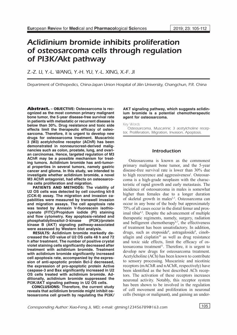
the CCK-8 assay. As shown in Figure 1, com-pared with control group, aclidinium bromide could significantly decrease the OD value of U2 OS cells 48 h and 72 h after treatment (p<0.05). This result suggested that aclidinium bromide inhibited the proliferation of U2 OS cells.
Aclidinium Bromide Suppressed the Invasion and Migration of U2 OS Cells
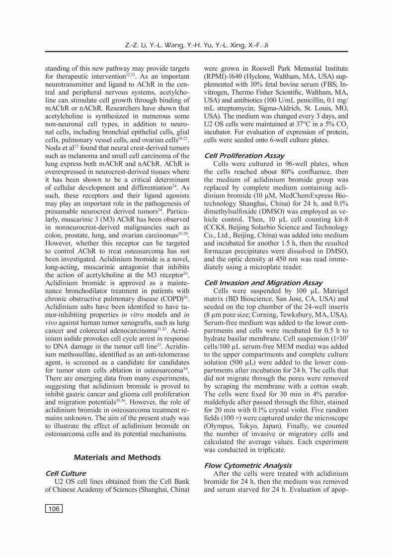
Then, we observed the effect of aclidinium bromide on U2 OS cells invasion and migration. In invasion assay, the number of positive crystal violet staining cells significantly decreased after treated with aclidinium bromide (p<0.05) (Figure 2). Similar findings were shown in migration as-say that the number of positive cells significantly decreased in aclidinium bromide treated group (p<0.05) (Figure 2). These results suggested that the ability of invasion and migration in aclidinium bromide treated group significantly decreased.
Induction of Apoptosis in U2 OS Cells by Aclidinium Bromide Exposure
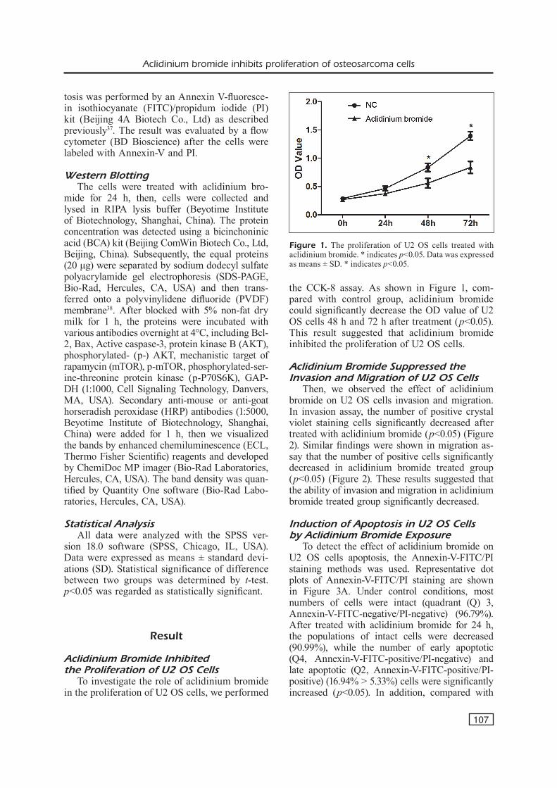
To detect the effect of aclidinium bromide on U2 OS cells apoptosis, the Annexin-V-FITC/PI staining methods was used. Representative dot plots of Annexin-V-FITC/PI staining are shown in Figure 3A. Under control conditions, most numbers of cells were intact (quadrant (Q) 3, Annexin-V-FITC-negative/PI-negative) (96.79%). After treated with aclidinium bromide for 24 h, the populations of intact cells were decreased (90.99%), while the number of early apoptotic (Q4, Annexin-V-FITC-positive/PI-negative) and late apoptotic (Q2, Annexin-V-FITC-positive/PI- positive) (16.94% > 5.33%) cells were significantly increased (p<0.05). In addition, compared with
Figure 1. The proliferation of U2 OS cells treated with aclidinium bromide. * indicates p<0.05. Data was expressed as means ± SD. * indicates p<0.05.
Z.-Z. Li, Y.-L. Wang, Y.-H. Yu, Y.-L. Xing, X.-F. Ji
108
Figure 2. Aclidinium bromide significantly decreased the invasion and migration of U2 OS cells. The migrated and invaded cell number of U2 OS cells was quantified. Data was expressed as means ± SD. * indicates p<0.05.
Figure 3. Apoptosis is induced by aclidini-um bromide treatment in U2 OS cells. A, The proportions of living and dead cells were determined using flow cytometric analysis of Annexin-V/FITC and PI-labeled cells. Cells showing Annexin-V/FITC and PI double labeling (Q2) indicate those that have already died by apoptosis. Live cells were unlabeled with an Annexin-V/FITC and PI (Q3), while Annexin-V/FITC labeling (Q4) rep-resents the population of early apoptosis. B, Western blot analysis of apoptosis protein expression in human U2 OS cells. Data was expressed as means ± SD. * indicates p<0.05.
A
B
Aclidinium bromide inhibits proliferation of osteosarcoma cells
109
control group, the expression of anti-apoptotic pro-tein Bcl-2 in aclidinium bromide group was sig-nificantly decreased (p<0.05), and the expression of proapoptotic protein Bax and Active caspase-3 was significantly increased (p<0.05) (Figure 3). All these results suggested that aclidinium bro-mide could induce apoptosis in U2 OS cells.
Aclidinium Bromide Inhibited the Activation of PI3K/AKT Pathway in U2 OS Cells
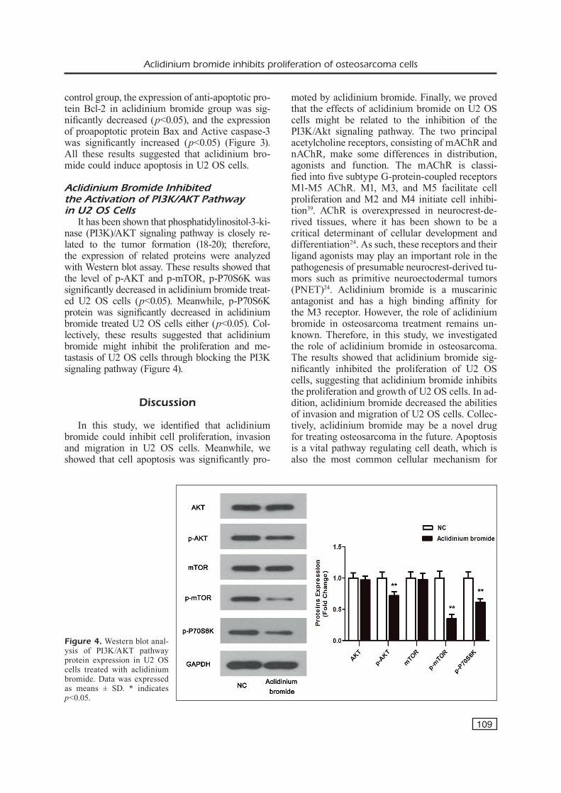
It has been shown that phosphatidylinositol-3-ki-nase (PI3K)/AKT signaling pathway is closely re-lated to the tumor formation (18-20); therefore, the expression of related proteins were analyzed with Western blot assay. These results showed that the level of p-AKT and p-mTOR, p-P70S6K was significantly decreased in aclidinium bromide treat-ed U2 OS cells (p<0.05). Meanwhile, p-P70S6K protein was significantly decreased in aclidinium bromide treated U2 OS cells either (p<0.05). Col-lectively, these results suggested that aclidinium bromide might inhibit the proliferation and me-tastasis of U2 OS cells through blocking the PI3K signaling pathway (Figure 4).
Discussion
In this study, we identified that aclidinium bromide could inhibit cell proliferation, invasion and migration in U2 OS cells. Meanwhile, we showed that cell apoptosis was significantly pro-
moted by aclidinium bromide. Finally, we proved that the effects of aclidinium bromide on U2 OS cells might be related to the inhibition of the PI3K/Akt signaling pathway. The two principal acetylcholine receptors, consisting of mAChR and nAChR, make some differences in distribution, agonists and function. The mAChR is classi-fied into five subtype G-protein-coupled receptors M1-M5 AChR. M1, M3, and M5 facilitate cell proliferation and M2 and M4 initiate cell inhibi-tion39. AChR is overexpressed in neurocrest-de-rived tissues, where it has been shown to be a critical determinant of cellular development and differentiation24. As such, these receptors and their ligand agonists may play an important role in the pathogenesis of presumable neurocrest-derived tu-mors such as primitive neuroectodermal tumors (PNET)24. Aclidinium bromide is a muscarinic antagonist and has a high binding affinity for the M3 receptor. However, the role of aclidinium bromide in osteosarcoma treatment remains un-known. Therefore, in this study, we investigated the role of aclidinium bromide in osteosarcoma. The results showed that aclidinium bromide sig-nificantly inhibited the proliferation of U2 OS cells, suggesting that aclidinium bromide inhibits the proliferation and growth of U2 OS cells. In ad-dition, aclidinium bromide decreased the abilities of invasion and migration of U2 OS cells. Collec-tively, aclidinium bromide may be a novel drug for treating osteosarcoma in the future. Apoptosis is a vital pathway regulating cell death, which is also the most common cellular mechanism for
Figure 4. Western blot anal-ysis of PI3K/AKT pathway protein expression in U2 OS cells treated with aclidinium bromide. Data was expressed as means ± SD. * indicates p<0.05.
Z.-Z. Li, Y.-L. Wang, Y.-H. Yu, Y.-L. Xing, X.-F. Ji
110
naphthalimide inhibition in cancer cell40,41. Insuf-ficient apoptosis could result in uncontrolled cell proliferation, which has been shown to be involved in cancer42-44. Ji et al45 synthesized a triazolonaph-thalimide, LSS-11, and found that it exhibited strong cytotoxicity by inducing cell cycle arrest and apoptosis in selected human colon cancer cell lines, which was accompanied by DNA damage response. In our study, we found that after treat-ment with aclidinium bromide for 24 h, the pop-ulations of intact cells were decreased, while the numbers of early apoptotic and late apoptotic cells were significantly increased. Meanwhile, the ex-pression of anti-apoptotic protein Bcl-2 in aclidin-ium bromide group was significantly decreased, and the expression of proapoptotic protein Bax and Active caspase-3 was significantly increased after treatment with aclidinium bromide for 24 h. Therefore, we hypothesized that aclidinium bro-mide may inhibit U2 OS cells proliferation by pro-moting the cell apoptosis. The PI3K/AKT pathway is an important signaling pathway that contributes to cellular growth. For instance, the PI3K/AKT pathway is involved in survival in different cell types, which is activated by receptors of growth factor and stimulates cell growth, and differentia-tion46-48. A serine/threonine kinase AKT is located downstream of class I and class III PI3K49,50. In the context of infection, activation of this pathway is associated with an increase in proliferation, cell survival, and cell migration, as well as en-hanced protein synthesis via mTOR activation51-54. AKT activation is known to regulate cell cycle progression, cell death, and cell growth. In our study, aclidinium bromide exposure inhibited the phosphorylation of AKT and its downstream pro-tein mTOR in U2 OS cells. Moreover, p-P70S6K protein was significantly decreased in aclidinium bromide-treated U2 OS cells. p-P70S6K is close-ly related to cell proliferation and cell cycle55. p-P70S6K was reported to be an analogous sup-pression of the phosphorylation of mTOR56. It has been shown that the PI3K signaling pathway is closely related to tumor formation; therefore, the expression of related proteins was analyzed with western blot assay. These results showed that the level of p-AKT and p-mTOR was significantly decreased in aclidinium bromide treated U2 OS cells. Meanwhile, p-P70S6K protein was signifi-cantly decreased in aclidinium bromide treated U2 OS cells. Collectively, these results suggested that aclidinium bromide might inhibit the proliferation and metastasis of U2 OS cells through blocking the PI3K signaling pathway.
Conclusions
We provided a scientific basis for aclidinium bromide application to the treatment of osteosarco-ma in the future. Aclidinium bromide inhibited U2 OS cells proliferation and growth by suppressing PI3K pathway to mediate apoptosis. However, in vivo experiments, which might prove its drug ef-ficacy, will be better in the future.
Ethics Committee approval
Not applicable.
Conflict of InterestsThe Authors declare that they have no conflict of interest.
References
1) Jawad Mu, Cheung MC, Clarke J, koniaris lg, sCully sp. Osteosarcoma: improvement in sur-vival limited to high-grade patients only. J Cancer Res Clin Oncol 2011; 137: 597-607.
2) huvos ag. The importance of the open surgical biopsy in the diagnosis and treatment of bone and soft-tissue tumors. Hematol Oncol Clin North Am 1995; 9: 541-544.
3) reChl h, kirChhoff C, wortler k, lenze u, topfer a, von eisenhart-rothe r. [Diagnosis of malig-nant bone and soft tissue tumors]. Orthopade 2011; 40: 931-941; 942-933.
4) wittig JC, BiCkels J, prieBat d, Jelinek J, kel-lar-graney k, shMookler B, Malawer MM. Osteo-sarcoma: a multidisciplinary approach to diagno-sis and treatment. Am Fam Physician 2002; 65: 1123-1132.
5) park sk, lee is, Cho kh, lee yh, yi Jh, Choi ku. Osteosarcoma of pelvic bones: imaging features. Clin Imaging 2017; 41: 59-64.
6) yuan g, Chen J, wu d, gao C. Neoadjuvant che-motherapy combined with limb salvage surgery in patients with limb osteosarcoma of Enneking stage II: a retrospective study. Onco Targets Ther 2017; 10: 2745-2750.
7) harrison dJ, geller ds, gill Jd, lewis vo, gorliCk r. Current and future therapeutic approaches for osteosarcoma. Exp Rev Anticancer Ther 2018; 18: 39-50.
8) ferreira de oliveira JMp, paCheCo ar, Coutinho l, oliveira h, pinho s, alMeida l, fernandes e, santos C. Combination of etoposide and fisetin results in anti-cancer efficiency against osteosarcoma cell models. Arch Toxicol 2018; 92: 1205-1214.
9) hu t, fei z, wei n. Chemosensitive effects of As-tragaloside IV in osteosarcoma cells via induction of apoptosis and regulation of caspase-depen-
Aclidinium bromide inhibits proliferation of osteosarcoma cells
111
dent Fas/FasL signaling. Pharmacol Rep 2017; 69: 1159-1164.
10) dai g, yu l, yang J, Xia k, zhang z, liu g, gao t, guo w. The synergistic antitumor effect of ci-nobufagin and cisplatin in human osteosarcoma cell line in vitro and in vivo. Oncotarget 2017; 8: 85150-85168.
11) selle f, gligorov J, soares dg, lotz Jp. [High-dose chemotherapy as a strategy to overcome drug resistance in solid tumors]. Bull Cancer 2016; 103: 861-868.
12) zhou C, wen zX, shi dM, Xie zp. Muscarinic ace-tylcholine receptors involved in the regulation of neural stem cell proliferation and differentiation in vitro. Cell Biol Int 2004; 28: 63-67.
13) Quik M, Chan J, patriCk J. alpha-Bungarotoxin blocks the nicotinic receptor mediated increase in cell number in a neuroendocrine cell line. Brain Res 1994; 655: 161-167.
14) grando sa, kist da, Qi M, dahl Mv. Human keratinocytes synthesize, secrete, and degrade acetylcholine. J Invest Dermatol 1993; 101: 32-36.
15) reinheiMer t, Bernedo p, klapproth h, oelert h, zeiske B, raCke k, wessler i. Acetylcholine in isolated airways of rat, guinea pig, and human: species differences in role of airway mucosa. Am J Physiol 1996; 270: L722-728.
16) proskoCil BJ, sekhon hs, Jia y, savChenko v, Blakely rd, lindstroM J, spindel er. Acetylcholine is an autocrine or paracrine hormone synthesized and secreted by airway bronchial epithelial cells. Endocrinology 2004; 145: 2498-2506.
17) wessler i, reinheiMer t, klapproth h, sChneider fJ, raCke k, haMMer r. Mammalian glial cells in culture synthesize acetylcholine. Naunyn Schmie-debergs Arch Pharmacol 1997; 356: 694-697.
18) sekhon hs, proskoCil BJ, Clark Ja, spindel er. Prenatal nicotine exposure increases connective tissue expression in foetal monkey pulmonary vessels. Eur Respir J 2004; 23: 906-915.
19) haBerBerger rv, BodenBenner M, kuMMer w. Ex-pression of the cholinergic gene locus in pulmo-nary arterial endothelial cells. Histochem Cell Biol 2000; 113: 379-387.
20) pfeil u, vollerthun r, kuMMer w, lips ks. Ex-pression of the cholinergic gene locus in the rat placenta. Histochem Cell Biol 2004; 122: 121-130.
21) Mayerhofer a, kunz l. A non-neuronal cholinergic system of the ovarian follicle. Ann Anat 2005; 187: 521-528.
22) wessler i, kirkpatriCk CJ, raCke k. Non-neuronal acetylcholine, a locally acting molecule, widely dis-tributed in biological systems: expression and func-tion in humans. Pharmacol Ther 1998; 77: 59-79.
23) noda s, laMMerding-koppel M, oettling g, drews u. Characterization of muscarinic receptors in the human melanoma cell line SK-Mel-28 via calcium mobilization. Cancer Lett 1998; 133: 107-114.
24) sChlauder sM, steffensen ts, Morgan M, letson dg, pledger wJ, Ma l, Bui MM. Assessment of muscarinic and nicotinic acetylcholine receptor
expression in primitive neuroectodermal tumor/ewing family of tumor and desmoplastic small round cell tumor: an immunohistochemical and Western blot study of tissue microarray and cell lines. Fetal Pediatr Pathol 2008; 27: 83-97.
25) wess J. Molecular biology of muscarinic acetyl-choline receptors. Crit Rev Neurobiol 1996; 10: 69-99.
26) Cheng k, ziMniak p, raufMan Jp. Transactivation of the epidermal growth factor receptor mediates cholinergic agonist-induced proliferation of H508 human colon cancer cells. Cancer Res 2003; 63: 6744-6750.
27) yagle k, lu h, guizzetti M, Moller t, Costa lg. Activation of mitogen-activated protein kinase by muscarinic receptors in astroglial cells: role in DNA synthesis and effect of ethanol. Glia 2001; 35: 111-120.
28) rayford w, noBle MJ, austenfeld Ma, weigel J, MeBust wk, shah gv. Muscarinic cholinergic receptors promote growth of human prostate can-cer cells. Prostate 1997; 30: 160-166.
29) oppitz M, MoBus v, BroCk s, drews u. Muscarinic receptors in cell lines from ovarian carcinoma: negative correlation with survival of patients. Gynecol Oncol 2002; 85: 159-164.
30) Bateman ed, Chapman Kr, Singh d, d’urzo ad, Molins e, leselBauM a, gil eg. Aclidinium bro-mide and formoterol fumarate as a fixed-dose combination in COPD: pooled analysis of symp-toms and exacerbations from two six-month, mul-ticentre, randomised studies (ACLIFORM and AUGMENT). Respir Res 2015; 16: 92.
31) Cheng Mk, Modi C, Cookson JC, hutChinson i, heald ra, MCCarroll aJ, Missailidis s, tanious f, wilson wd, Mergny Jl, laughton Ca, stevens Mf. Antitumor polycyclic acridines. 20. Search for DNA quadruplex binding selectivity in a series of 8,13-dimethylquino[4,3,2-kl]acridinium salts: telomere-targeted agents. J Med Chem 2008; 51: 963-975.
32) rizzo a, iaChettini s, zizza p, Cingolani C, porru M, artuso s, stevens M, huMMersone M, BiroCCio a, salvati e, leonetti C. Identification of novel RHPS4-derivative ligands with improved toxico-logical profiles and telomere-targeting activities. J Exp Clin Cancer Res 2014; 33: 81.
33) Missailidis s, stanslas J, Modi C, ellis MJ, roBins ra, laughton Ca, stevens Mf. Antitumor polycy-clic acridines. Part 12. Physical and biological properties of 8,13-diethyl-6-methylquino[4,3,2-kl]acridinium iodide: a lead compound in anticancer drug design. Oncology Res 2002; 13: 175-189.
34) saini v, hose Cd, Monks a, nagashiMa k, han B, newton dl, Millione a, shah J, hollingshead Mg, hite kM, Burkett Mw, delosh rM, silvers te, sCudiero da, shoeMaker rh. Identification of CBX3 and ABCA5 as putative biomarkers for tu-mor stem cells in osteosarcoma. PLoS One 2012; 7: e41401.
35) wang y, Cui p, liu J, wu h, Ma J. Aclidinium bromide inhibits the growth and metastasis of
Z.-Z. Li, Y.-L. Wang, Y.-H. Yu, Y.-L. Xing, X.-F. Ji
112
gastric cancer MKN28 cells via the PI3K signaling pathway. Mol Med Rep 2018; 18: 2263-2268.
36) huang sh, zhang t, zhao Cg, Qin J, Qi p, li ft, he XJ. Aclidinium bromide inhibits proliferation, migration and invasion but promotes apoptosis of human glioma cells via PI3K/AKT signaling pathway. Neoplasma 2018; 65: 865-871.
37) Qing y, liang y, du Q, fan p, Xu h, Xu y, shi n. Apoptosis induced by trimethyltin chloride in hu-man neuroblastoma cells SY5Y is regulated by a balance and cross-talk between NF-kappaB and MAPKs signaling pathways. Arch Toxicol 2013; 87: 1273-1285.
38) fukunaga y, itoh h, doi k, tanaka t, yaMashita J, Chun th, inoue M, Masatsugu k, sawada n, saito t, hosoda k, kook h, ueda M, nakao k. Thiazo-lidinediones, peroxisome proliferator-activated receptor gamma agonists, regulate endothelial cell growth and secretion of vasoactive peptides. Atherosclerosis 2001; 158: 113-119.
39) song p, sekhon hs, Jia y, keller Ja, BlusztaJn Jk, Mark gp, spindel er. Acetylcholine is synthesized by and acts as an autocrine growth factor for small cell lung carcinoma. Cancer Res 2003; 63: 214-221.
40) zhang g, an y, lu X, zhong h, zhu y, wu Y, Ma f, yang J, liu y, zhou z, peng y, Chen z. A Novel naphthalimide compound restores p53 function in non-small cell lung cancer by reor-ganizing the bak.bcl-xl complex and triggering transcriptional regulation. J Biol Chem 2016; 291: 4211-4225.
41) tan s, sun d, lyu J, sun X, wu f, li Q, yang y, liu J, wang X, Chen z, li h, Qian X, Xu y. Antiprolif-erative and apoptosis-inducing activities of novel naphthalimide-cyclam conjugates through dual topoisomerase (topo) I/II inhibition. Bioorganic Medicinal Chemistry 2015; 23: 5672-5680.
42) khan hy, zuBair h, ullah Mf, ahMad a, hadi sM. A prooxidant mechanism for the anticancer and chemopreventive properties of plant polyphenols. Curr Drug Targets 2012; 13: 1738-1749.
43) sui X, kong n, ye l, han w, zhou J, zhang Q, he C, pan h. p38 and JNK MAPK pathways control the balance of apoptosis and autophagy in re-sponse to chemotherapeutic agents. Cancer Lett 2014; 344: 174-179.
44) ivanova d, Bakalova r, lazarova d, gadJeva v, zhelev z. The impact of reactive oxygen species on anticancer therapeutic strategies. Adv Clin Exp Med 2013; 22: 899-908.
45) Ji l, yang s, li s, liu s, tang s, liu z, Meng X, yu s. A novel triazolonaphthalimide induces apoptosis and inhibits tumor growth by targeting
DNA and DNA-associated processes. Oncotar-get 2017; 8: 37394-37408.
46) songyang z, BaltiMore d, Cantley lC, kaplan dr, franke tf. Interleukin 3-dependent survival by the Akt protein kinase. Proc Natl Acad Sci U S A 1997; 94: 11345-11350.
47) geng w, zhang hy. Research on the mechanism of HP mediated PI3K/AKT/GSK3beta pathways in gastric cancer. Eur Rev Med Pharmacol Sci 2017; 21: 33-37.
48) wang zM, zhong Cy, zhao gJ. Polyphenol epigal-locatechin-3-gallate alleviates high glucose-in-duced H9C2 cell damage through PI3K/Akt path-way. Eur Rev Med Pharmacol Sci 2017; 21: 4236-4242.
49) petiot a, ogier-denis e, BloMMaart ef, MeiJer aJ, Codogno p. Distinct classes of phosphatidylinosi-tol 3’-kinases are involved in signaling pathways that control macroautophagy in HT-29 cells. J Biol Chem 2000; 275: 992-998.
50) Bhaskar pt, hay n. The two TORCs and Akt. Dev Cell 2007; 12: 487-502.
51) nagy ta, frey Mr, yan f, israel da, polk dB, peek rM, Jr. Helicobacter pylori regulates cellular migration and apoptosis by activation of phospha-tidylinositol 3-kinase signaling. J Infect Dis 2009; 199: 641-651.
52) suzuki M, MiMuro h, kiga k, fukuMatsu M, ishiJiMa n, Morikawa h, nagai s, koyasu s, gilMan rh, kersulyte d, Berg de, sasakawa C. Helicobacter pylori CagA phosphorylation-independent func-tion in epithelial proliferation and inflammation. Cell Host Microbe 2009; 5: 23-34.
53) sokolova o, vieth M, gnad t, Bozko pM, nauMann M. Helicobacter pylori promotes eukaryotic protein translation by activating phosphatidylinositol 3 kinase/mTOR. Int J Biochem Cell Biol 2014; 55: 157-163.
54) li Q, Mou lJ, tao l, Chen w, sun Xt, Xia Xf, wu Xy, shi Xl. Inhibition of mTOR suppresses human gallbladder carcinoma cell proliferation and enhances the cytotoxicity of 5-fluorouracil by downregulating MDR1 expression. Eur Rev Med Pharmacol Sci 2016; 20: 1699-1706.
55) Xin p, li C, zheng y, peng Q, Xiao h, huang y, zhu X. Efficacy of the dual PI3K and mTOR in-hibitor NVP-BEZ235 in combination with imatinib mesylate against chronic myelogenous leukemia cell lines. Drug Des Devel Ther 2017; 11: 1115-1126.
56) zeng h, fu r, yan l, huang J. Lycorine induces apoptosis of A549 cells via AMPK-mammalian tar-get of rapamycin (mTOR)-S6K signaling pathway. Medical Science monitor 2017; 23: 2035-2041.