acls ekg review - american heart associations certificationsabcertifications.com/documents/ekg...
TRANSCRIPT

1
ACLS
EKG REVIEW

2
Rate 60-100 bpm
Rate 40-60 bpm
Rate 20-40 bpm

3
A
Sinus Rhythm
B
Sinus Brady
C
Sinus Tach
D

4
Sinus Rhythm w/ PAC (look for the inverted P wave. Upright P waves generally are
Above the baseline.
E
SVT ( No P waves narrow complex) Treatment if stable think meds ( Adenosine
6mg, 12mg,12mg)
Unstable think (Synchronized cardioversion without delay.)
F
Atrial Fibrillation ( No regular Ps and variable rate)
G

5
Atrial Flutter
H
Wandering Atrial Pacemaker ( WAP) / if > 100 = Multifocal Tach ( MAT)
I
6
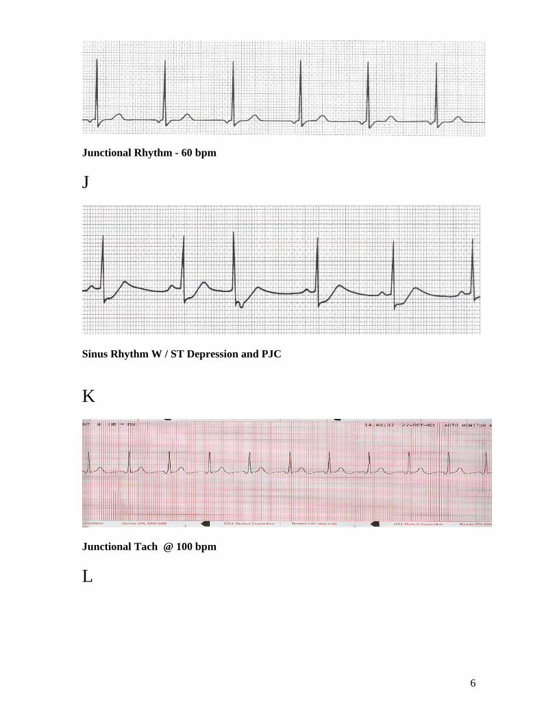
Junctional Rhythm - 60 bpm
J
Sinus Rhythm W / ST Depression and PJC
K
Junctional Tach @ 100 bpm
L
7
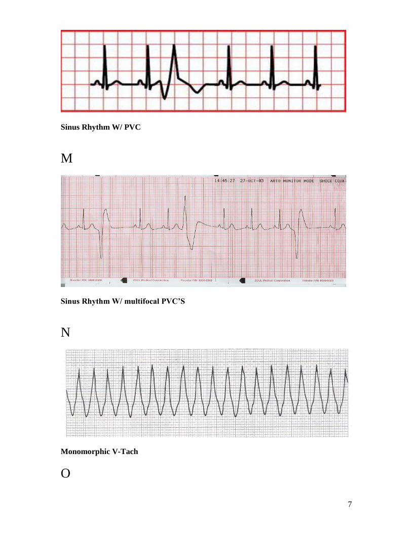
Sinus Rhythm W/ PVC
M
Sinus Rhythm W/ multifocal PVC’S
N
Monomorphic V-Tach
O

8
Poly V-Tach
P
Poly V-Tach / Torsades de points ( prolong QT)
Q
Poly V-Tach / Torsades de points
R
9
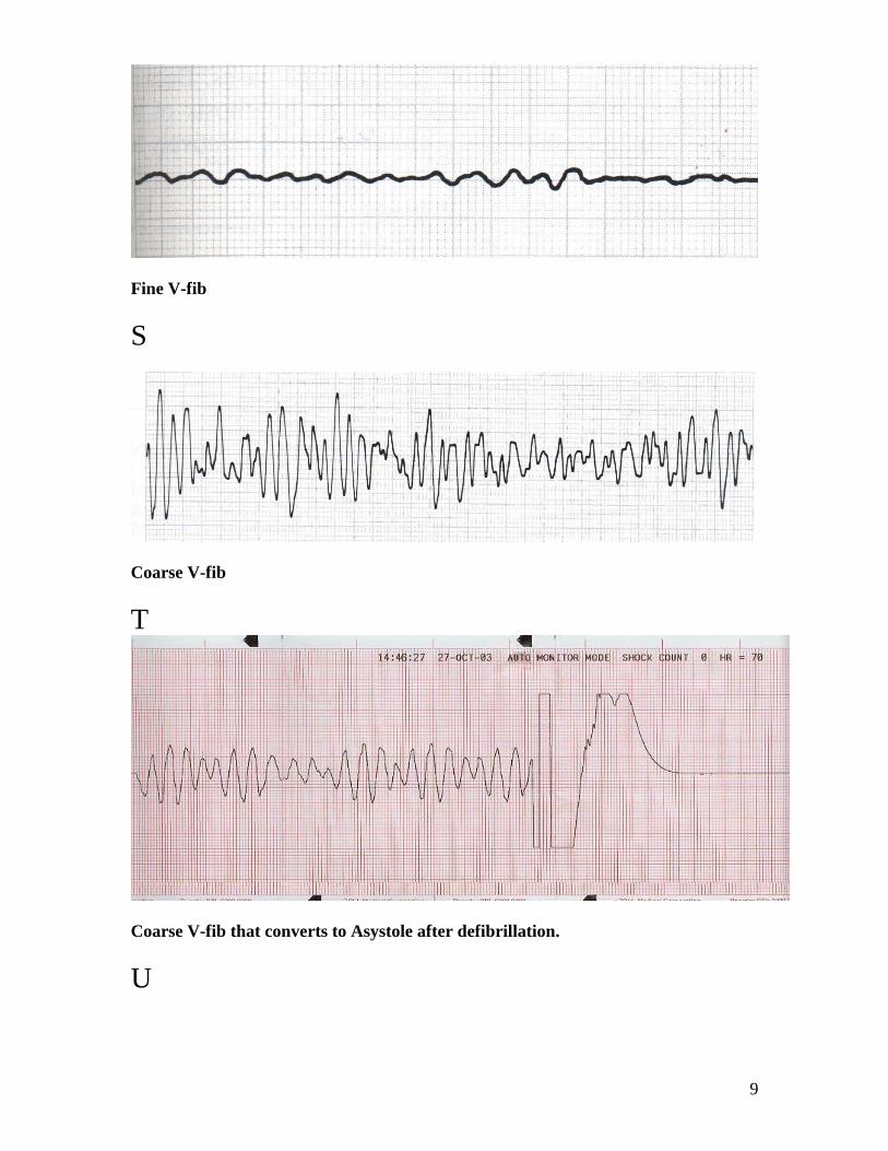
Fine V-fib
S
Coarse V-fib
T
Coarse V-fib that converts to Asystole after defibrillation.
U

10
1st degree ( P wave distant from the Q)
V
1st degree
W
2nd
degree type 1
X

11
2nd
degree type 1
Y
2nd
degree type 2 ( P wave will march out same space)
Z
2nd
degree type 2
AA
12
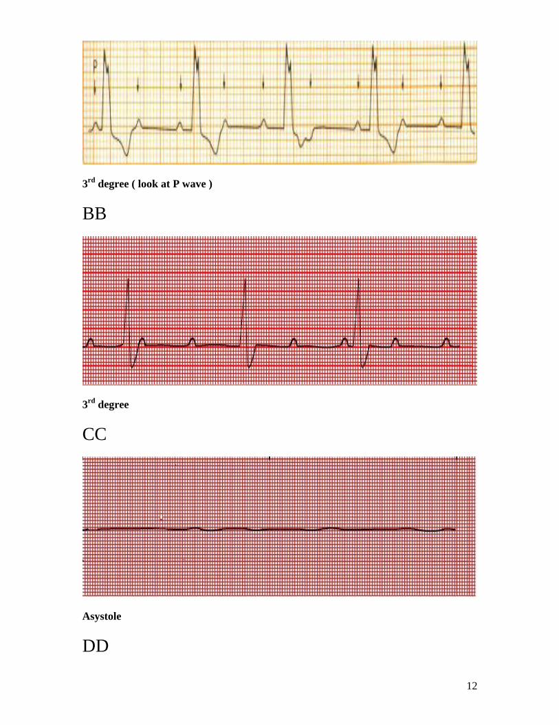
3rd
degree ( look at P wave )
BB
3rd
degree
CC
Asystole
DD

13
STEMI
EE
Non STEMI
14
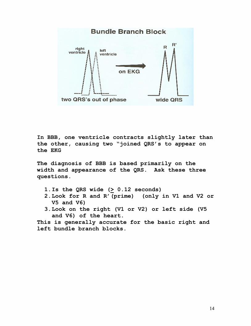
In BBB, one ventricle contracts slightly later than
the other, causing two “joined QRS’s to appear on
the EKG
The diagnosis of BBB is based primarily on the
width and appearance of the QRS. Ask these three
questions.
1. Is the QRS wide (> 0.12 seconds) 2. Look for R and R’(prime) (only in V1 and V2 or
V5 and V6)
3. Look on the right (V1 or V2) or left side (V5 and V6) of the heart.
This is generally accurate for the basic right and
left bundle branch blocks.
15
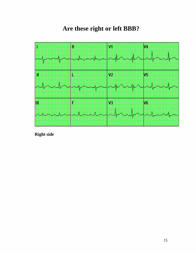
Are these right or left BBB?
Right side

16
Left Side