acquired macular disorders macular disorders chapter17 age-related macular degeneration 334...
TRANSCRIPT
Acquired Macular Disorders
17Chapter
Age-related macular degeneration 334Polypoidal choroidal vasculopathy 341Age-related macular hole 342Central serous retinopathy 344Cystoid macular oedema 345Macular epiretinal membrane 347Degenerative myopia 348Angioid streaks 350Choroidal folds 351Hypotony maculopathy 351Vitreomacular traction syndrome 352Solar maculopathy 352Idiopathic choroidal neovascularization 352
PROPERTY OF E
LSEVIE
R
SAMPLE C
ONTENT - NOT FIN
AL
Acquired Macular Disorders334
Age-related macular degeneration
Drusen
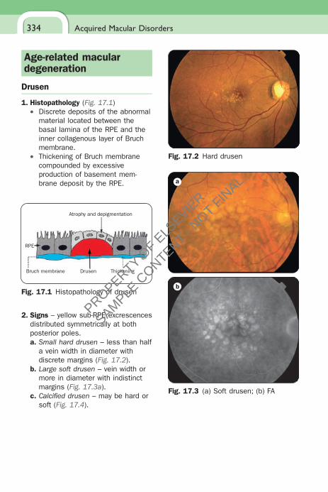
1. Histopathology (Fig. 17.1)• Discrete deposits of the abnormal
material located between the basal lamina of the RPE and the inner collagenous layer of Bruch membrane.
• Thickening of Bruch membrane compounded by excessive production of basement mem-brane deposit by the RPE.
Atrophy and depigmentation
RPE
Bruch membrane Drusen Thickening
Fig. 17.1 Histopathology of drusen
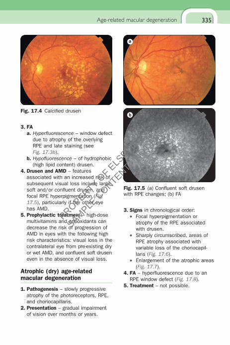
2. Signs – yellow sub-RPE excrescences distributed symmetrically at both posterior poles.a. Small hard drusen – less than half
a vein width in diameter with discrete margins (Fig. 17.2).
b. Large soft drusen – vein width or more in diameter with indistinct margins (Fig. 17.3a).
c. Calcifi ed drusen – may be hard or soft (Fig. 17.4).
Fig. 17.2 Hard drusen
a
b
Fig. 17.3 (a) Soft drusen; (b) FA
PROPERTY OF E
LSEVIE
R
SAMPLE C
ONTENT - NOT FIN
AL
335
3. FAa. Hyperfl uorescence – window defect
due to atrophy of the overlying RPE and late staining (see Fig. 17.3b).
b. Hypofl uorescence – of hydrophobic (high lipid content) drusen.
4. Drusen and AMD – features associated with an increased risk of subsequent visual loss include large soft and/or confl uent drusen, and focal RPE hyperpigmentation (Fig. 17.5), particularly if the other eye has AMD.
5. Prophylactic treatment – high-dose multivitamins and antioxidants can decrease the risk of progression of AMD in eyes with the following high risk characteristics: visual loss in the contralateral eye from pre-existing dry or wet AMD, and confl uent soft drusen even in the absence of visual loss.
Atrophic (dry) age-related macular degeneration
1. Pathogenesis – slowly progressive atrophy of the photoreceptors, RPE, and choriocapillaris.
2. Presentation – gradual impairment of vision over months or years.
3. Signs in chronological order:• Focal hyperpigmentation or
atrophy of the RPE associated with drusen.
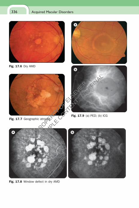
• Sharply circumscribed, areas of RPE atrophy associated with variable loss of the choriocapil-laris (Fig. 17.6).
• Enlargement of the atrophic areas (Fig. 17.7).
4. FA – hyperfl uorescence due to an RPE window defect (Fig. 17.8).
5. Treatment – not possible.
Fig. 17.4 Calcifi ed drusen
a
b
Fig. 17.5 (a) Confl uent soft drusen with RPE changes; (b) FA
Age-related macular degeneration
PROPERTY OF E
LSEVIE
R
SAMPLE C
ONTENT - NOT FIN
AL
Acquired Macular Disorders336
Fig. 17.6 Dry AMD
Fig. 17.7 Geographic atrophy
a b
Fig. 17.8 Window defect in dry AMD
a
b
Fig. 17.9 (a) PED; (b) ICG
PROPERTY OF E
LSEVIE
R
SAMPLE C
ONTENT - NOT FIN
AL
337
Retinal pigment epithelial detachment
1. Pathogenesis – reduction of hydraulic conductivity of the thickened Bruch membrane impeding movement of fl uid from the RPE towards the choroid.
2. Presentation – metamorphopsia and impairment of central vision.
3. Signs – circumscribed, dome-shaped elevation at the posterior pole (Fig. 17.9a).
4. ICG – oval hypofl uorescence with a faint ring of surrounding hyperfl uores-cence (Fig. 17.9b).
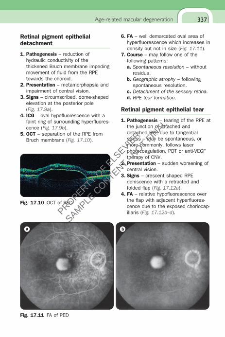
5. OCT – separation of the RPE from Bruch membrane (Fig. 17.10).
Fig. 17.10 OCT of PED
Age-related macular degeneration
a b
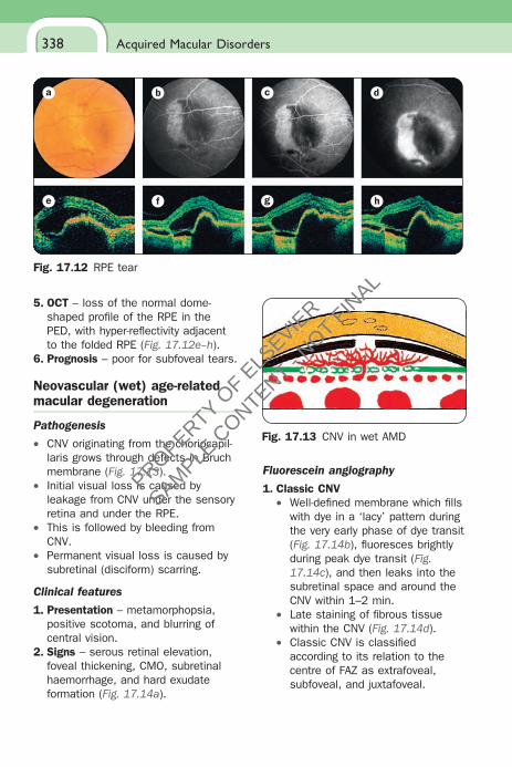
Fig. 17.11 FA of PED
6. FA – well demarcated oval area of hyperfl uorescence which increases in density but not in size (Fig. 17.11).
7. Course – may follow one of the following patterns:a. Spontaneous resolution – without
residua.b. Geographic atrophy – following
spontaneous resolution.c. Detachment of the sensory retina.d. RPE tear formation.
Retinal pigment epithelial tear
1. Pathogenesis – tearing of the RPE at the junction of attached and detached RPE due to tangential stress – may be spontaneous, or more commonly, follows laser photocoagulation, PDT or anti-VEGF therapy of CNV.
2. Presentation – sudden worsening of central vision.
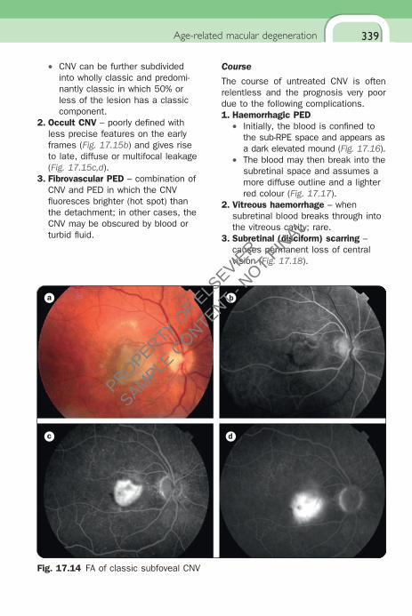
3. Signs – crescent shaped RPE dehiscence with a retracted and folded fl ap (Fig. 17.12a).
4. FA – relative hypofl uorescence over the fl ap with adjacent hyperfl uores-cence due to the exposed choriocap-illaris (Fig. 17.12b–d).
PROPERTY OF E
LSEVIE
R
SAMPLE C
ONTENT - NOT FIN
AL
Acquired Macular Disorders338
a b c d
e f g h
Fig. 17.12 RPE tear
Fig. 17.13 CNV in wet AMD
5. OCT – loss of the normal dome-shaped profi le of the RPE in the PED, with hyper-refl ectivity adjacent to the folded RPE (Fig. 17.12e–h).
6. Prognosis – poor for subfoveal tears.
Neovascular (wet) age-related macular degeneration
Pathogenesis
• CNV originating from the choriocapil-laris grows through defects in Bruch membrane (Fig. 17.13).
• Initial visual loss is caused by leakage from CNV under the sensory retina and under the RPE.
• This is followed by bleeding from CNV.
• Permanent visual loss is caused by subretinal (disciform) scarring.
Clinical features
1. Presentation – metamorphopsia, positive scotoma, and blurring of central vision.
2. Signs – serous retinal elevation, foveal thickening, CMO, subretinal haemorrhage, and hard exudate formation (Fig. 17.14a).
Fluorescein angiography
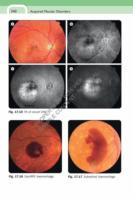
1. Classic CNV• Well-defi ned membrane which fi lls
with dye in a ‘lacy’ pattern during the very early phase of dye transit (Fig. 17.14b), fl uoresces brightly during peak dye transit (Fig. 17.14c), and then leaks into the subretinal space and around the CNV within 1–2 min.
• Late staining of fi brous tissue within the CNV (Fig. 17.14d).
• Classic CNV is classifi ed according to its relation to the centre of FAZ as extrafoveal, subfoveal, and juxtafoveal.
PROPERTY OF E
LSEVIE
R
SAMPLE C
ONTENT - NOT FIN
AL
339
• CNV can be further subdivided into wholly classic and predomi-nantly classic in which 50% or less of the lesion has a classic component.
2. Occult CNV – poorly defi ned with less precise features on the early frames (Fig. 17.15b) and gives rise to late, diffuse or multifocal leakage (Fig. 17.15c,d).
3. Fibrovascular PED – combination of CNV and PED in which the CNV fl uoresces brighter (hot spot) than the detachment; in other cases, the CNV may be obscured by blood or turbid fl uid.
a b
c d
Fig. 17.14 FA of classic subfoveal CNV
Age-related macular degeneration
Course
The course of untreated CNV is often relentless and the prognosis very poor due to the following complications.1. Haemorrhagic PED
• Initially, the blood is confi ned to the sub-RPE space and appears as a dark elevated mound (Fig. 17.16).
• The blood may then break into the subretinal space and assumes a more diffuse outline and a lighter red colour (Fig. 17.17).
2. Vitreous haemorrhage – when subretinal blood breaks through into the vitreous cavity; rare.
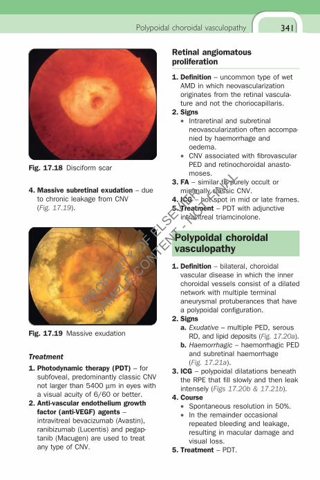
3. Subretinal (disciform) scarring – causes permanent loss of central vision (Fig. 17.18).
PROPERTY OF E
LSEVIE
R
SAMPLE C
ONTENT - NOT FIN
AL
Acquired Macular Disorders340
a b
c d
Fig. 17.15 FA of occult CNV
Fig. 17.16 Sub-RPE haemorrhage Fig. 17.17 Subretinal haemorrhage
PROPERTY OF E
LSEVIE
R
SAMPLE C
ONTENT - NOT FIN
AL
341
4. Massive subretinal exudation – due to chronic leakage from CNV (Fig. 17.19).
Fig. 17.18 Disciform scar
Fig. 17.19 Massive exudation
Treatment
1. Photodynamic therapy (PDT) – for subfoveal, predominantly classic CNV not larger than 5400 µm in eyes with a visual acuity of 6/60 or better.
2. Anti-vascular endothelium growth factor (anti-VEGF) agents – intravitreal bevacizumab (Avastin), ranibizumab (Lucentis) and pegap-tanib (Macugen) are used to treat any type of CNV.
Retinal angiomatous proliferation
1. Defi nition – uncommon type of wet AMD in which neovascularization originates from the retinal vascula-ture and not the choriocapillaris.
2. Signs• Intraretinal and subretinal
neovascularization often accompa-nied by haemorrhage and oedema.
• CNV associated with fi brovascular PED and retinochoroidal anasto-moses.
3. FA – similar to purely occult or minimally classic CNV.
4. ICG – hot spot in mid or late frames.5. Treatment – PDT with adjunctive
intravitreal triamcinolone.
Polypoidal choroidal vasculopathy
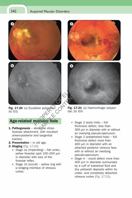
1. Defi nition – bilateral, choroidal vascular disease in which the inner choroidal vessels consist of a dilated network with multiple terminal aneurysmal protuberances that have a polypoidal confi guration.
2. Signsa. Exudative – multiple PED, serous
RD, and lipid deposits (Fig. 17.20a).b. Haemorrhagic – haemorrhagic PED
and subretinal haemorrhage (Fig. 17.21a).
3. ICG – polypoidal dilatations beneath the RPE that fi ll slowly and then leak intensely (Figs 17.20b & 17.21b).
4. Course• Spontaneous resolution in 50%.• In the remainder occasional
repeated bleeding and leakage, resulting in macular damage and visual loss.
5. Treatment – PDT.
Polypoidal choroidal vasculopathy
PROPERTY OF E
LSEVIE
R
SAMPLE C
ONTENT - NOT FIN
AL
Acquired Macular Disorders342
a
b
Fig. 17.20 (a) Exudative polypoidal; (b) ICG
a
b
Fig. 17.21 (a) Haemorrhagic polypoi-dal; (b) ICG
Age-related macular hole
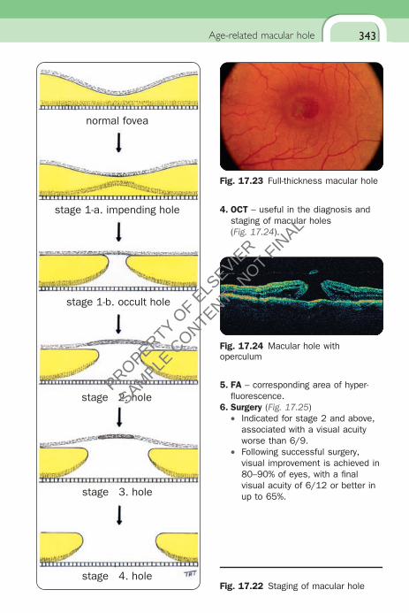
1. Pathogenesis – abnormal vitreo-foveolar attachment, with resultant antero-posterior and tangential traction.
2. Presentation – in old age.3. Staging (Fig. 17.22)
• Stage 1a (impending) – fl at umbo, yellow foveolar spot 100–200 µm in diameter with loss of the foveolar refl ex.
• Stage 1b (occult) – yellow ring with a bridging interface of vitreous cortex.
• Stage 2 (early hole) – full-thickness defect, less than 300 µm in diameter with or without an overlying pseudo-operculum.
• Stage 3 (established hole) – full-thickness defect more than 400 µm in diameter with an attached posterior vitreous face with or without an overlying pseudo-operculum.
• Stage 4 – round defect more than 400 µm in diameter surrounded by a cuff of subretinal fl uid and tiny yellowish deposits within its crater, and completely detached vitreous cortex (Fig. 17.23).
PROPERTY OF E
LSEVIE
R
SAMPLE C
ONTENT - NOT FIN
AL
343
normal fovea
stage 4. hole
stage 3. hole
stage 2. hole
stage 1-b. occult hole
stage 1-a. impending hole
Fig. 17.22 Staging of macular hole
Age-related macular hole
4. OCT – useful in the diagnosis and staging of macular holes (Fig. 17.24).
Fig. 17.23 Full-thickness macular hole
Fig. 17.24 Macular hole with operculum
5. FA – corresponding area of hyper-fl uorescence.
6. Surgery (Fig. 17.25)• Indicated for stage 2 and above,
associated with a visual acuity worse than 6/9.
• Following successful surgery, visual improvement is achieved in 80–90% of eyes, with a fi nal visual acuity of 6/12 or better in up to 65%.
PROPERTY OF E
LSEVIE
R
SAMPLE C
ONTENT - NOT FIN
AL
Acquired Macular Disorders344
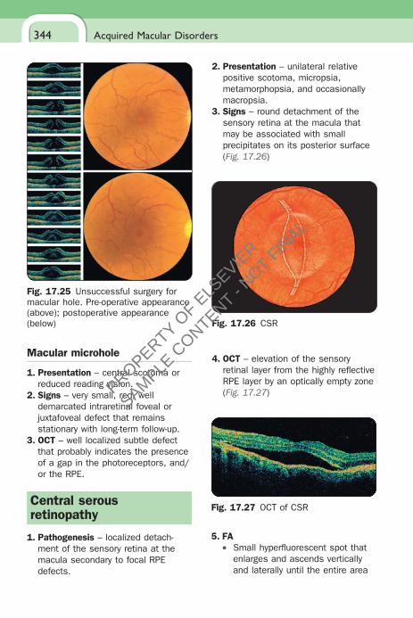
Macular microhole
1. Presentation – central scotoma or reduced reading vision.
2. Signs – very small, red, well demarcated intraretinal foveal or juxtafoveal defect that remains stationary with long-term follow-up.
3. OCT – well localized subtle defect that probably indicates the presence of a gap in the photoreceptors, and/or the RPE.
Central serous retinopathy
1. Pathogenesis – localized detach-ment of the sensory retina at the macula secondary to focal RPE defects.
2. Presentation – unilateral relative positive scotoma, micropsia, metamorphopsia, and occasionally macropsia.
3. Signs – round detachment of the sensory retina at the macula that may be associated with small precipitates on its posterior surface (Fig. 17.26)
Fig. 17.25 Unsuccessful surgery for macular hole. Pre-operative appearance (above); postoperative appearance (below) Fig. 17.26 CSR
Fig. 17.27 OCT of CSR
5. FA• Small hyperfl uorescent spot that
enlarges and ascends vertically and laterally until the entire area
4. OCT – elevation of the sensory retinal layer from the highly refl ective RPE layer by an optically empty zone (Fig. 17.27)
PROPERTY OF E
LSEVIE
R
SAMPLE C
ONTENT - NOT FIN
AL
345
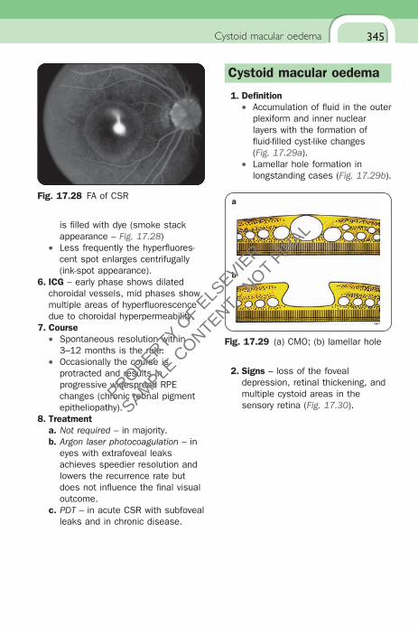
Fig. 17.28 FA of CSR
Cystoid macular oedema
is fi lled with dye (smoke stack appearance – Fig. 17.28)
• Less frequently the hyperfl uores-cent spot enlarges centrifugally (ink-spot appearance).
6. ICG – early phase shows dilated choroidal vessels, mid phases show multiple areas of hyperfl uorescence due to choroidal hyperpermeability.
7. Course• Spontaneous resolution within
3–12 months is the rule.• Occasionally the course is
protracted and results in progressive widespread RPE changes (chronic retinal pigment epitheliopathy).
8. Treatmenta. Not required – in majority.b. Argon laser photocoagulation – in
eyes with extrafoveal leaks achieves speedier resolution and lowers the recurrence rate but does not infl uence the fi nal visual outcome.
c. PDT – in acute CSR with subfoveal leaks and in chronic disease.
Cystoid macular oedema
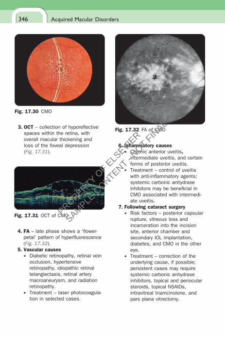
1. Defi nition• Accumulation of fl uid in the outer
plexiform and inner nuclear layers with the formation of fl uid-fi lled cyst-like changes (Fig. 17.29a).
• Lamellar hole formation in longstanding cases (Fig. 17.29b).
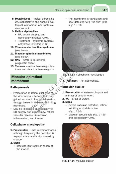
2. Signs – loss of the foveal depression, retinal thickening, and multiple cystoid areas in the sensory retina (Fig. 17.30).
a
b
Fig. 17.29 (a) CMO; (b) lamellar hole
PROPERTY OF E
LSEVIE
R
SAMPLE C
ONTENT - NOT FIN
AL
Acquired Macular Disorders346
4. FA – late phase shows a ‘fl ower-petal’ pattern of hyperfl uorescence (Fig. 17.32).
5. Vascular causes• Diabetic retinopathy, retinal vein
occlusion, hypertensive retinopathy, idiopathic retinal telangiectasis, retinal artery macroaneurysm, and radiation retinopathy.
• Treatment – laser photocoagula-tion in selected cases.
Fig. 17.30 CMO
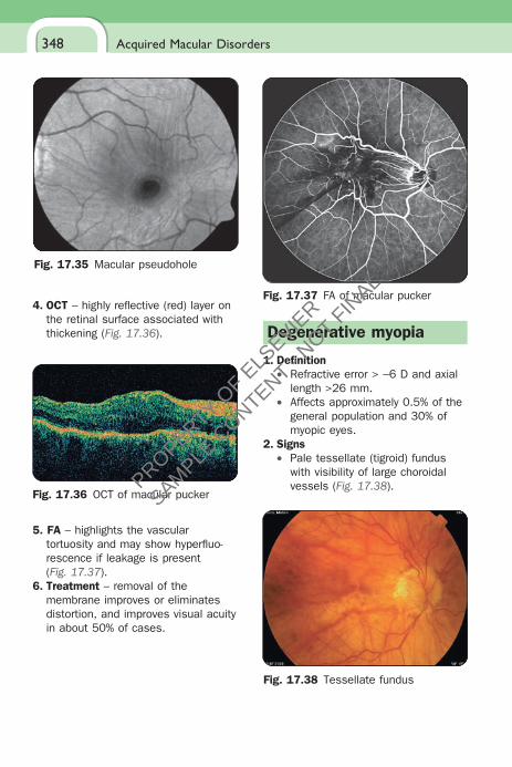
3. OCT – collection of hyporefl ective spaces within the retina, with overall macular thickening and loss of the foveal depression (Fig. 17.31).
Fig. 17.31 OCT of CMO
Fig. 17.32 FA of CMO
6. Infl ammatory causes• Chronic anterior uveitis,
intermediate uveitis, and certain forms of posterior uveitis.
• Treatment – control of uveitis with anti-infl ammatory agents; systemic carbonic anhydrase inhibitors may be benefi cial in CMO associated with intermedi-ate uveitis.
7. Following cataract surgery• Risk factors – posterior capsular
rupture, vitreous loss and incarceration into the incision site, anterior chamber and secondary IOL implantation, diabetes, and CMO in the other eye.
• Treatment – correction of the underlying cause, if possible; persistent cases may require systemic carbonic anhydrase inhibitors, topical and periocular steroids, topical NSAIDs, intravitreal triamcinolone, and pars plana vitrectomy.
PROPERTY OF E
LSEVIE
R
SAMPLE C
ONTENT - NOT FIN
AL
347Macular epiretinal membrane
8. Drug-induced – topical adrenaline 2% (especially in the aphakic eye), topical latanoprost, and systemic nicotinic acid.
9. Retinal dystrophies• RP, gyrate atrophy, and
dominantly inherited CMO.• Treatment – systemic carbonic
anhydrase inhibitors in RP.10. Vitreomacular traction syndrome
(see below).11. Macular epiretinal membranes
(see below).12. CNV – CMO is an adverse
prognostic factor.13. Tumours – retinal haemangioblas-
toma and choroidal haemangioma.
Macular epiretinal membrane
Pathogenesis
• Proliferation of retinal glial cells at the vitreoretinal interface that have gained access to the retinal surface through breaks in the internal limiting membrane.
• May be idiopathic or secondary to RD surgery and cryotherapy, retinal vascular disease, intraocular infl ammation, and trauma.
Cellophane maculopathy
1. Presentation – mild metamorphopsia although frequently the condition is asymptomatic and is discovered by chance.
2. Signs• Irregular light refl ex or sheen at
the macula.
• The membrane is translucent and best detected with ‘red-free’ light (Fig. 17.33).
Fig. 17.33 Cellophane maculopathy
3. Treatment – not appropriate.
Macular pucker
1. Presentation – metamorphopsia and blurring of central vision.
2. VA – 6/12 or worse.3. Signs
• Severe vascular distortion, retinal wrinkling and white striae (Fig. 17.34).
• Macular pseudo-hole (Fig. 17.35) and occasionally CMO.
Fig. 17.34 Macular pucker
PROPERTY OF E
LSEVIE
R
SAMPLE C
ONTENT - NOT FIN
AL
Acquired Macular Disorders348
4. OCT – highly refl ective (red) layer on the retinal surface associated with thickening (Fig. 17.36).
Fig. 17.35 Macular pseudohole
Fig. 17.36 OCT of macular pucker
Fig. 17.37 FA of macular pucker
5. FA – highlights the vascular tortuosity and may show hyperfl uo-rescence if leakage is present (Fig. 17.37).
6. Treatment – removal of the membrane improves or eliminates distortion, and improves visual acuity in about 50% of cases.
Degenerative myopia
1. Defi nition• Refractive error > −6 D and axial
length >26 mm.• Affects approximately 0.5% of the
general population and 30% of myopic eyes.
2. Signs• Pale tessellate (tigroid) fundus
with visibility of large choroidal vessels (Fig. 17.38).
Fig. 17.38 Tessellate fundus
PROPERTY OF E
LSEVIE
R
SAMPLE C
ONTENT - NOT FIN
AL
349
• ‘Lacquer cracks’ consist of ruptures in the RPE–Bruch membrane–choriocapillaris complex (Fig. 17.39).
• Focal chorioretinal atrophy with visibility of the sclera (Fig. 17.40).
• Staphylomas due to expansion of the globe and scleral thinning.
Fig. 17.39 Lacquer cracks
Degenerative myopia
Fig. 17.40 Chorioretinal atrophy
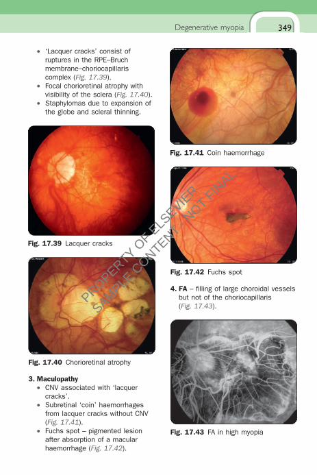
Fig. 17.41 Coin haemorrhage
3. Maculopathy• CNV associated with ‘lacquer
cracks’.• Subretinal ‘coin’ haemorrhages
from lacquer cracks without CNV (Fig. 17.41).
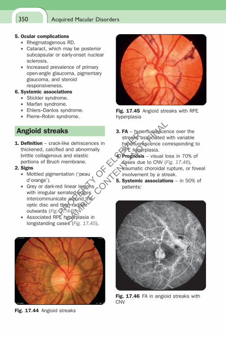
• Fuchs spot – pigmented lesion after absorption of a macular haemorrhage (Fig. 17.42).
Fig. 17.42 Fuchs spot
Fig. 17.43 FA in high myopia
4. FA – fi lling of large choroidal vessels but not of the choriocapillaris (Fig. 17.43).
PROPERTY OF E
LSEVIE
R
SAMPLE C
ONTENT - NOT FIN
AL
Acquired Macular Disorders350
5. Ocular complications• Rhegmatogenous RD.• Cataract, which may be posterior
subcapsular or early-onset nuclear sclerosis.
• Increased prevalence of primary open-angle glaucoma, pigmentary glaucoma, and steroid responsiveness.
6. Systemic associations• Stickler syndrome.• Marfan syndrome.• Ehlers–Danlos syndrome.• Pierre–Robin syndrome.
Angioid streaks
1. Defi nition – crack-like dehiscences in thickened, calcifi ed and abnormally brittle collagenous and elastic portions of Bruch membrane.
2. Signs• Mottled pigmentation (‘peau
d’orange’).• Grey or dark-red linear lesions
with irregular serrated edges intercommunicate around the optic disc and then radiate outwards (Fig. 17.44).
• Associated RPE hyperplasia in longstanding cases (Fig. 17.45).
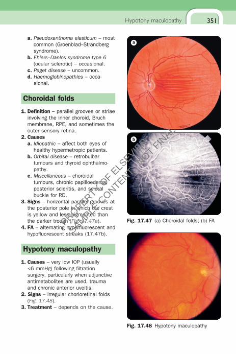
Fig. 17.44 Angioid streaks
Fig. 17.45 Angioid streaks with RPE hyperplasia
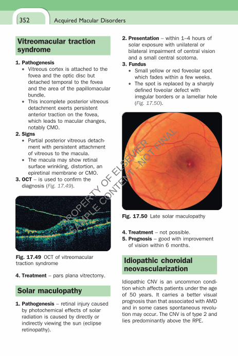
Fig. 17.46 FA in angioid streaks with CNV
3. FA – hyperfl uorescence over the streaks associated with variable hypofl uorescence corresponding to RPE hyperplasia.
4. Prognosis – visual loss in 70% of cases due to CNV (Fig. 17.46), traumatic choroidal rupture, or foveal involvement by a streak.
5. Systemic associations – in 50% of patients:
PROPERTY OF E
LSEVIE
R
SAMPLE C
ONTENT - NOT FIN
AL
351Hypotony maculopathy
a. Pseudoxanthoma elasticum – most common (Groenblad–Strandberg syndrome).
b. Ehlers–Danlos syndrome type 6 (ocular sclerotic) – occasional.
c. Paget disease – uncommon.d. Haemoglobinopathies – occa-
sional.
Choroidal folds
1. Defi nition – parallel grooves or striae involving the inner choroid, Bruch membrane, RPE, and sometimes the outer sensory retina.
2. Causesa. Idiopathic – affect both eyes of
healthy hypermetropic patients.b. Orbital disease – retrobulbar
tumours and thyroid ophthalmo-pathy.
c. Miscellaneous – choroidal tumours, chronic papilloedema, posterior scleritis, and scleral buckle for RD.
3. Signs – horizontal parallel grooves at the posterior pole in which the crest is yellow and less pigmented than the darker trough (Fig. 17.47a).
4. FA – alternating hyperfl uorescent and hypofl uorescent streaks (17.47b).
Hypotony maculopathy
1. Causes – very low IOP (usually <6 mmHg) following fi ltration surgery, particularly when adjunctive antimetabolites are used, trauma and chronic anterior uveitis.
2. Signs – irregular chorioretinal folds (Fig. 17.48).
3. Treatment – depends on the cause.
a
b
Fig. 17.47 (a) Choroidal folds; (b) FA
Fig. 17.48 Hypotony maculopathy
PROPERTY OF E
LSEVIE
R
SAMPLE C
ONTENT - NOT FIN
AL
Acquired Macular Disorders
E
352
Vitreomacular traction syndrome
1. Pathogenesis• Vitreous cortex is attached to the
fovea and the optic disc but detached temporal to the fovea and the area of the papillomacular bundle.
• This incomplete posterior vitreous detachment exerts persistent anterior traction on the fovea, which leads to macular changes, notably CMO.
2. Signs• Partial posterior vitreous detach-
ment with persistent attachment of vitreous to the macula.
• The macula may show retinal surface wrinkling, distortion, an epiretinal membrane or CMO.
3. OCT – is used to confi rm the diagnosis (Fig. 17.49).
2. Presentation – within 1–4 hours of solar exposure with unilateral or bilateral impairment of central vision and a small central scotoma.
3. Fundus• Small yellow or red foveolar spot
which fades within a few weeks.• The spot is replaced by a sharply
defi ned foveolar defect with irregular borders or a lamellar hole (Fig. 17.50).
Fig. 17.49 OCT of vitreomacular traction syndrome
4. Treatment – pars plana vitrectomy.
Solar maculopathy
1. Pathogenesis – retinal injury caused by photochemical effects of solar radiation is caused by directly or indirectly viewing the sun (eclipse retinopathy).
Fig. 17.50 Late solar maculopathy
4. Treatment – not possible.5. Prognosis – good with improvement
of vision within 6 months.
Idiopathic choroidal neovascularization
Idiopathic CNV is an uncommon condi-tion which affects patients under the age of 50 years. It carries a better visual prognosis than that associated with AMD and in some cases spontaneous revolu-tion may occur. The CNV is of type 2 and lies predominantly above the RPE.
Ch017-S3135.indd 352 2/13/2009 2:22:08 PM
PROPERTY OF E
LSEVIE
R
SAMPLE C
ONTENT - NOT FIN
AL