activation of human primary visual cortex during visual recall: a
TRANSCRIPT

Proc. Natl. Acad. Sci. USAVol. 90, pp. 11802-11805, December 1993Neurobiology
Activation of human primary visual cortex during visual recall:A magnetic resonance imaging study
(blood flow/blood oxygenation/human cognition/functional neuroimaging/echo-planar imaging)
D. LE BIHAN*t, R. TURNERt, T. A. ZEFFIRO§, C. A. CUtNOD¶, P. JEZZARDt, AND V. BONNEROT**The Warren G. Magnuson Clinical Center Diagnostic Radiology Department, tLaboratory of Cardiac Energetics of the National Heart, Lung and BloodInstitute, NMedical Neurology Branch of the National Institute of Neurological Disorders and Stroke, and fLaboratory of Diagnostic Radiology Research,National Institutes of Health, Bethesda, MD 20892
Communicated by Mortimer Mishkin, August 30, 1993 (received for review April 15, 1993)
ABSTRACT The degree to which the processes involved invisual perception and visual imagery share a common neu-roanatomical substrate is unclear. Physiological evidence forlocalization of visual imagery early in the visual pathwayswould have important bearing on current theories of visualprocessing. A magnetic resonance imaging technique sensitiveto regional changes in blood oxygenation was used to obtainfunctional activation maps in the human visual cortex. Duringrecall of a visual stimulus, focal increases in signal related tochanges in blood flow were detected in Vl and V2 cortex in fiveof seven subjects. These experiments show that the same areasof the early visual cortex that are excited by visual stimulationare also activated during mental representation of the samestimulus. Some of the processes used in topographicallymapped cortical areas during visual perception may also beutilized during visual recall.
The role of the cortical visual system in the processesunderlying mental imagery has long been debated. An issueof particular interest concerns the degree to which theprocesses that are operative in visual perception are the sameprocesses involved in the generation of visual imagery andwhether visual representations are shared between imageryand perceptual processes (1-3). The finding that brain activityin relation to mental imagery occurs in at least some of thesame locations as those seen during perception would bestrong evidence that these two mental experiences sharesome common physiological mechanisms. Previous clinicaland experimental studies provide support for spatial colocal-ization of these processes (4, 5). The clinical evidence is thefinding that lesions to the occipital lobe in the human thatresult in disorders of visual perception can also result invisual imagery deficits (6, 7). The most compelling experi-mental evidence comes from functional neuroimaging studiesdemonstrating concordance between the neuroanatomicalregions in occipital cortex activated by visual perception andimagery tasks in the same individual. Although the details ofthe results differ among experiments, all have found extra-striate visual areas that have been activated both when thestimulus is perceived and when it is recalled (8-11). Incontrast, activation in striate cortex related to visual recallhas been more difficult to demonstrate. This may result fromthe limited spatial and temporal resolution inherent in thesetechniques, so that precise response localization has not beenpossible. The one previous positron emission tomographystudy that investigated visual recall using tomographic mea-surements at higher resolution demonstrated small but sta-tistically nonsignificant activations in the calcarine fissure(12). Furthermore, techniques that involve ionizing radiationexposure to subjects preclude the sort of repetitive studies
The publication costs of this article were defrayed in part by page chargepayment. This article must therefore be hereby marked "advertisement"in accordance with 18 U.S.C. §1734 solely to indicate this fact.
that would provide greater sensitivity. Recent advances inmagnetic resonance imaging (MRI) have demonstrated thefeasibility offunctional neuroimaging at a temporal resolutionof seconds and spatial resolution of millimeters (13-17). Thiscompletely noninvasive procedure provides a powerfulmeans to reinvestigate the question of the role of primaryvisual cortex in the generation of mental imagery.The biophysical basis for the measurements made with
functional MRI differs from those underlying other neuroim-aging techniques. Transient changes in regional cerebralblood flow and oxygen extraction produce differences in thebalance between paramagnetic deoxyhemoglobin and dia-magnetic oxyhemoglobin in red cells (18-20). Changes in thisbalance, closely related to oxygen saturation, result in tran-sient magnetic-susceptibility contrast that may be monitoreddirectly and noninvasively by using fast MRI acquisitionschemes. This method has been employed in the human brainto visualize activation of primary visual cortex by externalsensory stimuli and sensorimotor cortex during voluntarymovement (14-17). Using this technique, we report clearevidence that the primary visual cortex in the calcarinefissure can be activated during both visual perception andvisual recall.
METHODSBrain activation maps calculated with actual photic stimula-tion were compared with those obtained when the subjectwas later asked to recall the same visual stimulus. Sevennormal subjects (five men and two women) ranging in agefrom 26 to 46 years underwent dynamic MRI. Studies wereperformed on a clinical 1.5-T whole-body MRI scanner(General Electric Medical Systems, Milwaukee, WI). Toincrease signal-to-noise ratio a 7.5-cm radiofrequency sur-face coil placed under the back ofthe head was used for signalreception. Cushions placed around the subject's head wereused to minimize subject movement between scans. Activa-tion studies were performed first with echo-planar imaging(21, 22) using a home-designed head (27-cm diameter) z-gra-dient coil. Acquisition parameters were as follows: echo time= 40 ms, repetition rate of 20 images per minute with anin-plane resolution of 2.5 x 2.5 mm2. Images were obtainedin coronal orientation, approximately perpendicular to thecalcarine fissures (Fig. 1). Later, a conventional gradient-echo sequence was used [spoiled gradient-echo acquisitionsequence in the steady state (GRASS) sequence, repetitiontime = 71 ms, echo time = 40 ms, flip angle = 400, 10 imagesper minute] in two ofour responding subjects. This sequence,which allows an in-plane resolution of 1.5 x 0.8 mm2 and
Abbreviations: MRI, magnetic resonance imaging; GRASS, gradi-ent-echo acquisition in the steady state.tTo whom reprint requests should be addressed at: DiagnosticRadiology Department, Clinical Center, Building 10, Room 1C660,National Institutes of Health, Bethesda, MD 20892.
11802

Proc. Natl. Acad. Sci. USA 90 (1993) 11803
FIG. 1. (Upper) Sagittal Ti-weighted MRI image. Such imageswere acquired for localization and selection of coronal (echo-planarimaging) and oblique-transverse (spoiled GRASS) slices. The obliq-uity of the transverse slices (line in Upper) was chosen to include alarge part ofthe calcarine fissure. (Lower) This oblique slice is shownwith high-resolution and Ti-weighted contrast.
acquisition of images in an axial-oblique orientation, follow-ing the calcarine fissures (Fig. 1), gave a better identificationof striate cortex. The visual stimulation was provided bylight-proof binocular goggles (model SlOVS, Grass Instru-ments, Quincy, MA) fitted with red light-emitting diodesgeometrically arranged as two square patterns flashing at 16Hz (23). With these goggles, the stimulus was diffuse andexcited both visual fields within a large angle. The tasksequence consisted of a single prestimulation of the visualcortex for 30 s during which time the subjects were asked tomemorize the stimulus. The stimulus was then switchedalternately on and offtwice for periods of24 s. Between thesetwo actual photic stimulation episodes, the subjects were
asked instead to recall the previously seen stimulus for 24 s,
controlled by vocal command. The subjects were in completedarkness during the experiment and were asked to keep theireyes open for the duration of data acquisition.
RESULTSRegions of interest (pixel-by-pixel irregular regions) were
placed on the areas of maximum response during actual
stimulation on the medial surface of the occipital lobe in thebank of the calcarine fissure. These areas of maximumresponse lay within 1-2 cm ofthe occipital pole, 0-2 cm frommidline, which includes cortex within Vl and V2 regions (24,25). This position may result from the diffusiveness of thestimulus. Moreover, the positron emission tomography studyof retinotopic organization of Vl by Fox et al. (26) shows asimilar localization of response using visual stimuli similar toour own. The exact position and shape of each region ofinterest were defined for each individual from the activationmap calculated during actual stimulation. Typical size forregions of interest was 10-20 mm2. The same regions ofinterest were used to determine the amplitude ofthe responseduring the recalled stimulus as well.
Fig. 2 displays the measured MRI signal changes in a regionof interest in the striate cortex in one of our subjects duringvisual stimulation conditions and rest (this blank trial wasgiven as a control without the instruction to imagine) (Fig.2A) and when asked to recall the stimulus between the twoactual stimulation episodes (Fig. 2B). During actual photicstimulation, each of our seven subjects showed a significantincrease in MRI signal occurring bilaterally within the cal-carine fissure (unpaired Mann-Whitney test, P < 0.004, withan average increase of 2.8 ± 1.1% (Table 1).More interestingly, significant signal increases were also
observed in five of our seven subjects in the same regionswhen the subject was recalling the stimulus (unpaired Mann-Whitney test, P < 0.01) (Table 1). However, the amplitude ofthe response to the recalled stimulus, when present, was notas large as the response to the actual stimulus (1.5 ± 0.6%,n = 5). In two of our subjects, otherwise unremarkable, nosignificant signal increase from baseline could be demon-strated during the recall condition, although a brief signalincrease could be seen. However, we had no way to screenfor attention and quality of the visual recall performance.Measurements within the visual cortex, but beyond Vi andprobably V2, showed no significant response and very stablesignal intensity over the acquisition period (variations of<0.2% of the mean signal intensity).
Activation maps, A(x, y), were calculated on a voxel-by-voxel basis as the normal deviate statistical variable Z,
A(x, y) =2 Jj(x, y)/n -2 I#(x, y)/m]/SD, [ll
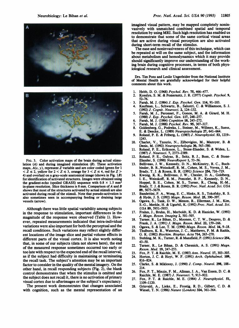
in which the mean of n images (typically 5) acquired in actualor imagined stimulation state, J(x, y), was compared with themean ofm "baseline" images, I(x, y) (typically 5). SD is thestandard deviation of the measurements. Functional activa-tion images from one subject are shown in Fig. 3. Regions inthe primary visual cortex showed highly localized changes ofintensity during actual stimulation (Fig. 3A), in agreementwith previous studies (13-17). The recalled stimulus activa-tion map (Fig. 3B) clearly shows that primary visual cortex isalso activated, but with less intensity, when the subject isasked to recall the stimulus.
DISCUSSIONThese variations in MRI intensity directly reflect changes inthe amount of intravascular deoxyhemoglobin. The overallincrease in signal intensity during actual or recalled stimu-lation is believed to originate from the increase in blood flowand blood volume leading to an increase in oxygen supply(18-20) which overcomes the increase in oxygen extraction(27). In all subjects, the rise time ofthe response to activationpatterns was several seconds, in agreement with opticalimaging measurements of cortical signal changes associatedwith hemodynamic perturbations (28). In all cases, during the
Neurobiology: Le Bihan et al.

11804 Neurobiology: Le Bihan et al.
0:25 0:40 0:55 1:10 1:25 1:40 1:55 2:10 2:25 2:40 2:55
Time (min:sec)
0:54 1:24
Time (min:sec)
1:54 2:24
LUS
FIG. 2. Plot of MRI signal intensity relative changes versus time for primary visual cortex and a cortical region outside the striate cortex(echo-planar imaging data). In A, the subject was resting between the two actual stimuli (blank trial) (a, visual striate cortex). In B, the samesubject was asked to recall the stimulus (+, visual striate cortex; *, non-striate cortex).
period of rest immediately following actual and imaginedstimulation the signal from the excited areas dropped belowits resting value (Fig. 2). This origin of this drop in signalintensity below baseline level is still unclear but may repre-
sent the known undershoot in blood flow in the posteriorcerebral artery following visual activation or continuedhigher metabolism and oxygen consumption by tissue afterblood flow returns to baseline.
Table 1. Comparison of activation during actual and imagined stimulationActual stimulation Imagined stimulation
Subject % change from baseline P % change from baseline P1 3.6 0.004 1.4 0.0042 1.1 0.004 0.5 0.013 1.9 0.0009 (0.04) 0.5 (NS)4 2.5 0.0008 1.7 0.00085 4.1 0.0003 1.6 0.00076 2.4 0.002 (0.4) 0.1 (NS)7 4.1 0.003 2.4 0.003Mean 2.8 ± 1.1 (n = 7) 1.5 ± 0.6(n = 5)
1.0 ± 0.9 (n = 7)We determined the regions of interest with the greatest change for the actual stimulus (coronal
echo-planar imaging data). The activation ratios for the actual stimulation and the imagined stimulationwere calculated according to Eq. 1. Statistical analysis was performed for each region of interest byusing a Mann-Whitney test (unpaired, one-tailed) between the average signal in the baseline images andthe average signal in the images acquired during actual or imagined stimulation. For comparison ofresponse amplitudes, statistics for the recall condition are also given for the responding subjects only.NS, not significant.
A5%
4% -
CDIm 3%
0'a 2%
._CDcn- 1%
-1%
C
0-
-1%
-2% l-l-0:10
B6%
4%
2%
0%
0)0)
0
C)0)050)a.
sTIMULUS STIMULU%P.OFF OFF
...STIMUOFF
.............. .t
IA -hi TIT CN A,;IL L:-2%1
-4% L0:24
I I I I I I I I t I t I I I I I r I t I t t
Proc. NatL Acad. Sci. USA 90 (1993)
L.. r
Proc. Natl. Acad. Sci. USA 90 (1993) 11805
FiG. 3. Color activation maps of the brain during actual stimu-lation (A) and during imagined stimulation (B). These activationmaps, A(x, y), represent Z variable and are color coded (green for 1< Z s 2, yellow for 2 < Z s 3, orange for 3 < Z s 4, red for Z >4) and overlaid on a gray-scale anatomical image (shown in Fig. 1B)for identification ofactivated structures. Images were obtained usingthe gradient-echo (spoiled GRASS) sequence with 0.8 x 1.5 mm2in-plane resolution. Slice thickness is 8 mm. Comparison ofA and Bshows that most of the structures activated by actual stimuli are alsoactivated during recall of the stimuli. Note that pseudo-activation isalso sometimes seen in accompanying feeding or draining largevessels (arrow).
Although there was little spatial variability among subjectsin the response to stimulation, important differences in themagnitude of the response were observed (Table 1). How-ever, repeated measurements indicated that intra-individualvariations were also important for both the perceptual and therecall conditions. Such variations may reflect slightly differ-ent locations of the image slice and partial volume effects indifferent parts of the visual cortex. It is also worth notingthat, in some of our subjects (data not shown here), the endof the measured response sometimes occurred too early ortoo late with respect to the expected end ofthe recall interval,as if the subject had difficulty in maintaining or terminatingthe recall task. The subject's attention may be an importantfactor to consider in the quality ofthe mental imagery. On theother hand, in recall responding subjects (Fig. 2), the blankcontrol demonstrates that when the stimulus is omitted andthe subject does not recall it, there is no activation ofprimaryvisual cortex due to afterimages or the subject's expectancy.The present work demonstrates that changes associated
with cognition, such as the mental representation of an
imagined visual pattern, may be mapped completely nonin-vasively with unmatched combined spatial and temporalresolution by using MRI. Such high resolution has enabled usto demonstrate that some of the same cortical visual areasthat are active during visual perception are also activatedduring short-term recall of the stimulus.The ease and noninvasiveness ofthis technique, which can
be repeated at will on the same subject, and the informationabout metabolism and hemodynamics which it may provideshould significantly improve our understanding of the work-ing brain during cognitive processes, in terms of both phys-iological research and clinical assessment.
Drs. Tim Pons and Leslie Ungerleider from the National Instituteof Mental Health are gratefully acknowledged for their helpfulcomments about this work.
1. Hebb, D. 0. (1968) Psychol. Rev. 75, 466-477.2. Kosslyn, S. M. & Pomerantz, J. R. (1977) Cognit. Psychol. 9,
52-76.3. Farah, M. J. (1984) J. Exp. Psychol. Gen. 114, 91-103.4. Kaufman, L., Schwartz, B., Salustri, C. & Williamson, S. J.
(1992) J. Cognit. Neurosci. 2, 124-132.5. Farah, M. J., Peronnet, F., Gonon, M. A. & Girard, M. H.
(1988) J. Exp. Psychol. Gen. 117, 248-257.6. Farah, M. J. (1984) Cognition 18, 245-272.7. Farah, M. J. (1988) Psychol. Rev. 95, 307-317.8. Goldenberg, G., Podreka, I., Steiner, M., Willmes, K., Suess,
E. & Deecke, L. (1989) Neuropsychologia 27, 641-664.9. Roland, P. E. & Friberg, L. (1985) J. Neurophysiol. 53, 1219-
1243.10. Charlot, V., Tzourio, N., Zilbovigius, M., Mazoyer, B. &
Denis, M. (1992) Neuropsychologia 30, 565-580.11. Roland, P. E., Eriksson, L., Stone-Elander, S. & Widen, L.
(1987) J. Neurosci. 7, 2373-2389.12. Roland, P. E., Gulyas, B., Seitz, R. J., Bom, C. & Stone-
Elander, S. (1990) NeuroReport 1, 53-56.13. Belliveau, J. W., Kennedy, D. N., McKinstry, R. C., Buch-
binder, B. R.,Weisskoff, R. M., Cohen, M. S., Vevea, J. M.,Brady, T. J. & Rosen, B. R. (1991) Science 254, 716-719.
14. Kwong, K. K., Beliiveau, J. W., Chesler, D. A., Goldberg,I. E., Weisskoff, R. M., Poncelet, B. P., Kennedy, D. N.,Hoppel, B. E., Cohen, M. S., Turner, R., Cheng, H.-M.,Brady, T. J. & Rosen, B. R. (1992) Proc. Natl. Acad. Sci. USA89, 5675-5679.
15. Bandettini, P. A., Wong, E. C., Hinks, R. S., Tokofsky, R. S.& Hyde, J. S. (1992) Magn. Reson. Med. 25, 390-397.
16. Ogawa, S., Tank, D. W., Menon, R., Ellerman, J. M., Kim,S.-G., Merkle, H. & Ugurbil, K. (1992) Proc. Natl. Acad. Sci.USA 89, 5951-5955.
17. Frahm, J., Bruhn, H., Merboldt, K. D. & Hanicke, W. (1992)J. Magn. Reson. Imaging 2, 501-505.
18. Turner, R., Le Bihan, D., Moonen, C. T. W., Despres, D. &Frank, J. A. (1991) J. Magn. Reson. Med. 22, 159-166.
19. Ogawa, S. & Lee, T. M. (1990) Magn. Reson. Med. 16, 9-18.20. Thulborn, K. R., Waterton, J. C., Matthews, P. M. & Radda,
G. K. (1982) Biochim. Biophys. Acta 714, 265-270.21. Stehling, M. K., Turner, R. & Mansfield, P. (1991) Science 254,
43-50.22. Turner, R., Le Bihan, D. & Chesnick, A. S. (1991) Magn.
Reson. Med. 19, 247-253.23. Fox, P. T. & Raichle, M. E. (1985) Ann. Neurol. 17, 303-305.24. Horton, J. C. & Hoyt, W. F. (1991) Arch. Ophthalmol. 109,
816-824.25. Clarke, S. & Miklossy, J. (1990) J. Comp. Neurol. 298, 188-
214.26. Fox, P. T., Miezin, F. M., Ailman, J. A., Van Essen, D. C. &
Raichle, M. E. (1987) J. Neurosci. 7, 913-922.27. Fox, P. T. & Raichle, M. E. (1984) J. Neurophysiol. 51,
1109-1120.28. Grinvald, A., Lieke, E., Frostig, R. D., Gilbert, C. D. &
Wiesel, T. N. (1986) Nature (London) 324, 361-364.
Neurobiology: Le Bihan et al.