activation of phosphatidylinositol-3 kinase, amp-activated kinase and akt substrate-160 kda by...
TRANSCRIPT

Activation of phosphatidylinositol-3 kinase, AMP-activated kinase and Akt substrate-160 kDa by trans-10, cis-12 conjugated linoleic acid mediates skeletal muscle glucose uptake
trans-10, cis-12共役リノール酸による PI3K、 AMPK、 AS160の活性化は、骨格筋の糖取り込みを調節する。
Journal of Nutritional Biochemistry 24 (2013) 445–456
2015/01/05 U4 眞野 僚
【背景と目的】共役リノール酸は、リノール酸からステアリン酸を合成する過程で生成されるリノール酸の異性体であり、リノール酸由来の CLAには、 cis-9,trans-11-CLAと trans-10, cis-12-CLAが 1対 1で含まれている。また近年では、 CLAがインスリン抵抗性の改善作用を示すことが分かってきたが、骨格筋に対してどのように働くかについては明確な知見は得られていない。そこで本研究では、 CLAの異性体が、骨格筋の GLUT4の膜移行に直接作用するであろうという仮説の検証と、 c9,t11-CLAと t10,c12-CLAの異性体特異的な糖取り込みに与える影響を比較した。
【略語】ACC acetyl-CoA carboxylase AICAR 5-aminoimidazole-4-carboxamide 1 β-D-ribonucleoside AMPK AMP-activated protein kinase CLA conjugated linoleic acidGSVs GLUT4 storage vesicles eEF2 eukaryotic translation elongation factor 2 MAPK mitogen-activated protein kinase PI3-kinase phosphatidylinositol3-kinaseSAPK/JNK stress-activated protein kinase/c-jun NH2-terminal kinase
【骨格筋における糖取り込みのメカニズム】

Fig. 2. CLA isomers facilitate GLUT4 trafficking: (A and B) Effect of cytochalasin B on CLA isomer-induced glucose uptake. [3H]-2-deoxyglucose uptake was measured in serum-deprived L6 myotubes pre-incubated without or with cytochalasin B (1 μM) for 15 min. These cells were treated with either insulin (100 nM) or 60 μM (A) or 240 μM (B) of individual CLA isomers. Basal glucose uptake (null)=126±7 pmol. Values are expressed as mean±S.E. (n=3). *P<.05 without cytochalasin B; different letters, P<.05 relative to each other. (C) Plasma membrane GLUT4 localisation. Wide field fluorescence microscopy images depict the localisation of GLUT4. The cells indicated with a white arrow (↑) in the inset are shown magnified. Serum-deprived L6 myotubes were treated (conditions described for panel A) and immunostained with anti-GLUT4 antibody. White triangles (Δ) in the images indicate the GLUT4 vesicles localised in the cell periphery. Scale bar, 10 μm. (D) Effect of CLA isomers on GLUT4. EGFP localisation. L6 myoblasts expressing GLUT4. EGFP was treated in the absence or presence of insulin (100 nM) or individual CLA isomers (60 μM) for 15 min, washed, fixed, permeablized and immunostained with anti-GLUT4 antibody. White triangles (Δ) in the images indicate the GLUT4 localised in the cell periphery. Scale bar, 10 μm. (E) Sub-cellular fractionation. Serum-deprived L6 myotubes were incubated without or with insulin or individual CLA isomers (60 μM) for 15 min and then washed, lysed and fractionated. Membrane and cytoplasm fractions were immunoblotted for GLUT4 and IGF-1α.
Fig. 1. CLA isomers stimulate glucose uptake by L6 myotubes. (A) Glucose uptake in response to CLA isomer concentration. [3H]-2-deoxyglucose uptake was measured in serumdeprived L6 myotubes treated with varying concentrations of c9,t11 and t10,c12-CLA as described in experimental procedures. Glucose uptake (basal) at 0 μM for c9,t11 and t10,c12-CLA was 71±7 and 98±21 pmol, respectively. Values are expressed as mean±S.E. (n=3). *P<.05 vs. basal glucose uptake (vehicle treatment control); #P<.05 vs. t10,c12-CLA at same concentration. (B) Time course of CLA isomer-induced glucose uptake. [3H]-2-deoxyglucose uptake was measured in serum-deprived L6 myotubes treated with insulin (100 nM), c9,t11-CLA (60 μM) and t10,c12-CLA (60 μM) for varying times. Glucose uptake (basal) at 0 min for insulin, c9,t11 and t10,c12-CLA was 118±17, 109±12 and 104±12 pmol, respectively. Values are expressed as mean±S.E. (n=3). *P<.05 vs. basal glucose uptake (vehicle treatment control). (C) Effects of CLA on 2-NBDG uptake by L6 myotubes. Wide field fluorescence microscopy images depict the localisation of 2-NBDG. 2-NBDG was visualized in serum-deprived L6 myotubes after treatment for 15 min with insulin (100 nM), individual CLA isomers (60 μM) or CLA–mix (1:1; 60 μM total) as described in experimental procedures. Scale bar, 10 μm. (D) Quantification of CLA isomer-stimulated 2-NBDG uptake. The relative fluorescence intensity of 2-NBDG per 10 cells per field were calculated after normalising the images for background non-specific fluorescence. The data are presented as mean±S.E. (n=3); different letters, Pb.05 relative to each other. (E) Effect of CLA isomers conjugated to BSA on [3H]-2-deoxyglucose uptake. Serum-deprived L6 myotubes treated without or with individual CLA isomers (60 μM) that were either unconjugated or conjugated to BSA, as described in experimental procedures. Values are expressed as mean±S.E. (n=3). *P<.05 vs. basal glucose uptake (vehicle treatment control); #P<.05 vs. each other.
CLAの添加濃度、時間は、 60 µM、 15分とし、 BSAは使わない。また、この条件で糖取り込み量が増加した。
CLAによる骨格筋の糖取り込みの上昇は、 GLUT4の膜移行を増加させることで引き起こされることが分かった。

Fig. 4. Effect of CLA isomers on insulin-unresponsive cell signaling. (A) Serum-deprived L6 myotubes were treated for 15 min with insulin (100 nM) or individual CLA isomers (60 μM), washed and lysed. Immunoblots of phospho-AMPKα-Thr172, AMPKα, phospho-LKB1-Ser428, β-actin, phospho-ACC-Ser79, and ACC, are shown in the left panel. Band intensities were quantified and are expressed as arbitrary units relative to loading control in the right panel. Values are expressed as mean±S.E. (n=3). Different letters, P<.05 vs. relative to each other.(B and C) Effect of the AMPK activator AICAR (1 mM) and AMPK inhibitor dorsomorphin (1 μM) on glucose uptake (B) and phosphorylation of AS160 (C) in response to treatment with either insulin (100 nM) or 60 μM of individual CLA isomers. Representative blots of phospho-AS160-Thr642 used to obtain relative band intensities are shown. Basal glucose uptake (null)=126±7 pmol. The data are plotted as mean±S.E. (n=3). *P<.05 vs. respective control. #P<.05 vs. respective AICAR treatment. (D) Effect of AMPK on glucose uptake. [3H]-2- deoxyglucose uptake was measured in L6 myotubes infected with either wild-type AMPKα or dominant-negative AMPKα adenovirus for 36 h prior to treatment with either insulin (100 nM) or 60 μMof individual CLA isomers. Basal glucose uptake (null)=109±5 pmol. Values are expressed as mean±S.E. (n=3). *P<.05 vs. respective control; #P<.05 vs. respective AMPKα (WT). (E) Effect of AMPK inhibition on CLA-induced AS160 phosphorylation. L6 myotubes were infected with either wild-type AMPKα or dominant-negative AMPKα adenovirus for 36 h prior to addition of insulin (100 nM) or CLA isomers (60 μM) for 15 min. Immunoblots of phospho-AS160-Thr642 and AMPK are shown in top panel and the respective band intensities are expressed as mean±S.E. (n=3). *P<.05 vs. respective AMPKα (WT).
Fig. 3. Isomer-specific effects of c9,t11 and t10,c12-CLA on cell signaling: Serum-deprived L6 myotubes were treated for 15 min with insulin (100 nM) or individual CLA isomers (60 μM), washed and lysed. (A) Effect of CLA isomers on p44/42 MAPK and SAPK/JNK phosphorylation: Total cell lysates were immunoblotted for phospho-p44/42 MAPK-Thr202/Tyr204 and p44/42 MAPK, phospho-SAPK/JNK-Thr183/Tyr185, β-actin as indicated. (B, C and D) Effect of CLA isomers on insulin-responsive signal transduction. Total cell lysates were immunoblotted for phospho-IRβ-Tyr1345, β-actin, phospho-PI3-kinase-p85/p55-Tyr458/Tyr199, phospho-Akt-Ser473, phospho-AS160-Thr642, IRS1-Ser636/639 and eEF2 as indicated. Band intensities of phospho-PI3-kinase-p85/p55-Tyr458/Tyr199 (C) and phospho-AS160-Thr642 (D) were quantified and are expressed as arbitrary units relative to loading control. Values are expressed as mean±S.E. (n=3). *P<.05 vs. null. (E and F) Effect of PI3-kinase inhibition on glucose uptake and AS160 phosphorylation. LY294002 (10 μM) was added to serum-deprived L6 myotubes 15 min prior to treatment. [3H]-2-deoxyglucose uptake (E) was measured in response to either insulin (100 nM) or 60 μM of individual CLA isomers. Values are expressed as mean±S.E. (n=3). *P<.05 vs. respective control. Western blotting was used to monitor phosphorylation of AS160 in the presence of LY294002 (F). Representative blots of phospho-AS160-Thr642 are shown. Relative band intensities are plotted as mean±S.E. (n=3). *P<.05 vs. respective control.
インスリンシグナル経路においては、 CLAは PI3KとAS160のリン酸化を増加させるが、 IR、 Akt、 IRS-1には関与しない。
インスリンシグナル経路以外では、 CLAは AMPKを経て、 AS160のリン酸化を制御することで、糖取り込みを制御する。
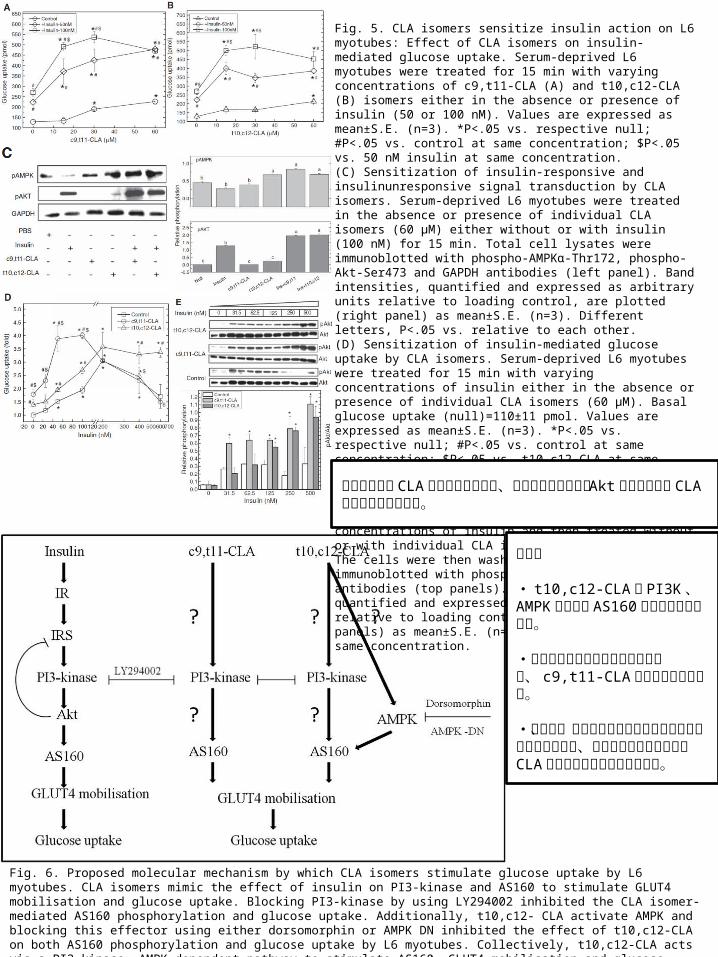
Fig. 6. Proposed molecular mechanism by which CLA isomers stimulate glucose uptake by L6 myotubes. CLA isomers mimic the effect of insulin on PI3-kinase and AS160 to stimulate GLUT4 mobilisation and glucose uptake. Blocking PI3-kinase by using LY294002 inhibited the CLA isomer-mediated AS160 phosphorylation and glucose uptake. Additionally, t10,c12- CLA activate AMPK and blocking this effector using either dorsomorphin or AMPK DN inhibited the effect of t10,c12-CLA on both AS160 phosphorylation and glucose uptake by L6 myotubes. Collectively, t10,c12-CLA acts via a PI3-kinase, AMPK-dependent pathway to stimulate AS160, GLUT4 mobilisation and glucose uptake, whereas the mechanism of c9,t11-CLA remain unclear.
Fig. 5. CLA isomers sensitize insulin action on L6 myotubes: Effect of CLA isomers on insulin-mediated glucose uptake. Serum-deprived L6 myotubes were treated for 15 min with varying concentrations of c9,t11-CLA (A) and t10,c12-CLA (B) isomers either in the absence or presence of insulin (50 or 100 nM). Values are expressed as mean±S.E. (n=3). *P<.05 vs. respective null; #P<.05 vs. control at same concentration; $P<.05 vs. 50 nM insulin at same concentration. (C) Sensitization of insulin-responsive and insulinunresponsive signal transduction by CLA isomers. Serum-deprived L6 myotubes were treated in the absence or presence of individual CLA isomers (60 μM) either without or with insulin (100 nM) for 15 min. Total cell lysates were immunoblotted with phospho-AMPKα-Thr172, phospho-Akt-Ser473 and GAPDH antibodies (left panel). Band intensities, quantified and expressed as arbitrary units relative to loading control, are plotted (right panel) as mean±S.E. (n=3). Different letters, P<.05 vs. relative to each other. (D) Sensitization of insulin-mediated glucose uptake by CLA isomers. Serum-deprived L6 myotubes were treated for 15 min with varying concentrations of insulin either in the absence or presence of individual CLA isomers (60 μM). Basal glucose uptake (null)=110±11 pmol. Values are expressed as mean±S.E. (n=3). *P<.05 vs. respective null; #P<.05 vs. control at same concentration; $P<.05 vs. t10,c12-CLA at same concentration. (E) Sensitization of insulin-mediated Akt activation by CLA isomers. Serum-deprived L6 myotubes were preincubated for 15 min with varying concentrations of insulin and then treated without or with individual CLA isomers (60 μM) for 15 min. The cells were then washed, lysed and immunoblotted with phospho-Akt-Ser473 and Akt antibodies (top panels). Band intensities, quantified and expressed as arbitrary units relative to loading control, are plotted (bottom panels) as mean±S.E. (n=3). *P<.05 vs. control at same concentration.
インスリンと CLAの同時添加により、インスリン誘導性の Aktのリン酸化を CLAが強める働きをする。
まとめ
・ t10,c12-CLAは PI3K、 AMPKを介して AS160のリン酸化に関わる。
・高濃度のインスリンによる脱感作は、 c9,t11-CLAによって妨げられる。
・しかし、メカニズムが完全に解明されたわけではないので、他の内分泌組織も含めたCLAの働きを研究する必要がある。