activation of protein tyrosine kinase pyk2 by the m1 muscarinic acetylcholine receptor
TRANSCRIPT
Activation of Protein Tyrosine Kinase PYK2 by the m1 Muscarinic Acetylcholine ReceptorAuthor(s): Jason S. Felsch, Teresa G. Cachero and Ernest G. PeraltaSource: Proceedings of the National Academy of Sciences of the United States of America,Vol. 95, No. 9 (Apr. 28, 1998), pp. 5051-5056Published by: National Academy of SciencesStable URL: http://www.jstor.org/stable/44664 .
Accessed: 08/05/2014 14:04
Your use of the JSTOR archive indicates your acceptance of the Terms & Conditions of Use, available at .http://www.jstor.org/page/info/about/policies/terms.jsp
.JSTOR is a not-for-profit service that helps scholars, researchers, and students discover, use, and build upon a wide range ofcontent in a trusted digital archive. We use information technology and tools to increase productivity and facilitate new formsof scholarship. For more information about JSTOR, please contact [email protected].
.
National Academy of Sciences is collaborating with JSTOR to digitize, preserve and extend access toProceedings of the National Academy of Sciences of the United States of America.
http://www.jstor.org
This content downloaded from 169.229.32.137 on Thu, 8 May 2014 14:04:28 PMAll use subject to JSTOR Terms and Conditions
Proc. Natl. Acad. Sci. USA Vol. 95, pp. 5051-5056, April 1998 Cell Biology
Activation of protein tyrosine kinase PYK2 by the ml muscarinic acetylcholine receptor JASON S. FELSCH, TERESA G. CACHERO, AND E"RNEST G. PERALTA*
Department of Molecular and Cellular Biology, Harvard University, Cambridge, MA 02138
Communicated by Daniel Branton, Harvard University, Cambridge, MA, February 23, 1998 (received for review November 20, 1997)
ABSTRACT Several G protein-coupled receptors are known to direct the tyrosine phosphorylation, and in some cases the activation, of diverse tyrosine kinases. Using a stable cell line approach, we characterize the activation of PYK2, a tyrosine kinase structurally related to focal adhesion kinase, by the G protein-coupled ml muscarinic acetylcholine recep- tor. We find that PYK2 tyrosine kinase activity is critical for the ml receptor-stimulated tyrosine phosphorylation of PYK2. Furthermore, we identify two tyrosine residues that are subject to phosphorylation in response to muscarinic signal- ing and show that this phosphorylation induces two cytosolic proteins, c-Src and Grb2, to bind to PYK2. This is the first demonstration of the significance played by distinct PYK2 tyrosine residues in G protein-coupled signaling to this ki- nase. By comparison, though ml receptors induce the tyrosine phosphorylation of the cytoskeletal protein paxillin, the as- sociation of paxillin with PYK2 is unaffected by miuscarinic signaling. We also provide evidence that PYK2 specifically phosphorylates the carboxyl-terminal cytosolic portion of the potassium channel Kv1.2 in a manner regulated by the ml receptor. These results delineate molecular events attending the ml muscarinic receptor stimulation of this tyrosine kinase and establish PYK2 as an effector of the ml muscarinic receptor in the regulation of multiple cell functions.
Muscarinic acetylcholine receptors perform crucial functions throughout the central and peripheral nervous systems, con- trolling many physiological responses to the neurotransmitter acetylcholine (1). The five subtypes of receptors (ml-m5) are members of the family of seven transmembrane receptors, which couple to heterotrimeric guanine nucleotide-binding (G) proteins. The receptor subtypes can be divided into two functional groups. The ml, m3, and m5 subtypes couple to the Gcq/11 class of G proteins to stimulate phospholipase Cf, which hydrolyzes phosphatidylinositol 4,5-bisphosphate to generate the second messengers diacylglycerol and inositol 1,4,5-trisphosphate (1). These second messengers activate protein kinase C (PKC) and elicit Ca2+ release from intracel- lular stores, respectively (1). The m2 and m4 receptor subtypes inhibit adenylyl cyclase and activate phospholipases and in- ward rectifying potassium channels (1, 2).
Several protein tyrosine kinases have been proposed as functional targets of G protein-coupled receptors (GPCRs), including the Src family kinases (3, 4). Indeed the catalytic activities of Src family kinases are elevated by stimnulation of Gcq/1 -coupled receptors (3, 5). Focal adhesion kinase (FAK) and the related protein tyrosine kinase (PTK) PYK2 have also been proposed as targets of regulation by select GPCRs (3, 4, 6-10). These PTKs are best known for their involvement in the cell-cell and cell-matrix signaling events mediated by cell surface receptors termed integrins (11, 12). Members of the
The publication costs of this article were defrayed in part by page charge payment. This article must therefore be hereby marked "advertisement" in accordance with 18 U.S.C. ?1734 solely to indicate this fact.
C) 1998 by The National Academy of Sciences 0027-8424/98/955051-6$2.00/0 PNAS is available online at http://www.pnas.org.
FAK family of tyrosine kinases may participate in T and B cell antigen receptor signaling (12, 13), mitogenesis (14-16), and neuronal signaling events (9, 15). As with the Src family kinases, several neurotransmitters cause an increase in the tyrosine phosphorylation of FAK and PYK2 (4, 6, 8, 10, 17). However, as with the Src family kinases, neither the mecha- nism by which this signaling occurs nor its relevance for cellular physiology is well understood.
One example of a G protein-coupled neurotransmitter signaling pathway that requires tyrosine kinase activity is the ml muscarinic acetylcholine (ml) receptor-induced suppres- sion of the delayed rectifier potassium channel Kvl.2 (18, 19). Recently, PYK2 was shown to play a role in the phorbol ester-induced suppression of Kvl.2 (15). It therefore seems plausible that PYK2 may be involved in the tyrosine kinase- dependent suppression of Kvl.2 by ml receptors. However, it is not known if ml receptor signaling in any way regulates PYK2. Furthermore, although several GPCRs can induce the tyrosine phosphorylation of PYK2, it has not been shown that any GPCR can regulate the catalytic activity of PYK2. Like the Src family kinases for which tyrosine phosphorylation can be both stimulatory and inhibitory (20), the catalytic activity of FAK family kinases may be independent of tyrosine phos- phorylation (21-23).
In studying the activation of a tyrosine kinase by GPCRs two common challenges need to be addressed. On one hand, choosing to study a cell line or a primary tissue preparation that expresses a tyrosine kinase endogenously limits the pre- cision with which one can examine the participation of key structural elements of the kinase in a signaling mechanism. On the other hand, transiently transfecting cells with structurally altered derivatives of the kinase usually results in overexpres- sion of the kinase at levels that surpass the ability of the cell to support its regulation by GPCRs. For example, in cells expressing the ml muscarinic receptor and overexpressing transiently transfected PYK2 muscarinic stimulation does not affect the phosphorylation state of PYK2 (J.S.F., unpublished observation). Therefore, to investigate whether PYK2 is reg- ulated by the ml muscarinic acetylcholine receptor, we gen- erated stable cell lines expressing the ml receptor and epitope- tagged PYK2 variants. These cell lines express PYK2 con- structs at stable levels that are consistently regulated by the G protein-coupled ml muscarinic receptor.
We demonstrate here that ml receptor stimulation not only leads to the rapid and robust tyrosine phosphorylation of PYK2 but also enhances the catalytic activity of the kinase. This finding contrasts early indications that muscarinic signal- ing does not stimulate PYK2 (15). Pharmacological analyses indicate that the signal transduction pathway linking ml receptors to PYK2 may involve both Ca2' and PKC. We examined the events attending the GPCR regulation of this
Abbreviations: FAK, focal adhesion kinase; GPCR, G protein-coupled receptor; GST, glutathione S-transferase; HSV, herpes simplex virus; MAPK, mitogen-activated protein kinase; PKC, protein kinase C; PMA, phorbol 12-myristate 13-acetate; PTK, protein tyrosine kinase. *To whom reprint requests should be addressed. e-mail: peralta@ husc.harvard.edu.
5051
This content downloaded from 169.229.32.137 on Thu, 8 May 2014 14:04:28 PMAll use subject to JSTOR Terms and Conditions
5052 Cell Biology: Felsch et al. Proc. Natl. Acad. Sci. USA 95 (1998)
tyrosine kinase to understand the mechanism by which this signaling occurs. We show that ml receptor-induced tyrosine phosphorylation of PYK2 depends critically upon the intrinsic catalytic activity of PYK2. We also identify two PYK2 tyrosine residues that are phosphorylated in response to muscarinic signaling and show that these residues support the association between PYK2 and either c-Src or Grb2 in a muscarinic receptor-dependent manner. By comparison, the ml receptor signaling that induces tyrosine phosphorylation of the cy- toskeletal protein paxillin does not affect paxillin's association with PYK2. Furthermore, we provide evidence that PYK2 specifically phosphorylates the carboxy terminal cytosolic por- tion Kv1.2 and that this phosphorylation is significantly en- hanced by activation of the ml receptor. These results establish PYK2 as a target of muscarinic signaling with implications for a variety of cell functions.
EXPERIMENTAL PROCEDURES
Stable Expression of PYK2 in Human Embryonic Kidney 293 Cells. A plasmid [plasmid cytomegalovirus (pCMV)- PYK2-herpes simplex virus (HSV)] encoding rat PYK2 se- quence (24) under transcriptional control of the cytomegalo- virus early promoter was constructed. Selected point muta- tions (Y402F, K457A, and Y881F) were introduced into this plasmid by Kunkel mutagenesis, identified by the introduction or removal of a restriction site, and confirmed by sequencing. All PYK2 coding sequences terminated with the HSV epitope, QPELAPEDPED. Human embryonic kidney 293 cells ex- pressing the human ml receptor were cotransfected with a pCMV-PYK2-HSV plasmid and pBCMGHyg by the Ca2+ phosphate method (18). Clonal lines selected for their resis- tance to 250 ,ug/ml hygromycin were screened for their expression of the 115-kDa epitope-tagged construct (PYK2- HSV) by Western blot analysis of cell lysates generated as below. Resulting cell lines were subsequently maintained and propagated as described (18). Cells were starved by reducing the serum content in the media to 0.2% 24 hr before use.
Immunoprecipitations and Western Blot Analysis. Conflu- ent cultures of stable cell lines were treated with vehicle, atropine, carbachol, phorbol 12-myristate 13-acetate (PMA), or A21387 (Sigma) as indicated at 37?C. Cell lysates were prepared and immunoprecipitated essentially as described (5). PYK2-HSV and paxillin were immunoprecipitated by incuba- tion with 1 ,ug of anti-HSV (Novagen) or 1 ,ug of anti-paxillin (Transduction Laboratories, Lexington, KY) at 4?C for 1 hr. After subsequent incubation with protein A Sepharose, the immune complex was pelleted. The immunoprecipitations were washed with lysis buffer and either analyzed by using Western blots or subjected to in vitro kinase assays. Western blot analysis was performed essentially as described (18). Primary anti-HSV, anti-paxillin, anti-c-Src, and anti-Grb2 (Santa Cruz Biotechnology) antibodies were prepared in blocking buffer at 100 ng/ml. Horseradish peroxidase- conjugates of goat anti-mouse or goat anti-rabbit served as the secondary antibody at 1 [kg/ml. Phosphotyrosine content was assessed with horseradish peroxidase-conjugated anti- phosphotyrosine antibody 4G10 (Upstate Biotechnology, Lake Placid, NY) diluted to 1 ,ug/ml.
In Vitro Kinase Assays. Autophosphorylation reactions, tyrosine kinase assays using the exogenous peptide substrate poly(Glu,Tyr)4:1 (Sigma), and quantification of 32p incorpora- tion were analyzed by SDS/PAGE and autoradiography as described (24). To ascertain that equivalent amounts of kinase were present in each of the autophosphorylation and poly- (Glu,Tyr)4:1 kinase reactions, the reaction mixtures were de- termined by Western blot analysis with antibodies against the HSV epitope. For kinase assays using glutathione S- transferase (GST)-fusion proteins as substrates, the reactions were carried out as above except that immune complex beads
were agitated for 10 min at 22?C in 40 [lI of freshly prepared buffer containing 40 ,g of purified GST-fusion protein, 40 [Ci (1 Ci = 37 GBq) of [-y-32P]ATP, 20 mM TriP HCI, 1 mM DTT, 1% Triton X-100, 0.004% Sarcosyl, and 0.1 mM MnCl2 at pH 7.4. These gels were fixed and stained with Coomassie blue before autoradiography.
Preparation of GST-Fusion Proteins. For bacterial expres- sion of GST-fusion proteins, plasmids encoding either the amino- or carboxy-terminal cytoplasmic regions of rat Kvl.2, were constructed with a PCR strategy that fused the cytosolic amino (amino acids 1-163) and carboxy (amino acids 412-499) termini of Kvl.2 (25) to GST by using the vector pGEX-2T (Pharmacia) to form pGEX-2T-Kvl.2-N and pGEX-2T- Kvl.2-C. These plasmids along with the control pGEX-2T were transformed into E. coli strain BL21 and selected for resistance to 50 ,ug/ml ampicillin. Cultures inoculated with these bacteria were treated for 2 hr with 0.5 mM isopropyl
3-D-thiogalactoside and the induced fusion proteins were solubilized as described (2). Solubilized fusion proteins were applied to a glutathione agarose column, washed, and eluted with 1 mM glutathione. Purified fusion proteins diluted to 5 ml with elution buffer were dialyzed twice against elution buffer lacking glutathione and twice against 20 mM Tris'HCl (pH 7.4), 1 mM DTT, 1% Triton X-100, and 0.01% Sarcosyl. The dialysates were centrifuged at 15,000 x g for 20 min at 4?C to remove insoluble material. The supernatants were then con- centrated to greater than 2 mg/ml using a Centricon-10 centrifugal concentrator (Amicon).
RESULTS
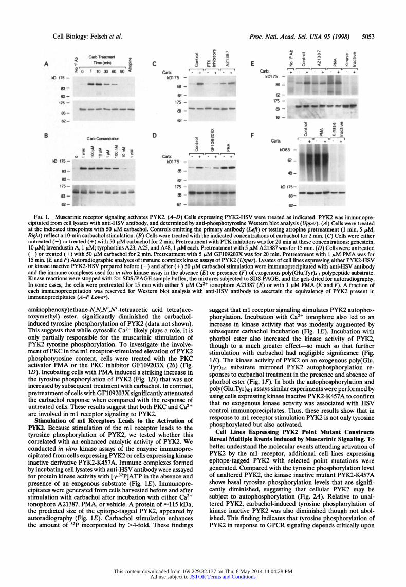
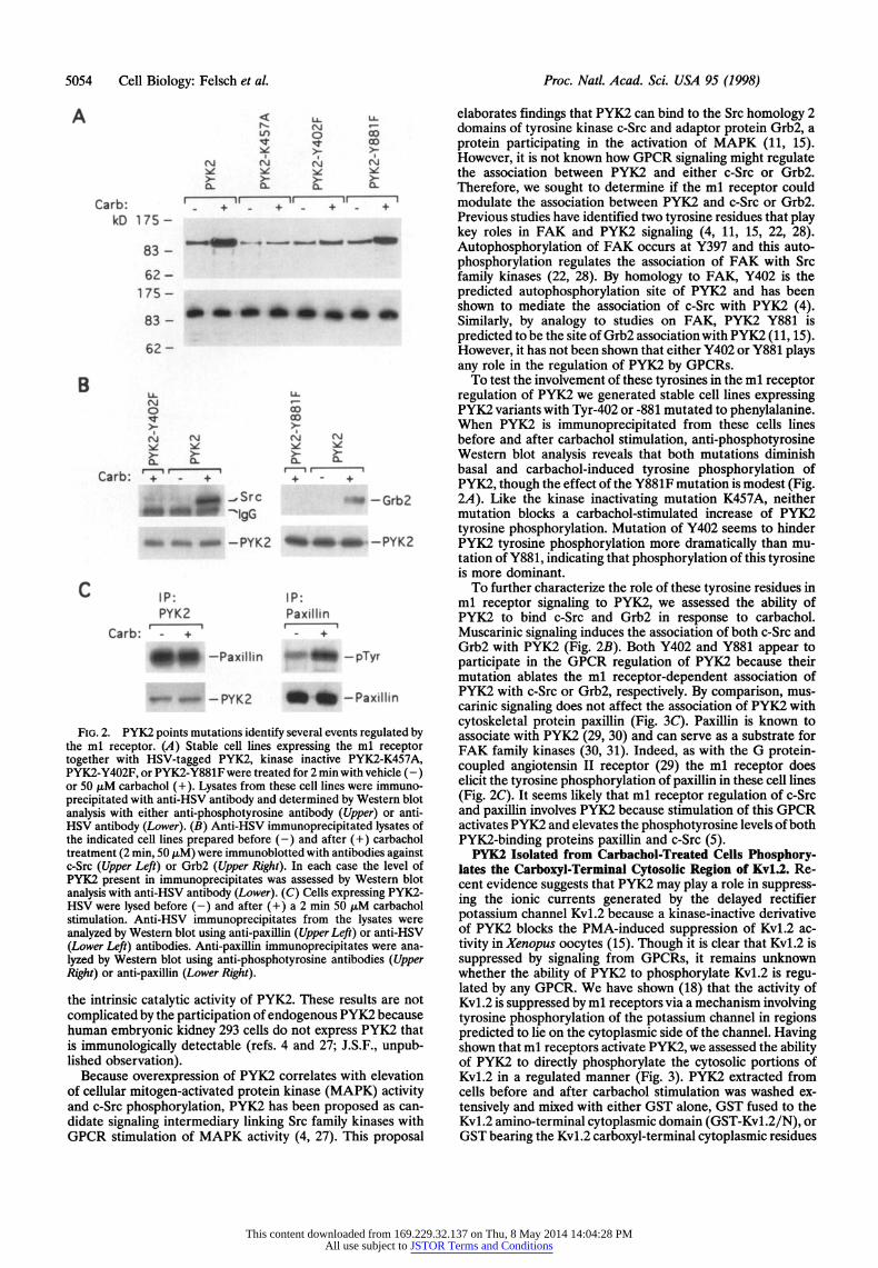
Stimulation of the ml Muscarinic Receptor Leads to the Tyrosine Phosphorylation of PYK2. To test whether or not PYK2 was regulated by ml muscarinic receptors, cells express- ing both the human ml receptor and rat PYK2 bearing an HSV epitope were treated with the muscarinic receptor agonist carbachol. PYK2 protein was immunoprecipitated with anti- HSV antibody from lysates of treated or untreated cells and subjected to Western blot analysis with anti-phosphotyrosine antibody (Fig. 1 A and B Upper). Carbachol caused a striking increase in the level of tyrosine phosphorylated PYK2 (Fig. 1A). Pretreatment with the muscarinic antagonist atropine completely blocked the carbachol-dependent tyrosine phos- phorylation of PYK2 (Fig. IA). The duration of increased PYK2 tyrosine phosphorylation in response to carbachol, which persists for >1 hr, is comparable to the long-lived tyrosine phosphorylation of FAK in response to neuropeptide- activated GPCRs (6). Such a long-lived response contrasts the much shorter-lived GPCR-induced tyrosine phosphorylation of Src family kinases that terminates within minutes (3). Dose response experiments illustrate that the increase in tyrosine phosphorylation of PYK2 in response to carbachol is concen- tration dependent (Fig. iB). Furthermore, preincubation with PTK inhibitors significantly diminishes both basal and carba- chol-stimulated increases in the phosphotyrosine content of PYK2 relative to untreated controls (Fig. IC Upper).
Pharmacological Analyses of ml Receptor Signaling to PYK2. Among the cellular events frequently associated with Gcq/1i-coupled GPCRs is the elevation of cytosolic Ca2+ levels and the activation of PKC (1). We therefore examined the role of Ca2+ and PKC in the ml receptor-dependent stimulation of PYK2 tyrosine phosphorylation (Fig. 1 C and D). Treatment of cells expressing epitope-tagged PYK2 with the Ca21 iono- phore A21387 increased the basal phosphotyrosine content of PYK2 compared with untreated controls (Fig. IC). However, ionophore treatment failed to evoke a maximal response because subsequent treatment with carbachol further in- creased PYK2 tyrosine phosphorylation (Fig. lC). As in previous experiments with other GPCRs (10, 15, 29), pretreat- ing the cells with calcium chelators, such as 1,2-bis(o-
This content downloaded from 169.229.32.137 on Thu, 8 May 2014 14:04:28 PMAll use subject to JSTOR Terms and Conditions
Cell Biology: Felsch et al. Proc. Natl. Acad. Sci. USA 95 (1998) 5053
C wbTmis miW e S~~~ 0- 9
_____
__
E
^
__"" 0 10 iowa _ _____ so . l"
MD 176 -l kM B75 k_ 75
|~~~~~~~ .. . .._ - . . .. ... . .. .. ..
~ ii. - 1k175 - U- ~ ~ ~ ~ ~ ~ ~ ~ ~ ~ ~~~~~~~~~~~~1 a- _
B _ D F
*+ ~~~~~~~~~~kOl 7585B<>: -~
175- IdJ175 - D _ e- S B - _ ~ ~~~ ~~~~~~~~~~~ - _ /
|~~~~~~~~~~~2 - 42- lzi- _ 17 U -- al 7- _
Ja- _- U# - - _
FIG. 1. Muscarinic receptor signaling activates PYK2. (A-D) Cells expressing PYK2-HSV were treated as indicated. PYK2 was immunopre- cipitated from cell lysates with anti-HSV antibody, and determined by anti-phosphotyrosine Western blot analysis (Upper). (A) Cells were treated at the indicated timepoints with 50 ,uM carbachol. Controls omitting the primary antibody (Left) or testing atropine pretreatment (1 min, 5 ,M; Right) reflect a 10-min carbachol stimulation. (B) Cells were treated with the indicated concentrations of carbachol for 2 min. (C) Cells were either untreated (-) or treated (+) with 50 ,uM carbachol for 2 min. Pretreatment with PTK inhibitors was for 20 min at these concentrations: genestein, 10 ,uM; lavendustin A, 1 ,uM; tyrphostins A23, A25, and A48, 1 ,uM each. Pretreatment with 5 ,uM A21387 was for 15 min. (D) Cells were untreated (-) or treated (+) with 50 ,uM carbachol for 2 min. Pretreatment with 5 zM GF109203X was for 20 min. Pretreatment with 1 AM PMA was for 15 min. (E and F) Autoradiographic analyses of immune complex kinase assays of PYK2 (Upper). Lysates of cell lines expressing either PYK2-HSV or kinase inactive PYK2-HSV prepared before (-) and after (+) 50 ,uM carbachol stimulation were immunoprecipitated with anti-HSV antibody and the immune complexes used for in vitro kinase assay in the absence (E) or presence (F) of exogenous poly(Glu,Tyr)4:l polypeptide substrate. Kinase reactions were stopped with 2x SDS/PAGE sample buffer, the mixtures subjected to SDS-PAGE, and the gels dried for autoradiography. In some cases, the cells were pretreated for 15 min with either 5 ,uM Ca2+ ionophore A21387 (E) or with 1 ,uM PMA (E and F). A fraction of each immunoprecipitation was reserved for Western blot analysis with anti-HSV antibody to ascertain the equivalency of PYK2 present in immunoprecipitates (A-F Lower).
aminophenoxy)ethane-N,N,N',N'-tetraacetic acid tetra(ace- toxymethyl) ester, significantly diminished the carbachol- induced tyrosine phosphorylation of PYK2 (data not shown). This suggests that while cytosolic Ca2+ likely plays a role, it is only partially responsible for the muscarinic stimulation of PYK2 tyrosine phosphorylation. To investigate the involve- ment of PKC in the ml receptor-stimulated elevation of PYK2 phosphotyrosine content, cells were treated with the PKC activator PMA or the PKC inhibitor GF109203X (26) (Fig. ID). Incubating cells with PMA induced a striking increase in the tyrosine phosphorylation of PYK2 (Fig. 1D) that was not increased by subsequent treatment with carbachol. In contrast, pretreatment of cells with GF109203X significantly attenuated the carbachol response when compared with the response of untreated cells. These results suggest that both PKC and Ca2+ are involved in ml receptor signaling to PYK2.
Stimulation of ml Receptors Leads to the Activation of PYK2. Because stimulation of the ml receptor leads to the tyrosine phosphorylation of PYK2, we tested whether this correlated with an enhanced catalytic activity of PYK2. We conducted in vitro kinase assays of the enzyme immunopre- cipitated from cells expressing PYK2 or cells expressing kinase inactive derivative PYK2-K457A. Immune complexes formed by incubating cell lysates with anti-HSV antibody were assayed for protein kinase activity with [y-32P]ATP in the absence and presence of an exogenous substrate (Fig. 1E). Immunopre- cipitates were generated from cells harvested before and after stimulation with carbachol after incubation with either Ca2+ ionophore A21387, PMA, or vehicle. A protein of -1l15 kDa, the predicted size of the epitope-tagged PYK2, appeared by autoradiography (Fig. 1E). Carbachol stimulation enhances the amount of 32p incorporated by >4-fold. These findings
suggest that ml receptor signaling stimulates PYK2 autophos- phorylation. Incubation with Ca2+ ionophore also led to an increase in kinase activity that was modestly augmented by subsequent carbachol incubation (Fig. 1E). Incubation with phorbol ester also increased the kinase activity of PYK2, though to a much greater effect-so much so that further stimulation with carbachol had negligible significance (Fig. 1E). The kinase activity of PYK2 on an exogenous poly(Glu, Tyr)4:1 substrate mirrored PYK2 autophosphorylation re- sponses to carbachol treatment in the presence and absence of phorbol ester (Fig. 1F). In both the autophosphorylation and poly(Glu,Tyr)4:1 assays similar experiments were performed by using cells expressing kinase inactive PYK2-K457A to confirm that no exogenous kinase activity was associated with HSV control immunoprecipitates. Thus, these results show that in response to ml receptor stimulation PYK2 is not only tyrosine phosphorylated but also activated.
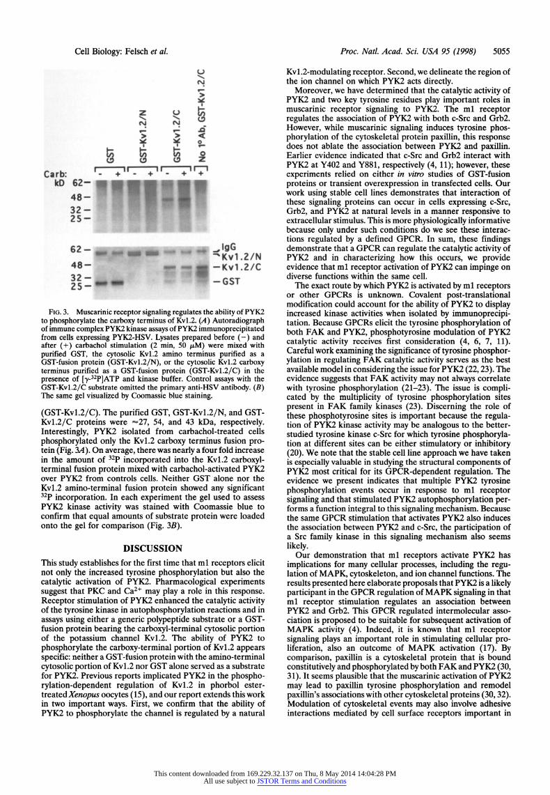
Cell Lines Expressing PYK2 Point Mutant Constructs Reveal Multiple Events Induced by Muscarinic Signaling. To better understand the molecular events attending activation of PYK2 by the ml receptor, additional cell lines expressing epitope-tagged PYK2 with selected point mutations were generated. Compared with the tyrosine phosphorylation level of unaltered PYK2, the kinase inactive mutant PYK2-K457A shows basal tyrosine phosphorylation levels that are signifi- cantly diminished, suggesting that cellular PYK2 may be subject to autophosphorylation (Fig. 2A). Relative to unal- tered PYK2, carbachol-induced tyrosine phosphorylation of kinase inactive PYK2 was also diminished though not abol- ished. This finding indicates that tyrosine phosphorylation of PYK2 in response to GPCR signaling depends critically upon
This content downloaded from 169.229.32.137 on Thu, 8 May 2014 14:04:28 PMAll use subject to JSTOR Terms and Conditions
5054 Cell Biology: Felsch et al Proc. Natl. Acad. Sci. USA 95 (1998)
A IA L
N4 N N
Carb: + + + + kD 175-
833-
62 175-
62-
B X-
4.~~~~~~4 c
N N N N W. ... l..
Carb: 7' + + +
._Src _-G b2
_-P PYK2 _-PY 2
C IP: IP: PYK2 Paxillin
Carb: - + -
_Paxilin -- -pyr
UIIE-PYK2 -Paxillin
FIG. 2. PYK2 points mutations identify several events regulated by the ml receptor. (A) Stable cell lines expressing the ml receptor together with HSV-tagged PYK2, kinase inactive PYK2-K457A, PYK2-Y402F, or PYK2-Y881F were treated for 2 min with vehicle (-) or 50 ,uM carbachol (+). Lysates from these cell lines were immuno- precipitated with anti-HSV antibody and determined by Western blot analysis with either anti-phosphotyrosine antibody (Upper) or anti- HSV antibody (Lower). (B) Anti-HSV immunoprecipitated lysates of the indicated cell lines prepared before (-) and after (+) carbachol treatment (2 min, 50 ,uM) were immunoblotted with antibodies against c-Src (Upper Left) or Grb2 (Upper Right). In each case the level of PYK2 present in immunoprecipitates was assessed by Western blot analysis with anti-HSV antibody (Lower). (C) Cells expressing PYK2- HSV were lysed before (-) and after (+) a 2 min 50 ,uM carbachol stimulation. Anti-HSV immunoprecipitates from the lysates were analyzed by Westem blot using anti-paxillin (Upper Left) or anti-HSV (Lower Left) antibodies. Anti-paxillin immunoprecipitates were ana- lyzed by Western blot using anti-phosphotyrosine antibodies (Upper Right) or anti-paxillin (Lower Right).
the intrinsic catalytic activity of PYK2. These results are not complicated by the participation of endogenous PYK2 because human embryonic kidney 293 cells do not express PYK2 that is immunologically detectable (refs. 4 and 27; J.S.F., unpub- lished observation).
Because overexpression of PYK2 correlates with elevation of cellular mitogen-activated protein kinase (MAPK) activity and c-Src phosphorylation, PYK2 has been proposed as can- didate signaling intermediary linking Src family kinases with GPCR stimulation of MAPK activity (4, 27). This proposal
elaborates findings that PYK2 can bind to the Src homology 2 domains of tyrosine kinase c-Src and adaptor protein Grb2, a protein participating in the activation of MAPK (11, 15). However, it is not known how GPCR signaling might regulate the association between PYK2 and either c-Src or Grb2. Therefore, we sought to determine if the ml receptor could modulate the association between PYK2 and c-Src or Grb2. Previous studies have identified two tyrosine residues that play key roles in FAK and PYK2 signaling (4, 11, 15, 22, 28). Autophosphorylation of FAK occurs at Y397 and this auto- phosphorylation regulates the association of FAK with Src family kinases (22, 28). By homology to FAK, Y402 is the predicted autophosphorylation site of PYK2 and has been shown to mediate the association of c-Src with PYK2 (4). Similarly, by analogy to studies on FAK, PYK2 Y881 is predicted to be the site of Grb2 association with PYK2 (11, 15). However, it has not been shown that either Y402 or Y881 plays any role in the regulation of PYK2 by GPCRs.
To test the involvement of these tyrosines in the ml receptor regulation of PYK2 we generated stable cell lines expressing PYK2 variants with Tyr-402 or -881 mutated to phenylalanine. When PYK2 is immunoprecipitated from these cells lines before and after carbachol stimulation, anti-phosphotyrosine Western blot analysis reveals that both mutations diminish basal and carbachol-induced tyrosine phosphorylation of PYK2, though the effect of the Y881F mutation is modest (Fig. 2A). Like the kinase inactivating mutation K457A, neither mutation blocks a carbachol-stimulated increase of PYK2 tyrosine phosphorylation. Mutation of Y402 seems to hinder PYK2 tyrosine phosphorylation more dramatically than mu- tation of Y881, indicating that phosphorylation of this tyrosine is more dominant.
To further characterize the role of these tyrosine residues in ml receptor signaling to PYK2, we assessed the ability of PYK2 to bind c-Src and Grb2 in response to carbachol. Muscarinic signaling induces the association of both c-Src and Grb2 with PYK2 (Fig. 2B). Both Y402 and Y881 appear to participate in the GPCR regulation of PYK2 because their mutation ablates the ml receptor-dependent association of PYK2 with c-Src or Grb2, respectively. By comparison, mus- carinic signaling does not affect the association of PYK2 with cytoskeletal protein paxillin (Fig. 3C). Paxillin is known to associate with PYK2 (29, 30) and can serve as a substrate for FAK family kinases (30, 31). Indeed, as with the G protein- coupled angiotensin II receptor (29) the ml receptor does elicit the tyrosine phosphorylation of paxillin in these cell lines (Fig. 2C). It seems likely that ml receptor regulation of c-Src and paxillin involves PYK2 because stimulation of this GPCR activates PYK2 and elevates the phosphotyrosine levels of both PYK2-binding proteins paxillin and c-Src (5).
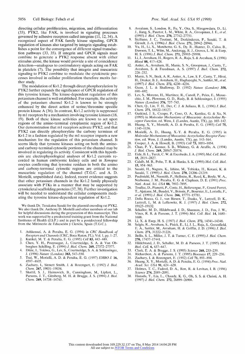
PYK2 Isolated from Carbachol-Treated Cells Phosphory- lates the Carboxyl-Terminal Cytosolic Region of Kv1.2. Re- cent evidence suggests that PYK2 may play a role in suppress- ing the ionic currents generated by the delayed rectifier potassium channel Kvl.2 because a kinase-inactive derivative of PYK2 blocks the PMA-induced suppression of Kvl.2 ac- tivity in Xenopus oocytes (15). Though it is clear that Kvl.2 is suppressed by signaling from GPCRs, it remains unknown whether the ability of PYK2 to phosphorylate Kvl.2 is regu- lated by any GPCR. We have shown (18) that the activity of Kvl.2 is suppressed by ml receptors via a mechanism involving tyrosine phosphorylation of the potassium channel in regions predicted to lie on the cytoplasmic side of the channel. Having shown that ml receptors activate PYK2, we assessed the ability of PYK2 to directly phosphorylate the cytosolic portions of Kv1.2 in a regulated manner (Fig. 3). PYK2 extracted from cells before and after carbachol stimulation was washed ex- tensively and mixed with either GST alone, GST fused to the Kv1.2 amino-terminal cytoplasmic domain (GST-Kv1.2/N), or GST bearing the Kv1.2 carboxyl-terminal cytoplasmic residues
This content downloaded from 169.229.32.137 on Thu, 8 May 2014 14:04:28 PMAll use subject to JSTOR Terms and Conditions
Cell Biology: Felsch et al. Proc. Natl. Acad. Sci. USA 95 (1998) 5055
U
N
N N
f
Caur b: +r_ +r kD 62-
48- 32- 25-
62- IgG 48-K =Kvl.2/N
48-v-Kvl .2/C 32 - GST 25-
FIG. 3. Muscarinic receptor signaling regulates the ability of PYK2 to phosphorylate the carboxy terminus of Kv1.2. (A) Autoradiograph of immune complex PYK2 kinase assays of PYK2 immunoprecipitated from cells expressing PYK2-HSV. Lysates prepared before (-) and after (+) carbachol stimulation (2 min, 50 ,uM) were mixed with purified GST, the cytosolic Kv1.2 amino terminus purified as a GST-fusion protein (GST-Kv1.2/N), or the cytosolic Kv1.2 carboxy terminus purified as a GST-fusion protein (GST-Kv1.2/C) in the presence of [y-32P]ATP and kinase buffer. Control assays with the GST-Kvl.2/C substrate omitted the primary anti-HSV antibody. (B) The same gel visualized by Coomassie blue staining.
(GST-Kvl.2/C). The purified GST, GST-Kvl.2/N, and GST- Kvl.2/C proteins were -27, 54, and 43 kDa, respectively. Interestingly, PYK2 isolated from carbachol-treated cells phosphorylated only the Kvl.2 carboxy terminus fusion pro- tein (Fig. 3A). On average, there was nearly a four fold increase in the amount of 32p incorporated into the Kvl.2 carboxyl- terminal fusion protein mixed with carbachol-activated PYK2 over PYK2 from controls cells. Neither GST alone nor the Kvl.2 amino-terminal fusion protein showed any significant 32p incorporation. In each experiment the gel used to assess PYK2 kinase activity was stained with Coomassie blue to confirm that equal amounts of substrate protein were loaded onto the gel for comparison (Fig. 3B).
DISCUSSION This study establishes for the first time that ml receptors elicit not only the increased tyrosine phosphorylation but also the catalytic activation of PYK2. Pharmacological experiments suggest that PKC and Ca2+ may play a role in this response. Receptor stimulation of PYK2 enhanced the catalytic activity of the tyrosine kinase in autophosphorylation reactions and in assays using either a generic polypeptide substrate or a GST- fusion protein bearing the carboxyl-terminal cytosolic portion of the potassium channel Kvl.2. The ability of PYK2 to phosphorylate the carboxy-terminal portion of Kvl.2 appears specific: neither a GST-fusion protein with the amino-terminal cytosolic portion of Kvl.2 nor GST alone served as a substrate for PYK2. Previous reports implicated PYK2 in the phospho- rylation-dependent regulation of Kvl.2 in phorbol ester- treated Xenopus oocytes (15), and our report extends this work in two important ways. First, we confirm that the ability of PYK2 to phosphorylate the channel is regulated by a natural
Kvl.2-modulating receptor. Second, we delineate the region of the ion channel on which PYK2 acts directly.
Moreover, we have determined that the catalytic activity of PYK2 and two key tyrosine residues play important roles in muscarinic receptor signaling to PYK2. The ml receptor regulates the association of PYK2 with both c-Src and Grb2. However, while muscarinic signaling induces tyrosine phos- phorylation of the cytoskeletal protein paxillin, this response does not ablate the association between PYK2 and paxillin. Earlier evidence indicated that c-Src and Grb2 interact with PYK2 at Y402 and Y881, respectively (4, 11); however, these experiments relied on either in vitro studies of GST-fusion proteins or transient overexpression in transfected cells. Our work using stable cell lines demonstrates that interaction of these signaling proteins can occur in cells expressing c-Src, Grb2, and PYK2 at natural levels in a manner responsive to extracellular stimulus. This is more physiologically informative because only under such conditions do we see these interac- tions regulated by a defined GPCR. In sum, these findings demonstrate that a GPCR can regulate the catalytic activity of PYK2 and in characterizing how this occurs, we provide evidence that ml receptor activation of PYK2 can impinge on diverse functions within the same cell.
The exact route by which PYK2 is activated by ml receptors or other GPCRs is unknown. Covalent post-translational modification could account for the ability of PYK2 to display increased kinase activities when isolated by immunoprecipi- tation. Because GPCRs elicit the tyrosine phosphorylation of both FAK and PYK2, phosphotyrosine modulation of PYK2 catalytic activity receives first consideration (4, 6, 7, 11). Careful work examining the significance of tyrosine phosphor- ylation in regulating FAK catalytic activity serves as the best available model in considering the issue for PYK2 (22,23). The evidence suggests that FAK activity may not always correlate with tyrosine phosphorylation (21-23). The issue is compli- cated by the multiplicity of tyrosine phosphorylation sites present in FAK family kinases (23). Discerning the role of these phosphotyrosine sites is important because the regula- tion of PYK2 kinase activity may be analogous to the better- studied tyrosine kinase c-Src for which tyrosine phosphoryla- tion at different sites can be either stimulatory or inhibitory (20). We note that the stable cell line approach we have taken is especially valuable in studying the structural components of PYK2 most critical for its GPCR-dependent regulation. The evidence we present indicates that multiple PYK2 tyrosine phosphorylation events occur in response to ml receptor signaling and that stimulated PYK2 autophosphorylation per- forms a function integral to this signaling mechanism. Because the same GPCR stimulation that activates PYK2 also induces the association between PYK2 and c-Src, the participation of a Src family kinase in this signaling mechanism also seems likely.
Our demonstration that ml receptors activate PYK2 has implications for many cellular processes, including the regu- lation of MAPK, cytoskeleton, and ion channel functions. The results presented here elaborate proposals that PYK2 is a likely participant in the GPCR regulation of MAPK signaling in that ml receptor stimulation regulates an association between PYK2 and Grb2. This GPCR regulated intermolecular asso- ciation is proposed to be suitable for subsequent activation of MAPK activity (4). Indeed, it is known that ml receptor signaling plays an important role in stimulating cellular pro- liferation, also an outcome of MAPK activation (17). By comparison, paxillin is a cytoskeletal protein that is bound constitutively and phosphorylated by both FAK and PYK2 (30, 31). It seems plausible that the muscarinic activation of PYK2 may lead to paxillin tyrosine phosphorylation and remodel paxillin's associations with other cytoskeletal proteins (30, 32). Modulation of cytoskeletal events may also involve adhesive interactions mediated by cell surface receptors important in
This content downloaded from 169.229.32.137 on Thu, 8 May 2014 14:04:28 PMAll use subject to JSTOR Terms and Conditions
5056 Cell Biology: Felsch et al. Proc. Natl. Acad. Sci. USA 95 (1998)
directing cellular proliferation, migration, and differentiation (33). PYK2, like FAK, is involved in signaling processes governed by adhesive receptors called integrins (11, 12, 34). A recognized aspect of FAK family kinase signaling, GPCR regulation of kinases also targeted by integrin signaling estab- lishes a point for the convergence of different signal transduc- tion pathways (33, 35). If integrin and GPCR signals must combine to generate a PYK2 response absent with either stimulus alone, the kinase would provide a site of coincidence detection-analogous to costimulatory signals acting on FAK in platelets (7). The possibility that integrin and muscarinic signaling to PYK2 combine to modulate the cytokinetic pro- cesses involved in cellular proliferation therefore merits fur- ther study.
The modulation of Kv1.2 through direct phosphorylation by PYK2 further expands the significance of GPCR regulation of this tyrosine kinase. The kinase-dependent regulation of this potassium channel is well established. For example, the activity of the potassium channel Kv1.2 is known to be strongly enhanced by the direct action of serine/threonine specific protein kinase A (36). In contrast, Kv1.2 activity is suppressed by ml receptors by a mechanism involving tyrosine kinases (18, 19). Both of these kinase activities are known to act upon regions of the amino-terminal cytoplasmic region of Kv1.2. Our demonstration that ml receptors activate PYK2, and that PYK2 can directly phosphorylate the carboxy terminus of Kv1.2 in a fashion regulated by the ml receptor imparts a new mechanism for the regulation of this potassium channel. It seems likely that tyrosine kinases acting on both the amino- and carboxy-terminal cytosolic portions of the channel may be involved in regulating its activity. Consistent with this hypoth- esis are electrophysiological analyses of Kv1.2 currents re- corded in human embryonic kidney cells and in Xenopus oocytes confirming that tyrosine residues in both the amino and carboxy-terminal cytosolic regions are critical to the muscarinic regulation of the channel (T.G.C. and A. D. Morielli, unpublished data). Indeed, recent evidence suggests that other potassium channels in the Kv1.2 family physically associate with PTKs in a manner that may be supported by cytoskeletal scaffolding proteins (37, 38). Further investigation will be needed to understand the cellular components medi- ating the tyrosine kinase-dependent regulation of Kv1.2.
We thank Dr. Terakatsu Sasaki for the plasmid encoding rat PYK2. We also thank Dr. Anthony D. Morielli and other members of our lab for helpful discussions during the preparation of this manuscript. This work was supported by a predoctoral training grant from the National Institutes of Health (J.S.F.) and in part by a postdoctoral fellowship from the Ministerio de Educaci6n y Ciencia, Spain (T.G.C.).
1. Ashkenazi, A. & Peralta, E. G. (1994) in CRC Handbook of Receptors and Channels (CRC, Boca Raton, FL), Vol. 1, pp. 1-27.
2. Kunkel, M. T. & Peralta, E. G. (1995) Cell 83, 443-449. 3. Chen, Y. H., Pouyssegur, J., Courtneidge, S. A. & Van Ob-
berghen Schilling, E. (1994) J. Biol. Chem. 269, 27372-27377. 4. Dikic, I., Tokiwa, G., Lev, S., Courtneidge, S. A. & Schlessinger,
J. (1996) Nature (London) 383, 547-550. 5. Tsai, W., Morielli, A. D. & Peralta, E. G. (1997) EMBO J. 16,
4597-4605. 6. Zachary, I., Sinnett Smith, J. & Rozengurt, E. (1992) J. Biol.
Chem. 267, 19031-19034. 7. Shattil, S. J., Haimovich, B., Cunningham, M., Lipfert, L.,
Parsons, J. T., Ginsberg, M. H. & Brugge, J. S. (1994) J. Biol. Chem. 269, 14738-14745.
8. Avraham, S., London, R., Fu, Y., Ota, S., Hiregowdara, D., Li, J., Jiang, S., Pasztor, L. M., White, R. A., Groopman, J. E., et al. (1995) J. Biol. Chem. 270, 27742-27751.
9. Siciliano, J. C., Toutant, M., Derkinderen, P., Sasaki, T. & Girault, J.-A. (1996) J. Biol. Chem. 271, 28942-28946.
10. Yu, H., Li, X., Marchetto, G. S., Dy, R., Hunter, D., Calvo, B., Dawson, T. L., Wilm, M., Anderegg, R. J., Graves, L. M. & Earp, H. S. (1996) J. Biol. Chem. 271, 29993-29998.
11. Li, J., Avraham, H., Rogers, R. A., Raja, S. & Avraham, S. (1996) Blood 88, 417-428.
12. Astier, A., Avraham, H., Manie, S. N., Groopman, J., Canty, T., Avraham, S. & Freedman, A. S. (1997) J. Biol. Chem. 272, 228-232.
13. Manie, S. N., Beck, A. R., Astier, A., Law, S. F., Canty, T., Hirai, H., Druker, B. J., Avraham, H., Haghayeghi, N., Sattler, M., et al. (1997) J. Biol. Chem. 272, 4230-4236.
14. Guan, J. L. & Shalloway, D. (1992) Nature (London) 358, 690-692.
15. Lev, S., Moreno, H., Martinez, R., Canoll, P., Peles, E., Musac- chio, J. M., Plowman, G. D., Rudy, B. & Schlessinger, J. (1995) Nature (London) 376, 737-745.
16. Chen, Q., Lin, T. H., Der, C. J. & Juliano, R. L. (1996) J. Biol. Chem. 271, 18122-18127.
17. Gutkind, J. S., Crespo, P., Coso, 0. A., Kanilec, G. & Xu, N. (1995) in Molecular Mechanisms of Muscarinic Acetycholine Re- ceptor Function, ed. Wess, J. (Landes, Austin, TX), pp. 103-142.
18. Huang, X. Y., Morielli, A. D. & Peralta, E. G. (1993) Cell 75, 1145-1156.
19. Morielli, A. D., Huang, X.-Y. & Peralta, E. G. (1995) in Molecular Mechanisms of Muscarinic Acetycholine Receptor Func- tion, ed. Wess, J. (Landes, Austin, TX), pp. 143-164.
20. Cooper, J. A. & Howell, B. (1993) Cell 73, 1051-1054. 21. Chan, P. Y., Kanner, S. B., Whitney, G. & Aruffo, A. (1994)
J. Biol. Chem. 269, 20567-20574. 22. Eide, B. L., Turck, C. W. & Escobedo, J. A. (1995) Mol. Cell. Biol.
15, 2819-2827. 23. Calalb, M. B., Polte, T. R. & Hanks, S. K. (1995) Mol. Cell. Biol.
15, 954-963. 24. Sasaki, H., Nagura, K., Ishino, M., Tobioka, H., Kotani, K. &
Sasaki, T. (1995) J. Biol. Chem. 270, 21206-21219. 25. Paulmichl, M., Nasmith, P., Hellmiss, R., Reed, K., Boyle, W. A.,
Nerbonne, J. M., Peralta, E. G. & Clapham, D. E. (1991) Proc. Natl. Acad. Sci. USA 88, 7892-7895.
26. Toullec, D., Pianetti, P., Coste, H., Bellevergue, P., Grand Perret, T., Ajakane, M., Baudet, V., Boissin, P., Boursier, E., Loriolle, F., et al. (1991) J. Biol. Chem. 266, 15771-15781.
27. Della Rocca, G. J., van Biesen, T., Daaka, Y., Luttrell, D. K., Luttrell, L. M. & Lefkowitz, R. J. (1997) J. Biol. Chem. 272, 19125-19132.
28. Schaller, M. D., Hildebrand, J. D., Shannon, J. D., Fox, J. W., Vines, R. R. & Parsons, J. T. (1994) Mol. Cell. Biol. 14, 1680- 1688.
29. Li, X. & Earp, H. S. (1997) J. Biol. Chem. 272, 14341-14348. 30. Salgia, R., Avraham, S., Pisick, E., Li, J. L., Raja, S., Greenfield,
E. A., Sattler, M., Avraham, H. & Griffin, J. D. (1996) J. Biol. Chem. 271, 31222-31226.
31. Bellis, S. L., Miller, J. T. & Turner, C. E. (1995) J. Biol. Chem. 270, 17437-17441.
32. Hildebrand, J. D., Schaller, M. D. & Parsons, J. T. (1995) Mol. Biol. Cell. 6, 637-647.
33. Clark, E. A. & Brugge, J. S. (1995) Science 268, 233-239. 34. Richardson, A. & Parsons, J. T. (1995) Bioessays 17, 229-236. 35. Zachary, I. & Rozengurt, E. (1992) Cell 71, 891-894. 36. Huang, X. Y., Morielli, A. D. & Peralta, E. G. (1994) Proc. Natl.
Acad. Sci. USA 91, 624-628. 37. Holmes, T. C., Fadool, D. A., Ren, R. & Levitan, I. B. (1996)
Science 274, 2089-2091. 38. Hanada, T., Lin, L., Chandy, K. G., Oh, S. S. & Christi, A. H.
(1997) J. Biol. Chem. 272, 26899-26904.
This content downloaded from 169.229.32.137 on Thu, 8 May 2014 14:04:28 PMAll use subject to JSTOR Terms and Conditions