acute & chronic bronchitis & copd
DESCRIPTION
lectureTRANSCRIPT

KURSK STATE MEDICAL KURSK STATE MEDICAL UNIVERSITYUNIVERSITY
DEPARTMENT OF PROPAEDEUTICS OF INNER DEPARTMENT OF PROPAEDEUTICS OF INNER DISEASESDISEASES
Acute bronchitisAcute bronchitis
COPD (COLD)-COPD (COLD)-CHRONIC OBSTRUCTIVE CHRONIC OBSTRUCTIVE PULMONARY DISEASEPULMONARY DISEASE

Bronchitis -Bronchitis -an inflammation of the bronchi .an inflammation of the bronchi .
Type according to the course:Type according to the course: AcuteAcute Chronic Chronic
Type according to location:Type according to location: FocalFocal Diffuse Diffuse
Type according to inflammation:Type according to inflammation: CatarrhalCatarrhal MucopurulentMucopurulent PurulentPurulent FibrinousFibrinous HaemorrhagicHaemorrhagic

Acute bronchitisAcute bronchitis
An acute inflammation of the mucous An acute inflammation of the mucous membranes of the trachea and the membranes of the trachea and the
bronchial tree that follows infections bronchial tree that follows infections of the upper respiratory tractof the upper respiratory tract
(< 1month) (< 1month)

Acute bronchitisAcute bronchitisAetiologyAetiology
AdenovirusAdenovirus Influenza Influenza Para influenza Para influenza RhinovirusRhinovirus Coxsackie virus Coxsackie virus MycoplasmaMycoplasma Chlamidia bordetellaChlamidia bordetella Haemophilus influenzaHaemophilus influenza Moraxella catarrhalisMoraxella catarrhalis StreptococciStreptococci Fungi (rare) Fungi (rare)

Risk factorsRisk factors
COPDCOPD Chronic sinusitisChronic sinusitis Hypertrophy of the oropharynx and tonsils Hypertrophy of the oropharynx and tonsils Presence of the tracheostomaPresence of the tracheostoma AllergyAllergy ImmunodeficiencyImmunodeficiency SmokingSmoking AlcoholismAlcoholism Reflux-esophagitisReflux-esophagitis Air pollutionsAir pollutions Children and aged personsChildren and aged persons

Pathological anatomy.Pathological anatomy.
Hyperemia and swelling of the bronchial Hyperemia and swelling of the bronchial mucosamucosa
Hypersecretion of mucus Hypersecretion of mucus Diapedesis of leucocytesDiapedesis of leucocytes Desquamation of epithelium and formation Desquamation of epithelium and formation
of erosionsof erosions Inflammation may involve the subInflammation may involve the sub--and and
muscular layers of the bronchial walls and muscular layers of the bronchial walls and peribronchial interstitial tissues (grave peribronchial interstitial tissues (grave bronchitis)bronchitis)

SymptomsSymptoms Discomfort in the throat and retrosternal Discomfort in the throat and retrosternal
smarting. smarting. Hoarse voice.Hoarse voice. Intoxication: weakness, excess Intoxication: weakness, excess
perspiration, subfebrile fever, muscular perspiration, subfebrile fever, muscular pain.pain.
Cough dry or with expectoration of scant Cough dry or with expectoration of scant tenacious sputum; may be coarse, tenacious sputum; may be coarse, resonant, barking (in excruciating attacks).resonant, barking (in excruciating attacks).
Sputum 2-3 day of the disease: first - Sputum 2-3 day of the disease: first - mucopurulent, sometimes with streaks of mucopurulent, sometimes with streaks of scarlet blood; then - purulent.scarlet blood; then - purulent.

Objective examination:Objective examination: Temperature - normal or subfebrileTemperature - normal or subfebrile Dyspnoea & tachypnea Dyspnoea & tachypnea Palpation & Percussion: unchangedPalpation & Percussion: unchanged Auscultation: Auscultation:
harsh breathing harsh breathing
dry buzzing and whistling rales (wheezes & dry buzzing and whistling rales (wheezes & ronchi)ronchi)
During resolution (tenacious sputum is During resolution (tenacious sputum is thinned by the action of proteolytic thinned by the action of proteolytic enzymes):enzymes):
moist rales with dry ralesmoist rales with dry rales

Investigations:Investigations: X-ray: unchanged. X-ray: unchanged. The leukocyte count of the blood:The leukocyte count of the blood: rise 9000-11000 in one microlitre. rise 9000-11000 in one microlitre. ESR slightly increased. ESR slightly increased. Sputum: mucous / mucopurulent Sputum: mucous / mucopurulent
(sometimes with streaks of blood) contains (sometimes with streaks of blood) contains columnar epithelium and other cell columnar epithelium and other cell elements. elements.
Fibrin clots (bronchial casts) - in acute Fibrin clots (bronchial casts) - in acute fibrous bronchitis. Culture (to determine fibrous bronchitis. Culture (to determine aetiology). Viruses / Mycoplasma Ab.aetiology). Viruses / Mycoplasma Ab.
Functional pulmonary tests: FEVFunctional pulmonary tests: FEV1,1, PEF. PEF.

COPD (COLD)COPD (COLD)
Chronic obstructive pulmonary Chronic obstructive pulmonary (lung) disease - a condition with (lung) disease - a condition with
chronic obstruction to airflow chronic obstruction to airflow due to chronic bronchitis and / due to chronic bronchitis and /
or emphysema or emphysema
(most often present in (most often present in combination)combination)

COPDCOPDDefinition:Definition:
Chronic, slowly progressive Chronic, slowly progressive disorder characterized by disorder characterized by
airflow obstruction airflow obstruction
(FEV(FEV11 < 80% predicted, < 80% predicted,
FEVFEV11/VC ratio < 70%)/VC ratio < 70%) which does not change markedly which does not change markedly
over several monthsover several months

COPDCOPD Over 10% of all hospital admissionsOver 10% of all hospital admissions Males are more often affected than Males are more often affected than
females (20% of adult males): females (20% of adult males): 9.34/1000 men9.34/1000 men
7.33/1000 women 7.33/1000 women (WHO)(WHO) Age > 40 y.o.Age > 40 y.o.
The death rate – 25000 / yearThe death rate – 25000 / year (>20-fold higher than asthma).(>20-fold higher than asthma).
66thth place among the leading death causes in place among the leading death causes in the world (5the world (5thth – Europe, 4 – Europe, 4thth - USA) - USA)

AETIOLOGY AETIOLOGY Contributory factorsContributory factors
Smoking - Particularly of cigarette.Smoking - Particularly of cigarette.Pack years=1 packet of cigarette/day x number Pack years=1 packet of cigarette/day x number
of of years (1 pack- 20 cigarettes).years (1 pack- 20 cigarettes).Smoking index:Smoking index: <100 - mild smoker<100 - mild smoker 101-300 - moderate 101-300 - moderate > 300 - heavy smoker> 300 - heavy smoker Air pollution: Dust, Smoke, FumesAir pollution: Dust, Smoke, Fumes Infections.Infections. Familial and genetic factorsFamilial and genetic factors (deficient or absent serum levels of (deficient or absent serum levels of аа11--
antitripsin).antitripsin).

CHRONIC BRONCHITISCHRONIC BRONCHITIS
characterized by productive cough characterized by productive cough on most of the days for at least on most of the days for at least
3 consecutive months3 consecutive months
for > for > 2 consecutive years2 consecutive years
(exception of others causes of (exception of others causes of productive cough:productive cough:
bronchiectasis & chronic asthma)bronchiectasis & chronic asthma)

InfectionInfection
Morbidity, mortality & frequency of Morbidity, mortality & frequency of acute respiratory illnesses acute respiratory illnesses ((viruses, viruses, Mycoplasma, bacteria – Haemophilus Mycoplasma, bacteria – Haemophilus influenzae, Str. pneumoniaeinfluenzae, Str. pneumoniae)) higher higher in patients with chronic bronchitis.in patients with chronic bronchitis.
Rhinoviruses – often during Rhinoviruses – often during
exacerbation.exacerbation.

PATHOGENESISPATHOGENESISHypertrophy of the mucus-secreting Hypertrophy of the mucus-secreting
glands, an increase in the number of glands, an increase in the number of goblet cells in the bronchi and bronchiole goblet cells in the bronchi and bronchiole with a consequent decrease in ciliated with a consequent decrease in ciliated cells. cells.
Less efficient transport of the Less efficient transport of the increased mucus in the airways.increased mucus in the airways.
Mucosal oedema and permanent Mucosal oedema and permanent structural damage of the airway walls structural damage of the airway walls reduce the caliber of the air passages.reduce the caliber of the air passages.
Air is trapped in the alveoli because Air is trapped in the alveoli because the degree of obstruction is greater during the degree of obstruction is greater during expiration, which leads to over-distension expiration, which leads to over-distension of the alveoli resulting in disruption of of the alveoli resulting in disruption of their walls (emphysema)their walls (emphysema)

Reid index Reid index The airway epithelium is characterized by the The airway epithelium is characterized by the
squamous metaplasia, atrophy of ciliated cells, squamous metaplasia, atrophy of ciliated cells, hypertrophy of the mucus glands hypertrophy of the mucus glands
(Quantitation of the anatomic change)(Quantitation of the anatomic change)
Ratio of the thickness of submucosal Ratio of the thickness of submucosal glands to that of the bronchial wall. glands to that of the bronchial wall.
Normal = 0.44 Normal = 0.44 ++ 0.09 0.09
COPD = 0.52 COPD = 0.52 ++ 0.08 0.08

Morphology Morphology of normal airways and in bronchitis.of normal airways and in bronchitis.

CLINICAL FEATURESCLINICAL FEATURESSymptomsSymptoms
CoughCough Initially productive cough - during Initially productive cough - during
winter, winter, later - constant.later - constant. Tightness in the chest in the Tightness in the chest in the
morning (disappeared by morning (disappeared by coughing).coughing).
ExpectorationExpectorationSputum may be little, mucoid Sputum may be little, mucoid and tenacious or ½ cup of and tenacious or ½ cup of mucopurulent / purulent.mucopurulent / purulent.
BreathlessnessBreathlessnessexertional dyspnea, exertional dyspnea,
later – episodes of sleep apnea.later – episodes of sleep apnea.

OBJECTIVE EXAMINATIONOBJECTIVE EXAMINATION
““Blue Bloater”Blue Bloater”::
overweight,overweight,
edematous, edematous,
cyanotic. cyanotic.

Smoker’s signsSmoker’s signs90% COPD patients – tobacco smokers90% COPD patients – tobacco smokers
Tar stains Tar stains
(nicotine is colorless)(nicotine is colorless)
Hair discoloration Hair discoloration

Finger clubbing & Hippocratic nails Finger clubbing & Hippocratic nails
(in purulent infection)(in purulent infection)

RESPIRATORY SYSTEM RESPIRATORY SYSTEM EXAMINATIONEXAMINATION
Inspection:Inspection: 1) respiratory rate is normal or 1) respiratory rate is normal or
slightly increased.slightly increased. 2) there is no apparent usage of 2) there is no apparent usage of
accessory muscles.accessory muscles. 3) flapping tremor (asterixis)3) flapping tremor (asterixis) Palpation:Palpation: hyperinflated chest with hyperinflated chest with
reduced expansion.reduced expansion. Percussion:Percussion: resonant sound. resonant sound.

Auscultation:Auscultation:
Hush breathing Hush breathing (prolonged expiration)(prolonged expiration)
Coarse ronchi & Coarse ronchi &
wheezes wheezes may be may be
non-consonating non-consonating crackles crackles
(change in location / (change in location / intensity after a deep and intensity after a deep and productive cough)productive cough)

CARDIOVASCULAR SYSTEM CARDIOVASCULAR SYSTEM EXAMINATIONEXAMINATION
““Cor pulmonale” –Cor pulmonale” – Cardiac beat.Cardiac beat. Epigastric pulsation.Epigastric pulsation. Bounding pulse, tachycardia, dilatation of Bounding pulse, tachycardia, dilatation of
the peripheral veinsthe peripheral veins Right heart border shifted to the right.Right heart border shifted to the right. Accentuation of SAccentuation of S2. 2. In the presence of right In the presence of right
ventricular failure there are often an early ventricular failure there are often an early diastolic gallop and a holosystolic murmur, diastolic gallop and a holosystolic murmur, both of which are accentuated by both of which are accentuated by inspiration.inspiration.

DECOMPENSATED “COR DECOMPENSATED “COR PULMONALE”PULMONALE”
Central cyanosis (due to desaturation and Central cyanosis (due to desaturation and erythrocytosis).erythrocytosis).
Peripheral edema. Peripheral edema. Neck vein distantion, positive jugular Neck vein distantion, positive jugular
pulse.pulse. Enlargement of the liver. Positive Plash's Enlargement of the liver. Positive Plash's
sign (hepatojugular reflux)sign (hepatojugular reflux) Ascitis.Ascitis. Hydrothorax.Hydrothorax. Hydropericardium.Hydropericardium.

X-ray X-ray
Diaphragms - well roundedDiaphragms - well rounded Bronchovascular markings increased Bronchovascular markings increased
in the lower lung fieldsin the lower lung fields Cardiac silhouette enlargedCardiac silhouette enlarged Pulmonary arteries - more prominentPulmonary arteries - more prominent

ECG:ECG:
Increased P wave in III and AVF leads Increased P wave in III and AVF leads
(P-pulmonale)(P-pulmonale) Increased R wave in VIncreased R wave in V1-21-2.. Increased S wave in VIncreased S wave in V5-65-6.. Right limb block of His bundle.Right limb block of His bundle.
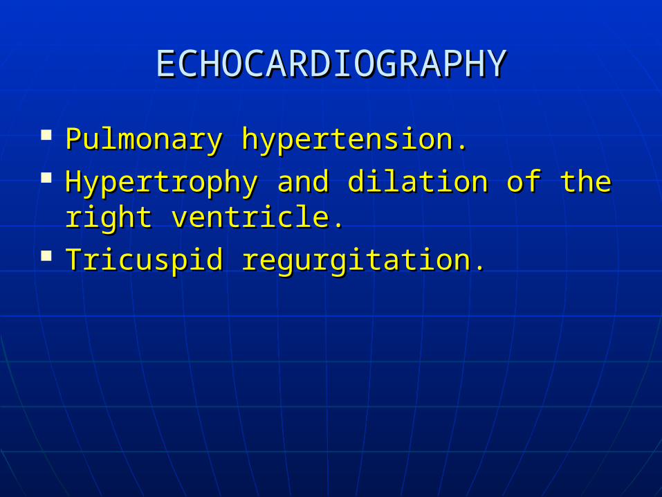
ECHOCARDIOGRAPHYECHOCARDIOGRAPHY
Pulmonary hypertension.Pulmonary hypertension. Hypertrophy and dilation of the right Hypertrophy and dilation of the right
ventricle.ventricle. Tricuspid regurgitation.Tricuspid regurgitation.

Ophthalmologic examinationOphthalmologic examination
Papilloedema –Papilloedema – result of increased result of increased cerebral and retinal cerebral and retinal blood flowblood flow
(CO(CO2 2 retention)retention)

Pulmonary function tests:Pulmonary function tests:
FEV1 reduced.FEV1 reduced. FEV1/VC FEV1/VC
decreased.decreased. PEF reduced.PEF reduced.

COMPLICATIONSCOMPLICATIONS
Secondary polycythemia.Secondary polycythemia. Pulmonary hypertension / Pulmonary hypertension /
right ventricular failure right ventricular failure
(cor pulmonale).(cor pulmonale). Hypoxia -> Pulmonary arteriolar Hypoxia -> Pulmonary arteriolar
vasoconstriction -> Pulmonary vasoconstriction -> Pulmonary hypertension.hypertension.
Type I / Type II respiratory failure.Type I / Type II respiratory failure.

EMPHYSEMAEMPHYSEMA
Distention of the Distention of the air spaces distal air spaces distal to the terminal to the terminal bronchiole with bronchiole with destruction of destruction of alveolar septaalveolar septa
Reduced lung Reduced lung elasticityelasticity

Types of emphysemaTypes of emphysema CentriacinarCentriacinar involving the respiratory involving the respiratory
bronchioles and alveolar ducts in bronchioles and alveolar ducts in the center of the acinus.the center of the acinus.
Panacinar Panacinar involving the entire acinusinvolving the entire acinus ParaseptalParaseptal involving alveolar ducts & sacs involving alveolar ducts & sacs
farther out in the acinusfarther out in the acinus

EmphysemaEmphysema
Centriacinar:Centriacinar: Result of chronic cigarette smokingResult of chronic cigarette smoking Upper lung zones involvementUpper lung zones involvement
Panacinar:Panacinar: A1-antitrypsin deficiency A1-antitrypsin deficiency Bases of the lungs involvementBases of the lungs involvement

CLINICAL FEATURESCLINICAL FEATURESSymptomsSymptoms
Increasing breathlessness - an Increasing breathlessness - an exertional dyspnea (long history).exertional dyspnea (long history).
Minimal cough with small amounts of Minimal cough with small amounts of mucoid sputum. Mucopurulent mucoid sputum. Mucopurulent exacerbations with infections exacerbations with infections (infrequent). (infrequent).

OBJECTIVE EXAMINATIONOBJECTIVE EXAMINATION
“ “Pink puffer”Pink puffer”
Tachypnea with Tachypnea with prolonged prolonged expiration trough expiration trough pursed lips / pursed lips / expiration with expiration with grunting soundgrunting sound
Lips tightly apposedLips tightly apposed
at height of inspiration,at height of inspiration,
Lips held narrowly apart during Lips held narrowly apart during expirationexpiration

OBJECTIVE EXAMINATIONOBJECTIVE EXAMINATIONRespiratory systemRespiratory system
Asthenic constitution with Asthenic constitution with weight loss.weight loss.
Barrel–shaped chest Barrel–shaped chest (increased anteroposterior (increased anteroposterior diameter).diameter).
Use of accessory muscles in Use of accessory muscles in respiration.respiration.
Tachypnea.Tachypnea. Prolonged expiration Prolonged expiration
through pursed lips.through pursed lips. Lower intercostal spaces Lower intercostal spaces
retract with each retract with each inspiration.inspiration.
Neck veins distended Neck veins distended during expiration.during expiration.

OBJECTIVE EXAMINATIONOBJECTIVE EXAMINATIONRespiratory systemRespiratory system
Palpation:Palpation: Increased rigidityIncreased rigidity Decreased vocal Decreased vocal
fremitusfremitus Diminished excursion Diminished excursion

OBJECTIVE EXAMINATIONOBJECTIVE EXAMINATION
Percussion:Percussion: Hyperresonant (bandbox) soundHyperresonant (bandbox) sound Upper borders protruded Upper borders protruded Lower borders: descendent Lower borders: descendent limited mobilitylimited mobility Decreased size of liver & cardiac dullnessDecreased size of liver & cardiac dullnessAuscultation:Auscultation: diminished vesicular diminished vesicular breathing breathing (diffuse dry rales in bronchitis)(diffuse dry rales in bronchitis)

OBJECTIVE EXAMINATIONOBJECTIVE EXAMINATIONCardiovascular systemCardiovascular system
Cardiac dullness severely reduced.Cardiac dullness severely reduced. Decreased heart sounds.Decreased heart sounds. Presystolic gallop accentuated during Presystolic gallop accentuated during
inspiration.inspiration.

Pulmonary function tests:Pulmonary function tests:
The TLC and RV are increased.The TLC and RV are increased. The VC is low. The VC is low. The maximal expiratory flow rates The maximal expiratory flow rates
are diminished. are diminished.

X-ray of the chest:X-ray of the chest:
Diaphragm is low Diaphragm is low and flattened.and flattened.
Bronchovascular Bronchovascular shadow do not shadow do not extend to the extend to the periphery of the periphery of the lungs.lungs.
Cardiac silchouette Cardiac silchouette is lengthened and is lengthened and narrowed.narrowed.
Overinflation.Overinflation.

Features Features Predominant emphysema Predominant bronchitis
Type A “Pink puffer” Type B “Blue bloater”
Age at time of diagnosis 60± 50±
Dyspnea Severe Mild
Cough After dyspnea starts Before dyspnea starts
Sputum Scanty, mucoid Copious, purulent
Bronchial infections Less frequent More frequentRespiratory insufficiency episodes Often terminal Repeated
X-ray "Hyperinflation" ± bullous changes,
small heart
Increased bronchovascular markings at bases, large
heart.
Chronic PaCO2
mmHg
35-40 50-60
Chronic PaO2 mmHg 65-75 45-60
Hematocrit % 35-45 50-55

Pulmonary hypertension
Features Features Predominant emphysema Predominant bronchitis
Type A “Pink puffer” Type B “Blue bloater”
Rest None to mild Moderate to severe
Exercise Moderate Worsens
Cor pulmonale Rare, except terminally Common
Elastic recoil Severely decreased Normal
Resistance Normal to slight increase High

Stages of COPDStages of COPD
Stage Stage 00High High risk risk
Stage 1Stage 1 Stage 2Stage 2 Stage 3Stage 3 Stage 4Stage 4
Risk Risk factorsfactors
Chronic Chronic productive productive coughcough
Normal Normal functional functional tests tests
FEVFEV11/FLVC/FLVC
<70%<70%
FEVFEV11>80%>80%
Chronic Chronic productive productive coughcough
FEVFEV11/FLVC/FLVC
<70%<70%
FEVFEV11>50%>50%
oror
FEVFEV11<80%<80%
Chronic Chronic productive productive coughcough
FEVFEV11/FLVC/FLVC
<70%<70%
FEVFEV11<50%<50%
oror
FEVFEV11>30%>30%
Chronic Chronic productive productive coughcough
FEVFEV11/FLVC/FLVC
<70%<70%
FEVFEV11<30%<30%
Chronic Chronic respiratory respiratory insufficiency insufficiency
&/or&/or
Right cardiac Right cardiac failurefailure

Treatment of COPDTreatment of COPD Stop smoking. Nutritional improvement. Stop smoking. Nutritional improvement.
Exercises Exercises Preventive vaccination against influenza Preventive vaccination against influenza
virus strainsvirus strains Pneumococcal polysaccharide vaccine Pneumococcal polysaccharide vaccine (once in life time) (once in life time) Early treatment of the infections Early treatment of the infections (broad spectrum antibiotics 7-10 days)(broad spectrum antibiotics 7-10 days) Bronchodilator drugs:Bronchodilator drugs: methylxantines, methylxantines, BB22-stimulating sympathomimetics, -stimulating sympathomimetics, anticholinergicsanticholinergics Corticosteroids Corticosteroids

Schematic representation of the morphology of normal airways and lung parenchyma Schematic representation of the morphology of normal airways and lung parenchyma and the changes produced in this structures by asthma, chronic bronchitis, and and the changes produced in this structures by asthma, chronic bronchitis, and emphysema.emphysema.
Mucous gland hyperplasia
Inflammatory cellular infiltration

Schematic representation of the morphology of normal airways and lung parenchyma Schematic representation of the morphology of normal airways and lung parenchyma and the changes produced in this structures by asthma, chronic bronchitis, and and the changes produced in this structures by asthma, chronic bronchitis, and emphysema.emphysema.
|______
_____|
IntrapulmonaryAirway