acute stroke management - twareat · recent ami, arterial puncture/lp within 7 days history of...
TRANSCRIPT

Saleh Alrashdi, MD Emergency consultant & Medical toxicologist
Chairman of Adult emergency King Fahad Medical City
Riyadh, Saudi Arabia [email protected]
Acute Stroke Management

Brain Attack!
Stroke � A sudden focal neurological
deficit or acute neurological impairment caused by the interruption of blood flow to a specific region of the brain
� 700,000 suffer a new
or repeat stroke in U.S. each year
� Third leading cause of
death in the US (> 150,000 yearly)

Stroke: Differential Diagnosis
� Syncope � Partial epileptic seizure
with Todd’s paresis � Migraine attack (aura) � Hypoglycaemia � Hysteria � Intoxication
� Subarachnoid haemorrhage � Neuroinfection � Neoplasm � Brain injury � Multiple sclerosis � Peripheral vertigo

Stroke / TIA Syndromes

Stroke / TIA Syndromes
� Anatomy of Cerebral Blood Flow � Anterior Circulation: 80% of cerebral blood flow
originates from the carotids which supplies the � Frontoparietal lobes � Anterior temporal lobes � Optic nerve and retina

Stroke / TIA Syndromes
� Posterior Circulation: 20 % of cerebral blood flow which originates from the vertebrobasilar arteries � Thalamus & Brainstem � Occipital cortex and Cerebellum � Upper Spinal cord & Auditory and Vestibular functions in ear
� Circle of Willis: connects the Anterior and Posterior circulations

Section 1 Slide 17
Causes of Ischaemic Stroke

Pathophysiology of Stroke
1- Ischemic Strokes: (thrombi or emboli)
� Cerebral Thrombi may result from: � Atherosclerosis (#1 cause) � Infective arteritis � Vasculitis � Hypercoaguable states � Post traumatic carotid or vertebral artery dissections

Pathophysiology of Stroke
� Cerebral emboli may result from: � Mural thrombus from heart (#1 cause) � Aortic plaques � Endocarditis � Long bone or Dysbaric injuries (fat / air emboli)

Stroke /TIA Syndromes
� Type of Stroke (rule of 2/3’s) � 2/3 of ALL Strokes will be ISCHEMIC
� 2/3 of these will be thrombotic � Therefore thrombotic ischemic strokes most common.
� Incidence of Stroke � Biggest Risk Factors
� Prior TIA ( 30 % will have stroke in 5 years) � HTN � Atherosclerosis � DM � Hyperlipidemia � Smoking

Ischemic Stroke Syndromes
� Thrombotic Syndromes � Usually slow, progressive onset � Sx develop shortly after awakening and are progressive
� Embolic Syndromes � Usually abrupt onset with maximal deficit that tends to
improve over time as the embolus breaks up.

Occlusive Stroke Syndromes
� Middle Cerebral Artery Occlusion (MCA) � Contralateral hemiplegia, hemianesthesia, and
homonymous hemianopsia � Upper extremity deficit >> Lower extremity � Aphasia (if dominant hemisphere involved)

Occlusive Stroke Syndromes
� Anterior Cerebral Artery Occlusion (ACA) � Contralateral leg, arm, paralysis � Lower Extremity deficit >> Upper extremity � Loss of frontal lobe control
� Incontinence

Occlusive Stroke Syndromes
� Posterior Cerebral Artery Occlusion (PCA) � Ipsilateral CN III palsy, visual loss � Contralateral hemiparesis and hemisensory loss � Memory loss

Occlusive Stroke Syndromes
� Vertebrobasilar Artery Occlusion (VBA) Keys: CN AND Cerebellar deficits that affect BOTH sides of the body,
with contralateral pain and temperature deficits. - Contralateral hemiplegia - Ipsilateral CN III palsy with Cerebellar findings.
- Nausea/Vomiting - Vertigo, Nystagmus, - Ataxia, Dysarthia

TIA’s (Transient Ischemic Attacks)
� Definition: A temporary loss of neurologic function, that resolves completely <24 hours.
� Clinically;
� Arm numbness, weakness � Facial droop, slurred speech � Sx resolved, or improve over time
� Main point: These patients at high risk for stroke if: � >50 � HTN, DM, Smoker, Prior TIA in last month � Any prior CVA…… ADMISSION IS THE RULE!!

Pathophysiology of Stroke
2- Hemorrhagic Strokes result from � Spontaneous rupture of berry aneurysm or AV
malformation � Rupture of arteriolar aneurysms secondary to:
� Hypertension � Congenital abnormality � Infection � Neoplasm
� Trauma (Epidural / Subdural Hematomas)

Hemorrhagic Syndromes (SAH, & Intracerebral)
� Subarachnoid Hemorrhage � Highest incidence in 35-65 year old. � Usually from the rupture of a berry aneurysm � Clinically:
� abrupt onset of “worst headache of life” � Nuchal rigidity, photophobia, vomiting, retinal hemorrhages.
� Diagnosis : CT + LP!!!! � CT only 92% sensitive within 24 hours of event, loses sensitivity
>24 hours out from headache. � 72 hours out CANNOT r/o without LP!

Subarachnoid Hemorrhage

Hemorrhagic Stroke Syndromes
� Intracerebral � Hypertensive intracerebral hemorrhage MOST common
cause. � Traumatic, contusion, coup/contracoup � Rupture of small blood vessels with bleeding inside the
brain parenchyma � Putamen � Cerebellar � Thalamic � Pontine ( 3 P’s – pinpoint pontine pupils)

Intracerebral Hemorrhage

Pre of hospital recognition TIA FAST
� Facial Weakness � Can person smile � Has mouth dropped
� Arm Weakness � Can person raise both arms
� Speech problems � Can person speak clearly and
understand what you say
� Test all three symptoms
� Stroke Association campaign to raise awareness
� Practice staff should be trained to inform doctor immediately if patient calls with symptoms identifiable
� Ambulance crews now trained to use FAST score to prioritise Calls and dispatch

Medical Stabilization
� Airway and Breathing � Protect from aspiration and hypoxemia
� Vitals / O2 � IV access (isotonic fluids only) � Labs
� Glucose, electrolytes � Consider cardiac markers, tox screen, coags
� EKG � Order Non-Contrast Head CT � Neurological Assessment
First 10 Minutes!

History / Exam
� History focus points � Onset and scenario � Significant comorbidities and
medications � Review contraindication list for
thrombolytics
n Exam focus points
n Obtain CT !
Within 25 Minutes!

CT Read
Hemorrhagic Ischemic
Within 45 Minutes!

Treatment of Stroke
� Hyperglycaemia � is a frequent finding in acute stroke
� Diabetes � Stress hyperglycaemia
� Raised blood glucose is positively associated with greater mortality and poor functional outcome
� Temperature regulation � Body temperature on admission predicts both the severity and long
term outcome � 10C rise independently predicts a 30% relative increase in long term
morbidity or death.

Treatment of Stroke
Penumbra
Infarction

Treatment of Stroke
� Protect the “Penumbra” � Keep SBP >90mm ( CPP = MAP – ICP)
� Goal keep CPP > 60mm Hg � Treat Fever ( Mild Hypothermia beneficial)
� Acetaminophen 650mg po or pr, cooling blanket � Oxygenate (Keep Sao2 >95%) � Elevate head of bed 30 deg. (Clear c-spine)
� Frequent repeat Neuro checks!! Reassess GCS!

Treatment of Stroke
� What type of stroke is Present?? � Bleed vs Ischemic
� Ischemic strokes � ASA 75-325mg � Patients with Systolic BP >220 , Diastolic>130 need BP control with
Nitroprusside or Labetolol. � DO NOT OVERTREAT BP or risk extending the infarct. � Consider Heparin if area of infarct small and neurologist agrees.
� No bolus, just infusion.

Treatment of Strokes
� Strokes with Edema, Mass Effect or Shift � Load with Dilantin 1 g @ rate no faster than 50mg/min. Acute
seizure prophylaxis still of benefit.
� Thrombolytics � Ischemic strokes ONLY with large deficit NOT improving. � Time from symptom onset <3 hours � No ABSOLUTE Contraindications!!

Thrombolytic Therapy for Acute Stroke Checklist
� Answer to ALL must be YES: � Age 18 or older � Clinical diagnosis of Acute Ischemic Stroke causing a
measurable NON improving neurologic deficit. � NO high clinical suspicion for SAH � Time of onset to treatment is < 3H.

Thrombolytic Therapy for Acute Ischemic Stroke Checklist
� Answer to ALL MUST be NO:
� Evidence of hemorrhage on CT � Active internal bleeding (GI/GU) within last 21 days. � Known bleeding diasthesis:
� Platelets<100,000 � Heparin within last 48 hours with elevated PTT � Warfarin use with PT > 15 seconds
� Within 3 months of IC injury, prior surgery or prior ischemic stroke. � Within 14 days of serious trauma, major surgery � Recent AMI, arterial puncture/LP within 7 days � History of prior ICH, AVM, tumor,or aneurysm or seizure at stroke � Systolic BP >185mmHg, or Diastolic BP >110Hg

Thrombolysis history
Date Thrombolytic dose
Time window
Mean time to
treatment
NINDS part I 1995 rt-PA 0.9 mg/kg <3hrs
NINDS part II 1995 rt-PA 0.9 mg/kg <3hrs 119 min
ECASS 1 1995 rt-PA 1.1 mg/kg <6hrs 264 minutes
ECASS 2 1998 rt-PA 0.9 mg/kg <6hrs
ATLANTIS 1999 rt-PA 0.9 mg/kg 3-5hrs* NA
MAST-E 1996 STK 1.5 m.U <6hrs
MAST-I 1995 STK 1.5 m.U <6hrs 268 minutes
ASK 1996 STK 1.5 m.U <4hrs
PROACT I & II 1998&1999 IA proUK <6hrs
EMS 1999 IA & IV rt-PA <6hrs
IMS 2002 IA & IV rt-PA <6hrs
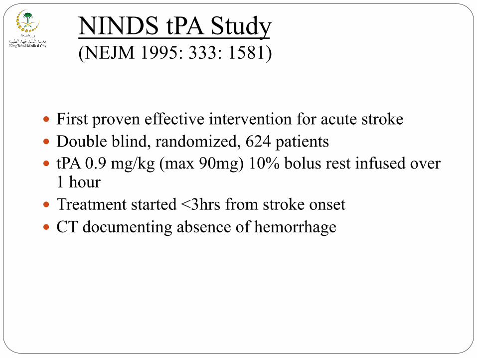
NINDS tPA Study (NEJM 1995: 333: 1581)
� First proven effective intervention for acute stroke � Double blind, randomized, 624 patients � tPA 0.9 mg/kg (max 90mg) 10% bolus rest infused over
1 hour � Treatment started <3hrs from stroke onset � CT documenting absence of hemorrhage


Blood Pressure Management
� Lowering BP may extend infarct � Look for 2° causes of HTN (pain, full bladder..) � Untreated, BP will decline w/in 24 hrs and return to
baseline in 4-7 days
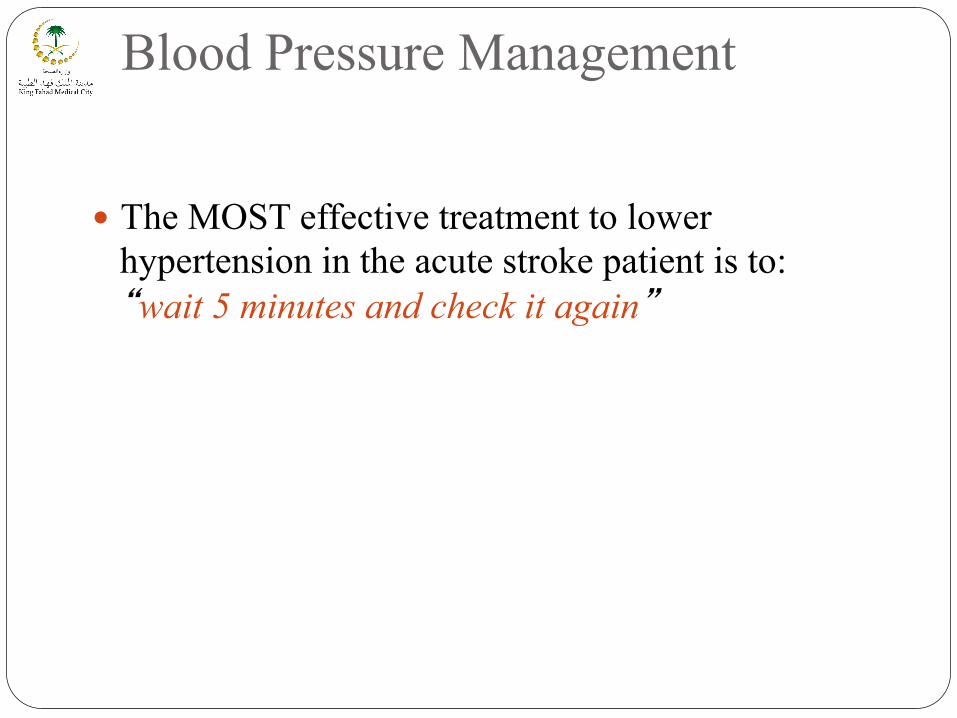
Blood Pressure Management
� The MOST effective treatment to lower hypertension in the acute stroke patient is to: “wait 5 minutes and check it again”

Blood Pressure Management
Mean blood pressure
Cerebral Blood Flow
50 150
CBF remains constant because of “autoregulation”. Beyond these limits CBF varies with blood pressure


ANTICOAGULATION
� Tested unfractionated heparin, LMW heparin, danaparoid � Most trials tested SQ therapy – only 1 trial of IV-adjusted dose
therapy � No evidence in halting neurological worsening � No evidence in improving neurological outcomes � No data to recommend emergent anticoagulation for most
patients with acute ischemic stroke

Decompressive Craniectomy

Decompressive Craniectomy
Indications: Worsening midline shift or uncal herniation especially in cases of massive middle cerebral artery strokes
Contraindications: � Age greater than 50 years � Prolonged severely low Glasgow coma scale Timing: within 48 hours of the injury, before the period of
maximal cerebral swelling.

Acute intraparenchymal hematoma
Cerebral Hemorrhage

Acute Management of Cerebral haemorrhage
� Acute treatment � Conservative � Surgical � Medical Therapy
� Recominant Factor VII
� Secondary Prevention � Antihypertensive
Treatment

BP Management
� Lower blood pressure to decrease risk of ongoing bleeding from ruptured small arteries
� Overaggressive treatment of blood pressure may decrease cerebral perfusion pressure and worsen brain injury

BP Management
� AHA recommends blood pressure be maintained below a mean arterial pressure of 130 mm Hg in persons with a history of hypertension
� If there is an ICP monitor: � ICP should be kept < 20 m Hg � Cerbral perfusion pressure (MAP-ICP) should be kept >
70 mm Hg

Secondary prevention of stroke & TIA
� For patients with TIA § Hypertension / Blood pressure § Ant platelet therapy § Statins § Carotid Endarterectomy for patients with symptomatic
carotid stenosis § Anticoagulant therapy for patients with atrial fibrillation § Smoking
Warrants more aggressive evaluation & management

Strategies for preventing stroke and reducing stroke disability
First stroke
blood pressure smoking lipids
mass popl. strategy
hypertension TIA Atrial fibrillation other vascular disease
high risk strategy
stroke mortality
acute treatment
Secondary prevention
recurrent stroke
Stroke related disability
Rehabilitation

Case 1 � 63 year old male with diabetes, hypertension & atrial
fibrillation presented with a 1.5 hour presentation of right hemiparesis and aphasia.
� What’s the 1st line of management � Then what?
� Glucose � Blood pressure
� What tests would you do 1st

Case 2 � 56 yr male with HTN suddenly c/o headache, Rt
hemiparesis, vomits in the ER. � Exam: BP 210/120 � Arousable. Aphasic. Lt gaze preference � CT ??

Case 2 CAT scan

Stroke Treatment 2010 and Beyond
H U
R
Call emergency
services
A+E stroke team
Activated (15 minutes)
Brain scan
Thrombolysis
Drugs
Full recovery
Stroke onset Secondary prevention

Thank you