ad-a084 105 army research inst of environmental medicine ... · ad-a084 105 army research inst of...
TRANSCRIPT

AD-A084 105 ARMY RESEARCH INST OF ENVIRONMENTAL MEDICINE NATICK -A F/S 6/1THE EFFECT OF CATHEPTI ENZYMES ON CHILLED BOVINE MUSCLE(U)
'FEB aS S H C OHEN . L R TRUSALUNI LE V ARIEIFMED/80 N

SWrUITY, ASPICATION OF THIS PAGE =ffDafa Entered) L EAD~ AC..... I[ NRPOR1. GOVT ACCESSION NO 3. RECIPIENT'S CATALOG NUMBER
M-6/800"I
'4 N ,tU S... ..... ...... -. TYPE OF REPORT & PERIOD COVERED
The Effect of Catheptic Enzymes on Chilled BovineMuscl _
6. PERFORMING ORG. REPORT NUMBER
L. CONTRACT OR GRANT NUMUER(e)
9. PERFORMING ORGANIZATION NAME AND ADDRESS 10. PROGRAM ELEMENT, PROJECT, TASK
AREA & wORK UNIT NUMBERS'
US Army Research Institute f Environmental
Medicine, Natick, MA 01760-
II. CONTROLLING OFFICE NAME AND ADDRESS -/ * i-- W.-,R1Y-Th " .
US Army Medical Research & Development Command( Febm 80'fWashington, DC 20314 N ---- "nr12
14. MONITORING AG ESS(I1 dillemt from Controlling Office) IS. SECURITY CLASS. (of thi report)
UNCLASSIFIEDSame ___________IS. DECLASSIFICATION/IDOWNGRADI NG
SCHEDULE
IS. DISTRIBUTION STATEMENT (of hi. Report)
Distribution of this document is unlimited D T ICELECTEMAY 13 1980
17. DISTRIBUTION STATEMENT (of the absrct metered In Block 20, If different from Report) I
N/A E
IS. SUPPLEMENTARY NOTES
i. To be presented at Scanning Electron Microscopy 1980 Annual Meeting byS. H. Cohen.2. Submitted for publication as a full length paper in Proceeding of SEM 1980.
19. KEY WORDS (Continue on reverse side If neceeurWy amd identify by block number)
Cathepsin, cold shortening, sarcomere, Z-band, laser diffraction, rigor,muscle, electron microscopy
2(. ABSTRACT (03irihue an evero side If neCeeary7 mod Identify by block number)
/ The toughening of meat which has been caused by cold shortening prior to theLJJ onset of rigor is of significant commercial importance. Various studies have
shown that catheptic enzymes produce degradative changes to meat which are verysimilar to those which occur during the natural aging process and which leadto a more tender meat product. Because of the tenderizing action of cathepsins,this study was undertaken to determine whether these enzymes could hasten thereversal of the cold shortening process. Samples which were cold shortened for -
147 EITIN ~l OVOSI OSOLTE S CUftTY LASS1FICATf#R OF THIS PAGE (When 004ta*red)

Ilk.
ECUNITY CLASIFICATION OF THIS PAGE(Whan Data Enatere
- 24 or 72 hrs were soaked in either a control solution or one containing-J catheptic enzymes. The sarcomere lengths of all samples were measured by
laser diffraction, transmission electron microscopy and scanning electronmicroscopy, while the ultrastructural appearance was assessed by both formsof electron microscopy. Results showed that the microstructural appearanceand significantly longer sarcomere lengths of the enzyme treated samples wereconsistent with a reversal of the cold shortening process.
qFeIImgTy CLASO.IFICATInN MF THIS PAGOzften Data Entered)

The Eff ect of Catheptic Enzymes on Chilled Bovine Muscle
S. H. Cohen and L. R. Trusal*
Biochemistry and Nutrition GroupFood Sciences LaboratoryUS Army Natick Research and Development CommandNatick, MA 01760
* Experimental Pathology DivisionUS Army Research Institute of Environmental MedicineNatick, MA 01760
Running Title: Catheptic Action on Chilled Muscle
Key Words: Catnepsin, cold shortening, sarcomere, Z-band, laserdilffraction, rigor, muscle, electron microscopy
Accession For
NTIS G1a4&IDXC TAB
Justification
Di st special
......... ) ~ Cod....

ABSTRACT
The toughening of meat which has been caused by cold shortening prior to the
onset of rigor is of significant commercial importance. Various studies have
shown that cathe.tic enzymes produce degradative changes to meat which are
very similar to those which occur during the natural aging process and which lead
to a more tender meat product. Because of the tenderizing action of cathepsins,
this study was undertak.en to determine whether these enzymes could hasten the
reversal of the cold shortening process. Samples which were cold shortened for
24 or 72 hrs were soaked in either a control solution or one containing catheptic
enzymes. The sarco-nere lengths of all samples were measured by laser
diffraction, transmissio.n electron microscopy and scanning electron microscopy,
while the ultrastruc-tiral appearance was assessed by both forms of electron
microscopy. Resu!s showed that the microstructural appearance and
significantly longer sacomere lengths of the enzyme treated samples were
consistent with a reversal of the cold shortening process.
I!

INTRODUCTION
The chilling of bovine skeletal muscle prior to the onset of rigor mortis
produces a reversible cold shortening effect (Locker and Hagyard, 1963) which
causes toughening in cook-ed meat when there is a shortening of between 20 to
50% (Marsh and Leet, 1966). However, when shortening exceeds 50%, the severe
contraction of the mycfibrilar ultrastructure causes fiber fracture leading to a
decline in toughness (,arsh, Leet and Dixon, 1974).
As a result of postmortem aging meat becomes tender, but this tenderness
is caused by factors tnlvke the supraphysiological shortening mentioned above.
Aged meat is tender because of the degrada:ion of Z discs (Davey and Gilbert,
1967) and disruption of the sarcolemma (Varriano-Marston et al, 1976) as well as
certain other fac-ors -icluding loss of Ca + 2 accmulating ability (West et al,
1974) and modifi'-t~on of collagen (Marsh, 1977). Eino and Stanley (1973 a,b)
and Robbins and Cohen (1976) found that catheptic enzymes produce degradative
changes to the bcvLe rnycfibrillar ultrastructure which were quite similar to
those which occur durir,- postmortem aging.
Since there are similarities between morphological changes within the
myofibrills caused by physiological contraction and cold shortening (Davey and
Gilbert, 1974), the adcd-ion of catheptic enzymes to cold shortened muscle might
act to reverse some of the effects induced by cold shortening. The purpose of
the present paper is to investigate this possibility.

MATERIAL AND METHODS
Within 15 min of slaughter, a 5 cm 2 strip of sternomandibularis muscle was
dissected from a cow of undetermined age. The muscle was divided into 3
smaller strips, one to be used for laser diffraction (LD) one for transmission
electron microscopy (TEM) and one for scanning electron microscopy (SEM).
Sarcomere leng-h reasuremen:s were determined by all three methods (LD,
SEM, TEM).
Sampling periods were as follows: at death control (C); 24 hr cold
shortened control (C 1; 24 h.- cold shortened enzyme treated (El); 72 hr cold
shortened con:rol (C2 ); and 72 hr cold shortened enzyme treated (E2).
The stribs were refr-eratec at 20C in an unrestrained condition within
freezer Tapping raD>er for 24 or 72 hrs. At these times the samples were soaked
overnight (16 "*-.s) - a 1 % KCI control solution or in a catheptic enzyme solution
(1.2 activity Lnis/ml) (CRob*ins and Cohen, 1976).
1. Laser Diff--action-Sarcomere Length Measurements
Muscle fi>er bundles from each sampling period were teased from the
larger portion of muscle and fixed for I hr in 2.5% glutaraldehyde in KCI-borate
burffer (pH 7.1) and then wased twice in the buffer solution (10 min each).
Next, individual fibers were placed into a drop of the buffer on a glass slide
and a coversli, placed ov- the drop. The slide was mounted on a modified
microscope stage and positioned so that the muscle fiber was in the path of a
laser beam (Spectra- Physics HeNe 632.8 nm) so that a diffraction pattern was
formed on a groun gJass screen 10 cm from the sample. The sarcomere length
was determined by measuring the distance between the Oth and Ist order
diffraction bands a-d using 'he formLla d sin-&= n where d sacromere length,
2

e angular separation between the 0th and 1st order bands, )~=the wavelength
of laser light (632-8 n-.n) and n = the order of particular diffraction band
(Cleworth and EcLm-n, 1969). A total of 50 sarcomere length measurements were
made for each sapi~period.
2. TEM Processing
Small muscie strips tak~en at each sampling period were macerated and
then fixed in 2-5%- gluaraldehyde in 0.10 M cacodylate - 0.11 M sucrose buffer
(pH 7.3) for 24 hrs. This was followed by three (30 min ea) rinses in cacodylate
sucrose buffer, Dcst fiaion- in 1% 0s04 in 0.04 M cacodylate - 0.14 M sucrose
buffer for 1 hr at -*0C, three buffer rinses (10 min ea) and dehydration in a
graded series of ETCHi (70, 95, 100 and 100%). After dehydration the strips were
transferred to prcpye: e oxide for 30 min and then into a mixture of propylene
oxide and Ep~Alie(1:1) [Moon 812 (25 ml), Araldite 6005 (15 ml), DDSA (55
ml), DMP-30 (2 n-I), [ 3P (3 mn!)1 for 1 hr. This was followed by Epon-Araldite
(1:3) for an additicnad -' hr befor'e final infiltration in Epon-Araldite. The resin
mixture was then ,otvner-ized for 48 hrs at 600C.
A trapezoid shaped b'ock face was hand trimmed, smoothed by a glass
knife with final ulrattln sectioning by diamond knif e. Sections were picked up
with 300 mesh cop~c gFics and stained with 5% uranyl acetate in 50% methanol
for 15 min f ollowed by 2% lead citrate for 7 min, and then viewed using a JEOL
Model IOOB (JEOL, Medford, -MA) at an accelerating voltage of 60-80 MV At
least 5 sections tfrn each sample were examined and measurements of 40
sarcomeres were rrace.
3. SEMA Processn'g
Following overrigpt wa-shing in the KCl-borate buffer solution, mentioned
in --he laser diffriacl-on ,reparation, samples were dehydrated in 70, 90, 95, and
3

100% ETOH for I hr each. Then the fibers were critical point dried from liquid
CO 2. Following this step the fibers were mounted on SEM stubs with double
sided sticky tape and sputter coated (Commonwealth Sci., Alexandria, VA) for 10
min with gold-palladium.
After sputtering the samples were placed in a Coates and Welter Model
100-2 field emission SFM (Sunnyvale, CA) for observation. Photographs were
taken using an emission current of 10 j A and an accelerating voltage of 15kV.
Twenty-five sarcomere leng-th measurements were made for each treatment
group.
4. Statistical 'aJy sis
Sarcomere length measurements obtained by laser diffraction, transmission
and scanning EM were subjected to a two way analysis of variance with
repetitions followed by the Tukey test for multiple comparison of means.
Significance was com.puted at the 95% confidence level (p < 0.05) for all mean
comparisons.
RESULTS AND DISCUSSION
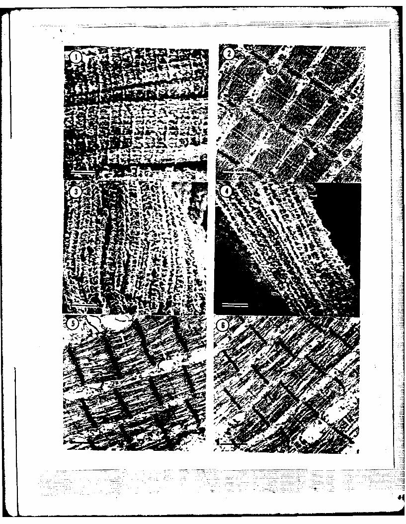
The SEM micrograph (Fig. 1) of a pre-rigor control (C) shows the even
register of sarcomeres with transverse (T) tubules overlying the Z-bands. In Fig.
2, the A, I and Z-bands as well as the mitochondria can be clearly distinguished
in the corresponding TE%i micrograph. The average sarcomere lengths of pre-
rigor, pre-cold shortened muscle, as seen in Table 1, ranged from 2.05 Jm (LD),
to 2.15 pim (SE.) to 2.29 Um (TEM) which is consistent with the at death
sarcomere lengths of 2.4 pim (TEM) determined by Henderson, Goll and Strorner
(1970).
After cold shortening for 24 hrs (CI), the values decreased to 1.03 Um
I== .. _4

(LD), 1.21 jitm (SEM) and 1.28 pm (TEM). Once again, the mean sarcomere length
as determined by TEM (1.23 Um) was almost identical to the value published by
Henderson, Goil and Stromer (1970) who obtained 1.3 pm (TEM) after cold
shortening for 24 hrs.
According to Locker and Hagyard (1963), the degree of muscle shortening
was approxirrately the same (47.7%) at O°C and 20 C. Our results show that
sarcomere lengths of muscle cold shortened at 20 C for 24 hrs and then soaked in
a control solu-.ion (20 C) ove-night, shortened between 43.7 and 48.8% (Table 1).
Chilling the sam.ples for 24 hrs resulted in severe contraction of the
myofib:ils and altered :he surface morphology considerably (Fig. 3). The degree
of contraction was suh tihat it was difficult to distinguish the untreated (C1)
sample from the treated sa.-nple (EI) (Fig. 4).
As seen in Fig. 5, dciling induced sarcomere contraction producing an
overlapping of myofibrllar filaments resulting in the disappearance of the I band
causing the sarccmere to rake on a concave appearance. Separation and
distortion of the individJal myofllaments also occurred. The overlapping of actin
and myosin fidamen-s by sliding across one another substantiates the sliding-
filament hypothesis of .arsh and Carse (1974) who explained that filament
overlapping is a signiicant stage in the cold-shortening process.
When the 24 hr cold shortened muscle was soaked in a catheptic enzyme
solution (E ), there was a significant increase (p < 0.05) in sarcomere length
(Table 1) in the LD and SEM samples but not the TEM sample. The degree of
chilling induced con-raction made it difficult to differentiate the untreated
sample (Fig. 5) fron -he enzyme treated one (Fig. 6) in the TEM micrographs.
After 72 hrs. the sarcomere lengths of the cold shortened muscles were
significamly longer (: <0.05) than the 24 hr group. (C1 ) when measured by all
5

three methods. The sarcomere lengths of the control samples (C 2) were 1.72 jim
(LD), 1.69 um (SEM) and 1.46 Vm (TM). However, the enzyme treated samples
(E2 ) had sarcomere lEg",s of 2.08 Um (LD), 2.06 pm (SEM) and 1.95 jim (TEM)
which ranged from 21 to 22 to 34% longer than the controls, respectively. The
significant differences were probably caused by the synergistic effect of soaking
in the catheptic enzymr.e solution.
While Eino and Stanley (1973a) found that the resolution of rigor usually
occured at five days pcst-rrtoem, we have found that the rigor process seems to
be largely resolved by 72 h-s (plus overnight soaking). The surface morphology of
the 72 hr control sampe (C2 ) has changed so there is no longer any difficulty in
differentiating the myofibri~lar features (Fig. 7). The enzyme treated sample
(E 2 ), although similar in appearance to the untreated sample (C 2 ) has areas,
especially in the :-1 b-d region, where a ce.-tain amount of degradation appears
to have taken place (Fg. 8). Eno and Stanley (1973a) and Varriano-Marston et
al. (1976) found similar ch&-i ges in naturally aged muscle as did Eino and Stanley
(1973b) and Robbins an' Cohen (1976) in muscle treated with catheptic enzyme.
The greates c:fferences between untreated (C2) and treated (E 2 ) samples
are seen in the TE.M micrographs (Figs. 9, 10) of the 72 hr samples. The
untreated sample (Fig. 9) is similar in appearance to Fig. 5, but the sacromere
has increased significartly (p < 0.05) in width, some degradation in the Z-band has
occurred and the sarco!emra is devoid of normal appearing mitochondria.
The contracion of the untreated 72 hr samples (C2 ) has been largely
reversed. Because of t e higher concen, tration of catheptic enzymes, the treated
(E2 ) myofibrils exhibi- the typical ultrastructural morphology found in aged
muscle (Davey and Dicsor, 1970). Furthermore, the sarcomere lengths of the
72 hr enzyme treared (E.2 ) sasrnle more closely approach those of the original
pre-rigor, pre-cold sho.ene- cortrol (C).
6

Although smistdcl analysis clearly showed significant differences
between untreated and enzy,ne treated samples no matter which preparative
technique was used (LD, SEM, TEM), the value differences of the three methods
was probably due :o the e ehricues themselves (Varriano-Marston, 1978). If one
is solely interested in measuring sarcomere lengths, the laser diffraction method
is the simplest, fastest and probably most accurate method. It also involves the
fewest rr ardpulF-tive procedw'es. On the other hand if information is desired
concerning surface s-r-c-r-ee or ultrastructure, then SEM and TEM examination
would be necessary.
TI s, there is !:.t-e du~t :hat cathepsins both aid and speed the processes
responsib:e for significantly increasing the sarcomere length in rigor meat by
degrading the Z-1 b-and region and sarcolemma microstructure. Our results
sugest thaz the acdEticn of catepic enzymes to cold shortened muscle hastens
:he change fren ric- -: aged muscle.
ACKNOWLEDGMENT
We are gratefu tc Dr. Frederick M. Robbins and John Walker for providing
us with the cathepic e-.zvrres and for very helpful technical advice and guidance
throughout this inves-igtio.. We also thank Ella Monro, Leonora Kundla and Dr.
Edward Ross for help. with the statistical analysis and Albert Guzman for his
excellent technical assistance.
7
REFERENCES
Cleworth, D. and K. A. P. Edman. Laser diffraction studies on single skeletal
muscle fiber. Science 163:1969, 296-298.
Davey, C. L. and M. R. Dickson. Studies in meat tenderness and ultrastructural
changes in meat -u-ing aging. J. Food Sci. 35:1970, 56-60.
Davey, C. L. and K. V. Gilbert. Structural changes in meat during aging. 3.
Food Techno0. 2:1967, 57-59.
Davey, C. L. and K. V. Gilbert. The mechanism of cold-induced shortening in
beef muscle. 3. F--ood Technol. 9:1974, 51-58.
Elino, M. F. and D. T. Stanley. Catheptic activity, textural properties and
surface uhzrastruc-e of post-mortem beef muscle. . Food Sci. 38:1973a,
45-50.
ELno, &I. F. and D. W. Sanley. Surface ultrastructure and tensile properties of
cathepsn and co'lag2niase treated muscle fibers. 3. Food Sci. 38:1973b,
51-55.
He-iderson, D. W., D. F Golf and M. H. Stromer. A comparison of shortening and
Z line degradation Ln post-mortem bovine, procine and rabbit muscle. Am.
3. Anat. 128:197C, 117-136.
Locker, R. .-. and C- 3. Hagyard. A cold shortening effect in beef muscles. 3.
Sci. Food Agric. 14: 1963, 787-793.
Marsh, B. B. Sy-nosi.n-,. The basis of quality in muscle foods. The basis of
tenderness in muscle foods. 3. Food Sci. 42:1977, 295-297.
Ma.-sh, B. B. and W. A. Carse. Meat tenderness and the sliding filament
hypothesis. . Foo)d Technol. 9:1974, 129-139.
Mash, B. B. and N. G. Leet. Studies in meat tenderness. III. The effects of
cold shorten.ng or. tenderness. 3. Food Sci. 311966, 450-459.
8

Marsh, B. B., N. G. Leet and M. R. Dickson. The ultrastructure and tenderness
of highly cold.-shartened muscle. I. Food Technol. 9:1974, 141-147.
Robbins, F. M. and S. H. Cohen. Effects of catheptic enzymes from spleen on
the micros-nucture of bovine semimembranosus muscle. J. Texture
Studies 7:1976, 137-142-
Varriano-Marston, E.., G. A. Davis, T. E. Hutchinson and I. Gordon. Scanning
electron microscopy of a-zed free and restrained bovine muscle. J. Food
Sci. 41:1976, 601-605.
Varriano-Marston, E-, E. A. Davis, T. E. Hutchinson and 3. Gordon. Postmortem
aging of b~ovine mnuscle: A comparison of two preparation techniques for
electron microscopy. J. Food Sci. 43:1978, 680-683.
West, R. L., P. W. Moeller, B. A. Link and W. A. Landmann. Loss of calcium
accumulati ab~l.r in the sarcoplasmic rtclmfollowing degradaionZ
by cathepsins. I. Fo~od Sci. 39:1974, 29-31.
9

Figure Legends
Fig. 1. SL% micrcgraph of pre-rigor muscle showing the register of sarco
meres (S) and transverse tubules (T) overlying the Z-I band region.
Bar = 5 tim
Fig. 2. TEM ricrcograph of pre-rigor muscle. Typical A, I and Z bands as
well as mitochondria (arrows) are easily distinguished. Bar = I Im
Fig. 3. SEM rricovraphs of untreated (C1) (Fig. 3) and enzyme treated (E1 )and 4.
(Fig. 4) 24 hr chilled muscle. Degree of contraction makes differen-
tiation of s7.rface features of both samples difficult. Bar = 5 pjm
Figs. 5. TEM rricrographs of untreated (C1) (Fig. 5) and enzyme treated (E)and 6.
(Fig. 6) 24 h chIled muscle. Note disappearance of I band, concavity
in A band region (arrows) and apparent separation of myofilaments in
both figu-es. Bar = I Pm
Fig. 7. SEM rricrovraphs of 72 hr muscle. Chilling-induced contraction isand S.
reduced L- an-teated sample (C2 ) (Fig. 7) and surface features can be
better diffeentiated. With enzyme-treatment (E2 ) some degradation
(arrows) has occurred. (Fig. 8) Bar = 5 urm
Fig. 9. TEIM rricrographs of 72 hr muscle. Untreated myofibrils (C2 ) (Fig. 9)and 10.
have d-dergone some aging-related changes including increased
sarcomere length and some degradation of the Z-1 band region.
Treatec sarcorneres (E2) show extensive degradation of the Z-I band
region (arrows), no separation of myofilaments and a return to convex
appear''ig sarcomers (Fig. 10). Bar = U rm.
10

0 09 N
1:+1
N0% i'
gO 0 -0
0% 0 :V z z > >
1:20 U0 0:L +1 1 4
V0 0 N
i-~~ ~ ~ eN -l NN> 0
r **z p N 9 . 0 0W"1J 0 :
12 P L! 1 +1 +1 0 0 0 D
WJ 0U 0 0
< CIO P 2 P
0 CD 0lw h0< Z Oz Oz c
z < 0) +0 N1 N+1.
w -j 2 0 0 0 0L/~ 0 0 0l w0
o~ Z1 le o N N
<< <<
LU~UJL C12 C'"2 O l9
0 0) V) V) tn
u- < -C 0z C zC 4w CC ~- C. < U 0 0< a < a<e q I P%
Z Z : -1 w N
I. - 0 w0 0: 0 0J
LrV )C P L L z 2
__ _ _ _ _ _ _ . . . . . . . - .... ..-- .... . .............
.... .........4
ar..
. o p

.... ., ....
.. ......
~~L I- Ig~t~
-r & - f 4 * a m -h o w
~J rihiP4L
e
L ___

The views, opinions, and findings contained in this report
are those of the authors and should not be construed as an official
Department of the Army position, policy, or decision, unless so
designated by other official documentation.