ad award number: w81xwh-10-1-0491 principal …
TRANSCRIPT
AD_________________
Award Number: W81XWH-10-1-0491 TITLE: M current-based therapies for nerve agent seizures PRINCIPAL INVESTIGATOR: Jaideep Kapur, M.D., Ph.D. CONTRACTING ORGANIZATION: University of Virginia Charlottesville, 22908 REPORT DATE: July 2013 TYPE OF REPORT: Final PREPARED FOR: U.S. Army Medical Research and Materiel Command Fort Detrick, Maryland 21702-5012 DISTRIBUTION STATEMENT: Approved for Public Release; Distribution Unlimited The views, opinions and/or findings contained in this report are those of the author(s) and should not be construed as an official Department of the Army position, policy or decision unless so designated by other documentation.
REPORT DOCUMENTATION PAGE Form Approved
OMB No. 0704-0188 Public reporting burden for this collection of information is estimated to average 1 hour per response, including the time for reviewing instructions, searching existing data sources, gathering and maintaining the data needed, and completing and reviewing this collection of information. Send comments regarding this burden estimate or any other aspect of this collection of information, including suggestions for reducing this burden to Department of Defense, Washington Headquarters Services, Directorate for Information Operations and Reports (0704-0188), 1215 Jefferson Davis Highway, Suite 1204, Arlington, VA 22202-4302. Respondents should be aware that notwithstanding any other provision of law, no person shall be subject to any penalty for failing to comply with a collection of information if it does not display a currently valid OMB control number. PLEASE DO NOT RETURN YOUR FORM TO THE ABOVE ADDRESS. 1. REPORT DATE July 2013
2. REPORT TYPEFinal
3. DATES COVERED 1 July 2010 – 30 June 2013
4. TITLE AND SUBTITLE
5a. CONTRACT NUMBER
M current-based therapies for nerve agent seizures 5b. GRANT NUMBER W81XWH-10-1-0491
5c. PROGRAM ELEMENT NUMBER
6. AUTHOR(S)
5d. PROJECT NUMBER
Jaideep Kapur, Jian Li Sun, Terry Zhang, Marko Todorovic, John Williamson
5e. TASK NUMBER
E-Mail: [email protected]
5f. WORK UNIT NUMBER
7. PERFORMING ORGANIZATION NAME(S) AND ADDRESS(ES)
8. PERFORMING ORGANIZATION REPORT NUMBER
University of Virginia Charlottesville, 22908
9. SPONSORING / MONITORING AGENCY NAME(S) AND ADDRESS(ES) 10. SPONSOR/MONITOR’S ACRONYM(S)U.S. Army Medical Research and Materiel Command Fort Detrick, Maryland 21702-5012 11. SPONSOR/MONITOR’S REPORT
NUMBER(S) 12. DISTRIBUTION / AVAILABILITY STATEMENT Approved for Public Release; Distribution Unlimited 13. SUPPLEMENTARY NOTES
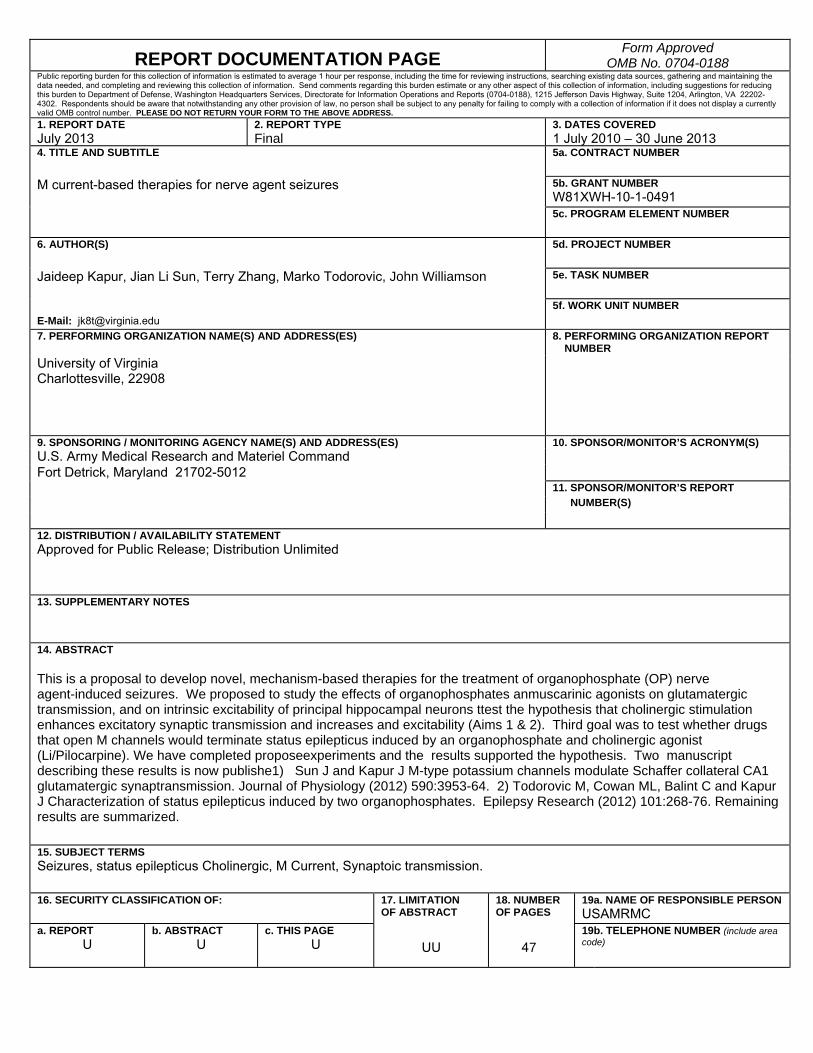
14. ABSTRACT This is a proposal to develop novel, mechanism-based therapies for the treatment of organophosphate (OP) nerve agent-induced seizures. We proposed to study the effects of organophosphates anmuscarinic agonists on glutamatergic transmission, and on intrinsic excitability of principal hippocampal neurons ttest the hypothesis that cholinergic stimulation enhances excitatory synaptic transmission and increases and excitability (Aims 1 & 2). Third goal was to test whether drugs that open M channels would terminate status epilepticus induced by an organophosphate and cholinergic agonist (Li/Pilocarpine). We have completed proposeexperiments and the results supported the hypothesis. Two manuscript describing these results is now publishe1) Sun J and Kapur J M-type potassium channels modulate Schaffer collateral CA1 glutamatergic synaptransmission. Journal of Physiology (2012) 590:3953-64. 2) Todorovic M, Cowan ML, Balint C and Kapur J Characterization of status epilepticus induced by two organophosphates. Epilepsy Research (2012) 101:268-76. Remaining results are summarized. 15. SUBJECT TERMS Seizures, status epilepticus Cholinergic, M Current, Synaptoic transmission.
16. SECURITY CLASSIFICATION OF:
17. LIMITATION OF ABSTRACT
18. NUMBER OF PAGES
19a. NAME OF RESPONSIBLE PERSONUSAMRMC
a. REPORT U
b. ABSTRACT U
c. THIS PAGEU
UU
47
19b. TELEPHONE NUMBER (include area code)
Table of Contents
Page
Introduction…………………………………………………………….………..….. 5
Body………………………………………………………………………………….. 5
Key Research Accomplishments………………………………………….……..16
Reportable Outcomes………………………………………………………………16
Conclusion…………………………………………………………………………… 16
References……………………………………………………………………………. 17
Appendices…………………………………………………………………………… 23 and PDF of
reprints
5
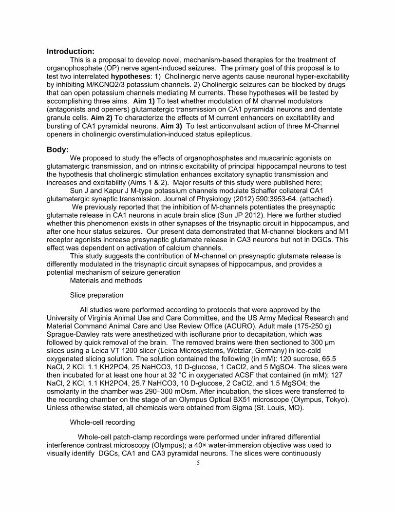
Introduction: This is a proposal to develop novel, mechanism-based therapies for the treatment of
organophosphate (OP) nerve agent-induced seizures. The primary goal of this proposal is to test two interrelated hypotheses: 1) Cholinergic nerve agents cause neuronal hyper-excitability by inhibiting M/KCNQ2/3 potassium channels. 2) Cholinergic seizures can be blocked by drugs that can open potassium channels mediating M currents. These hypotheses will be tested by accomplishing three aims. Aim 1) To test whether modulation of M channel modulators (antagonists and openers) glutamatergic transmission on CA1 pyramidal neurons and dentate granule cells. Aim 2) To characterize the effects of M current enhancers on excitabtility and bursting of CA1 pyramidal neurons. Aim 3) To test anticonvulsant action of three M-Channel openers in cholinergic overstimulation-induced status epilepticus.
Body: We proposed to study the effects of organophosphates and muscarinic agonists on
glutamatergic transmission, and on intrinsic excitability of principal hippocampal neurons to test the hypothesis that cholinergic stimulation enhances excitatory synaptic transmission and increases and excitability (Aims 1 & 2). Major results of this study were published here;
Sun J and Kapur J M-type potassium channels modulate Schaffer collateral CA1 glutamatergic synaptic transmission. Journal of Physiology (2012) 590:3953-64. (attached).
We previously reported that the inhibition of M-channels potentiates the presynaptic glutamate release in CA1 neurons in acute brain slice (Sun JP 2012). Here we further studied whether this phenomenon exists in other synapses of the trisynaptic circuit in hippocampus, and after one hour status seizures. Our present data demonstrated that M-channel blockers and M1 receptor agonists increase presynaptic glutamate release in CA3 neurons but not in DGCs. This effect was dependent on activation of calcium channels.
This study suggests the contribution of M-channel on presynaptic glutamate release is differently modulated in the trisynaptic circuit synapses of hippocampus, and provides a potential mechanism of seizure generation
Materials and methods
Slice preparation
All studies were performed according to protocols that were approved by the University of Virginia Animal Use and Care Committee, and the US Army Medical Research and Material Command Animal Care and Use Review Office (ACURO). Adult male (175-250 g) Sprague-Dawley rats were anesthetized with isoflurane prior to decapitation, which was followed by quick removal of the brain. The removed brains were then sectioned to 300 μm slices using a Leica VT 1200 slicer (Leica Microsystems, Wetzlar, Germany) in ice-cold oxygenated slicing solution. The solution contained the following (in mM): 120 sucrose, 65.5 NaCl, 2 KCl, 1.1 KH2PO4, 25 NaHCO3, 10 D-glucose, 1 CaCl2, and 5 MgSO4. The slices were then incubated for at least one hour at 32 °C in oxygenated ACSF that contained (in mM): 127 NaCl, 2 KCl, 1.1 KH2PO4, 25.7 NaHCO3, 10 D-glucose, 2 CaCl2, and 1.5 MgSO4; the osmolarity in the chamber was 290–300 mOsm. After incubation, the slices were transferred to the recording chamber on the stage of an Olympus Optical BX51 microscope (Olympus, Tokyo). Unless otherwise stated, all chemicals were obtained from Sigma (St. Louis, MO).
Whole-cell recording
Whole-cell patch-clamp recordings were performed under infrared differential interference contrast microscopy (Olympus); a 40× water-immersion objective was used to visually identify DGCs, CA1 and CA3 pyramidal neurons. The slices were continuously
6
perfused with ACSF solution that was saturated with 95% O2 and 5% CO2 at room temperature. Patch electrodes (final resistances, 3–5 MΩ) were pulled from borosilicate glass (Sutter Instruments, Novato, CA) on a horizontal Flaming-Brown microelectrode puller (Model P-97, Sutter Instruments). For voltage-clamp recordings, the electrode tips were filled with a filtered internal recording solution that consisted of the following components (in mM): 117.5 CsMeSO4, 10 HEPES, 0.3 EGTA, 15.5 CsCl, and 1.0 MgCl2; the pH was 7.3 (with CsOH), and the osmolarity was 310 mOsm. The electrode shank contained (in mM) 4 ATP Mg2+ salt, 0.3 GTP Na+ salt, and 5 QX-314. For current-clamp recording, the pipette solution contained the following (in mM): 135 K-gluconate, 2.5 NaCl, 10 HEPES, 0.5 EGTA, 4.0 MgATP, 0.4 NaGTP, 0.1 CaCl2; the pH was 7.3, and the osmolarity was 310 mOsm.
Neurons were voltage clamped at -60 mV using a PC-505B amplifier (Warner Instruments, Hamden, CT). Electrode capacitance was electronically compensated. Access resistance was continuously monitored, and if the series resistance increased by 20% at any time, the recording was terminated. Currents were filtered at 2 kHz, digitized using a Digidata 1322 digitizer (Molecular Devices, Sunnyvale, CA), and acquired using Clampex 10.2 software (Molecular Devices).
Spontaneous excitatory postsynaptic currents (sEPSCs) were recorded from CA1 pyramidal neurons after blocking the GABAA receptors with the antagonist picrotoxin (50 μM). In preliminary experiments, a combination of 6-cyano-7-nitroquinoxalene-2,3-dione (CNQX) and 2-amino-5-phosphonovaleric acid (APV) blocked all EPSCs. Miniature EPSCs (mEPSCs) were recorded by blocking action potentials with 1 μM TTX (Alomone labs, Jerusalem, Israel). All drugs were bath-applied via a peristaltic pump.
Data analysis
The offline digitized data were analyzed with MiniAnalysis (Synaptosoft, Decatur, GA) and Clampfit 10.2 (Molecular Devices). To detect sEPSCs and mEPSCs, a detection threshold was set at three times the root mean square (RMS) of the baseline noise. After detection, the frequency and peak amplitude of EPSCs from individual neurons were analyzed. Each detected event from the 20- to 30-min recording session was visually inspected to remove false detections. The Kolmogorov-Smirnov (K-S) test was used to compare amplitudes and inter-event intervals for continuously recorded EPSCs. The input resistance of CA3 neuron under current-clamp recording was analyzed by comparing the slope of current-voltage relationship and measured directly by Clampfit 10.2 at the peak of membrane potentials. The event frequency and amplitudedrug effects were compared using a paired Student’s t-test with a significance level of p < 0.05. Data values were expressed as means ± SEM unless otherwise noted.
Results
M-channel inhibition has no effect on mEPSCs in DGCs
We reported that inhibition of M-channels by M1 agonist and its blocker increases presynaptic miniature glutamate release in CA1 neurons (JP 2012). To study the function of M-channel on other synapses in hippocampus, we studied DGCs first. Surprisingly, inhibition of M-channel by M1 agonist McN-A-343 (McN, 10 µM) had no effect on mEPSC frequency (0.87 ± 0.24 Hz vs. 0.85 ± 0.17 Hz; n = 7, p > 0.05, Fig 1) and amplitude (16.50 ± 0.32 pA vs. 16.96 ± 0.26 pA; n = 7, p > 0.05, Fig 1). McN application had no shift in the cumulative distribution of the inter-event intervals (Fig 1) and amplitude of mEPSC (Fig 1). M-channel blocker XE991 also
7
had no effect on mEPSC frequency (1.35 ± 0.72 Hz vs. 1.36 ± 0.57 Hz; n = 6, p > 0.05, Fig 2) and amplitude (16.20 ± 3.16 pA vs. 16.00 ± 2.60 pA; n = 6, p > 0.05, Fig 2). XE991 application did not shift the cumulative distribution of the inter-event intervals (Fig 2) and amplitude of mEPSC (Fig). This result suggests M-channels do not affect miniature glutamate release at medial entorhinal cortex layers II (mECII)- dentate granule cell synapses.
M-channel inhibition increases the frequency of mEPSCs in CA3 neurons
We then studied the effect M-channel inhibition on mEPSC in mossy fiber-CA3 synapses. Whole-cell recording of mEPSC was performed in CA3 neurons of juvenile rats (24-35 days old). Inhibition of M-channel by M1 agonist McN increased mEPSC frequency in 6 of 11 neurons (1.18 ± 0.83 Hz vs. 2.30 ± 1.69 Hz; n = 6, p < 0.01, Fig), but did not change the amplitude of mEPSC (18.19 ± 0.19 pA vs. 18.14 ± 1.21 pA; n = 6, p > 0.05, Fig). McN application left shifted the cumulative distribution of the inter-event intervals (Fig 3), but did not shift the cumulative distribution of the amplitude of mEPSC (Fig 3). M-channel blocker XE991 also increased mEPSC frequency in 8 of 13 neurons (1.57 ± 0.45 Hz vs. 2.78 ± 0.89 Hz; n = 8, p < 0.05, Fig), but did not change the amplitude of mEPSC (19.86 ± 1.22 pA vs. 20.90 ± 1.92 pA; n = 8, p > 0.05, Fig). XE991 application left shifted the cumulative distribution of the inter-event intervals (Fig), but did not shift the cumulative distribution of the amplitude of mEPSC (Fig 3).
To confirm that the effect M1 agonist is mediated by M-channels, we incubated the slice with XE991 for 25 min. After 5 min baseline recording in XE991-containing medium, McN was applied. McN did not change the frequency of mEPSCs (baseline 0.86 ± 0.16 Hz, McN-A-343 0.76 ± 0.12 Hz; n = 10, p > 0.05, Fig 4) after the occlusion of XE991. This result suggested blocking M-channels prevented McN-A-343 enhancement of mEPSC frequency.
Our previous study suggested calcium influx through voltage-gated calcium channels mediates the effect of M-channel inhibition in CA1 neurons. To test if this is also happening in CA3 neuron, we incubated the slice with cadmium chloride (50 µM) for 25 min, XE991 was then applied. After block of calcium channels by cadmium, XE991 had no effect on mEPSC frequency in 10 of 12 neurons. This result suggested that the mechanism of M-channel on mEPSC frequency is similar in both Schaffer-collateral CA1 and mossy fiber CA3 synapses.
Discussion
M-channels selectively modulate glutamate release in hippocampal synapses We previously reported that inhibition of M-channels potentiates presynaptic miniature glutamate release in CA1 principle neurons. Here we found that inhibition of M-channels increased presynaptic miniature glutamate release in CA3 principle neurons but not in DGCs. These results suggest that M-channels selectively modulate glutamate release in hippocampal synapses.
Third goal was to test whether drugs that open M channels would terminate status epilepticus induced by an organophosphate and cholinergic agonist (Li/Pilocarpine). Two models of organophosphate-induced seizures were characterized and published in the Journal Epilepsy Research. A copy of the paper is attached ( appendix). The abstract follows.
8
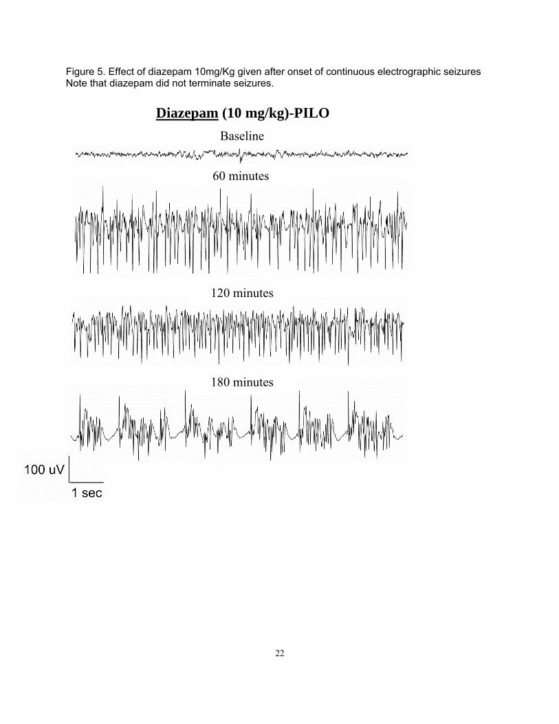
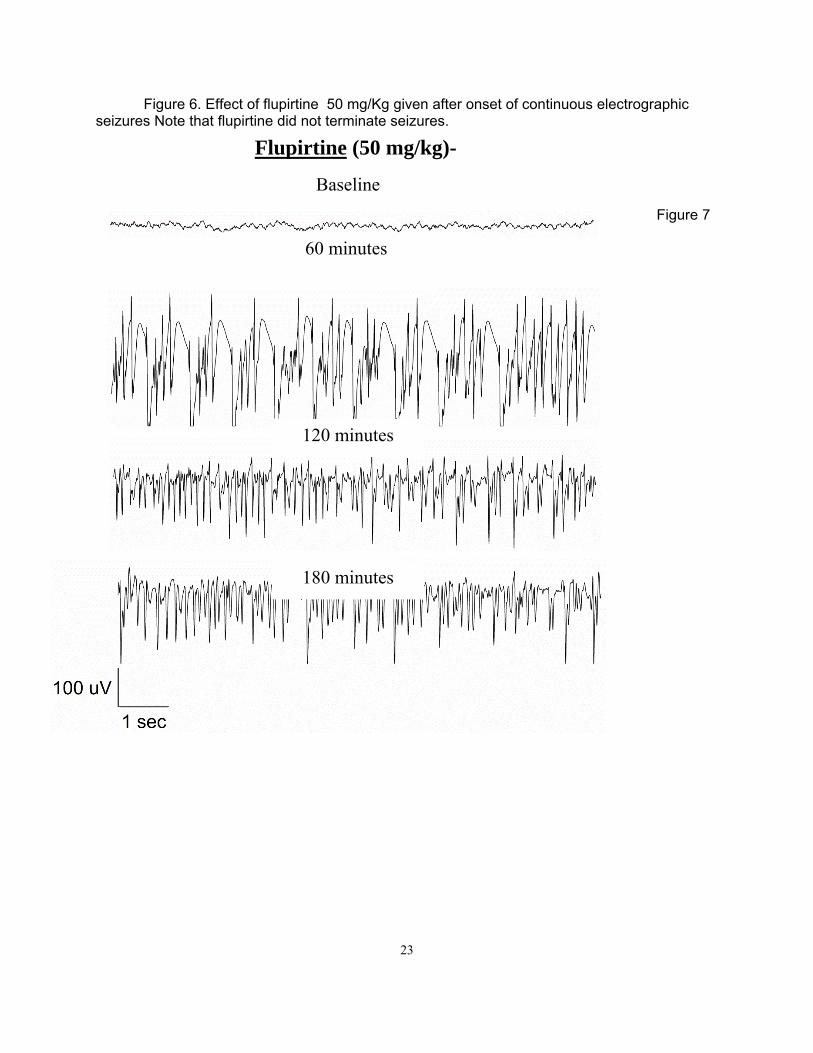
Materials and Methods Surgery Adult male Sprague-Dawley rats (Taconic) weighing 250-350 g were housed with food and water ad libitum. A bipolar insulated stainless steel electrode was implanted stereotaxically, under ketamine/xylazine anesthesia, in the over the cortex. The assembly was secured to the skull with dental acrylic, as previously described (Lothman et al. 1988). After a 5-7 day recovery period, the rats were administered 3 mmol/kg lithium chloride intraperitoneally (i.p.) 20 h later, SE was induced by i.p. injection of 50 mg/kg pilocarpine. Thirty minutes prior to pilocarpine administration, 2 mg/kg scopolamine was given to each rat to reduce the peripheral effects of the pilocarpine. SE was induced by continuous hippocampal stimulation (CHS) as previously described (Lothman et al., 1989; Borris et al., 2000) by stimulating the left ventral hippocampus with 10s trains of 50 Hz, 1 ms, 400 mA biphasic square wave current pulses delivered every 13 s for 90 min. The protocol to induce SE by DFP was previously reported (Todorovic, 2012). Briefly, 30 min after administration of atropine (2 mg/kg s.c, Sigma, St. Louis, MO) and 2 pralidoxime iodide (2-PAM, 50 mg/kg i.p, Sigma, St. Louis, MO), DFP (10 mg/kg s.c, Sigma, St. Louis, MO) was injected. Both 2-PAM and atropine were dissolved in solution within an hour of injection. DFP was mixed into cold saline immediately prior to injection. EEG activity was recorded continuously for at least 18 hrs following drug injection to determine the effect of the drug on prolonged SE. SE was considered terminated when the EEG returned to normal baseline or showed irregular spikes without recurrence of seizures in a subsequent observation period of five hours. Behavioral seizures were considered terminated when there was cessation of behavioral seizures and resumption of exploratory behavior. In some animals end of behavioral seizures was accompanied by sedation where animal lay still in the cage. Results Lithium-pilocarpine (PILO)-induced ESE All the animals used in the study demonstrated continuous SE for at least 4 hrs from the onset of continuous electrographic discharges, with seizures lasting 9 hrs or more in some animals (Figure 5). The mean duration to SE onset following PILO administration was 26.19 ± 1.7 min (n = 35 [or 28.54 ± 4.96 for n=5] animals). Electrographically, all the animals demonstrated continuous SE for at least 4 hrs and the EEG features of SE were similar to those described previously 1,2. Animals exhibited wet dog shakes, facial twitching and automatisms, chewing, staring, hind limb scratching, head bobbing, forelimb clonus, rearing, and rearing and falling with generalized convulsions. Animals spent most of their time exhibiting stage 3-5 seizures, briefly interrupted by stage 2 behaviors. Treatment was initiated 10 min after continuous electrographic seizures because previous studies demonstrated refractoriness to DIA at this time point 2. Behaviorally, this corresponds to a time point 10 minutes after first stage 5 seizure 3. Five rats, treated with normal saline 10 minutes after continuous electrographic seizure, continued to exhibit continuous electrographic seizures for the next 4 hours. The mean duration of SE in these animals was 6.99 ± 1.2 h. Refractoriness to DIA (ESE) was confirmed in another experiment when animals treated with 10 mg/kg DIA 10 minutes after the onset of continuous electrographic seizure continued to have seizures for the next 3 h (4.12 ± 0.8 h, n = 5, p > 0.05 compared to untreated animals, Figure 5B). In a previous study of neonatal SE induced by kainic acid-induced SE, 50 mg/kg FLU controlled SE 4. Therefore, we first tested whether 50 mg/kg dose of FLU would control PILO-induced ESE. The time-course of electrographic seizure control was studied at 15 min intervals in the first hour after treatment and on an hourly interval for the subsequent 4 hrs (Figure 6). FLU (50

9
mg/kg) failed to terminate seizures in any of the 5 animals tested and the mean duration of SE in these animals was 7.86 ± 1.5 h (p > 0.05 compared to untreated animals (Figure 6). We then tested whether a higher dose of FLU could terminate ESE. FLU (75 mg/kg) also failed to terminate seizures in any animal tested (n = 6) and the mean SE duration in this group was 8.07 ± 1.8 h (p > 0.05 compared to untreated animals). Thus, FLU treatment per se could not treat PILO-induced ESE. Combination therapy with 10 mg/kg diazepam Since FLU administration in the neonatal SE study was performed when DIA transiently controlled seizures 4, we next tested whether a combination of DIA with FLU could control PILO-induced ESE. Animals (n = 5) were treated 10 minutes after 10 min of continuous electrographic seizures with a combination of DIA (10 mg/kg) and FLU (50 mg/kg). Combination therapy of DIA and FLU terminated SE in all animals tested (n = 5). The mean duration of SE in this group (1.34 ± 0.92 h) was significantly less than in animals that were untreated, or treated with FLU alone at 50 or 75 mg/kg (p < 0.05, Tukey’s multiple comparison test, Figure 1D). Further, the time-course analysis of EEG changes revealed that in 3 out of 5 animals (60 %), combination therapy terminated SE within 1 hr (27 ± 21.6 min) and animals did not revert to SE in the subsequent observation period. We then tested whether DIA (10 mg/kg) in combination with a lower dose of FLU (25 mg/kg) could terminate PILO-induced ESE. This combination terminated SE in all animals treated (n = 5) within 5 h and the mean duration of SE was 3.13 ± 0.5 h (p > 0.05 Tukey’s multiple comparison test, Figure 7,8 ) (Note that unpaired t shows a p value of 0.01). Analysis of time-course change in EEG seizures revealed that SE was not terminated in any animal within an hour. When DIA (10mg/kg) was co-administered in combination with a higher dose of FLU (75 mg/kg), all 4 animals tested died within 15.6 ± 2.8 min. Thus combination treatment with FLU (25 or 50 mg/kg) and DIA (10 mg/kg) could terminate PILO-induced ESE (figure 8). Diisopropylfluorophosphate (DFP)-induced ESE We next tested the efficacy of FLU (50 mg/kg), alone or in combination with DIA (10 mg/kg) to terminate DFP-induced ESE. As described previously 5, 30 min after treatment with 2-PAM (50 mg/kg, s.c) and atropine (2 mg/kg, s.c), adult male rats were injected with DFP (10 mg/kg, i.p). The mean time to onset of electrographic SE was 18.5 ± 6.5 min (n=5) and the duration of SE was 8.56 ± 1.5 h. Treatment with DIA (10 mg/kg) 10 min following continuous electrographic seizures terminated seizures in only 3/5 animals (60%). When DIA was administered 30 min after the start of continuous EEG activity, SE did not terminate within 60 and 120 minutes of seizure onset in any of the 5 animals tested. First termination of SE in these animals first occurred 2.5 hours after treatment injection, and 60% (3/5) of rats came out of SE within the first 5 hours of DIA injection. Further, though DIA treatment at 30 minutes eventually terminated SE, the termination was marked by appearance arrhythmic spikes and the baseline EEG was not restored in any animal. In contrast to DIA, treatment with FLU (50 mg/kg) terminated SE in all animals tested (n=7), and the duration of SE was 1.71 ± 0.3 h (p < 0.05 compared to untreated animals, Tukey’s multiple comparison test). However, only 1/6 animals (16.7%) was seizure free within 60 min of SE onset. Combined treatment with DIA (10 mg/kg) and FLU (50 mg/kg) was highly effective in terminating DFP-induced SE. In 100% of animals (n = 9), DIA+FLU rapidly terminated SE; the mean duration of seizures was 18.6 ± 3.6 min (p < 0.05 compared to untreated, DIA alone, FLU alone, Tukey’s multiple comparison test,). Thus, FLU in combination with DIA is highly effective in terminating DFP-induced ESE.
10
Discussion The principal finding of this study is that a combination of diazepam and flupirtine terminates prolonged both cholinergic stimulation and electrical stimulation induced ESE in a synergistic fashion. Diazepam increased the efficacy of flupirtine. Molecular cloning of various genes for K+ channels, identification of channel gene mutations that lead to human benign familial neonatal convulsions (BFNC), and reconstitution studies suggest that M-current is mediated by KCNQ2/KCNQ3 channels6-8. Mutations in KCNQ2 channels are associated with BFNC9-12. Drugs that block M-currents, linoperidine and XE 991 cause seizures in hippocampal slices 13. KCNQ2 and KCNQ3 are the predominant KCNQ channels in the brain where they activate at subthreshold membrane potentials and generate a voltage-dependent outward current near resting membrane potential (Delmas and Brown, 2005). The high expression of KCNQ2/KCNQ3 channels at the axon initial segment – the site of action potential generation – allows the channel to limit neuronal excitability and play a critical role for these channels in modulation of burst generation and after-hyperpolarization and transmitter release (Yue and Yaari, 2004; Devaux et al., 2004, Chung et al., 2006, Pan et al., 2006,). There is also evidence for the expression of M-channels in presynaptic terminals (Cooper et al. 2001; Chung et al. 2006; Garcia-Pino et al. 2010) suggesting a role for these channels in modulating neurotransmitter release (Martire et al. 2004, Vervaeke et al., 2006). We recently demonstrated that at Schaffer collateral–CA1 pyramidal neuron synapses, M1 muscarinic activation or M-channel inhibition, enhanced action-potential-independent glutamate release (Sun and Kapur, 2013). The finding that flupirtine in combination with diazepam could potently terminate SE induced in multiple models, including surrogate nerve agent compounds, suggests that in addition to reducing presynaptic glutamate release, flupirtine likely enhanced the strength of inhibitory neurotransmission mediated by GABA-A receptors (Otto et al., 2002). These findings are clinically significant and raise the possibility of using ketamine as an adjunct to diazepam for the treatment of patients with refractory SE. Ketamine was initially introduced as an anesthetic but its undesired psychic effects have limited its use as an anesthetic. More recently, the drug has found use as an analgesic in a hospice setting and in the perioperative period (Bell et al. 2006;Legge et al. 2006). There are case reports of ketamine use to treat SE (Bleck et al. 2002;Mewasingh et al. 2003;Ubogu et al. 2003). However, it is difficult to compare clinical studies with those performed in experimental animals, because the range of doses of ketamine that were used in clinical studies may not be pharmacologically equivalent to those used in experimental animals. Once appropriate human dose range been determined, therapeutic efficacy and safety of ketamine in terminating refractory SE in humans should be determined in a prospective randomized clinical trial. Key Research Accomplishments:
We demonstrated that muscarinic stimulation causes enhanced synaptic transmission at Schaffer collateral CA1 pyramidal neuron synapses in the hippocampus.
We developed two models of organophosphate induced seizures and status epilepticus.
We tested potassium channel opener flupirtine in an organophosphate model and found it to be effective in terminating status epilepticus when combined with diazepam.

11
REPORTABLE OUTCOMES
Conclusions: These studies demonstrate that muscarinic stimulation of hippocampal neurons
increases their excitability and enhances neurotransmitter release at Schaffer collateral-CA1 synapses. This enhancement occurs by inhibition of M-type (KCNQ2/3) potassium channels. Drugs that open M –type potassium channels can counter the effect of muscarinic stimulation in hippocampal slices. Drugs that open M type channels were effective in terminating organophosphate-induced status epilepticus.
12
References
Allen, T. G. & Brown, D. A. (1993). M2 muscarinic receptor-mediated inhibition of the Ca2+ current in rat magnocellular cholinergic basal forebrain neurones. J Physiol 466, 173-189.
Bernheim, L., Mathie, A., & Hille, B. (1992). Characterization of muscarinic receptor subtypes inhibiting Ca2+ current and M-current in rat sympathetic neurons. Proc Natl Acad Sci U S A 89, 9544-9548.
Biervert, C., Schroeder, B.C., Kubisch, C., Berkovic, S. F., Propping, P., Jentsch, T. J., & Steinlein, O. K. (1998). A potassium channel mutation in neonatal human epilepsy. Science 279, 403-406.
Bouron, A. & Reuter, H. (1997). Muscarinic stimulation of synaptic activity by protein kinase C is inhibited by adenosine in cultured hippocampal neurons. Proc Natl Acad Sci U S A 94, 12224-12229.
Brown, D. A. & Adams, P. R. (1980). Muscarinic suppression of a novel voltage-sensitive K+ current in a vertebrate neurone. Nature 283, 673-676.
Brown, D. A. & Passmore, G. M. (2009). Neural KCNQ (Kv7) channels. Br J Pharmacol 156, 1185-1195.
Chu-Shore, C. J. & Thiele, E. A. (2010). New drugs for pediatric epilepsy. Semin Pediatr Neurol 17, 214-223.
Chung, H. J., Jan, Y. N., & Jan, L. Y. (2006). Polarized axonal surface expression of neuronal KCNQ channels is mediated by multiple signals in the KCNQ2 and KCNQ3 C-terminal domains. Proc Natl Acad Sci U S A 103, 8870-8875.
Cobb, S. R. & Davies, C. H. (2005). Cholinergic modulation of hippocampal cells and circuits. J Physiol 562, 81-88.
Cole, A. E. & Nicoll, R. A. (1983). Acetylcholine mediates a slow synaptic potential in hippocampal pyramidal cells. Science 221, 1299-1301.
Colino, A. & Halliwell, J. V. (1993). Carbachol potentiates Q current and activates a calcium-dependent non-specific conductance in rat hippocampus in vitro. Eur J neurosci 5, 1198-1209.
Constanti, A. & Brown, D. A. (1981). M-currents in voltage-clamped mammalian sympathetic neurones. Neurosci Lett 24, 289-294.
Cooper, E. C., Harrington, E., Jan, Y. N., & Jan, L. Y. (2001). M-channel KCNQ2 subunits are localized to key sites for control of neuronal network oscillations and synchronization in mouse brain. J Neurosci 21, 9529-9540.
Delmas, P. & Brown, D. A. (2005). Pathways modulating neural KCNQ/M (Kv7) potassium channels. Nat Rev Neurosci 6, 850-862.

13
Devaux, J. J., Kleopa, K. A., Cooper, E. C., & Scherer, S. S. (2004). KCNQ2 is a nodal K+ channel. J Neurosci 24, 1236-1244.
Dutar, P., Bassant, M. H., Senut, M. C., & Lamour, Y. (1995). The septohippocampal pathway: structure and function of a central cholinergic system. Physiol Rev 75, 393-427.
Emptage, N. J., Reid, C. A., & Fine, A. (2001). Calcium stores in hippocampal synaptic boutons mediate short-term plasticity, store-operated Ca2+ entry, and spontaneous transmitter release. Neuron 29, 197-208.
Fernandez de Sevilla, D., Núnñez, A., Borde, M., Malinow, R., & Buño W. (2008). Cholinergic-mediated IP3-receptor activation induces long-lasting synaptic enhancement in CA1 pyramidal neurons. J Neurosci 28, 1469-1478.
Frotscher, M. & Léránth, C. (1985). Cholinergic innervation of the rat hippocampus as revealed by choline acetyltransferase immunocytochemistry: a combined light and electron microscopic study. J Comp Neurol 239, 237-246.
Garcia-Pino, E., Caminos, E., & Juiz, J. M. (2010). KCNQ5 reaches synaptic endings in the auditory brainstem at hearing onset and targeting maintenance is activity-dependent. J Comp Neurol 518, 1301-1314.
Geiger, J., Weber, Y. G., Landwehrmeyer, B., Sommer, C., & Lerche, H. (2006). Immunohistochemical analysis of KCNQ3 potassium channels in mouse brain. Neurosci Lett 400, 101-104.
Giessel, A. J. & Sabatini, B. L. (2010). M1 muscarinic receptors boost synaptic potentials and calcium influx in dendritic spines by inhibiting postsynaptic SK channels. Neuron 68, 936-947.
Gu, N., Vervaeke, K., Hu, H., & Storm, J. F. (2005). Kv7/KCNQ/M and HCN/h, but not KCa2/SK channels, contribute to the somatic medium after-hyperpolarization and excitability control in CA1 hippocampal pyramidal cells. J. Physiol 566, 689-715.
Halliwell, J. V. (1990). Physiological mechanisms of cholinergic action in the hippocampus. Prog Brain Res 84, 255-272.
Hamilton, S. E., Loose, M. D., Qi, M., Levey, A. I., Hille, B., McKnight, G. S., Idzerda, R. L., & Nathanson, N. M. (1997). Disruption of the M1 receptor gene ablates muscarinic receptor-dependent M-current regulation and seizure activity in mice. Proc.Natl.Acad.Sci.U.S.A 94, 13311-13316.
14
Hernandez, C. C., Zaika, O., Tolstykh, G. P., & Shapiro, M. S. (2008). Regulation of neural KCNQ channels: signalling pathways, structural motifs and functional implications. J Physiol 586, 1811-1821.
Higley, M. J., Soler-Llavina, G. J., & Sabatini, B. L. (2009). Cholinergic modulation of multivesicular release regulates striatal synaptic potency and integration. Nat Neurosci 12, 1121-1128.
Jentsch, T. J. (2000). Neuronal KCNQ potassium channels:physislogy and role in disease. Nat Rev Neurosci 1, 21-30.
Kanaumi, T., Takashima, S., Iwasaki, H., Itoh, M., Mitsudome, A., & Hirose, S. (2008). Developmental changes in KCNQ2 and KCNQ3 expression in human brain: possible contribution to the age-dependent etiology of benign familial neonatal convulsions. Brain Dev 30, 362-369.
Kozhemyakin, M., Rajasekaran, K., & Kapur, J. (2010). Central cholinesterase inhibition enhances glutamatergic synaptic transmission. J Neurophysiol 103, 1748-1757.
Langmead, C. J., Watson, J., & Reavill, C. (2008). Muscarinic acetylcholine receptors as CNS drug targets. Pharmacol Ther 117, 232-243.
Léránth, C. & Frotscher, M. (1987). Cholinergic innervation of hippocampal GAD- and somatostatin-immunoreactive commissural neurons. J Comp Neurol 261, 33-47.
Madison, D. V., Lancaster, B., & Nicoll, R. A. (1987). Voltage clamp analysis of cholinergic action in the hippocampus. J Neurosci 7, 733-741.
Marino, M. J., Rouse, S. T., Levey, A. I., Potter, L. T., & Conn, P. J. (1998). Activation of the genetically defined M1 muscarinic receptor potentiates N-methyl-d-aspartate (NMDA) receptor currents in hippocampal pyramidal cells. Proc.Natl.Acad.Sci.U.S.A 95, 11465-11470.
Markram, H., & Segal, M. (1990). Acetylcholine potentiates responses to N-methyl-d-aspartate in the rat hippocampus. Neurosci Lett 113, 62-65.
Marrion, N. V., Smart, T. G., Marsh, S. J., & Brown, D. A. (1989). Muscarinic suppression of the M-Current in the rat sympathetic-ganglion is mediated by receptors of the M1-subtype. Br J Pharmacol 98, 557-573.
15
Martire, M., Castaldo, P., D'Amico, M., Preziosi, P., Annunziato, L., & Taglialatela, M. (2004). M- channels containing KCNQ2 subunits modulate norepinephrine, aspartate, and GABA release from hippocampal nerve terminals. J Neurosci 24, 592-597.
Morton, R. A. & Davies, C. H. (1997). Regulation of muscarinic acetylcholine receptor-mediated synaptic responses by adenosine receptors in the rat hippocampus. J Physiol 502, 75-90.
Pan, Z., Kao, T., Horvath, Z., Lemos, J., Sul, J. Y., Cranstoun, S. D., Bennett, V., Scherer, S. S., & Cooper, E. C. (2006). A common ankyrin-G-based mechanism retains KCNQ and NaV channels at electrically active domains of the axon. J Neurosci 26, 2599-2613.
Peretz, A., Sheinin, A., Yue, C., Degani-Katzav, N., Gibor, G., Nachman, R., Gopin, A., Tam, E., Shabat, D., Yaari, Y., & Attali, B. (2007). Pre- and postsynaptic activation of M-channels by a novel opener dampens neuronal firing and transmitter release. J Neurophysiol 97, 283-295.
Pitler, T. A. & Alger, B. E. (1990). Activation of the pharmacologically defined M3 muscarinic receptor depolarizes hippocampal pyramidal cells. Brain Res 534, 257-262.
Power, J. M. & Sah, P. (2002). Nuclear calcium signaling evoked by cholinergic stimulation in hippocampal CA1 pyramidal neurons. J Neurosci 22, 3454-3462.
Qian, J. & Noebels, J. L. (2000). Presynaptic Ca2+ influx at a mouse central synapse with Ca2+ channel subunit mutations. J Neurosci 20, 163-170.
Qian, J. & Saggau, P. (1997). Presynaptic inhibition of synaptic transmission in the rat hippocampus by activation of muscarinic receptors: involvement of presynaptic calcium influx. Br J Pharmacol 122, 511-519.
Raol, Y. H., Lapides, D. A., Keating, J. G., Brooks-Kayal, A. R., & Cooper, E. C. (2009). A KCNQ channel opener for experimental neonatal seizures and status epilepticus. Ann Neurol 65, 326-336.
Rasmussen, H. B., Frøkjær-Jensen, C., Jensen, C. S., Jensen, H. S., Jørgensen, N. K., Misonou, H., Trimmer, J. S., Olesen, S.P., & Schmitt, N. (2007). Requirement of subunit co-assembly and ankyrin-G for M-channel localization at the axon initial segment. J Cell Sci 120, 953-963.
Safiulina, V. F., Zacchi, P., Taglialatela, M., Yaari, Y., & Cherubini, E. (2008). Low expression of Kv7/M channels facilitates intrinsic and network bursting in the developing rat hippocampus. J Physiol 586, 5437-5453.
Segal, M. (1988). Synaptic activation of a cholinergic receptor in rat hippocampus. Brain Res 452, 79-86.

16
Selyanko, A. A., Delmas, P., Hadley, J. K., Tatulian, L., Wood, I. C., Mistry, M., London, B., & Brown, D. A. (2002). Dominant-negative subunits reveal potassium channel families that contribute to M-like potassium currents. J. Neurosci 22, RC212.
Shah, M. M., Migliore, M., Valencia, I., Cooper, E. C., & Brown, D. A. (2008). Functional significance of axonal Kv7 channels in hippocampal pyramidal neurons. Proc Natl Acad Sci U S A 105, 7869-7874.
Shah, M. M., Mistry, M., Marsh, S. J., Brown, D. A., & Delmas, P. (2002). Molecular correlates of the M-current in cultured rat hippocampal neurons. J Physiol 544, 29-37.
Shen, W., Hamilton, S. E., Nathanson, N. M., & Surmeier, D. J. (2005). Cholinergic suppression of KCNQ channel currents enhances excitability of striatal medium spiny neurons. J Neurosci 25, 7449-7458.
Sim, J. A. & Griffith, W. H. (1996). Muscarinic Inhibition of glutamatergic transmission onto rat magnocellular basal forebrain neurons in a thin-slice preparation. Eur J Neurosci 8, 880-891.
Smolders, I., Khan, G. M., Manil, J., Ebinger, G., & Michotte, Y. (1997). NMDA receptor-mediated pilocarpine-induced seizures: characterization in freely moving rats by microdialysis. Br J Pharmacol 121, 1171-1179.
Stephen, L. J. & Brodie, M. I. (2011). Pharmacotherapy of epilepsy newly approved and developmental agents. CNS Drugs 25, 89-107.
Tayebati, S. K., Amenta, F., El-Assouad, D., & Zaccheo, D. (2002). Muscarinic cholinergic receptor subtypes in the hippocampus of aged rats. Mech of Ageing Dev 123, 521-528.
Toselli, M., Lang, J., Costa, T., & Lux, H. D. (1989). Direct modulation of voltage-dependent calcium channels by muscarinic activation of a pertussis toxin-sensitive G-protein in hippocampal neurons. Pflugers Arch 415, 255-261.
Vervaeke, K., Gu, N., Agdestein, C., Hu, H., & Storm, J. F. (2006). Kv7/KCNQ/M-channels in rat glutamatergic hippocampal axons and their role in regulation of excitability and transmitter release. J Physiol 576, 235-256.
Vogt, K. E. & Regehr, W. G. (2001). Cholinergic modulation of excitatory synaptic transmission in the CA3 area of the hippocampus. J.Neurosci 21, 75-83.
Volpicelli, L. A. & Levey, A. I. (2004). Muscarinic acetylcholine receptor subtypes in cerebral cortex and hippocampus. Prog Brain Res 145, 59-66
17
Wang, H. S., Pan, Z., Shi, W., Brown, B. S., Wymore, R. S., Cohen, I. S., Dixon, J. E., & McKinnon, D. (1998). KCNQ2 and KCNQ3 potassium channel subunits: molecular correlates of the M-channel. Science 282, 1890-1893.
Weber, Y. G., Geiger, J., Kämpchen, K., Landwehrmeyer, B., Sommer, C., & Lerche, H. (2006). Immunohistochemical analysis of KCNQ2 potassium channels in adult and developing mouse brain. Brain Res 1077, 1-6.
Wheeler, D. B., Randall, A., & Tsien, R. W. (1994). Roles of N-type and Q-type Ca2+ channels in supporting hippocampal synaptic transmission. Science 264, 107-111.
Wolff, C., Gillard, M., Fuks, B., & Chatelain, P. (2005). [3H]linopirdine binding to rat brain membranes is not relevant for M-channel interaction. Eur J Pharmacol 518, 10-17.
Wu, Y. J. & Dworetzky, S. I. (2005). Recent developments on KCNQ potassium channel openers. Curr Med Chem 12, 453-460.
Yajeya, J., De La Fuente, A., Criado, J. M., Bajo, V., Sánchez-Riolobos, A., & Heredia, M. (2000). Muscarinic agonist carbachol depresses excitatory synaptic transmission in the rat basolateral amygdala in vitro. Synapse 38, 151-160.
Yue, C. & Yaari, Y. (2004). KCNQ/M Channels control spike afterdepolarization and burst generation in hippocampal neurons. J Neurosci 24, 4614-4624.
Zhang, H. M., Chen, S. R., & Pan, H. L. (2007). Regulation of glutamate release from primary afferents and interneurons in the spinal cord by muscarinic receptor subtypes. J. Neurophysiol. 97, 102-109.

18
Appendices Figure 1-17 & PDF reprints of two publications
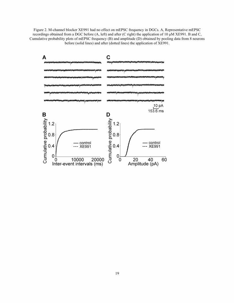
Figure1. M1 muscarinic had no effect on the frequency of mEPSCs recorded from Dentate granule cells (DGCs). A, Representative traces of mEPSCs recorded from a DGC before (left) and after (right) the application of 10 μM McN-A-343. B, Representative averaged traces of mEPSCs before (black) and during (grey) the application of McN-A-343 along with the overlay of the two traces. C and D, Cumulative probability plots of mEPSC frequency (C) and amplitude (D) obtained by pooling data from neurons before (solid lines) and after (dotted lines) application of McN-A-343.
19
Figure 2. M-channel blocker XE991 had no effect on mEPSC frequency in DGCs. A, Representative mEPSC recordings obtained from a DGC before (A, left) and after (C right) the application of 10 μM XE991. B and C,
Cumulative probability plots of mEPSC frequency (B) and amplitude (D) obtained by pooling data from 8 neurons before (solid lines) and after (dotted lines) the application of XE991.
20
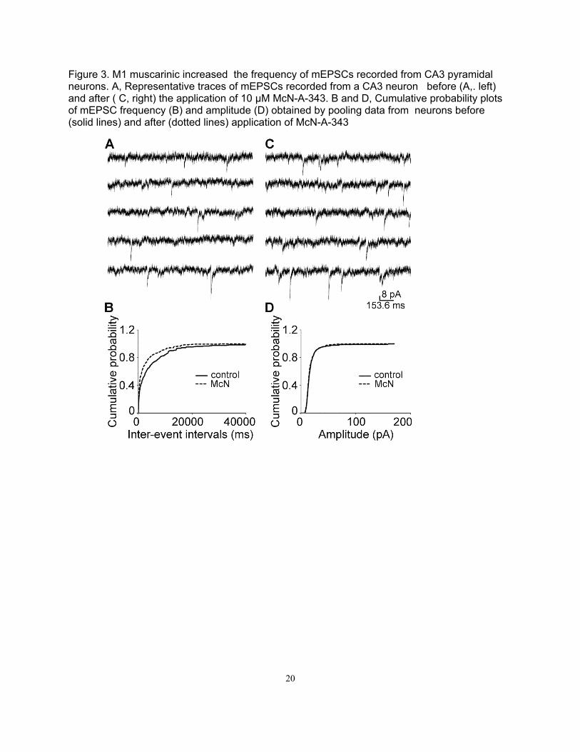
Figure 3. M1 muscarinic increased the frequency of mEPSCs recorded from CA3 pyramidal neurons. A, Representative traces of mEPSCs recorded from a CA3 neuron before (A,. left) and after ( C, right) the application of 10 μM McN-A-343. B and D, Cumulative probability plots of mEPSC frequency (B) and amplitude (D) obtained by pooling data from neurons before (solid lines) and after (dotted lines) application of McN-A-343

21
Figure 4. M channel antagonist increased the frequency of mEPSCs recorded from CA3 pyramidal neurons. A, Representative traces of mEPSCs recorded from a CA3 neuron before (A,left) and after (C, right) the application of 10 μM XE 991. B and D, Cumulative probability plots of mEPSC frequency (B) and amplitude (D) obtained by pooling data from neurons before (solid lines) and after (dotted lines) application of XE 991.
22
Figure 5. Effect of diazepam 10mg/Kg given after onset of continuous electrographic seizures Note that diazepam did not terminate seizures.
Diazepam (10 mg/kg)-PILO
Baseline
60 minutes
120 minutes
180 minutes
23
Figure 6. Effect of flupirtine 50 mg/Kg given after onset of continuous electrographic seizures Note that flupirtine did not terminate seizures.
Figure 7
Flupirtine (50 mg/kg)-
Baseline
60 minutes
120 minutes
180 minutes
24
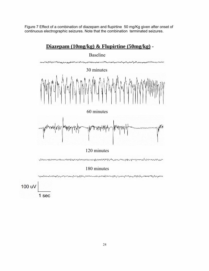
Figure 7 Effect of a combination of diazepam and flupirtine 50 mg/Kg given after onset of continuous electrographic seizures. Note that the combination terminated seizures.
Diazepam (10mg/kg) & Flupirtine (50mg/kg) -
Baseline
30 minutes
60 minutes
120 minutes
180 minutes
25
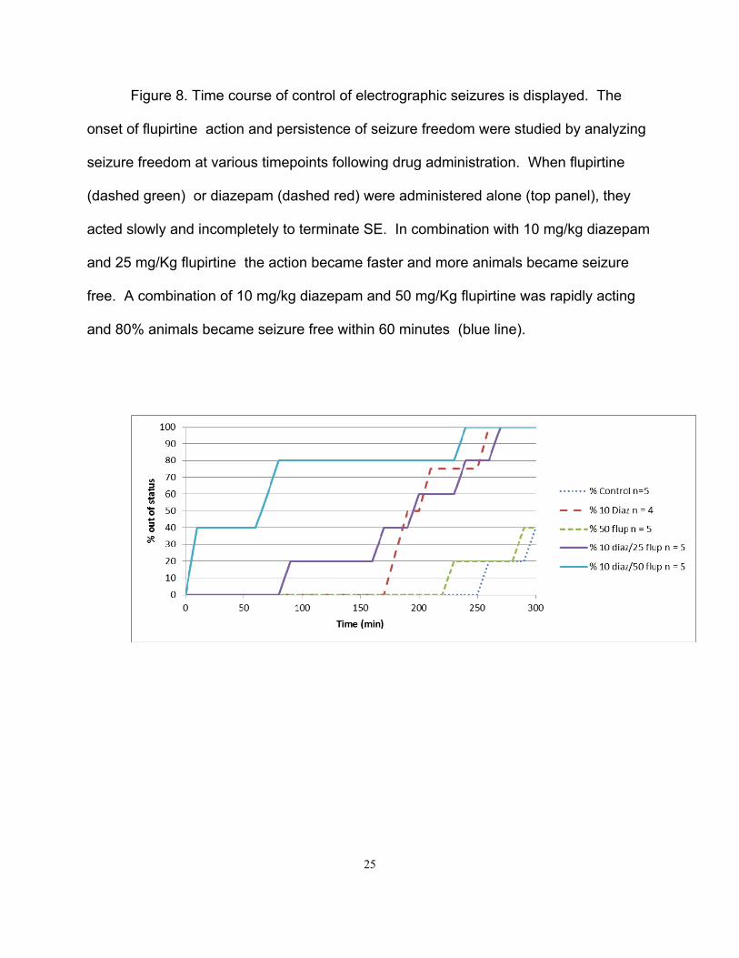
Figure 8. Time course of control of electrographic seizures is displayed. The
onset of flupirtine action and persistence of seizure freedom were studied by analyzing
seizure freedom at various timepoints following drug administration. When flupirtine
(dashed green) or diazepam (dashed red) were administered alone (top panel), they
acted slowly and incompletely to terminate SE. In combination with 10 mg/kg diazepam
and 25 mg/Kg flupirtine the action became faster and more animals became seizure
free. A combination of 10 mg/kg diazepam and 50 mg/Kg flupirtine was rapidly acting
and 80% animals became seizure free within 60 minutes (blue line).
26
References
1. Treiman, DM, Walton, NY, and Kendrick, C. A progressive sequence of electroencephalographic changes
during generalized convulsive status epilepticus. Epilepsy Res. 1990; 5:49-60.
2. Wang, NC, Good, LB, Marsh, ST, et al. EEG stages predict treatment response in experimental status
epilepticus. Epilepsia. 2009; 50:949-952.
3. Martin, BS and Kapur, J. A combination of ketamine and diazepam synergistically controls refractory status
epilepticus induced by cholinergic stimulation. Epilepsia. 2008; 49:248-255.
4. Raol, YH, Lapides, DA, Keating, JG, et al. A KCNQ channel opener for experimental neonatal seizures and
status epilepticus. Ann Neurol. 2009; 65:326-336.
5. Todorovic, MS, Cowan, ML, Balint, CA, et al. Characterization of status epilepticus induced by two
organophosphates in rats. Epilepsy Res. 2012; 101:268-276.
6. Shapiro, MS, Roche, JP, Kaftan, EJ, et al. Reconstitution of muscarinic modulation of the KCNQ2/KCNQ3
K(+) channels that underlie the neuronal M current. J Neurosci. 2000; 20:1710-1721.
7. Roche, JP, Westenbroek, R, Sorom, AJ, et al. Antibodies and a cysteine-modifying reagent show
correspondence of M current in neurons to KCNQ2 and KCNQ3 K+ channels. Br J Pharmacol.
2002; 137:1173-1186.
8. Cooper, EC and Jan, LY. M-channels: neurological diseases, neuromodulation, and drug development. Arch
Neurol. 2003; 60:496-500.
9. Singh, NA, Charlier, C, Stauffer, D, et al. A novel potassium channel gene, KCNQ2, is mutated in an
inherited epilepsy of newborns. Nat Genet. 1998; 18:25-29.
10. Singh, NA, Westenskow, P, Charlier, C, et al. KCNQ2 and KCNQ3 potassium channel genes in benign
familial neonatal convulsions: expansion of the functional and mutation spectrum. Brain. 2003;
126:2726-2737.
27
11. Borgatti, R, Zucca, C, Cavallini, A, et al. A novel mutation in KCNQ2 associated with BFNC, drug resistant
epilepsy, and mental retardation. Neurology. 2004; 63:57-65.
12. Biervert, C, Schroeder, BC, Kubisch, C, et al. A potassium channel mutation in neonatal human epilepsy.
Science. 1998; 279:403-406.
13. Qiu, C, Johnson, BN, and Tallent, MK. K+ M-current regulates the transition to seizures in immature and
adult hippocampus. Epilepsia. 2007; 48:2047-2058.
J Physiol 00.0 (2012) pp 1–12 1
The
Jou
rnal
of
Phys
iolo
gy
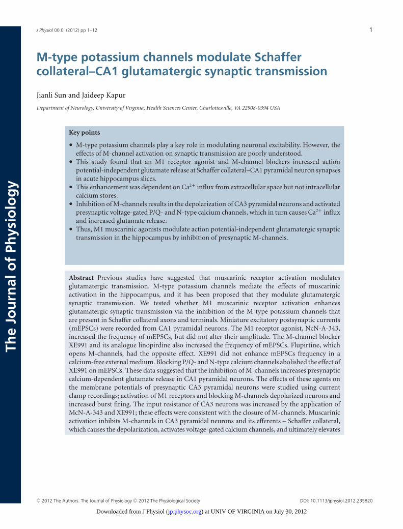
M-type potassium channels modulate Schaffercollateral–CA1 glutamatergic synaptic transmission
Jianli Sun and Jaideep Kapur
Department of Neurology, University of Virginia, Health Sciences Center, Charlottesville, VA 22908-0394 USA
Key points
• M-type potassium channels play a key role in modulating neuronal excitability. However, theeffects of M-channel activation on synaptic transmission are poorly understood.
• This study found that an M1 receptor agonist and M-channel blockers increased actionpotential-independent glutamate release at Schaffer collateral–CA1 pyramidal neuron synapsesin acute hippocampus slices.
• This enhancement was dependent on Ca2+ influx from extracellular space but not intracellularcalcium stores.
• Inhibition of M-channels results in the depolarization of CA3 pyramidal neurons and activatedpresynaptic voltage-gated P/Q- and N-type calcium channels, which in turn causes Ca2+ influxand increased glutamate release.
• Thus, M1 muscarinic agonists modulate action potential-independent glutamatergic synaptictransmission in the hippocampus by inhibition of presynaptic M-channels.
Abstract Previous studies have suggested that muscarinic receptor activation modulatesglutamatergic transmission. M-type potassium channels mediate the effects of muscarinicactivation in the hippocampus, and it has been proposed that they modulate glutamatergicsynaptic transmission. We tested whether M1 muscarinic receptor activation enhancesglutamatergic synaptic transmission via the inhibition of the M-type potassium channels thatare present in Schaffer collateral axons and terminals. Miniature excitatory postsynaptic currents(mEPSCs) were recorded from CA1 pyramidal neurons. The M1 receptor agonist, NcN-A-343,increased the frequency of mEPSCs, but did not alter their amplitude. The M-channel blockerXE991 and its analogue linopirdine also increased the frequency of mEPSCs. Flupirtine, whichopens M-channels, had the opposite effect. XE991 did not enhance mEPSCs frequency in acalcium-free external medium. Blocking P/Q- and N-type calcium channels abolished the effect ofXE991 on mEPSCs. These data suggested that the inhibition of M-channels increases presynapticcalcium-dependent glutamate release in CA1 pyramidal neurons. The effects of these agents onthe membrane potentials of presynaptic CA3 pyramidal neurons were studied using currentclamp recordings; activation of M1 receptors and blocking M-channels depolarized neurons andincreased burst firing. The input resistance of CA3 neurons was increased by the application ofMcN-A-343 and XE991; these effects were consistent with the closure of M-channels. Muscarinicactivation inhibits M-channels in CA3 pyramidal neurons and its efferents – Schaffer collateral,which causes the depolarization, activates voltage-gated calcium channels, and ultimately elevates
C© 2012 The Authors. The Journal of Physiology C© 2012 The Physiological Society DOI: 10.1113/jphysiol.2012.235820
) at UNIV OF VIRGINIA on July 30, 2012jp.physoc.orgDownloaded from J Physiol (
2 J. Sun and J. Kapur J Physiol 00.0
the intracellular calcium concentration to increase the release of glutamate on CA1 pyramidalneurons.
(Resubmitted 1 May 2012; accepted after revision 1 June 2012; first published online 6 June 2012)Corresponding author J. Kapur: Department of Neurology, Box 800394, University of Virginia-HSC, Charlottesville,VA 22908, USA. Email: [email protected]
Abbreviations M1, muscarinic type 1; mEPSCs, miniature excitatory postsynaptic currents; CNQX, 6-cyano-7-nitroquinoxalene-2,3-dione; K-S, Kolmogorov–Smirnov; NcN-A-343, (4-hydroxy-2-butynyl)-1-trimethylammonium-3-chlorocarbanilate chloride; sEPSC, spontaneous excitatory postsynaptic current; XE-991,10,10-bis(4-pyridi-nylmethyl)-9(10H)-anthracenone.
Introduction
Muscarinic acetylcholine receptors (mAChRs) areseven-transmembrane-domain G protein-coupledreceptors (GPCRs) that are widely expressed throughoutthe central nervous system. The M1 subtype is the pre-dominant mAChR in the cortex, hippocampus, striatumand thalamus (Langmead et al. 2008). Muscarinic receptoractivation has distinct effects on glutamatergic trans-mission in different neurons. It inhibits glutamatergictransmission in magnocellular neurons of the basalforebrain (Sim & Griffith, 1996), along with neurons inthe basolateral amygdala (Yajeya et al. 2000), striatum(Higley et al. 2009) and spinal cord (Zhang et al. 2007). Inthe CA3 region of the hippocampus, muscarinic activationinhibits associational-commissural synaptic transmissionvia presynaptic calcium channel inhibition; however, itenhances mossy fibre–CA3 pyramidal neuron synaptictransmission (Vogt & Regehr, 2001). The activation ofmuscarinic receptors induces a long-lasting synapticenhancement at Schaffer collateral–CA1 pyramidalneuron synapses by increasing the release of calcium frompostsynaptic endoplasmic reticulum stores both in vivoand in vitro (Fernandez de Sevilla et al. 2008). Muscarinicactivation also enhances glutamatergic transmissionin dentate granule cells (Kozhemyakin et al. 2010).M1 muscarinic receptor activation also inhibits M-typepotassium channels (Brown & Adams, 1980; Marrion et al.1989; Bernheim et al. 1992). M-type potassium channelsbelong to the Kv7 (KCNQ) K+ channel family (Wanget al. 1998; Selyanko et al. 2002). M-channels activate at asubthreshold membrane potential and do not inactivate,so they generate a steady voltage-dependent outwardcurrent near the resting membrane potential (Constanti& Brown, 1981; Delmas & Brown, 2005). Mutations of theKCNQ2 and KCNQ3 genes cause benign familial neonatalconvulsions (BFNC) (Biervert et al. 1998; Jentsch, 2000).The expression of KCNQ channels increases during earlydevelopment in rodent hippocampus (Shah et al. 2002;Geiger et al. 2006; Weber et al. 2006; Safiulina et al. 2008),but the expression of KCNQ2 and KCNQ3 has differentdevelopmental pattern in human brain (Kanaumi et al.2008). It has been suggested that the highest density ofKCNQ2 and KCNQ3 immunoreactivity in the CA1 region
is in the axon initial segments, where action potentialsare generated and the Kv7 channels co-localize with Na+
channels via binding to ankyrin G. This localization allowsM-channels to powerfully limit neuronal excitability(Devaux et al. 2004; Chung et al. 2006; Pan et al. 2006)and therefore function as a ‘brake’ on repetitive firingand play a key role in regulating the excitability ofvarious central and peripheral neurons (Yue & Yaari,2004; Gu et al. 2005; Shen et al. 2005; Brown & Randall,2009). Other studies have suggested that M-channels areexpressed in presynaptic terminals (Cooper et al. 2001;Chung et al. 2006; Garcia-Pino et al. 2010). Physiologicalstudies have suggested that M-channels regulate therelease of neurotransmitters. Drugs that block or openM-channels can regulate presynaptic fibre volley and theevoked EPSPs recorded from CA1 pyramidal neurons(Vervaeke et al. 2006). M-channel blocking by XE991increases the release of noradrenaline, and retigabine,an M-channel opener, decreases noradrenaline releasein cultured sympathetic neurons (Hernandez et al.2008). These drugs also regulate neurotransmitterrelease from hippocampal synaptosomes (Martireet al. 2004) and cultured hippocampal neurons (Peretzet al. 2007). However, the mechanism by whichM-channels regulate neurotransmitter release remainsunclear. The current study demonstrates that both M1muscarinic activation and M-channel inhibition enhanceaction-potential-independent glutamate release inSchaffer collateral–CA1 pyramidal neuron synapses. Thisaction appears to be dependent on the depolarizationof CA3 pyramidal neurons and the activation ofvoltage-gated calcium channels.
Methods
Slice preparation
All studies were performed according to protocols thatwere approved by the University of Virginia AnimalUse and Care Committee, and the US Army MedicalResearch and Material Command Animal Care andUse Review Office (ACURO). Adult male (175–250 g)Sprague–Dawley rats were anaesthetized with isoflurane
C© 2012 The Authors. The Journal of Physiology C© 2012 The Physiological Society
) at UNIV OF VIRGINIA on July 30, 2012jp.physoc.orgDownloaded from J Physiol (

J Physiol 00.0 M-type potassium channels modulate glutamatergic transmission in CA1 neurons 3
prior to decapitation, which was followed by quickremoval of the brain. The removed brains were thensectioned to 300 μm slices using a Leica VT 1200slicer (Leica Microsystems, Wetzlar, Germany) in ice-coldoxygenated slicing solution. The solution contained thefollowing (in mM): 120 sucrose, 65.5 NaCl, 2 KCl, 1.1KH2PO4, 25 NaHCO3, 10 D-glucose, 1 CaCl2, and 5MgSO4. The slices were then incubated for at least 1 hat 32C in oxygenated ACSF that contained (in mM): 127NaCl, 2 KCl, 1.1 KH2PO4, 25.7 NaHCO3, 10 D-glucose, 2CaCl2, and 1.5 MgSO4; the osmolarity in the chamberwas 290–300 mosmol l−1. After incubation, the sliceswere transferred to the recording chamber on the stageof an Olympus Optical BX51 microscope (Olympus,Tokyo, Japan). Unless otherwise stated, all chemicalswere obtained from Sigma-Aldrich (St Louis, MO,USA).
Whole-cell recording
Whole-cell patch-clamp recordings were performed underinfrared differential interference contrast microscopy(Olympus); a 40× water-immersion objective was usedto visually identify CA1 and CA3 pyramidal neurons.The slices were continuously perfused with ACSF solutionthat was saturated with 95% O2 and 5% CO2 at roomtemperature. Patch electrodes (final resistances, 3–5 M)were pulled from borosilicate glass (Sutter Instruments,Novato, CA, USA) on a horizontal Flaming-Brown micro-electrode puller (Model P-97, Sutter Instruments). Forvoltage-clamp recordings, the electrode tips were filledwith a filtered internal recording solution that consistedof the following components (in mM): 117.5 CsMeSO4, 10Hepes, 0.3 EGTA, 15.5 CsCl, and 1.0 MgCl2; the pH was7.3 (with CsOH), and the osmolarity was 310 mosmol l−1.The electrode shank contained (in mM) 4 Mg-ATP salt,0.3 Na-GTP salt, and 5 QX-314. For current-clamprecording, the pipette solution contained the following(in mM): 135 potassium gluconate, 2.5 NaCl, 10 Hepes,0.5 EGTA, 4.0 Mg-ATP, 0.4 Na-GTP, 0.1 CaCl2; the pHwas 7.3, and the osmolarity was 310 mosmol l−1. Neuronswere voltage clamped at −60 mV using a PC-505Bamplifier (Warner Instruments, Hamden, CT, USA).Electrode capacitance was electronically compensated.Access resistance was continuously monitored, and ifthe series resistance increased by 20% at any time,the recording was terminated. Currents were filteredat 2 kHz, digitized using a Digidata 1322 digitizer(Molecular Devices, Sunnyvale, CA, USA), and acquiredusing Clampex 10.2 software (Molecular Devices).Spontaneous excitatory postsynaptic currents (sEPSCs)were recorded from CA1 pyramidal neurons after blockingthe GABAA receptors with the antagonist picrotoxin(50 μM). In preliminary experiments, a combination
of 6-cyano-7-nitroquinoxalene-2,3-dione (CNQX) and2-amino-5-phosphonovaleric acid (APV) blocked allEPSCs. Miniature EPSCs (mEPSCs) were recorded byblocking action potentials with 1 μM TTX (Alomone labs,Jerusalem, Israel). All drugs were bath-applied via a peri-staltic pump.
Data analysis
The offline digitized data were analysed with Mini-Analysis (Synaptosoft, Decatur, GA, USA) and Clampfit10.2 (Molecular Devices). To detect sEPSCs and mEPSCs,a detection threshold was set at three times the root meansquare (RMS) of the baseline noise. After detection, thefrequency and peak amplitude of EPSCs from individualneurons were analysed. Each detected event from the20–30 min recording session was visually inspected toremove false detections. The Kolmogorov–Smirnov (K-S)test was used to compare amplitudes and inter-eventintervals for continuously recorded EPSCs. The inputresistance of CA3 neuron under current-clamp recordingwas analysed by comparing the slope of current–voltagerelationship and measured directly by Clampfit 10.2 atthe peak of membrane potentials. The drug effects werecompared using a paired Student’s t test with a significancelevel of P < 0.05. Data are expressed as means ± SEMunless otherwise noted.
Results
An M1 muscarinic agonist increases mEPSC frequencyin CA1 neurons
Previous studies suggested that muscarinic receptoractivation increased presynaptic glutamate release fromperforant path to dentate granule cells (Kozhemyakin et al.2010). In the present study, we investigated the effectof the M1 muscarinic receptor agonist McN-A-343 onglutamate release recorded from Schaffer collateral–CA1synapses. Application of McN-A-343 (10 μM) increasedthe frequency of mEPSCs that were recorded from CA1pyramidal neurons by 69.89 ± 8.28% (0.12 ± 0.03 Hz vs.0.21 ± 0.05 Hz; n = 7, P < 0.01), but it did not changetheir amplitudes (16.51 ± 1.14 pA vs. 16.79 ± 1.47 pA;n = 7, P = 0.38, Fig. 1A and B). McN-A-343 caused asignificant leftward shift in the cumulative distributionof the inter-event intervals but had no effect on thecumulative distribution of the amplitudes of the mEPSCs(K-S test, Fig. 1C and D). This suggested that the activationof M1 receptors increased action potential-independentglutamate release from the presynaptic terminals ofSchaffer collateral–CA1 synapses.
C© 2012 The Authors. The Journal of Physiology C© 2012 The Physiological Society
) at UNIV OF VIRGINIA on July 30, 2012jp.physoc.orgDownloaded from J Physiol (
4 J. Sun and J. Kapur J Physiol 00.0
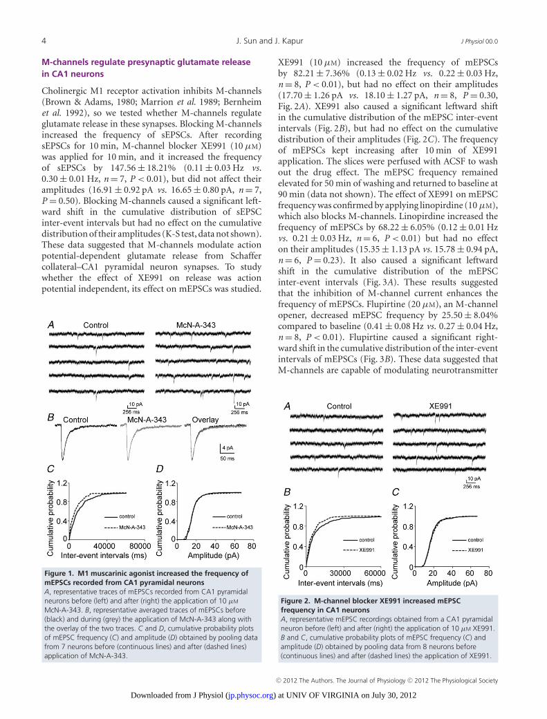
M-channels regulate presynaptic glutamate releasein CA1 neurons
Cholinergic M1 receptor activation inhibits M-channels(Brown & Adams, 1980; Marrion et al. 1989; Bernheimet al. 1992), so we tested whether M-channels regulateglutamate release in these synapses. Blocking M-channelsincreased the frequency of sEPSCs. After recordingsEPSCs for 10 min, M-channel blocker XE991 (10 μM)was applied for 10 min, and it increased the frequencyof sEPSCs by 147.56 ± 18.21% (0.11 ± 0.03 Hz vs.0.30 ± 0.01 Hz, n = 7, P < 0.01), but did not affect theiramplitudes (16.91 ± 0.92 pA vs. 16.65 ± 0.80 pA, n = 7,P = 0.50). Blocking M-channels caused a significant left-ward shift in the cumulative distribution of sEPSCinter-event intervals but had no effect on the cumulativedistribution of their amplitudes (K-S test, data not shown).These data suggested that M-channels modulate actionpotential-dependent glutamate release from Schaffercollateral–CA1 pyramidal neuron synapses. To studywhether the effect of XE991 on release was actionpotential independent, its effect on mEPSCs was studied.
Figure 1. M1 muscarinic agonist increased the frequency ofmEPSCs recorded from CA1 pyramidal neuronsA, representative traces of mEPSCs recorded from CA1 pyramidalneurons before (left) and after (right) the application of 10 μM
McN-A-343. B, representative averaged traces of mEPSCs before(black) and during (grey) the application of McN-A-343 along withthe overlay of the two traces. C and D, cumulative probability plotsof mEPSC frequency (C) and amplitude (D) obtained by pooling datafrom 7 neurons before (continuous lines) and after (dashed lines)application of McN-A-343.
XE991 (10 μM) increased the frequency of mEPSCsby 82.21 ± 7.36% (0.13 ± 0.02 Hz vs. 0.22 ± 0.03 Hz,n = 8, P < 0.01), but had no effect on their amplitudes(17.70 ± 1.26 pA vs. 18.10 ± 1.27 pA, n = 8, P = 0.30,Fig. 2A). XE991 also caused a significant leftward shiftin the cumulative distribution of the mEPSC inter-eventintervals (Fig. 2B), but had no effect on the cumulativedistribution of their amplitudes (Fig. 2C). The frequencyof mEPSCs kept increasing after 10 min of XE991application. The slices were perfused with ACSF to washout the drug effect. The mEPSC frequency remainedelevated for 50 min of washing and returned to baseline at90 min (data not shown). The effect of XE991 on mEPSCfrequency was confirmed by applying linopirdine (10 μM),which also blocks M-channels. Linopirdine increased thefrequency of mEPSCs by 68.22 ± 6.05% (0.12 ± 0.01 Hzvs. 0.21 ± 0.03 Hz, n = 6, P < 0.01) but had no effecton their amplitudes (15.35 ± 1.13 pA vs. 15.78 ± 0.94 pA,n = 6, P = 0.23). It also caused a significant leftwardshift in the cumulative distribution of the mEPSCinter-event intervals (Fig. 3A). These results suggestedthat the inhibition of M-channel current enhances thefrequency of mEPSCs. Flupirtine (20 μM), an M-channelopener, decreased mEPSC frequency by 25.50 ± 8.04%compared to baseline (0.41 ± 0.08 Hz vs. 0.27 ± 0.04 Hz,n = 8, P < 0.01). Flupirtine caused a significant right-ward shift in the cumulative distribution of the inter-eventintervals of mEPSCs (Fig. 3B). These data suggested thatM-channels are capable of modulating neurotransmitter
Figure 2. M-channel blocker XE991 increased mEPSCfrequency in CA1 neuronsA, representative mEPSC recordings obtained from a CA1 pyramidalneuron before (left) and after (right) the application of 10 μM XE991.B and C, cumulative probability plots of mEPSC frequency (C) andamplitude (D) obtained by pooling data from 8 neurons before(continuous lines) and after (dashed lines) the application of XE991.
C© 2012 The Authors. The Journal of Physiology C© 2012 The Physiological Society
) at UNIV OF VIRGINIA on July 30, 2012jp.physoc.orgDownloaded from J Physiol (
J Physiol 00.0 M-type potassium channels modulate glutamatergic transmission in CA1 neurons 5
release independent from action potentials. To confirmthat the effect of M1 agonist was mediated by M-channels,we incubated the slice with XE991 for 25 min. After5 min baseline recording in XE991-containing medium,McN-A-343 was applied, and its effect was blocked. Over-all, McN-A-343 did not change the frequency of mEPSCs(baseline 0.53 ± 0.07 Hz, McN-A-343 0.56 ± 0.08 Hz;n = 7, P = 0.38). In two cells, there was a modest (<10%)increase in frequency but the cumulative frequency plotof inter-event intervals from these cells was not shifted(Fig. 3C). This result suggested blocking M-channels pre-vented McN-A-343 enhancement of mEPSC frequency.
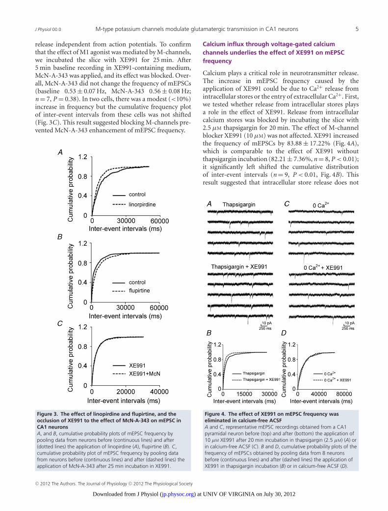
Figure 3. The effect of linopirdine and flupirtine, and theocclusion of XE991 to the effect of McN-A-343 on mEPSC inCA1 neuronsA, and B, cumulative probability plots of mEPSC frequency bypooling data from neurons before (continuous lines) and after(dotted lines) the application of linopirdine (A), flupirtine (B). C,cumulative probability plot of mEPSC frequency by pooling datafrom neurons before (continuous lines) and after (dashed lines) theapplication of McN-A-343 after 25 min incubation in XE991.
Calcium influx through voltage-gated calciumchannels underlies the effect of XE991 on mEPSCfrequency
Calcium plays a critical role in neurotransmitter release.The increase in mEPSC frequency caused by theapplication of XE991 could be due to Ca2+ release fromintracellular stores or the entry of extracellular Ca2+. First,we tested whether release from intracellular stores playsa role in the effect of XE991. Release from intracellularcalcium stores was blocked by incubating the slice with2.5 μM thapsigargin for 20 min. The effect of M-channelblocker XE991 (10 μM) was not affected. XE991 increasedthe frequency of mEPSCs by 83.88 ± 17.22% (Fig. 4A),which is comparable to the effect of XE991 withoutthapsigargin incubation (82.21 ± 7.36%, n = 8, P < 0.01);it significantly left shifted the cumulative distributionof inter-event intervals (n = 9, P < 0.01, Fig. 4B). Thisresult suggested that intracellular store release does not
Figure 4. The effect of XE991 on mEPSC frequency waseliminated in calcium-free ACSFA and C, representative mEPSC recordings obtained from a CA1pyramidal neuron before (top) and after (bottom) the application of10 μM XE991 after 20 min incubation in thapsigargin (2.5 μM) (A) orin calcium-free ACSF (C). B and D, cumulative probability plots of thefrequency of mEPSCs obtained by pooling data from 8 neuronsbefore (continuous lines) and after (dashed lines) the application ofXE991 in thapsigargin incubation (B) or in calcium-free ACSF (D).
C© 2012 The Authors. The Journal of Physiology C© 2012 The Physiological Society
) at UNIV OF VIRGINIA on July 30, 2012jp.physoc.orgDownloaded from J Physiol (

6 J. Sun and J. Kapur J Physiol 00.0
contribute to the enhancement of M-channel inhibitionon mEPSCs frequency.
Therefore, we tested whether the effect of XE991 onthe frequency of mEPSCs was dependent on extracellularCa2+ by recording mEPSCs in a calcium-free medium.The effect of XE991 on mEPSCs was eliminated inACSF that lacked calcium. Application of XE991 neitherchanged the frequency of mEPSCs (0.13 ± 0.02 Hz vs.0.12 ± 0.01 Hz, n = 8, P = 0.30, Fig. 4C) nor shifted thecumulative distribution of the inter-event intervals ofmEPSCs (Fig. 4D). These data suggested that the effect ofXE991 on spontaneous glutamate release was dependenton the influx of Ca2+.
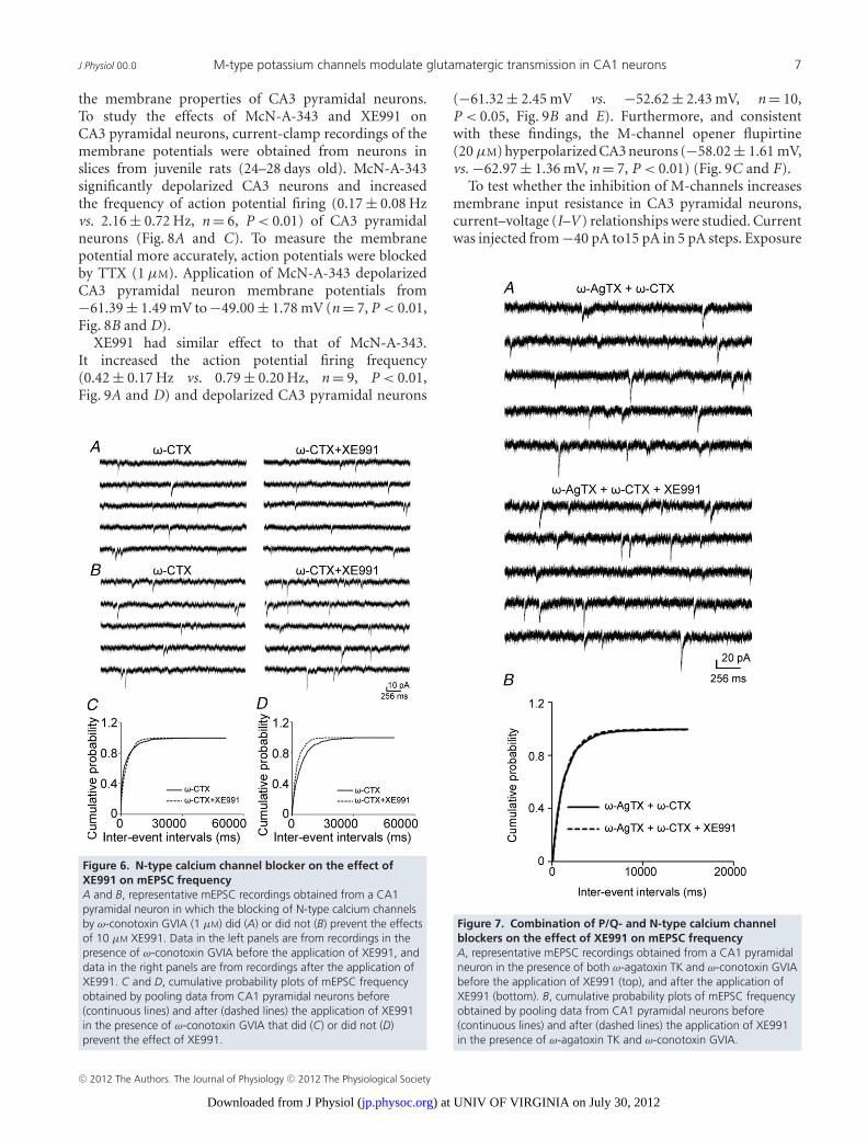
We also studied the effect of McN-A-343 in acalcium-free medium. The McN-A-343 enhancement ofmEPSCs was blocked in calcium-free medium (base-line, 0.40 ± 0.08 Hz, McN-A-343, 0.41 ± 0.07 Hz, n = 7,P = 0.36). There was no significant shift on the cumulativedistribution of inter-event intervals of mEPSCs (data notshown). This suggested that the effect of M1 receptoractivation on the frequency of mEPSC is largely mediatedby increasing calcium influx from extracellular space. Totest whether Ca2+ enters Schaffer collateral terminals byactivation of presynaptic voltage-gated calcium channelsdue to M-channel inhibition, we studied the effectof P/Q- and N-type calcium channel blockers on theXE991-mediated enhancement of mEPSC frequency.These two channels are expressed in the presynapticterminals of Schaffer collaterals (Wheeler et al. 1994;Qian & Noebels, 2000). Slices were incubated witheither ω-agatoxin TK (200 nM) or ω-conotoxin GVIA(1 μM) (Peptides International, Louisville, KY, USA) for15 min prior to data collection. In the presence ofω-agatoxin TK, which blocks P/Q-type calcium channels,the XE991-mediated enhancement of mEPSC frequencywas prevented in approximately half of the cells thatwere studied. In 5 out of 9 neurons, the applicationof XE991 did not increase the frequency of mEPSCs(0.23 ± 0.08 Hz vs. 0.20 ± 0.06 Hz, P = 0.46, Fig. 5A andC). In the other four CA1 pyramidal neurons, XE991still significantly increased the frequency of mEPSCs(0.16 ± 0.03 Hz vs. 0.31 ± 0.07 Hz, P < 0.05, Fig. 5B andD). Blocking N-type channels with ω-conotoxin GVIAalso prevented the effect of XE991 on the frequency ofmEPSCs in half of the CA1 pyramidal neurons from whichrecordings were made. In 4 of 8 neurons, the effect ofXE991 was eliminated (0.33 ± 0.05 Hz vs. 0.34 ± 0.04 Hz,P = 0.47, Fig. 6A and C). In the other four neurons,XE991 significantly increased the frequency of mEPSCs(0.24 ± 0.04 Hz vs. 0.43 ± 0.07 Hz, P < 0.05, Fig. 6B andD).
Next, we applied the blockers of two channelssimultaneously. Slices were incubated in ω-agatoxinTK and ω-conotoxin GVIA for 25–40 min, and after5 min baseline recording, XE991 was applied. The
mean frequency was not significantly increased (baseline,0.78 ± 0.08 Hz, XE991, 0.82 ± 0.09 Hz; n = 8, P = 0.08,Fig. 7A), and there was no significant shift of cumulativedistribution of inter-event intervals for each celland pooled data (Fig. 7B). This result suggested theenhancement of the frequency of mEPSCs by XE991 wasmediated by calcium influx though P/Q- and N-typecalcium channels.
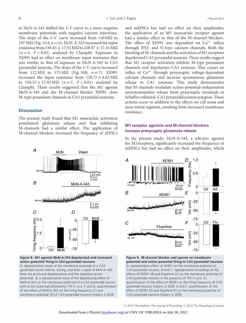
McN-A-343 and XE991 depolarize and increase actionpotential firing in CA3 neurons
The aforementioned data suggested that inhibition ofM-channels resulted in the depolarization of presynapticterminals of the CA3 pyramidal neuron, which thenactivated P/Q- or N-type calcium channels, therebycausing calcium influx and ultimately leading to increasedglutamate release. We tested the effect of M-channels on
Figure 5. P/Q-type calcium channel blocker on the effect ofXE991 on mEPSC frequencyA and B, representative mEPSC recordings obtained from a CA1pyramidal neuron in which the blocking of P/Q-type of calciumchannels by 200 nM ω-agatoxin TK did (A) or did not (B) prevent theeffect of 10 μM XE991. The left panels include data from recordingsin the presence of ω-agatoxin TK before the application of XE991,and the right panels include data from recordings after theapplication of XE991. C and D, cumulative probability plots ofmEPSC frequency obtained by pooling data from CA1 pyramidalneurons before (continuous lines) and after (dashed lines) theapplication of XE991 in the presence of ω-agatoxin TK that did (C) ordid not (D) prevent the effect of XE991.
C© 2012 The Authors. The Journal of Physiology C© 2012 The Physiological Society
) at UNIV OF VIRGINIA on July 30, 2012jp.physoc.orgDownloaded from J Physiol (
J Physiol 00.0 M-type potassium channels modulate glutamatergic transmission in CA1 neurons 7
the membrane properties of CA3 pyramidal neurons.To study the effects of McN-A-343 and XE991 onCA3 pyramidal neurons, current-clamp recordings of themembrane potentials were obtained from neurons inslices from juvenile rats (24–28 days old). McN-A-343significantly depolarized CA3 neurons and increasedthe frequency of action potential firing (0.17 ± 0.08 Hzvs. 2.16 ± 0.72 Hz, n = 6, P < 0.01) of CA3 pyramidalneurons (Fig. 8A and C). To measure the membranepotential more accurately, action potentials were blockedby TTX (1 μM). Application of McN-A-343 depolarizedCA3 pyramidal neuron membrane potentials from−61.39 ± 1.49 mV to −49.00 ± 1.78 mV (n = 7, P < 0.01,Fig. 8B and D).
XE991 had similar effect to that of McN-A-343.It increased the action potential firing frequency(0.42 ± 0.17 Hz vs. 0.79 ± 0.20 Hz, n = 9, P < 0.01,Fig. 9A and D) and depolarized CA3 pyramidal neurons
Figure 6. N-type calcium channel blocker on the effect ofXE991 on mEPSC frequencyA and B, representative mEPSC recordings obtained from a CA1pyramidal neuron in which the blocking of N-type calcium channelsby ω-conotoxin GVIA (1 μM) did (A) or did not (B) prevent the effectsof 10 μM XE991. Data in the left panels are from recordings in thepresence of ω-conotoxin GVIA before the application of XE991, anddata in the right panels are from recordings after the application ofXE991. C and D, cumulative probability plots of mEPSC frequencyobtained by pooling data from CA1 pyramidal neurons before(continuous lines) and after (dashed lines) the application of XE991in the presence of ω-conotoxin GVIA that did (C) or did not (D)prevent the effect of XE991.
(−61.32 ± 2.45 mV vs. −52.62 ± 2.43 mV, n = 10,P < 0.05, Fig. 9B and E). Furthermore, and consistentwith these findings, the M-channel opener flupirtine(20 μM) hyperpolarized CA3 neurons (−58.02 ± 1.61 mV,vs. −62.97 ± 1.36 mV, n = 7, P < 0.01) (Fig. 9C and F).
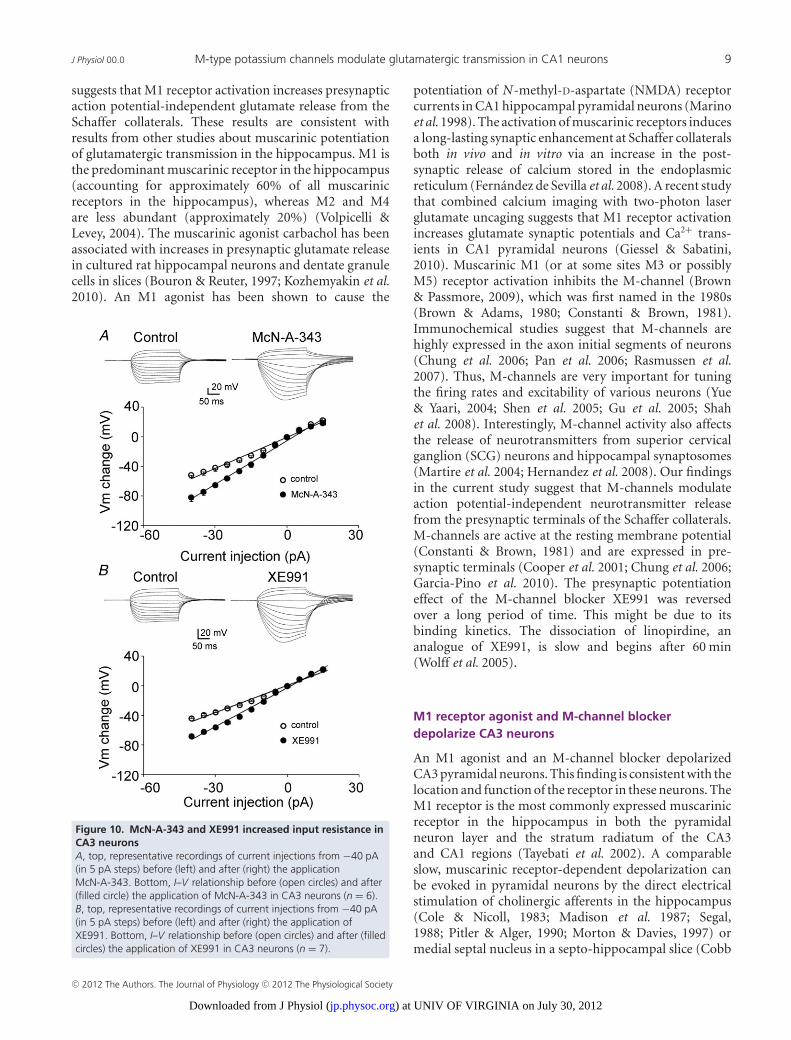
To test whether the inhibition of M-channels increasesmembrane input resistance in CA3 pyramidal neurons,current–voltage (I–V ) relationships were studied. Currentwas injected from −40 pA to15 pA in 5 pA steps. Exposure
Figure 7. Combination of P/Q- and N-type calcium channelblockers on the effect of XE991 on mEPSC frequencyA, representative mEPSC recordings obtained from a CA1 pyramidalneuron in the presence of both ω-agatoxin TK and ω-conotoxin GVIAbefore the application of XE991 (top), and after the application ofXE991 (bottom). B, cumulative probability plots of mEPSC frequencyobtained by pooling data from CA1 pyramidal neurons before(continuous lines) and after (dashed lines) the application of XE991in the presence of ω-agatoxin TK and ω-conotoxin GVIA.
C© 2012 The Authors. The Journal of Physiology C© 2012 The Physiological Society
) at UNIV OF VIRGINIA on July 30, 2012jp.physoc.orgDownloaded from J Physiol (
8 J. Sun and J. Kapur J Physiol 00.0
to McN-A-343 shifted the I–V curve to a more negativemembrane potentials with negative current injections.The slope of the I–V curve increased from 140 M to197 M (Fig. 10A, n = 6). McN-A-343 increased the inputresistance from 156.43 ± 17.51 M to 228.07 ± 17.16 M(n = 6, P < 0.01) analysed by Clampfit. Exposure toXE991 had an effect on membrane input resistance thatwas similar to that of exposure to McN-A-343 in CA3pyramidal neurons. The slope of the I–V curve increasedfrom 122 M to 175 M (Fig. 10B, n = 7). XE991increased the input resistance from 129.75 ± 8.63 Mto 194.53 ± 17.92 M (n = 7, P < 0.01) analysed byClampfit. These results suggested that the M1 agonistMcN-A-343 and the M-channel blocker XE991 closeM-type potassium channels in CA3 pyramidal neurons.
Discussion
The present study found that M1 muscarinic activationpotentiated glutamate release and that inhibitingM-channels had a similar effect. The application ofM-channel blockers increased the frequency of sEPSCs
Figure 8. M1 agonist McN-A-343 depolarized and increasedaction potential firing in CA3 pyramidal neuronsA, representative traces of the membrane potential of a CA3pyramidal neuron before, during, and after a wash of McN-A-343.Note the profound depolarization and the repetitive actionpotentials. B, a representative trace of the depolarizing effect ofMcN-A-343 on the membrane potential of a CA3 pyramidal neuronwith action potentials blocked by TTX (1 μM). C and D, quantificationof the effect of McN-A-343 on the firing frequency (C) andmembrane potential (D) of CA3 pyramidal neurons (means ± SEM).
and mEPSCs but had no effect on their amplitudes;the application of an M1 muscarinic receptor agonisthad a similar effect to that of the M-channel blockers.The effect of XE991 was dependent on Ca2+ influxthrough P/Q- and N-type calcium channels. Both theblocking of M-channels and the activation of M1 receptorsdepolarized CA3 pyramidal neurons. These results suggestthat M1 receptor activation inhibits M-type potassiumchannels and depolarizes CA3 neurons. This causes aninflux of Ca2+ through presynaptic voltage-dependentcalcium channels and increase spontaneous glutamaterelease to CA1 neurons. This study demonstratesthat M-channels modulate action-potential-independentneurotransmitter release from presynaptic terminals inSchaffer collateral–CA1 pyramidal neuron synapses. Theseactions occur in addition to the effects on cell soma andaxon initial segment, resulting from increased membraneresistance.
M1 receptor agonists and M-channel blockersincrease presynaptic glutamate release
In the present study, McN-A-343, a selective agonistfor M1receptors, significantly increased the frequency ofmEPSCs but had no effect on their amplitudes, which
Figure 9. M-channel blocker and opener on membranepotential and action potential firing in CA3 pyramidal neuronsA, representative effect of XE991 on the membrane potential ofCA3 pyramidal neurons. B and C, representative recordings of theeffects of XE991 (B) and flupirtine (C) on the membrane potential ofCA3 pyramidal neurons in the presence of TTX (1 μM). D,quantification of the effect of XE991 on the firing frequency of CA3pyramidal neurons (means ± SEM). E and F, quantification of theeffect of XE991 (E) and flupirtine (F) on the membrane potential ofCA3 pyramidal neurons (means ± SEM).
C© 2012 The Authors. The Journal of Physiology C© 2012 The Physiological Society
) at UNIV OF VIRGINIA on July 30, 2012jp.physoc.orgDownloaded from J Physiol (
J Physiol 00.0 M-type potassium channels modulate glutamatergic transmission in CA1 neurons 9
suggests that M1 receptor activation increases presynapticaction potential-independent glutamate release from theSchaffer collaterals. These results are consistent withresults from other studies about muscarinic potentiationof glutamatergic transmission in the hippocampus. M1 isthe predominant muscarinic receptor in the hippocampus(accounting for approximately 60% of all muscarinicreceptors in the hippocampus), whereas M2 and M4are less abundant (approximately 20%) (Volpicelli &Levey, 2004). The muscarinic agonist carbachol has beenassociated with increases in presynaptic glutamate releasein cultured rat hippocampal neurons and dentate granulecells in slices (Bouron & Reuter, 1997; Kozhemyakin et al.2010). An M1 agonist has been shown to cause the
Figure 10. McN-A-343 and XE991 increased input resistance inCA3 neuronsA, top, representative recordings of current injections from −40 pA(in 5 pA steps) before (left) and after (right) the applicationMcN-A-343. Bottom, I–V relationship before (open circles) and after(filled circle) the application of McN-A-343 in CA3 neurons (n = 6).B, top, representative recordings of current injections from −40 pA(in 5 pA steps) before (left) and after (right) the application ofXE991. Bottom, I–V relationship before (open circles) and after (filledcircles) the application of XE991 in CA3 neurons (n = 7).
potentiation of N-methyl-D-aspartate (NMDA) receptorcurrents in CA1 hippocampal pyramidal neurons (Marinoet al. 1998). The activation of muscarinic receptors inducesa long-lasting synaptic enhancement at Schaffer collateralsboth in vivo and in vitro via an increase in the post-synaptic release of calcium stored in the endoplasmicreticulum (Fernandez de Sevilla et al. 2008). A recent studythat combined calcium imaging with two-photon laserglutamate uncaging suggests that M1 receptor activationincreases glutamate synaptic potentials and Ca2+ trans-ients in CA1 pyramidal neurons (Giessel & Sabatini,2010). Muscarinic M1 (or at some sites M3 or possiblyM5) receptor activation inhibits the M-channel (Brown& Passmore, 2009), which was first named in the 1980s(Brown & Adams, 1980; Constanti & Brown, 1981).Immunochemical studies suggest that M-channels arehighly expressed in the axon initial segments of neurons(Chung et al. 2006; Pan et al. 2006; Rasmussen et al.2007). Thus, M-channels are very important for tuningthe firing rates and excitability of various neurons (Yue& Yaari, 2004; Shen et al. 2005; Gu et al. 2005; Shahet al. 2008). Interestingly, M-channel activity also affectsthe release of neurotransmitters from superior cervicalganglion (SCG) neurons and hippocampal synaptosomes(Martire et al. 2004; Hernandez et al. 2008). Our findingsin the current study suggest that M-channels modulateaction potential-independent neurotransmitter releasefrom the presynaptic terminals of the Schaffer collaterals.M-channels are active at the resting membrane potential(Constanti & Brown, 1981) and are expressed in pre-synaptic terminals (Cooper et al. 2001; Chung et al. 2006;Garcia-Pino et al. 2010). The presynaptic potentiationeffect of the M-channel blocker XE991 was reversedover a long period of time. This might be due to itsbinding kinetics. The dissociation of linopirdine, ananalogue of XE991, is slow and begins after 60 min(Wolff et al. 2005).
M1 receptor agonist and M-channel blockerdepolarize CA3 neurons
An M1 agonist and an M-channel blocker depolarizedCA3 pyramidal neurons. This finding is consistent with thelocation and function of the receptor in these neurons. TheM1 receptor is the most commonly expressed muscarinicreceptor in the hippocampus in both the pyramidalneuron layer and the stratum radiatum of the CA3and CA1 regions (Tayebati et al. 2002). A comparableslow, muscarinic receptor-dependent depolarization canbe evoked in pyramidal neurons by the direct electricalstimulation of cholinergic afferents in the hippocampus(Cole & Nicoll, 1983; Madison et al. 1987; Segal,1988; Pitler & Alger, 1990; Morton & Davies, 1997) ormedial septal nucleus in a septo-hippocampal slice (Cobb
C© 2012 The Authors. The Journal of Physiology C© 2012 The Physiological Society
) at UNIV OF VIRGINIA on July 30, 2012jp.physoc.orgDownloaded from J Physiol (

10 J. Sun and J. Kapur J Physiol 00.0
& Davies, 2005). The medial septal nucleus providesthe major source of cholinergic innervations to thehippocampus (Dutar et al. 1995) and presents a directsynaptic input to both principal neurons and interneurons(Frotscher & Leranth, 1985; Leranth & Frotscher, 1987).Muscarinic acetylcholine receptors modulate several ionicconductances in addition to the M-current, includingIAHP (the Ca2+-activated K+ current that is responsiblefor slowing action potential discharges) and I leak (thebackground leak current) (Halliwell, 1990). Activationof mAChRs also potentiates two mixed cation currents(Ih, the hyperpolarization-activated cation current; andI cat, the Ca2+-dependent non-specific cation current)(Halliwell, 1990; Colino & Halliwell, 1993) and theN-methyl-D-aspartate (NMDA) receptor (Markram &Segal, 1990). This may explain why, in our study, the M1agonist McN-A-343 had a stronger depolarization effectthan the M-channel blocker XE991 in CA3 pyramidalneurons.
Muscarinic receptor and intracellular calcium level
Current study suggested that M1 receptor activationdepolarizes presynaptic membrane and activatesvoltage-gated calcium channel to increase intracellularcalcium level and neurotransmitter release by inhibition ofM-channels. Previous studies suggested that muscarinicstimulation inhibits voltage-gated calcium channels(Toselli et al. 1989; Qian & Saggau, 1997). Howeverthese studies did not classify the subtype of muscarnicreceptor that mediates this effect. It was reported thatthe inhibitory effect of muscarinic receptor activationon calcium channels was mediated by M4 subtype in ratsympathetic neurons (Bernheim et al. 1992) and by M2subtype in rat magocellular cholinergic basal forebrainneurons (Allen & Brown, 1993). There is no report tosuggest that M1 subtype muscarinic receptor inhibitscalcium channels.
Muscarinic receptors activate inositol1,4,5-trisphosphate (IP3) receptor to trigger calciumrelease from endoplasmic reticulum (ER) stores inCA1 pyramidal neurons, and appears to be actionpotential independent (Power & Sah, 2002). Anotherstudy suggested this effect is mediated by M1 receptor(Fernandez de Sevilla et al. 2008). The role of thesestores in enhancing synaptic release remains uncertain.Although spontaneous transmitter release (in the absenceof action potentials) is largely mediated by calcium releasefrom internal stores (Emptage et al. 2001), our studysuggests the modulation of spontaneous transmitterrelease could be mediated by calcium influx thoughvoltage-gated calcium channels.
Overall, this study demonstrated that M1 muscarinicreceptor activation inhibits M-type potassium channels,thereby increasing excitatory glutamate neurotransmitter
release from the presynaptic terminals of CA3 neuronsby increasing the rate of calcium influx throughvoltage-dependent calcium channels. This effect of M1receptor activation could contribute to the generation ofseizures.
References
Allen TG & Brown DA (1993). M2 muscarinicreceptor-mediated inhibition of the Ca2+ current in ratmagnocellular cholinergic basal forebrain neurones. J Physiol466, 173–189.
Bernheim L, Mathie A & Hille B (1992). Characterization ofmuscarinic receptor subtypes inhibiting Ca2+ current andM-current in rat sympathetic neurons. Proc Natl Acad Sci US A 89, 9544–9548.
Biervert C, Schroeder BC, Kubisch C, Berkovic SF, Propping P,Jentsch TJ & Steinlein OK (1998). A potassium channelmutation in neonatal human epilepsy. Science 279, 403–406.
Bouron A & Reuter H (1997). Muscarinic stimulation ofsynaptic activity by protein kinase C is inhibited byadenosine in cultured hippocampal neurons. Proc Natl AcadSci U S A 94, 12224–12229.
Brown DA & Adams PR (1980). Muscarinic suppression of anovel voltage-sensitive K+ current in a vertebrate neurone.Nature 283, 673–676.
Brown DA & Passmore GM (2009). Neural KCNQ (Kv7)channels. Br J Pharmacol 156, 1185–1195.
Brown JT & Randall AD (2009). Activity-dependent depressionof the spike after-depolarization generates long-lastingintrinsic plasticity in hippocampal CA3 pyramidal neurons.The Journal of Physiology 587, 1265–1281.
Chung HJ, Jan YN & Jan LY (2006). Polarized axonal surfaceexpression of neuronal KCNQ channels is mediated bymultiple signals in the KCNQ2 and KCNQ3 C-terminaldomains. Proc Natl Acad Sci U S A 103, 8870–8875.
Cobb SR & Davies CH (2005). Cholinergic modulation ofhippocampal cells and circuits. J Physiol 562, 81–88.
Cole AE & Nicoll RA (1983). Acetylcholine mediates a slowsynaptic potential in hippocampal pyramidal cells. Science221, 1299–1301.
Colino A & Halliwell JV (1993). Carbachol potentiates Qcurrent and activates a calcium-dependent non-specificconductance in rat hippocampus in vitro. Eur J Neurosci 5,1198–1209.
Constanti A & Brown DA (1981). M-currents involtage-clamped mammalian sympathetic neurones.Neurosci Lett 24, 289–294.
Cooper EC, Harrington E, Jan YN & Jan LY (2001). M-channelKCNQ2 subunits are localized to key sites for control ofneuronal network oscillations and synchronization in mousebrain. J Neurosci 21, 9529–9540.
Delmas P & Brown DA (2005). Pathways modulating neuralKCNQ/M (Kv7) potassium channels. Nat Rev Neurosci 6,850–862.
Devaux JJ, Kleopa KA, Cooper EC & Scherer SS (2004).KCNQ2 is a nodal K+ channel. J Neurosci 24, 1236–1244.
C© 2012 The Authors. The Journal of Physiology C© 2012 The Physiological Society
) at UNIV OF VIRGINIA on July 30, 2012jp.physoc.orgDownloaded from J Physiol (
J Physiol 00.0 M-type potassium channels modulate glutamatergic transmission in CA1 neurons 11
Dutar P, Bassant MH, Senut MC & Lamour Y (1995). Theseptohippocampal pathway: structure and function of acentral cholinergic system. Physiol Rev 75, 393–427.
Emptage NJ, Reid CA & Fine A (2001). Calcium stores inhippocampal synaptic boutons mediate short-termplasticity, store-operated Ca2+ entry, and spontaneoustransmitter release. Neuron 29, 197–208.
Fernandez de Sevilla D, Nunnez A, Borde M, Malinow R &Buno W. (2008). Cholinergic-mediated IP3-receptoractivation induces long-lasting synaptic enhancement inCA1 pyramidal neurons. J Neurosci 28, 1469–1478.
Frotscher M & Leranth C (1985). Cholinergic innervation ofthe rat hippocampus as revealed by choline acetyltransferaseimmunocytochemistry: a combined light and electronmicroscopic study. J Comp Neurol 239, 237–246.
Garcia-Pino E, Caminos E & Juiz JM (2010). KCNQ5 reachessynaptic endings in the auditory brainstem at hearing onsetand targeting maintenance is activity-dependent. J CompNeurol 518, 1301–1314.
Geiger J, Weber YG, Landwehrmeyer B, Sommer C & Lerche H(2006). Immunohistochemical analysis of KCNQ3 potassiumchannels in mouse brain. Neurosci Lett 400, 101–104.
Giessel AJ & Sabatini BL (2010). M1 muscarinic receptors boostsynaptic potentials and calcium influx in dendritic spines byinhibiting postsynaptic SK channels. Neuron 68, 936–947.
Gu N, Vervaeke K, Hu H & Storm JF (2005). Kv7/KCNQ/Mand HCN/h, but not KCa2/SK channels, contribute to thesomatic medium after-hyperpolarization and excitabilitycontrol in CA1 hippocampal pyramidal cells. J Physiol 566,689–715.
Halliwell JV ( 1990). Physiological mechanisms of cholinergicaction in the hippocampus. Prog Brain Res 84, 255–272.
Hernandez CC, Zaika O, Tolstykh GP & Shapiro MS (2008).Regulation of neural KCNQ channels: signalling pathways,structural motifs and functional implications. J Physiol 586,1811–1821.
Higley MJ, Soler-Llavina GJ & Sabatini BL (2009). Cholinergicmodulation of multivesicular release regulates striatalsynaptic potency and integration. Nat Neurosci 12,1121–1128.
Jentsch TJ (2000). Neuronal KCNQ potassiumchannels:physislogy and role in disease. Nat Rev Neurosci 1,21–30.
Kanaumi T, Takashima S, Iwasaki H, Itoh M, Mitsudome A &Hirose S (2008). Developmental changes in KCNQ2 andKCNQ3 expression in human brain: possible contribution tothe age-dependent etiology of benign familial neonatalconvulsions. Brain Dev 30, 362–369.
Kozhemyakin M, Rajasekaran K & Kapur J (2010). Centralcholinesterase inhibition enhances glutamatergic synaptictransmission. J Neurophysiol 103, 1748–1757.
Langmead CJ, Watson J & Reavill C (2008). Muscarinicacetylcholine receptors as CNS drug targets. Pharmacol Ther117, 232–243.
Leranth C & Frotscher M (1987). Cholinergic innervation ofhippocampal GAD- and somatostatin-immunoreactivecommissural neurons. J Comp Neurol 261, 33–47.
Madison DV, Lancaster B & Nicoll RA (1987). Voltage clampanalysis of cholinergic action in the hippocampus. J Neurosci7, 733–741.
Marino MJ, Rouse ST, Levey AI, Potter LT & Conn PJ (1998).Activation of the genetically defined M1 muscarinic receptorpotentiates N-methyl-D-aspartate (NMDA) receptorcurrents in hippocampal pyramidal cells. Proc Natl Acad SciU S A 95, 11465–11470.
Markram H & Segal M (1990). Acetylcholine potentiatesresponses to N-methyl-D-aspartate in the rat hippocampus.Neurosci Lett 113, 62–65.
Marrion NV, Smart TG, Marsh SJ & Brown DA (1989).Muscarinic suppression of the M-current in the ratsympathetic-ganglion is mediated by receptors of theM1-subtype. Br J Pharmacol 98, 557–573.
Martire M, Castaldo P, D’Amico M, Preziosi P, Annunziato L &Taglialatela M (2004). M-channels containing KCNQ2subunits modulate norepinephrine, aspartate, and GABArelease from hippocampal nerve terminals. J Neurosci 24,592–597.
Morton RA & Davies CH (1997). Regulation of muscarinicacetylcholine receptor-mediated synaptic responses byadenosine receptors in the rat hippocampus. J Physiol 502,75–90.
Pan Z, Kao T, Horvath Z, Lemos J, Sul JY, Cranstoun SD,Bennett V, Scherer SS & Cooper EC (2006). A commonankyrin-G-based mechanism retains KCNQ and NaVchannels at electrically active domains of the axon. J Neurosci26, 2599–2613.
Peretz A, Sheinin A, Yue C, Degani-Katzav N, Gibor G,Nachman R, Gopin A, Tam E, Shabat D, Yaari Y & Attali B(2007). Pre- and postsynaptic activation of M-channels by anovel opener dampens neuronal firing and transmitterrelease. J Neurophysiol 97, 283–295.Pitler TA & Alger BE (1990). Activation of the pharmacologicallydefined M3 muscarinic receptor depolarizes hippocampalpyramidal cells. Brain Res 534, 257–262.
Power JM & Sah P (2002). Nuclear calcium signaling evoked bycholinergic stimulation in hippocampal CA1 pyramidalneurons. J Neurosci 22, 3454–3462.
Qian J & Noebels JL (2000). Presynaptic Ca2+ influx at a mousecentral synapse with Ca2+ channel subunit mutations. JNeurosci 20, 163–170.
Qian J & Saggau P (1997). Presynaptic inhibition of synaptictransmission in the rat hippocampus by activation ofmuscarinic receptors: involvement of presynaptic calciuminflux. Br J Pharmacol 122, 511–519.
Rasmussen HB, Frøkjær-Jensen C, Jensen CS, Jensen HS,Jørgensen NK, Misonou H, Trimmer JS, Olesen SP &Schmitt N (2007). Requirement of subunit co-assembly andankyrin-G for M-channel localization at the axon initialsegment. J Cell Sci 120, 953–963.
Safiulina VF, Zacchi P, Taglialatela M, Yaari Y & Cherubini E(2008). Low expression of Kv7/M channels facilitatesintrinsic and network bursting in the developing rathippocampus. J Physiol 586, 5437–5453.
Segal M ( 1988). Synaptic activation of a cholinergic receptor inrat hippocampus. Brain Res 452, 79–86.
Selyanko AA, Delmas P, Hadley JK, Tatulian L, Wood IC,Mistry M, London B & Brown DA (2002).Dominant-negative subunits reveal potassium channelfamilies that contribute to M-like potassium currents.J Neurosci 22, RC212.
C© 2012 The Authors. The Journal of Physiology C© 2012 The Physiological Society
) at UNIV OF VIRGINIA on July 30, 2012jp.physoc.orgDownloaded from J Physiol (
12 J. Sun and J. Kapur J Physiol 00.0
Shah MM, Migliore M, Valencia I, Cooper EC & Brown DA(2008). Functional significance of axonal Kv7 channels inhippocampal pyramidal neurons. Proc Natl Acad Sci U S A105, 7869–7874.
Shah MM, Mistry M, Marsh SJ, Brown DA & Delmas P (2002).Molecular correlates of the M-current in cultured rathippocampal neurons. J Physiol 544, 29–37.
Shen W, Hamilton SE, Nathanson NM & Surmeier DJ (2005).Cholinergic suppression of KCNQ channel currentsenhances excitability of striatal medium spiny neurons. JNeurosci 25, 7449–7458.
Sim JA & Griffith WH (1996). Muscarinic Inhibition ofglutamatergic transmission onto rat magnocellular basalforebrain neurons in a thin-slice preparation. Eur J Neurosci8, 880–891.
Tayebati SK, Amenta F, El-Assouad D & Zaccheo D (2002).Muscarinic cholinergic receptor subtypes in thehippocampus of aged rats. Mech of Ageing Dev 123, 521–528.
Toselli M, Lang J, Costa T & Lux HD (1989). Directmodulation of voltage-dependent calcium channels bymuscarinic activation of a pertussis toxin-sensitive G-proteinin hippocampal neurons. Pflugers Arch 415, 255–261.
Vervaeke K, Gu N, Agdestein C, Hu H & Storm JF (2006).Kv7/KCNQ/M-channels in rat glutamatergic hippocampalaxons and their role in regulation of excitability andtransmitter release. J Physiol 576, 235–256.
Vogt KE & Regehr WG (2001). Cholinergic modulation ofexcitatory synaptic transmission in the CA3 area of thehippocampus. J Neurosci 21, 75–83.
Volpicelli LA & Levey AI (2004). Muscarinic acetylcholinereceptor subtypes in cerebral cortex and hippocampus. ProgBrain Res 145, 59–66.
Wang HS, Pan Z, Shi W, Brown BS, Wymore RS, Cohen IS,Dixon JE & McKinnon D (1998). KCNQ2 and KCNQ3potassium channel subunits: molecular correlates of theM-channel. Science 282, 1890–1893.
Weber YG, Geiger J, Kampchen K, Landwehrmeyer B, SommerC & Lerche H (2006). Immunohistochemical analysis ofKCNQ2 potassium channels in adult and developing mousebrain. Brain Res 1077, 1–6.
Wheeler DB, Randall A & Tsien RW (1994). Roles of N-typeand Q-type Ca2+ channels in supporting hippocampalsynaptic transmission. Science 264, 107–111.
Wolff C, Gillard M, Fuks B & Chatelain P (2005).[3H]Linopirdine binding to rat brain membranes is notrelevant for M-channel interaction. Eur J Pharmacol 518,10–17.
Yajeya J, De La Fuente A, Criado JM, Bajo V, Sanchez-RiolobosA & Heredia M (2000). Muscarinic agonist carbacholdepresses excitatory synaptic transmission in the ratbasolateral amygdala in vitro. Synapse 38, 151–160.
Yue C & Yaari Y (2004). KCNQ/M Channels control spikeafterdepolarization and burst generation in hippocampalneurons. J Neurosci 24, 4614–4624.
Zhang HM, Chen SR & Pan HL (2007). Regulation ofglutamate release from primary afferents and interneuronsin the spinal cord by muscarinic receptor subtypes. JNeurophysiol 97, 102–109.
Author contributions
J.S. and J.K. designed the study. J.S. performed the experimentsand analysed the data. J.S. and J.K. interpreted the data, draftedand revised the article.
Acknowledgements
This study was supported by peer reviewed Medical ResearchProgram of the Department of Defense PR 093963 and NINDS(NIH) RO1 NS 40337.
C© 2012 The Authors. The Journal of Physiology C© 2012 The Physiological Society
) at UNIV OF VIRGINIA on July 30, 2012jp.physoc.orgDownloaded from J Physiol (

ARTICLE IN PRESS+ModelEPIRES-4759; No. of Pages 9
Epilepsy Research (2012) xxx, xxx—xxx
jou rn al h om epa ge: www.elsev ier .com/ locate /ep i lepsyres
Characterization of status epilepticus induced bytwo organophosphates in rats
Marko S. Todorovic, Morgan L. Cowan, Corrinee A. Balint, Chengsan Sun,Jaideep Kapur ∗
Department of Neurology, University of Virginia Health Sciences Center, Charlottesville, VA 22908, USA
Received 10 August 2011; received in revised form 8 April 2012; accepted 17 April 2012
KEYWORDSOrganophosphate;Status epilepticus;EEG;Seizures;Benzodiazepine;Diazepam
Summary Organophosphates (OPs) inhibit the enzyme cholinesterase and cause accumulationof acetylcholine, and are known to cause seizures and status epilepticus (SE) in humans. Theanimal models of SE caused by organophosphate analogs of insecticides are not well character-ized. SE caused by OPs paraoxon and diisopropyl fluorophosphate (DFP) in rats was characterizedby electroencephalogram (EEG), behavioral observations and response to treatment with thebenzodiazepine diazepam administered at various stages of SE. A method for SE induction usingintrahippocampal infusion of paraoxon was also tested. Infusion of 200 nmol paraoxon into thehippocampus caused electrographic seizures in 43/52 (82.7%) animals tested; and of these ani-mals, 14/43 (30%) had self-sustaining seizures that lasted 4—18 h after the end of paraoxoninfusion. SE was also induced by peripheral subcutaneous injection of diisopropyl fluorophos-phate (DFP, 1.25 mg/kg) or paraoxon (1.00 mg/kg) to rats pretreated with atropine (2 mg/kg)and 2-pralidoxime (2-PAM, 50 mg/kg) 30 min prior to OP injection. SE occurred in 78% paraoxon-treated animals and in 79% of DFP-treated animals. Diazepam (10 mg/kg) was administered10 min and 30 min after the onset of continuous EEG seizures induced by paraoxon and it ter-minated SE in a majority of animals at both time points. DFP-induced SE was terminated in
60% animals when diazepam was administered 10 min after the onset of continuous EEG seizureactivity but diazepam did not terminate SE in any animal when it was administered 30 min afterthe onset of continuous seizures. These studies demonstrate that both paraoxon and DFP caninduce SE in rats but refractoriness to diazepam is a feature of DFP induced SE.ts re
© 2012 Elsevier B.V. All righPlease cite this article in press as: Todorovic, M.S., et al.,organophosphates in rats. Epilepsy Res. (2012), http://dx.doi.
∗ Corresponding author at: Department of Neurology, Box 800394,University of Virginia HSC, Charlottesville, VA 22908-800394, USA.Tel.: +1 434 924 5312; fax: +1 434 982 1726.
E-mail address: [email protected] (J. Kapur).
I
StSa2
0920-1211/$ — see front matter © 2012 Elsevier B.V. All rights reserved.http://dx.doi.org/10.1016/j.eplepsyres.2012.04.014
served.
ntroduction
tatus epilepticus (SE) is a neurological emergency charac-
Characterization of status epilepticus induced by twoorg/10.1016/j.eplepsyres.2012.04.014
erized by recurrent or continuous self-sustaining seizures.E can contribute to morbidity, sustained neuronal injurynd contribute to mortality (Fujikawa et al., 2000; Fujikawa,005). Understanding mechanisms of SE in humans is a

IN+ModelE
2
pptteesoue1c
ehancEurMaalup
ucafGmoa2entctBaec
mrv
M
S
AaUwl(
ae−crdtweitit
I
FaP(0Sic0va
ebwotias1
P
E2ptpc1uctmmo
lfa
ARTICLEPIRES-4759; No. of Pages 9
articular challenge because patients have to be treatedromptly and underlying neurological insult contributes tohe pathology. Animal models have been used to understandhe pathophysiology of SE and test novel therapies (Chent al., 2007; Wasterlain et al., 1993). Current animals mod-ls of SE are based on chemical stimulation of cholinergicystem by muscarinic agonists such as pilocarpine, agonistsf glutamate receptors such as kainate and electrical stim-lation of limbic structures (Turski et al., 1983; Honchart al., 1983; Lothman et al., 1981, 1989; Mazarati et al.,998b). However, there are few instances of these toxinsausing human SE.
Organophosphates (OPs) are potent inhibitors of thenzyme cholinesterase, and several of these cause SE inumans and experimental animals. OPs such as parathionnd malathion are used as insecticides and there areumerous reports of SE in humans induced by these insecti-ides (Garcia et al., 2003; Hoffmann and Papendorf, 2006).xtremely potent OPs, such as soman, sarin and VX are alsosed as nerve agents for chemical warfare and in civilian ter-orist attacks, and they cause SE (McDonough and Shih, 1997;orita et al., 1995; Nozaki et al., 1995). However, OP nervegents sarin, soman, VX, etc. are restricted use chemicalsnd SE induced by these agents has been studied in defenseabs. Therefore, acceptable surrogate OP agents must besed for civilian research to understand the mechanisms,athophysiology and treatment of OP induced SE.
Several organophosphates are available for civilianse including diisopropyl fluorophosphate (DFP), paraoxon,hlorfenvinphos or dichlorvos. Chlorfenvinphos does notppear to cause seizures, and dichlorvos primarily causesatalities by central respiratory depression (Bird et al., 2003;ralewicz et al., 1989). On the other hand DFP and paraoxonodels have been have been used to study neuropathol-
gy, drug response and calcium homeostasis neuropathologyssociated with OPs (Deshpande et al., 2010; Harrison et al.,004; Kadriu et al., 2011; Li et al., 2011; Zaja-Milatovict al., 2009; Zhu et al., 2010). However, these studies didot characterize the evolution SE with EEG. Thus the timeo initiation of seizures, duration of seizures and their EEGharacteristics remain unknown. Furthermore, they did notest responsiveness to benzodiazepines, such as diazepam.enzodiazepines are the mainstay of treatment of seizuresnd OP induced seizures (Alldredge et al., 2001; Treimant al., 1998; Treiman, 2007). These drugs fail in 35—45% ofases and better treatments are needed.
We characterized SE induced by paraoxon and DFP byeans of EEG and behavior. We also characterized the
esponse to treatment with benzodiazepine diazepam atarious stages of SE.
aterials and methods
urgery
ll procedures on animals were performed according to protocol approved by the institutional Animal Care and
Please cite this article in press as: Todorovic, M.S., et al.,organophosphates in rats. Epilepsy Res. (2012), http://dx.doi.
se Committee. Adult male Sprague-Dawley rats (Taconic)eighing 175—300 g were housed with food and water ad
ibitum. The animals were anesthetized with ketamine50 mg/kg) and xylazine (10 mg/kg) for implantation. For
tsdb
PRESSM.S. Todorovic et al.
nimals undergoing intrahippocampal infusion, a bipolarlectrode was implanted in the left ventral hippocampus (AP5.3, ML −4.9, DV −5.0 to dura; incisor bar −3.3). A guide
annula (Plastics One, Roanoke, VA) was implanted into theight ventral hippocampus (AP −5.8, ML +4.6, DV −2.5 fromura; incisor bar −3.3), alongside a second bipolar elec-rode. A teflon coated 0.01 inch diameter stainless-steelire positioned near the frontal sinus served as a groundlectrode. For animals undergoing peripheral injection stud-es, three supra-dural cortical electrodes were placed overhe cortex. In both procedures, the electrodes were insertednto a strip connector and then secured to the skull with den-al acrylic as previously described (Lothman et al., 1988).
ntrahippocampal infusion
ollowing a 1 week recovery, animals were connected via cable to a data acquisition system (Stellate Systems).araoxon was suspended in 4% hydroxypropyl-B-cyclodextrinSigma, St. Louis, MO). An injection needle connected to a.1 mL Hamilton syringe and driven by an infusion pump (KDcientific, Portland, OR) was back filled with paraoxon andnserted such that it extended 3 mm below the end of theannula guide. Paraoxon was then infused at a rate of either.5 L/min or 1 L/min for a total volume of 20 L. EEG andideo monitoring began 10 min prior to paraoxon infusionnd continued for 24 h after infusion completion.
EEG recordings were subsequently reviewed for the pres-nce of seizures as well as SE. Seizures were characterizedy the appearance of high frequency (>2 Hz), rhythmic spikeave discharges with amplitudes at least three times thatf the baseline EEG. Animals were considered to have SE ifhere was continuous epileptiform activity for 30 min, dur-ng which spike frequencies were more than 2 Hz, and spikemplitude was at least 3 times the background. Behavioraleizures were scored according to the Racine scale (Racine,972).
eripheral injection
EG recording was initiated prior to administration of mg/kg atropine (Sigma, St. Louis, MO) and 50 mg/kg 2ralidoxime (2-PAM) iodide (Sigma, St. Louis, MO) intraperi-oneally. After 30 min, DFP (Sigma, St. Louis, MO) oraraoxon (Chem Service, West Chester, PA) was injected sub-utaneously (SC). In some experiments animals were given0 mg/kg diazepam 10 min or 30 min after onset of contin-ous SE activity. A non-diazepam injected group served asontrols. 2-PAM and atropine were both dissolved in solu-ion within 1 h of injection. DFP and paraoxon were bothixed into cold saline immediately prior to injection. Ani-als were monitored via EEG and video for 24 h following
rganophosphate injection.Epileptiform activity was monitored and defined as fol-
ows. Seizures were defined as the appearance of highrequency (>2 Hz), rhythmic spike wave discharges withmplitudes at least 3 times that of the baseline EEG. The cri-
Characterization of status epilepticus induced by twoorg/10.1016/j.eplepsyres.2012.04.014
erion for the onset of SE was the occurrence of continuouseizure activity for 10 min, during which spike frequencieso not drop below 2 Hz. The end of SE was characterizedy non-uniform spike frequencies that remained lower than
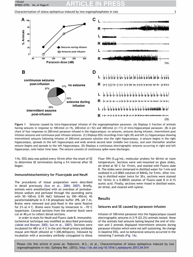
ARTICLE IN PRESS+ModelEPIRES-4759; No. of Pages 9
Characterization of status epilepticus induced by two organophosphates in rats 3
Figure 1 Seizures caused by intra-hippocampal infusion of the organophosphate paraoxon. (A) Displays % fraction of animalshaving seizures in response to 100 nmol (n = 9), 200 nmol (n = 52) and 300 nmol (n = 11) of intra-hippocampal paraoxon. (B) A piechart of four responses to 200 nmol paraoxon infused in the hippocampus: no seizures, seizures during infusion, intermittent postinfusion seizures and continuous post infusion seizures. (C) Displays EEG recordings from right (R) and left (L) hippocampus showingintermittent seizures following infusion of 200 nmol paraoxon solution into the right hippocampus. A seizure begins in the righthippocampus, spreads to the left hippocampus and ends several second later (middle two traces), and soon thereafter another
ys a
inuo
FtaBoifaa
R
S
Ieo
seizure begins and spreads to the left hippocampus. (D) Displahippocampi, note faster time base. The seizure consists of cont
1 Hz. EEG data was polled every 10 min after the onset of SEto determine SE termination during a 5 h interval after SEbegan.
Immunohistochemistry for Fluorojade and NeuN
The procedures of tissue preparation were describedin detail previously (Sun et al., 2004, 2007). Briefly,animals were anesthetized with an overdose of pentobar-bitone sodium and perfused through the ascending aortawith 50—100 mL 0.9% NaCl followed by 350—450 mL 4%paraformaldehyde in 0.1 M phosphate buffer (PB, pH 7.4).Brains were removed and post-fixed in the same fixativefor 2 h at 4 C. Brains were frozen by immersion in −70 Cisopentane. Coronal sections from the anterior block werecut at 40 m to collect dorsal sections.
In order to stain for NeuN and Fluoro-Jade B, immunohis-tochemical technique was modified as described previously
Please cite this article in press as: Todorovic, M.S., et al.,organophosphates in rats. Epilepsy Res. (2012), http://dx.doi.
(Jakab and Bowyer, 2002; Sun et al., 2007). Sections wereincubated for 48 h at 4 C in the anti-NeuN primary antibodymouse anti-NeuN (diluted at 1:200,Millipore), followed byincubation with a secondary antibody conjugated to Alexa
spir
continuous electrographic seizures occurring in right and leftus spike-wave discharges.
luor 594 (5 g/mL; molecular probes) for 60 min at roomemperature. Sections were wet-mounted on glass slides,ir-dried at 50 C for 15 min, and stained with Fluoro-Jade. The slides were immersed in distilled water for 1 min andxidized in a 0.006% solution of KMnO4 for 5 min. After rins-ng in distilled water twice for 30 s, sections were stainedor 10 min in a 0.0003% solution of Fluoro-Jade B in 0.1%cetic acid. Finally, sections were rinsed in distilled water,ir-dried, and cleared with xylene.
esults
eizures and SE caused by paraoxon infusion
nfusion of 100 nmol paraoxon into the hippocampus causedlectrographic seizures in 2/9 (22.2%) animals tested. Nonef the animals had seizures lasting beyond the end of infu-
Characterization of status epilepticus induced by twoorg/10.1016/j.eplepsyres.2012.04.014
ion and 2 animals displayed intermittent seizures duringaraoxon infusion which were not self-sustaining. No changen baseline EEG, and no behavioral seizures occurred in theemaining 7 animals (Fig. 1A).

ARTICLE IN PRESS+ModelEPIRES-4759; No. of Pages 9
4 M.S. Todorovic et al.
Figure 2 Images of hippocampal sections from animals that had prolonged seizures following intra-hippocampal paraoxon adminis-tration, which were stained for neuronal injury stain Fluoro-Jade B (green) and neuronal marker NeuN (red). (A) A low magnificationimage of the hippocampus showing drug infusion site close to the CA3 region of the hippocampus. (B) A section of the hippocampusdisplaying green Fluorojade positive CA3, CA1 pyramidal neurons and neurons in the subiculum. (C) A higher magnification imageo mpu
ctsddwao
iotahhfaa
peta4toem4
cttrotuTl
ris
iethnawpClw
P
Pmpfdwaqrcla
m5op
f Fluorojade positive neurons in the CA3 region of the hippoca
Infusion of 200 nmol paraoxon into the hippocampusaused electrographic seizures in 43/52 (82.7%) animalsested. In 32 animals seizures lasted beyond the end of infu-ion (61.5%) and 11 animals displayed intermittent seizuresuring paraoxon infusion which were not self-sustaining, andid not continue through the end of infusion (Fig. 1B). Thereas no change in baseline EEG in remaining 9 animals. Thesenimals did not display behavioral characteristics indicativef seizure activity.
Among animals with seizures lasting beyond paraoxonnfusion, two distinct types of electrographic seizure activityccurred: intermittent and continuous seizures. Intermit-ent seizures, which occurred in 18 animals, frequentlyppeared first in the paraoxon infusion site in the rightippocampus, and then spread to the contra-lateral (left)ippocampus (Fig. 1C). Seizures consisted of rhythmic highrequency spike wave discharges that evolved in frequencynd amplitude. There were brief periods of suppression ofctivity between seizures.
Continuous seizures occurred in 14 animals followingaraoxon infusion, and these consisted of sustained bilat-ral discharges of repetitive spike patterns evolving overime (Fig. 1D). Continuous electrographic seizures startedt approximately 15 min post infusion, and persisted for—18 h. These prolonged self-sustaining seizures consti-uted SE. Paraoxon-induced SE was further confirmed uponbservation of behavioral seizures. Animals experiencing SExhibited freezing, staring, blinking, and hyper-exploratoryovement, as well as wet-dog shaking movements for
—18 h.Infusion of 300 nmol paraoxon into the hippocampus
aused electrographic seizures in 10/11 (90.9%) of animalsested. The seizures in all of these animals continued beyondhe end of infusion in form of SE with behavioral seizuresanging from 2 to 5 on the Racine scale. The majorityf animals in SE (7) died of respiratory arrest, several of
Please cite this article in press as: Todorovic, M.S., et al.,organophosphates in rats. Epilepsy Res. (2012), http://dx.doi.
hese exhibited symptoms of peripheral cholinergic stim-lation, including muscle contractions and fasciculations.he remaining 3 animals survived, exhibiting seizures that
asted long beyond the end of infusion. Only 1 animal
bbia
s.
eceiving paraoxon 300 nM infusion displayed neither behav-oral characteristics indicative of seizure activity, nor EEGeizure activity (9.0%).
The location of the cannula was confirmed by section-ng the hippocampus. In many animals, the cannula andlectrode tracts were localized by sectioning the brain inhe plane of the electrode. In other animals, immuno-istochemistry for neuronal stain Neun and staining foreuro-degeneration dye Fluorojade J, was performed 3 daysfter SE caused by infusion of paraoxon. The site of infusionas at the ventricular border the CA3 layer of the hippocam-us (Fig. 2A). Fluorojade positive neurons were present inA1and CA3 regions and of the hippocampus and the subicu-
um (Fig. 2B and C). Occasional Fluorojade positive cellsere present in the hilus.
eripheral injection of paraoxon
reliminary experiments were performed to optimize theodel. Paraoxon (0.35 mg/kg) administered by the intra-eritoneal route caused widespread muscle contractions,asciculations, rare tonic convulsions, respiratory arrest andeath in all four animals tested. Pretreatment of animalsith scopolamine (4 mg/kg) protected against peripheralnd systemic effects of paraoxon (n = 4 animals). Subse-uently, based on the work of Deshpande et al. (2010) oximeeactivator 2-PAM and muscarinic antagonist atropine wasombined with peripheral paraoxon administration. In pre-iminary experiments we tested other doses of these agentsnd confirmed that most optimal doses were used.
Paraoxon (1 mg/kg) was administered SC to 23 ani-als 30 min after pre-treatment with 2 mg/kg atropine and
0 mg/kg 2-PAM and prolonged seizures were observed. SEccurred in 17 of the 23 animals (74%) with animals dis-laying a combination of chewing, head-bobbing, single and
Characterization of status epilepticus induced by twoorg/10.1016/j.eplepsyres.2012.04.014
ilateral limb clonus and rearing, leading to constant fullody tremors. In 3 animals there was no effect from the OPnjection and 3 animals died from respiratory arrest withoutny seizures.
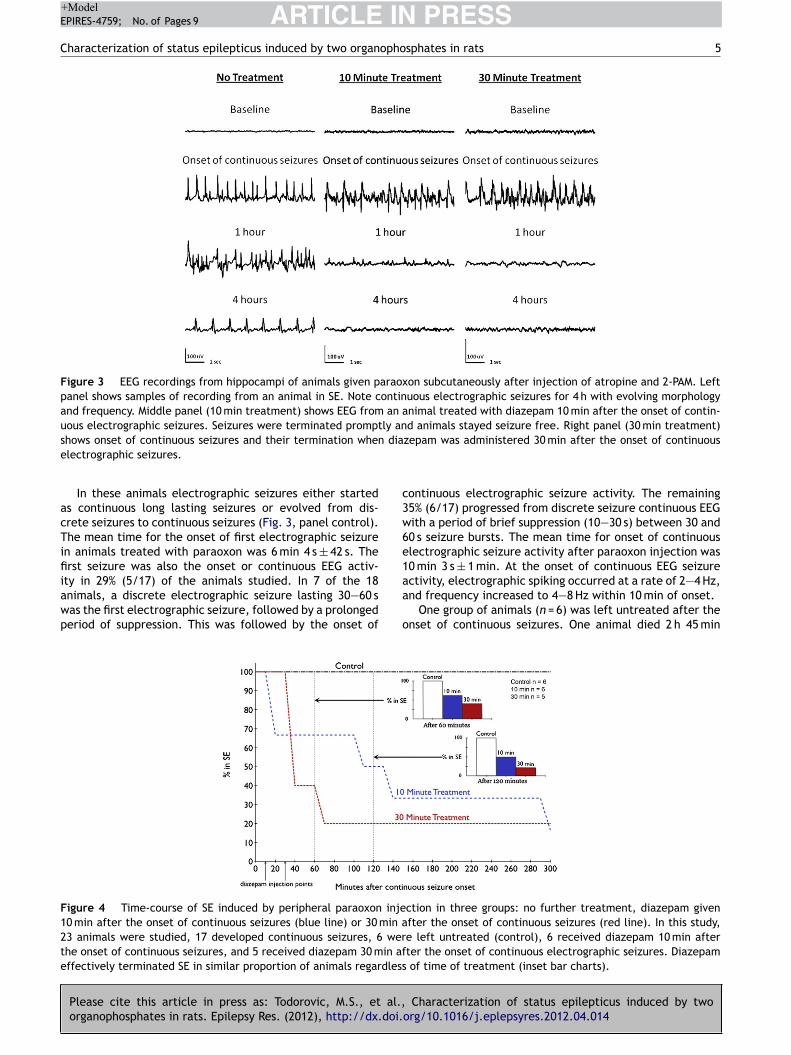
ARTICLE IN PRESS+ModelEPIRES-4759; No. of Pages 9
Characterization of status epilepticus induced by two organophosphates in rats 5
Figure 3 EEG recordings from hippocampi of animals given paraoxon subcutaneously after injection of atropine and 2-PAM. Leftpanel shows samples of recording from an animal in SE. Note continuous electrographic seizures for 4 h with evolving morphologyand frequency. Middle panel (10 min treatment) shows EEG from an animal treated with diazepam 10 min after the onset of contin-uous electrographic seizures. Seizures were terminated promptly and animals stayed seizure free. Right panel (30 min treatment)
dia
c3w6e1
shows onset of continuous seizures and their termination whenelectrographic seizures.
In these animals electrographic seizures either startedas continuous long lasting seizures or evolved from dis-crete seizures to continuous seizures (Fig. 3, panel control).The mean time for the onset of first electrographic seizurein animals treated with paraoxon was 6 min 4 s ± 42 s. Thefirst seizure was also the onset or continuous EEG activ-
Please cite this article in press as: Todorovic, M.S., et al.,organophosphates in rats. Epilepsy Res. (2012), http://dx.doi.
ity in 29% (5/17) of the animals studied. In 7 of the 18animals, a discrete electrographic seizure lasting 30—60 swas the first electrographic seizure, followed by a prolongedperiod of suppression. This was followed by the onset of
aa
o
Figure 4 Time-course of SE induced by peripheral paraoxon inje10 min after the onset of continuous seizures (blue line) or 30 min a23 animals were studied, 17 developed continuous seizures, 6 werthe onset of continuous seizures, and 5 received diazepam 30 min afeffectively terminated SE in similar proportion of animals regardless
zepam was administered 30 min after the onset of continuous
ontinuous electrographic seizure activity. The remaining5% (6/17) progressed from discrete seizure continuous EEGith a period of brief suppression (10—30 s) between 30 and0 s seizure bursts. The mean time for onset of continuouslectrographic seizure activity after paraoxon injection was0 min 3 s ± 1 min. At the onset of continuous EEG seizure
Characterization of status epilepticus induced by twoorg/10.1016/j.eplepsyres.2012.04.014
ctivity, electrographic spiking occurred at a rate of 2—4 Hz,nd frequency increased to 4—8 Hz within 10 min of onset.
One group of animals (n = 6) was left untreated after thenset of continuous seizures. One animal died 2 h 45 min
ction in three groups: no further treatment, diazepam givenfter the onset of continuous seizures (red line). In this study,e left untreated (control), 6 received diazepam 10 min afterter the onset of continuous electrographic seizures. Diazepam
of time of treatment (inset bar charts).

ARTICLE IN PRESS+ModelEPIRES-4759; No. of Pages 9
6 M.S. Todorovic et al.
Figure 5 SE induced by peripheral injection of DFP and response to diazepam treatment. Samples of recording from an animal inSE show continuous electrographic seizures for 4 h. Recordings an animal treated with diazepam 10 min after the onset of continuouselectrographic seizures (middle panel, 10 min treatment) demonstrate prompt termination of SE. When diazepam was administered30 min after the onset of continuous electrographic seizures (right panel 30 min treatment) SE continued with only a mild suppressiono disc(
awrew
tctmwsw9aws
aw(hcg
D
Pmadwa
ooidpl
i(rnaT4sIoswiciwoD
3rt
f amplitude of spikes but continued high frequency spike-wave4 h).
fter the onset of continuous seizures. The mean SE durationas 10 h 15 min ± 1 h 42 min in the remaining five animals,
anging from 7 to 17 h. The SE ended with the frequency ofpileptiform activity falling below 1 Hz in all 5 of the animalsithin 18 h of onset.
A group of animals (n = 6) in paraoxon induced SE wasreated with 10 mg/kg diazepam 10 min after the onset ofontinuous seizure activity (Fig. 3, middle panel). Diazepamerminated continuous seizures at this time point (Fig. 4,iddle panel). In two of these animals, SE was terminatedithin the first 10 min of treatment (Fig. 4). In these animals,
pike frequency dropped from 4—6 Hz to 0 and baseline EEGas restored. Seizures did not recur. In two animals SE ended0—150 min after onset (Fig. 4). The spike frequency gradu-lly dropped from 4 to 6 Hz with poly-spikes to single spikesith a frequency of less than 1 Hz (Fig. 3). All animals were
eizure free by 7 h. There was no mortality within 24 h.Animals (n = 5) treated with 10 mg/kg of diazepam 30 min
fter continuous EEG activity also responded to treatment,ith (60%) becoming seizure free within 10 min of treatment
Fig. 3, right panel). Only one of the five animals was stillaving seizures 70 min after continuous seizures began, andontinuous seizures lasted for 12 h (Fig. 4). No animal in thisroup died during the 24 h period.
FP
reliminary experiments were performed to optimize DFPodel of SE. Rats that were pretreated with 1 mg/kg
Please cite this article in press as: Todorovic, M.S., et al.,organophosphates in rats. Epilepsy Res. (2012), http://dx.doi.
tropine 30 min and then given 1.25 mg/kg DFP SC (n = 4) allied from respiratory arrest, whereas 50% of rats pretreatedith 1 mg/kg atropine and 25 mg/kg 2-PAM (n = 4) survivednd exhibited prolonged seizure activity. Higher DFP dose
d8
d
harges (1 h) and complex poly-spike-wave rhythmic discharges
f 1.5 mg/kg combined with a pretreatment of larger dosesf atropine (2 mg/kg) and 2-PAM (50 mg/kg) caused deathn 2 animals. These observations and previously publishedata suggested that 1.25 mg/kg DFP SC in cold saline with aretreatment of 2 mg/kg atropine and 50 mg/kg 2-PAM wereikely to produce SE in animals.
This combination was used to study DFP induced SE, andt caused continuous seizure activity and SE in 15 out of 1979%) of the animals studied, with the remainder dying ofespiratory arrest. Animals in SE group exhibited a combi-ation of chewing, head-bobbing, single and bi-limb clonusnd rearing, leading to constant full body tremors and SE.he mean time to first EEG seizure in this group was 21 min3 s ± 3 min 25 s. In 7 of 15 animals, the first electrographiceizure was also the onset of continuous seizure activity.n 7 other animals, seizure progression went from periodsf discrete seizures, separated by normal EEG followed byeizures lasting 10—30 s with periods of suppression, afterhich seizures merged into continuous epileptiform activ-
ty. In one animal, a brief discrete seizure was followed byontinuous seizure activity. Continuous epileptiform activ-ty started at a frequency of 2 Hz and accelerated to 4—6 Hzithin 10 min. The latency to the development of continu-us electrographic activity was 24 min 41 s ± 3 min 48 s afterFP injection.
In 5 animals left untreated after the DFP induced SE, survived to 5 h after continuous seizure onset and theemaining died. Seizures stopped in one animal 4.5 h afterhe onset of continuous seizure activity, while the other two
Characterization of status epilepticus induced by twoorg/10.1016/j.eplepsyres.2012.04.014
isplayed electrographic seizures which lasted for more than h.
DFP induced seizures were much more resistant toiazepam treatment when given at 30 min than when
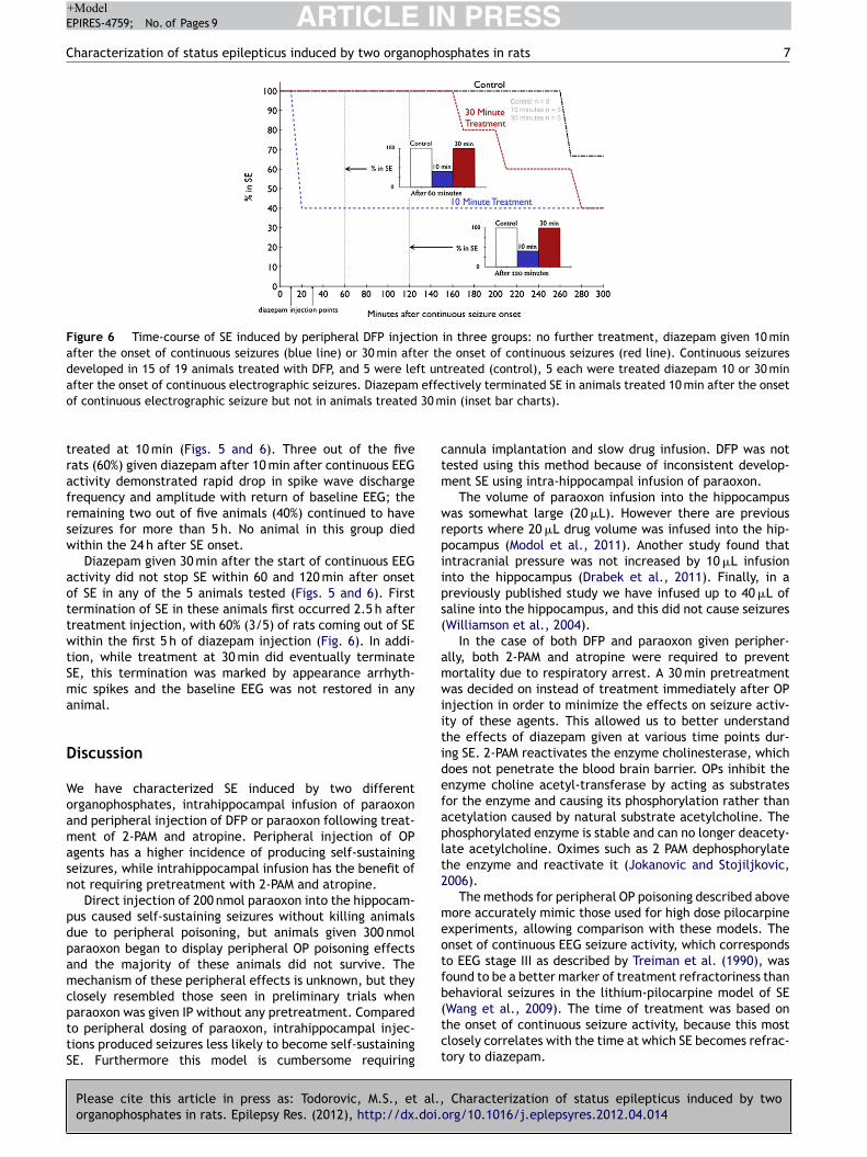
ARTICLE IN PRESS+ModelEPIRES-4759; No. of Pages 9
Characterization of status epilepticus induced by two organophosphates in rats 7
Figure 6 Time-course of SE induced by peripheral DFP injection in three groups: no further treatment, diazepam given 10 minafter the onset of continuous seizures (blue line) or 30 min after the onset of continuous seizures (red line). Continuous seizuresdeveloped in 15 of 19 animals treated with DFP, and 5 were left untreated (control), 5 each were treated diazepam 10 or 30 min
effe 30 m
ctm
wrpiips(
amwiitidefaplt2
meotfb
after the onset of continuous electrographic seizures. Diazepamof continuous electrographic seizure but not in animals treated
treated at 10 min (Figs. 5 and 6). Three out of the fiverats (60%) given diazepam after 10 min after continuous EEGactivity demonstrated rapid drop in spike wave dischargefrequency and amplitude with return of baseline EEG; theremaining two out of five animals (40%) continued to haveseizures for more than 5 h. No animal in this group diedwithin the 24 h after SE onset.
Diazepam given 30 min after the start of continuous EEGactivity did not stop SE within 60 and 120 min after onsetof SE in any of the 5 animals tested (Figs. 5 and 6). Firsttermination of SE in these animals first occurred 2.5 h aftertreatment injection, with 60% (3/5) of rats coming out of SEwithin the first 5 h of diazepam injection (Fig. 6). In addi-tion, while treatment at 30 min did eventually terminateSE, this termination was marked by appearance arrhyth-mic spikes and the baseline EEG was not restored in anyanimal.
Discussion
We have characterized SE induced by two differentorganophosphates, intrahippocampal infusion of paraoxonand peripheral injection of DFP or paraoxon following treat-ment of 2-PAM and atropine. Peripheral injection of OPagents has a higher incidence of producing self-sustainingseizures, while intrahippocampal infusion has the benefit ofnot requiring pretreatment with 2-PAM and atropine.
Direct injection of 200 nmol paraoxon into the hippocam-pus caused self-sustaining seizures without killing animalsdue to peripheral poisoning, but animals given 300 nmolparaoxon began to display peripheral OP poisoning effectsand the majority of these animals did not survive. Themechanism of these peripheral effects is unknown, but theyclosely resembled those seen in preliminary trials when
Please cite this article in press as: Todorovic, M.S., et al.,organophosphates in rats. Epilepsy Res. (2012), http://dx.doi.
paraoxon was given IP without any pretreatment. Comparedto peripheral dosing of paraoxon, intrahippocampal injec-tions produced seizures less likely to become self-sustainingSE. Furthermore this model is cumbersome requiring
(tct
ctively terminated SE in animals treated 10 min after the onsetin (inset bar charts).
annula implantation and slow drug infusion. DFP was notested using this method because of inconsistent develop-ent SE using intra-hippocampal infusion of paraoxon.The volume of paraoxon infusion into the hippocampus
as somewhat large (20 L). However there are previouseports where 20 L drug volume was infused into the hip-ocampus (Modol et al., 2011). Another study found thatntracranial pressure was not increased by 10 L infusionnto the hippocampus (Drabek et al., 2011). Finally, in areviously published study we have infused up to 40 L ofaline into the hippocampus, and this did not cause seizuresWilliamson et al., 2004).
In the case of both DFP and paraoxon given peripher-lly, both 2-PAM and atropine were required to preventortality due to respiratory arrest. A 30 min pretreatmentas decided on instead of treatment immediately after OP
njection in order to minimize the effects on seizure activ-ty of these agents. This allowed us to better understandhe effects of diazepam given at various time points dur-ng SE. 2-PAM reactivates the enzyme cholinesterase, whichoes not penetrate the blood brain barrier. OPs inhibit thenzyme choline acetyl-transferase by acting as substratesor the enzyme and causing its phosphorylation rather thancetylation caused by natural substrate acetylcholine. Thehosphorylated enzyme is stable and can no longer deacety-ate acetylcholine. Oximes such as 2 PAM dephosphorylatehe enzyme and reactivate it (Jokanovic and Stojiljkovic,006).
The methods for peripheral OP poisoning described aboveore accurately mimic those used for high dose pilocarpine
xperiments, allowing comparison with these models. Thenset of continuous EEG seizure activity, which correspondso EEG stage III as described by Treiman et al. (1990), wasound to be a better marker of treatment refractoriness thanehavioral seizures in the lithium-pilocarpine model of SE
Characterization of status epilepticus induced by twoorg/10.1016/j.eplepsyres.2012.04.014
Wang et al., 2009). The time of treatment was based onhe onset of continuous seizure activity, because this mostlosely correlates with the time at which SE becomes refrac-ory to diazepam.
IN+ModelE
8
ciaeierg2its1
ddp2Sdacatdd
poooiptrotDO
A
TNNNa
R
A
B
C
D
D
F
F
G
G
H
H
H
J
J
J
J
K
K
K
L
ARTICLEPIRES-4759; No. of Pages 9
Both DFP and paraoxon are OPs, which increase con-entration of acetylcholine levels in the brain rapidly bynhibiting its breakdown by the enzyme cholinesterase (Shihnd McDonough, 1997). This elevation in acetylcholine lev-ls is likely to activate muscarinic and nicotinic receptorsn the hippocampus (Harrison et al., 2004; Kozhemyakint al., 2010). It was recently suggested that muscariniceceptor activation by paraoxon causes increased release oflutamate from presynaptic terminals (Kozhemyakin et al.,010). In vitro models of recurrent bursting suggest thatncreased presynaptic release (frequency of EPSC) can con-ribute to the development of neuronal synchrony andeizures (Mangan and Kapur, 2004; Traub and Dingledine,990).
DFP-induced SE became resistant to treatment withiazepam as SE progressed. This phenomenon has beenescribed previously in SE induced by electrical stimulation,ilocarpine, lithium-pilocarpine and soman (Jones et al.,002; Kapur and Macdonald, 1997; Mazarati et al., 1998a;hih et al., 1999). However, the paraoxon model for SEoes not exhibit the same phenomenon, making it less suit-ble as a surrogate for military OP poisoning studies. Theurrent recommendation of 2-PAM, atropine and diazepams a treatment for OP poisoning is incomplete because ofhe time dependent nature of its treatment. These studiesemonstrate the importance of finding novel methods forealing with and treating victims of OP poisoning.
Previous studies of seizures caused by organophos-hate cholinesterase inhibitors have largely been carriedut in defense labs using agents such as sarin VX andther nerve agents (McDonough and Shih, 1997). However,rganophosphate poisoning needs to be studied in civil-an research laborites because organophosphate pesticideoisoning afflicts civilians and civilian populations can beargets of nerve agent attacks. A World Health Organizationeport suggested that there were more than 2 million casesf accidental or intentional organophosphate poisoning inhe world each year (Jeyaratnam, 1990). Development ofFP and paraoxon models can lead to better treatments forP poisoning.
cknowledgments
he research is supported by the CounterACT Program,ational Institutes of Health Office of the Director, and theational Institute Neurological Disorders and Stroke, Grantumbers NIH-NINDS UO1 NS58204 and RO1 NS040337 andlso by the Department of Defense grant PR093963.
eferences
lldredge, B.K., Gelb, A.M., Isaacs, S.M., Corry, M.D., Allen, F.,Ulrich, S., Gottwald, M.D., O’Neil, N., Neuhaus, J.M., Segal,M.R., Lowenstein, D.H., 2001. A comparison of lorazepam,diazepam, and placebo for the treatment of out-of-hospital sta-tus epilepticus. N. Engl. J. Med. 345, 631—637.
ird, S.B., Gaspari, R.J., Dickson, E.W., 2003. Early death due tosevere organophosphate poisoning is a centrally mediated pro-
Please cite this article in press as: Todorovic, M.S., et al.,organophosphates in rats. Epilepsy Res. (2012), http://dx.doi.
cess. Acad. Emerg. Med. 10, 295—298.hen, J.W., Naylor, D.E., Wasterlain, C.G., 2007. Advances in
the pathophysiology of status epilepticus. Acta Neurol. Scand.Suppl. 186, 7—15.
L
PRESSM.S. Todorovic et al.
eshpande, L.S., Carter, D.S., Blair, R.E., DeLorenzo, R.J., 2010.Development of a prolonged calcium plateau in hippocampalneurons in rats surviving status epilepticus induced by theorganophosphate diisopropylfluorophosphate. Toxicol. Sci. 116,623—631.
rabek, T., Janata, A., Jackson, E.K., End, B., Stezoski, J., Vagni,V.A., Janesko-Feldman, K., Wilson, C.D., van, R.N., Tisherman,S.A., Kochanek, P.M., 2011. Microglial depletion using intrahip-pocampal injection of liposome-encapsulated clodronate inprolonged hypothermic cardiac arrest in rats. Resuscitation,http://dx.doi.org/10.1016/j.resuscitation.2011.09.016(Epubahead of print).
ujikawa, D.G., 2005. Prolonged seizures and cellular injury: under-standing the connection. Epilepsy Behav. 7 (Suppl. 3), S3—S11.
ujikawa, D.G., Itabashi, H.H., Wu, A., Shinmei, S.S., 2000. Statusepilepticus-induced neuronal loss in humans without systemiccomplications or epilepsy. Epilepsia 41, 981—991.
arcia, S.J., Abu-Qare, A.W., Meeker-O’Connell, W.A., Borton,A.J., Abou-Donia, M.B., 2003. Methyl parathion: a review ofhealth effects. J. Toxicol. Environ. Health B: Crit. Rev. 6,185—210.
ralewicz, S., Tomas, T., Socko, R., 1989. Effects of single expo-sure to chlorphenvinphos, an organophosphate insecticide, onelectrical activity (EEG) of the rat brain. Pol. J. Occup. Med. 2,309—320.
arrison, P.K., Sheridan, R.D., Green, A.C., Scott, I.R., Tatter-sall, J.E., 2004. A guinea pig hippocampal slice model oforganophosphate-induced seizure activity. J. Pharmacol. Exp.Ther. 310, 678—686.
offmann, U., Papendorf, T., 2006. Organophosphate poison-ings with parathion and dimethoate. Intensive Care Med. 32,464—468.
onchar, M.P., Olney, J.W., Sherman, W.R., 1983. Systemic choliner-gic agents induce seizures and brain damage in lithium-treatedrats. Science 220, 323—325.
akab, R.L., Bowyer, J.F., 2002. Parvalbumin neuron circuits andmicroglia in three dopamine-poor cortical regions remain sensi-tive to amphetamine exposure in the absence of hyperthermia,seizure and stroke. Brain Res. 958, 52—69.
eyaratnam, J., 1990. Acute pesticide poisoning: a major globalhealth problem. World Health Stat. Q 43, 139—144.
okanovic, M., Stojiljkovic, M.P., 2006. Current understanding of theapplication of pyridinium oximes as cholinesterase reactivatorsin treatment of organophosphate poisoning. Eur. J. Pharmacol.553, 10—17.
ones, D.M., Esmaeil, N., Maren, S., Macdonald, R.L., 2002. Char-acterization of pharmacoresistance to benzodiazepines in therat Li-Pilocarpine model of status epilepticus. Epilepsy Res. 50,301—312.
adriu, B., Guidotti, A., Costa, E., Davis, J.M., Auta, J., 2011. Acuteimidazenil treatment after the onset of DFP-induced seizure ismore effective and longer lasting than midazolam at prevent-ing seizure activity and brain neuropathology. Toxicol. Sci. 120,136—145.
apur, J., Macdonald, R.L., 1997. Rapid seizure-induced reductionof benzodiazepine and Zn2+ sensitivity of hippocampal dentategranule cell GABAA receptors. J. Neurosci. 17, 7532—7540.
ozhemyakin, M., Rajasekaran, K., Kapur, J., 2010. Centralcholinesterase inhibition enhances glutamatergic synaptic trans-mission. J. Neurophysiol. 103, 1748—1757.
i, Y., Lein, P.J., Liu, C., Bruun, D.A., Tewolde, T., Ford, G., Ford,B.D., 2011. Spatiotemporal pattern of neuronal injury inducedby DFP in rats: a model for delayed neuronal cell death followingacute OP intoxication. Toxicol. Appl. Pharmacol. 253, 261—269.
Characterization of status epilepticus induced by twoorg/10.1016/j.eplepsyres.2012.04.014
othman, E.W., Bertram, E.H., Bekenstein, J.W., Perlin, J.B., 1989.Self-sustaining limbic status epilepticus induced by ‘continuous’hippocampal stimulation: electrographic and behavioral charac-teristics. Epilepsy Res. 3, 107—119.
IN+Model
opho
S
T
T
T
T
T
W
W
W
Z
Z
ARTICLEEPIRES-4759; No. of Pages 9
Characterization of status epilepticus induced by two organ
Lothman, E.W., Salerno, R.A., Perlin, J.B., et al., 1988. Screeningand characterization of antiepileptic drugs with rapidly recurringhippocampal seizures in rats. Epilepsy Res. 2, 367—379.
Lothman, E.W., Collins, R.C., Ferrendelli, J.A., 1981. Kainic acid-induced limbic seizures: electrophysiologic studies. Neurology31, 806—812.
Mangan, P.S., Kapur, J., 2004. Factors underlying bursting behaviorin a network of cultured hippocampal neurons exposed to zeromagnesium. J. Neurophysiol. 91, 946—957.
Mazarati, A.M., Baldwin, R.A., Sankar, R., Wasterlain, C.G., 1998a.Time-dependent decrease in the effectiveness of antiepilep-tic drugs during the course of self-sustaining status epilepticus.Brain Res. 814, 179—185.
Mazarati, A.M., Wasterlain, C.G., Sankar, R., Shin, D., 1998b. Selfsustaining status epilepticus after brief electrical stimulation ofthe perforant path. Brain Res. 801, 251—253.
McDonough Jr., J.H., Shih, T.M., 1997. Neuropharmacological mech-anisms of nerve agent-induced seizure and neuropathology.Neurosci. Biobehav. Rev. 21, 559—579.
Modol, L., Darbra, S., Pallares, M., 2011. Neurosteroids infusioninto the CA1 hippocampal region on exploration, anxiety-like behaviour and aversive learning. Behav. Brain Res. 222,223—229.
Morita, H., Yanagisawa, N., Nakajima, T., Shimizu, M., Hirabayashi,H., Okudera, H., Nohara, M., Midorikawa, Y., Mimura, S.,1995. Sarin poisoning in Matsumoto, Japan. Lancet 346,290—293.
Nozaki, H., Aikawa, N., Shinozawa, Y., Hori, S., Fujishima, S.,Takuma, K., Sagoh, M., 1995. Sarin poisoning in Tokyo subway.Lancet 345, 980—981.
Racine, R.J., 1972. Modification of seizure activity by electricalstimulation. II. Motor seizure. Electroencephalogr. Clin. Neuro-physiol. 32, 281—294.
Shih, T.M., McDonough Jr., J.H., 1997. Neurochemical mechanismsin soman-induced seizures. J. Appl. Toxicol. 17, 255—264.
Shih, T., McDonough Jr., J.H., Koplovitz, I., 1999. Anticonvul-sants for soman-induced seizure activity. J. Biomed. Sci. 6,
Please cite this article in press as: Todorovic, M.S., et al.,organophosphates in rats. Epilepsy Res. (2012), http://dx.doi.
86—96.Sun, C., Sieghart, W., Kapur, J., 2004. Distribution of alpha1,
alpha4, gamma2, and delta subunits of GABAA receptors in hip-pocampal granule cells. Brain Res. 1029, 207—216.
PRESSsphates in rats 9
un, C., Mtchedlishvili, Z., Bertram, E.H., Erisir, A., Kapur, J.,2007. Selective loss of dentate hilar interneurons contributesto reduced synaptic inhibition of granule cells in an electricalstimulation-based animal model of temporal lobe epilepsy. J.Comp. Neurol. 500, 876—893.
raub, R.D., Dingledine, R., 1990. Model of synchronized epilep-tiform bursts induced by high potassium in CA3 region of rathippocampal slice. Role of spontaneous EPSPs in initiation. J.Neurophysiol. 64, 1009—1018.
reiman, D.M., 2007. Treatment of convulsive status epilepticus.Int. Rev. Neurobiol. 81, 273—285.
reiman, D.M., Meyers, P.D., Walton, N.Y., Collins, J.F., Colling, C.,Rowan, A.J., Handforth, A., Faught, E., Calabrese, V.P., Uth-man, B.M., Ramsay, R.E., Mamdani, M.B., 1998. A comparison offour treatments for generalized convulsive status epilepticus. N.Engl. J. Med. 339, 792—798.
reiman, D.M., Walton, N.Y., Kendrick, C., 1990. A progressivesequence of electroencephalographic changes during general-ized convulsive status epilepticus. Epilepsy Res. 5, 49—60.
urski, W.A., Cavalheiro, E.A., Schwarz, M., Czuczwar, S.J., Klein-rok, Z., Turski, L., 1983. Limbic seizures produced by pilocarpinein rats: behavioural, electroencephalographic and neuropatho-logical study. Behav. Brain Res. 9, 315—335.
ang, N.C., Good, L.B., Marsh, S.T., Treiman, D.M., 2009. EEGstages predict treatment response in experimental status epilep-ticus. Epilepsia 50, 949—952.
asterlain, C.G., Fujikawa, D.G., Penix, L., Sankar, R., 1993.Pathophysiological mechanisms of brain damage from statusepilepticus. Epilepsia 34 (Suppl. 1), S37—S53.
illiamson, J., Mtchedlishvili, Z., Kapur, J., 2004. Characterizationof the convulsant action of pregnenolone sulfate. Neuropharma-cology 46, 856—864.
aja-Milatovic, S., Gupta, R.C., Aschner, M., Milatovic, D., 2009.Protection of DFP-induced oxidative damage and neurodegen-eration by antioxidants and NMDA receptor antagonist. Toxicol.Appl. Pharmacol. 240, 124—131.
hu, H., O’Brien, J.J., O’Callaghan, J.P., Miller, D.B., Zhang,
Characterization of status epilepticus induced by twoorg/10.1016/j.eplepsyres.2012.04.014
Q., Rana, M., Tsui, T., Peng, Y., Tomesch, J., Hendrick, J.P.,Wennogle, L.P., Snyder, G.L., 2010. Nerve agent exposure elicitssite-specific changes in protein phosphorylation in mouse brain.Brain Res. 1342, 11—23.