ad award number: w81xwh-11-1-0264 title: role of merlin ... · immunofluorescence-based analysis to...
TRANSCRIPT

AD_________________
Award Number: W81XWH-11-1-0264 TITLE: Role of merlin/NF2 in mTOR signaling and meningioma growth PRINCIPAL INVESTIGATOR: Vijaya Ramesh, Ph.D. Anat Stemmer-Rachamimov, M.D. CONTRACTING ORGANIZATION: Massachusetts General Hospital Boston, MA 02114 REPORT DATE: April 2012 TYPE OF REPORT: Annual PREPARED FOR: U.S. Army Medical Research and Materiel Command Fort Detrick, Maryland 21702-5012 DISTRIBUTION STATEMENT: Approved for Public Release; Distribution Unlimited The views, opinions and/or findings contained in this report are those of the author(s) and should not be construed as an official Department of the Army position, policy or decision unless so designated by other documentation.

REPORT DOCUMENTATION PAGE Form Approved
OMB No. 0704-0188 Public reporting burden for this collection of information is estimated to average 1 hour per response, including the time for reviewing instructions, searching existing data sources, gathering and maintaining the data needed, and completing and reviewing this collection of information. Send comments regarding this burden estimate or any other aspect of this collection of information, including suggestions for reducing this burden to Department of Defense, Washington Headquarters Services, Directorate for Information Operations and Reports (0704-0188), 1215 Jefferson Davis Highway, Suite 1204, Arlington, VA 22202-4302. Respondents should be aware that notwithstanding any other provision of law, no person shall be subject to any penalty for failing to comply with a collection of information if it does not display a currently valid OMB control number. PLEASE DO NOT RETURN YOUR FORM TO THE ABOVE ADDRESS. 1. REPORT DATE April 2012
2. REPORT TYPEAnnual
3. DATES COVERED 15 March 2011 – 14 March 2012
4. TITLE AND SUBTITLE
5a. CONTRACT NUMBER
Role of merlin/NF2 in mTOR signaling and meningioma growth 5b. GRANT NUMBER W81XWH-11-1-0264
5c. PROGRAM ELEMENT NUMBER
6. AUTHOR(S)
5d. PROJECT NUMBER
Vijaya Ramesh, Ph.D. Anat Stemmer-Rachamimov, M.D.
5e. TASK NUMBER
E-Mail: [email protected]
5f. WORK UNIT NUMBER
7. PERFORMING ORGANIZATION NAME(S) AND ADDRESS(ES)
8. PERFORMING ORGANIZATION REPORT NUMBER
Massachusetts General Hospital Boston, MA 02114
9. SPONSORING / MONITORING AGENCY NAME(S) AND ADDRESS(ES) 10. SPONSOR/MONITOR’S ACRONYM(S)U.S. Army Medical Research and Materiel Command Fort Detrick, Maryland 21702-5012 11. SPONSOR/MONITOR’S REPORT NUMBER(S) 12. DISTRIBUTION / AVAILABILITY STATEMENT Approved for Public Release; Distribution Unlimited 13. SUPPLEMENTARY NOTES
14. ABSTRACT The scope of this research project is to mechanistically define how merlin regulates mTORC1 signaling, to examine signaling downstream of mTORC2 and to validate the efficacy of mTOR inhibitors in both in vitro and in vivo preclinical models. The results obtained during assay development of an unbiased kinome screening clearly establish that Rheb is required for mTORC1 activation mediated by NF2 loss, supporting our hypothesis that NF2 may function through TSC1-TSC2 protein complex. Similar to TSC proteins, merlin negatively regulates mTORC1 and positively regulates mTORC2 However, contrary to activation of mTORC1, the attenuated mTORC2 signaling profiles exhibited by normal arachnoid and Schwann cells in response to acute merlin loss are not consistently reflected in NF2-deficient meningiomas and schwannomas, suggesting that additional genetic events may have been acquired in tumors after initial merlin loss. Our results show that mTOR kinase inhibitor such as Torin1 is more effective in blocking signaling and inhibiting proliferation of benign (WHO grdae1) and atypical (WHO grade 2) meningioma cells. A manuscript describing these results is now in press and expected to be published soon. We have shown that implantation of benign meningioma cells form tumors and may serve as valuable preclinical model for NF2. 15. SUBJECT TERMS None provided.
16. SECURITY CLASSIFICATION OF:
17. LIMITATION OF ABSTRACT
18. NUMBER OF PAGES
19a. NAME OF RESPONSIBLE PERSONUSAMRMC
a. REPORT U
b. ABSTRACT U
c. THIS PAGEU
UU
19
19b. TELEPHONE NUMBER (include area code)
Role of Merlin/NF2 in mTOR Signaling and Meningioma Growth P.I. Vijaya Ramesh
3
Table of Contents
Page
Introduction 4
Body 4
Key Research Accomplishments 7
Reportable Outcomes 8
Conclusion 8
References 8
Appendices 9

Role of Merlin/NF2 in mTOR Signaling and Meningioma Growth P.I. Vijaya Ramesh
4
Introduction
Neurofibromatosis 2 (NF2) is a dominantly inherited disorder characterized by multiple benign nervous system tumors, including schwannomas and meningiomas. Although merlin is implicated in a wide range of cellular activities, the precise growth inhibitory mechanism in human arachnoidal and Schwann cells, and how its loss results in tumor formation from these specific cell types in NF2 remains poorly understood. We believe that elucidating the cell/context-dependent functions of merlin will be critical in understanding the growth mechanisms of NF2-associated meningiomas and schwannomas. The tumor suppressor product of NF2 encodes merlin, a member of the ezrin-radixin-moesin (ERM) protein family that functions to link membrane proteins to the cortical actin cytoskeleton.
Employing human-derived, merlin-deficient meningiomas and merlin-suppressed arachnoidal cells, the non-neoplastic cell counterpart of meningiomas, we recently identified merlin as a novel negative regulator of mammalian target of rapamycin complex 1 (mTORC1) signaling. We believe that merlin functions upstream of the Tuberous Sclerosis Complex (TSC) proteins to regulate mTORC1. However, unlike in NF1, it does not regulate mTORC1 via the established PI3K/Akt- or MAPK/ERK-mediated TSC2 inactivation and may instead regulate TSC/mTORC1 signaling in a novel fashion (James et al., 2009). The scope of this research is to mechanistically define how merlin regulates mTORC1 signaling, to obtain insights as to whether aberrant activation of mTORC1 signaling and signaling downstream of mTORC2 can explain the benign nature of tumors in NF2, and to validate the efficacy of mTOR inhibitors in both in vitro and in vivo preclinical models. Body Research Accomplishments Task 1: To define the mechanism(s) by which merlin regulates mTORC1 Hypothesis: NF2 regulation of TORC1 is dependent on an intact TSC complex. Absence of merlin in meningiomas could result in degradation/decrease of TSC complex activity through novel phosphorylation sites in TSC1 and/or TSC2, which are mediated by as yet unidentified kinases in merlin-deficient cells. Loss-of-function kinome screen
We are in the final stages of assay development for performing a high-throughput, loss-of-function kinome screen to identify kinases involved in NF2-regulated mTORC2 signaling. A library of specific, short-hairpin RNAs (shRNAs), developed by The RNAi Consortium (TRC; Broad Institute/MIT), will be used to suppress ~800 known kinases in NF2-deficient arachnoidal (AC) and meningioma cells. For this screen, distinct shRNAs that target each kinase are introduced into NF2-suppressed arachnoidal cells (ACs) or a patient-derived, NF2-deficient benign meningioma cell line (BenMen1) by lentiviral infection in a 384-well format. In order to avoid false-positive effects, at least 5 distinct shRNAs are used for each individual target kinase, followed by immunofluorescence-based analysis to look for decrease of mTORC1 pathway activation. A two-fold decrease in mTORC1 signaling observed with at least 2 of the 5 shRNAs would be scored as positive. Specific kinases that lead to decreased pathway activation when suppressed will be considered as strong candidates for causing constitutive activation of mTORC1 in response to NF2

Role of Merlin/NF2 in mTOR Signaling and Meningioma Growth P.I. Vijaya Ramesh
5
loss. A standard fluorescent staining readout used to define the activation state of the mTORC1 pathway is phosphorylated S6 (P-S6Ser240/244). In addition, DAPI staining is also used to tag each cell nucleus in order to quantitate the level of P-S6 staining/cell. NF2 regulates the mTORC1 pathway in a Rheb-dependent manner
During the assay development phase, we carried out RNAi-mediated suppression of four established mTORC1 pathway targets (5 shRNAs/target) in NF2-suppressed ACs under serum-deprived conditions. These targets included mTORC1 complex components mTOR and Raptor; p70S6K, a direct target of mTORC1 and upstream kinase for phosphorylating S6 at Ser240/244 and Rheb, a small GTPase that activates mTORC1 in response to inactivation of TSC1-TSC2 complex. Immunofluorescence screening and quantitation was performed using the Acumen eX3 laser scanning image cytometer. As predicted, decreased P-S6Ser240/244 staining was observed using shRNAs for mTOR (2/5 shRNAs), Raptor (3/5 shRNAs), and S6K (2/5 shRNAs) compared to a null control shRNA. Importantly, hairpins targeting the mTORC1 regulator Rheb (3/5 shRNAs) also demonstrated downregulation of P-S6 (Figures 1 and 2). These results suggest that the NF2-mediated regulation of mTORC1 is Rheb-dependent, further supporting the likelihood that NF2 functions upstream of TSC1-TSC2, and loss of NF2 may lead to inactivation of the TSC complex.
Figure 1. Representative figure shows DAPI staining (blue) of individual cell nuclei along with P-S6 (green) used as an mTORC1 pathway readout. Left panel shows NF2-suppressed ACs infected with a null control shRNA, which retain the constitutive P-S6 activation. Right panel shows NF2-suppressed ACs infected with a Rheb-specific shRNA where abnormal P-S6 activation is reversed (decreased green signal).
Figure 2. Preliminary
results show downregulation of P-S6
staining following shRNA-mediated suppression of several mTORC1-related pathway targets (using 5 shRNAs/target) including mTOR, Rheb, p70S6K and Raptor compared with null control shRNA

Role of Merlin/NF2 in mTOR Signaling and Meningioma Growth P.I. Vijaya Ramesh
6
Task 2: To define the mechanisms that limit the malignancy potential of benign meningiomas and vestibular schwannomas in NF2 when mTORC1 is activated Hypothesis: mTORC1-dependent feedback mechanisms regulating IRS-1 and PDGFR functions, as well as impaired mTORC2 activity, may be responsible for the observed attenuation of Akt activation upon merlin loss in arachnoidal cells. The lack of Akt activation will affect the survival properties of merlin negative meningioma cells, which will be examined by treating benign and atypical meningioma cells with compounds that inhibit mTORC1 and Akt. Regulation of mTORC2 signaling in NF2-deficient target cell types
Previously, we demonstrated that merlin loss results in hyperactivation of mTORC1 in vitro and in vivo and that Akt signaling is impaired in response to insulin stimulation through an mTORC1-mediated negative feedback loop (James et al., 2009). Deregulation of mTORC1 signaling in merlin-deficient arachnoid cells is reminiscent of TSC deficiency in cells/tumors suggesting that growth control mechanisms may be overlapping in TSC and NF2 tumor suppressor syndromes. The TSC1-TSC2 complex, in addition to its role in inhibiting mTORC1, was shown to interact with mTORC2 and positively regulates its kinase activity (Huang et al., 2008). We have examined the regulation of mTORC2 signaling by merlin in NF2 target cell types and tumors. Our results show that merlin positively regulates the kinase activity of mTORC2, a second functionally distinct mTOR complex, and that downstream phosphorylation of mTORC2 substrates, including Akt, is reduced upon acute merlin deficiency in cells. In response to general growth factor stimulation, Akt signaling is attenuated in merlin-suppressed human arachnoid and Schwann cells through mechanisms mediated by hyperactive mTORC1 and impaired mTORC2. Moreover, Akt signaling is impaired differentially in a cell type-dependent manner in response to distinct growth factor stimuli (Figure 1 and 2 in the appended paper). However, contrary to activation of mTORC1, the attenuated mTORC2 signaling profiles exhibited by normal arachnoid and Schwann cells in response to acute merlin loss were not consistently reflected in NF2-deficient meningiomas and schwannomas with chronic merlin loss, suggesting additional genetic events may have been acquired in tumors after initial merlin loss (Figure 4 and Table1 in the appended paper). This finding contrasts with TSC, which exhibits attenuated mTORC2 signaling profiles in both cells and tumors. Furthermore, we tested the efficacy of mTOR pathway inhibitors including the mTORC1 inhibitor rapamycin; an ATP-competitive mTOR inhibitor Torin1 that potently inhibits both mTORC1 and mTORC2 complex; and the dual PI3K/mTOR inhibitor PI-103 on primary benign and atypical meningioma cells. We observed that Torin1 was more effective in blocking mTORC1 and Akt activation in meningioma cells in vitro than rapamycin and PI-103, and more effective than rapamycin in inhibiting cell proliferation (Figure 5 and 6 in the appended paper). A manuscript detailing these results is published and is included in the Appendix of this progress report.
Task 3: To test mTOR/PI3K inhibitors in a preclinical model of NF2-associated benign meningiomas Hypothesis: Intracranial implantation in mice of hTERT-immortalized, merlin-deficient human arachnoidal cells and patient-derived meningioma cells with aberrant mTORC1 activation will result in benign meningiomas resembling human NF2, which will be an appropriate in vivo preclinical model for biological and therapeutic studies. We have proposed the development of a relevant in vivo preclinical meningioma mouse model for use in determining the efficacy of mTOR and dual mTOR/PI3K inhibitors as potential therapeutics for NF2. To establish an appropriate NF2 meningioma tumor model we have utilized human

Role of Merlin/NF2 in mTOR Signaling and Meningioma Growth P.I. Vijaya Ramesh
7
immortalized normal arachnoid and patient-derived meningioma cells that we have engineered to stably express GFP and firefly luciferase by lentiviral-mediated infection (LV-GFP-Fluc) for use in monitoring tumor progression. For intracranial implantation experiments in nude mice, immortalized normal arachnoidal cells were used as control cells, and merlin-deficient arachnoidal cells or immortalized benign meningioma cells (BenMen1) cells were used for developing suitable tumor models. In preliminary studies, we observed by bioluminescence imaging (BLI) that merlin-deficient cells implanted at the cerebral convexity was optimal to the skull base location in sustaining the growth of implanted cells. In a pilot study of 20 mice, 10 each of nu/nu or NOD/SCID immunodeficient mice, we determined that both mice strains were equally sufficient to support the development of meningioma tumors. In initial studies, in collaboration with Dr. H. Wakimoto of MGH Neurosurgery, we have implanted approximately 0.75 x 10⁶ BenMen1 cells in a 5 µl volume at the cerebral convexity and followed progression of tumor development by BLI through 7 months. Representative BLI and H&E staining images of a mouse brain sacrificed 6.5 months post implantation demonstrate meningioma development (Figure 3).
Key Research Accomplishments § Rheb is required for NF2 loss to activate mTORC1 signaling, strengthening the idea that NF2 functions through Rheb-TSC1-TSC2 complex. § NF2 positively regulates mTORC2 signaling and acute loss of merlin in human arachnoidal and Schwann cells results in decrease in phosphorylation of mTORC2 targets. § NF2-associated meningiomas with chronic loss of merlin do not completely resemble acute loss of merlin in human arachnoidal cells, suggesting that either compensatory mechanisms or additional genetic events occur in meningiomas subsequent to initial merlin loss.
Figure 3. Bioluminescence imaging and histological staining of a brain from a mouse implanted with immortalized merlin-deficient benign meningioma cells. Representative nu/nu mouse sacrificed at 6.5 months post-implantation of BenMen1 cells. BLI imaging (2.70 E+07) demonstrates Fluc-expressing meningioma cells (A) and meningioma growth by H&E staining (B).
meningioma
A. B.

Role of Merlin/NF2 in mTOR Signaling and Meningioma Growth P.I. Vijaya Ramesh
8
§ Torin1, an ATP-competitive mTOR kinase inhibitor is more effective than rapamycin in inhibiting signaling and proliferation of benign and atypical meningioma cells. Reportable Outcome Marianne F. James, Elizabeth Stivison, Roberta Beauchamp, Sangyeul Han, James F. Gusella, Margaret R. Wallace, Anat Stemmer-Rachamimov, and Vijaya Ramesh. Signaling events downstream of mammalian target of rapamycin complex 2 (mTORC2) in NF2- deficient target cell types. (2011) Abstract presented at the Children’s Tumor Foundation meeting. Marianne F. James, Elizabeth Stivison, Roberta Beauchamp, Sangyeul Han, Hua Li, Margaret R. Wallace, James F. Gusella, Anat Stemmer-Rachamimov, and Vijaya Ramesh. Regulation of mTORC2 signaling in NF2-deficient target cell types (2012) Mol. Cancer Res. 10:649-659. Conclusion We are in the final stages of assay development for the unbiased large-scale kinome screen that we will be undertaking in NF2-deficient arachnoidal and meningioma cells (Task1). The results obtained during assay development clearly establish that Rheb is required for mTORC1 activation mediated by NF2 loss, supporting our hypothesis that NF2 may function through the TSC1-TSC2 protein complex. As a complementary strategy, we will examine a set of kinase inhibitors for blocking mTORC1 activation mediated by NF2 loss. Most of the experiments proposed under Task 2 are completed and a manuscript describing these results is now in press and expected to be published soon. Our results show that mTOR kinase inhibitor such as Torin1 is more effective than rapamycin in blocking signaling and inhibiting proliferation of benign (WHO grade1) and atypical (WHO grade2) meningioma cells. We have shown that implantation of benign meningioma cells in mice form tumors and may serve as a valuable preclinical model for NF2 (Task 3). In addition to taking approaches to improve this model we would consider other relevant models, which may become available. References James MF, Han S, Polizzano C, Plotkin SR, Manning, BD, Stemmer-Rachamimov, AO, et al. NF2/merlin is a novel negative regulator of mTOR complex 1, and activation of mTORC1 is associated with meningioma and schwannoma growth. Mol Cell Biol 2009; 29: 4250-61. Huang J, Dibble CC, Matsuzaki M, Manning BD. The TSC1-TSC2 complex is required for proper activation of mTOR complex 2. Mol Cell Biol 2008; 28: 4104-15.

Signaling and Regulation
Regulation of mTOR Complex 2 Signaling inNeurofibromatosis 2–Deficient Target Cell Types
Marianne F. James1, Elizabeth Stivison1, Roberta Beauchamp1, Sangyeul Han1, Hua Li3,Margaret R.Wallace3,James F. Gusella1, Anat O. Stemmer-Rachamimov2, and Vijaya Ramesh1
AbstractInactivating mutations in the neurofibromatosis 2 (NF2) tumor suppressor gene results in the development of
schwannomas and meningiomas. Using NF2-deficient meningioma cells and tumors, together with the normalcellular counterparts that meningiomas derive, arachnoid cells, we identified merlin as a novel negative regulator ofmTOR complex 1 (mTORC1). We now show that merlin positively regulates the kinase activity of mTORC2, asecond functionally distinct mTOR complex, and that downstream phosphorylation of mTORC2 substrates,includingAkt, is reduced upon acutemerlin deficiency in cells. In response to general growth factor stimulation, Aktsignaling is attenuated inmerlin RNA interference-suppressed human arachnoid and Schwann cells bymechanismsmediated by hyperactivemTORC1 and impairedmTORC2.Moreover, Akt signaling is impaired differentially in acell type–dependent manner in response to distinct growth factor stimuli. However, contrary to activation ofmTORC1, the attenuated mTORC2 signaling profiles exhibited by normal arachnoid and Schwann cells inresponse to acute merlin loss were not consistently reflected in NF2-deficient meningiomas and schwannomas,suggesting additional genetic events may have been acquired in tumors after initial merlin loss. This findingcontrasts with another benign tumor disorder, tuberous sclerosis complex, which exhibits attenuated mTORC2signaling profiles in both cells and tumors. Finally, we examined rapamycin, as well as the mTOR kinase inhibitor,Torin1, targeting bothmTOR complexes to identify the most efficacious class of compounds for blockingmTOR-mediated signaling and proliferation in merlin-deficient meningioma cells. These studies may ultimately aid in thedevelopment of suitable therapeutics for NF2-associated tumors.Mol Cancer Res; 10(5); 649–59. �2012 AACR.
IntroductionGermline mutations of the neurofibromatosis 2 (NF2)
gene are associated with NF2, a severe, inherited tumorsyndrome characterized by bilateral vestibular schwanno-mas, often in combination with other cranial and spinalschwannomas andmeningiomas (1). Biallelic inactivation ofNF2 is also the initiating event in the development of themajority (�60%) of sporadic meningiomas and almost allschwannomas, tumors that arise from cell types of neuralcrest origin. Specifically, meningiomas and schwannomas
develop respectively from arachnoid cells of the meningescovering the brain and spinal chord, and from Schwann cellsthat ensheath and myelinate peripheral nerves. Meningio-mas comprise approximately 30% of all brain neoplasms andalthough some can often be effectively treated with surgeryand radiation, an important subset remains inoperable or hasrecurrence rates of up to 20% over 10 years. To date, mostmedical therapies have generally been ineffective indicatingthat an improved understanding of the molecular patho-genesis of these tumors is needed to benefit the developmentof new treatments (1).NF2 encodes the tumor suppressor product, merlin, a
member of the ezrin, radixin, moesin (ERM) family ofmembrane-cytoskeletal linker proteins (2, 3), which regu-lates membrane organization and numerous actin cytoskel-etal-based cellular processes including cell adhesion, cell–cellcontact, membrane transport, and signal transduction path-ways (4). Although merlin overlaps functionally in part withERM proteins, merlin is distinguished by its tumor sup-pressor activity. Merlin is suggested to control cell prolifer-ation by mediating contact inhibition of growth throughmechanisms that include formation of stable adherens junc-tions or promoting efficient activity, expression, and/ortransport of specific growth factor receptors (5, 6). Conse-quently, merlin loss induces signaling of numerous
Authors' Affiliations: 1Center for Human Genetic Research; 2Departmentof Neurology, Molecular Neuro-Oncology Laboratory, Division of Neuro-pathology, Massachusetts General Hospital, Boston, Massachusetts; and3Department ofMolecular Genetics andMicrobiology, University of Florida,Gainesville, Florida
Note: Supplementary data for this article are available at Molecular CancerResearch Online (http://mcr.aacrjournals.org/).
Corresponding Authors: Vijaya Ramesh, Center for Human GeneticResearch, Massachusetts General Hospital, Richard B. Simches ResearchBuilding, 185CambridgeStreet, Boston,MA02114.Phone: 617-724-9733;Fax: 617-726-3655; E-mail: [email protected]; andMarianneF. James, E-mail: [email protected]
doi: 10.1158/1541-7786.MCR-11-0425-T
�2012 American Association for Cancer Research.
MolecularCancer
Research
www.aacrjournals.org 649
American Association for Cancer Research Copyright © 2012 on May 17, 2012mcr.aacrjournals.orgDownloaded from
Published OnlineFirst March 16, 2012; DOI:10.1158/1541-7786.MCR-11-0425-T

mitogenic pathways including Ras/mitogen-activated pro-tein kinase (MAPK), Rac, phosphoinositide 3-kinase(PI3K), Hippo/Mst1 (7–12), as well as E3 ubiquitin ligaseactivity (13) in multiple cell types. Although merlin isimplicated in a wide range of cellular activities in differentcell types, it is unclear which of these functions are essentialfor inhibiting cell proliferation and tumor growth in men-ingeal and Schwann cells.To investigate the mechanism(s) by which merlin loss
results in tumor growth, we have developed in vitro NF2model systems using human meningeal- and Schwann-derived cells to more accurately reflect the environment ofNF2 tumorigenesis. Using primary human merlin-deficientmeningioma and merlin RNA interference (RNAi)-sup-pressed arachnoid cells, we recently identified merlin as anovel negative regulator of the mTOR complex 1(mTORC1; ref. 14), a large multisubunit protein complexthat integrates signals from growth factors, nutrients andenergy to coordinate many key cellular processes includinggrowth (cell mass gain), and proliferation. mTOR, anevolutionarily conserved Ser/Thr kinase exists in one of 2distinct functional complexes, mTORC1 and mTORC2,which consists of discrete sets of proteins and appears to carryout nonoverlapping functions (15). mTORC1 regulates cellgrowth and proliferation by promoting increased translationand protein synthesis through phosphorylation of effectorproteins, S6 kinase (S6K), and the eukaryotic initiationfactor 4E-binding protein 1 (4E-BP1; ref. 16). mTORC1is potently inhibited by the tuberous sclerosis complex(TSC) tumor suppressors TSC1 and TSC2 that togetherregulate the GTP-loading state of Rheb (Ras homologueenriched in brain), a key upstream activator of mTORC1(15, 17). Rapamycin acutely and specifically inhibitsmTORC1, whereas its effects on mTORC2 are morevariable and generally requires prolonged treatment. Inaddition to their differential sensitivity to rapamycin,mTORC1 and mTORC2 are activated in different waysand possess distinct substrate specificity (18).Although much less is known about the regulation and
function of mTORC2, its best characterized function is thephosphorylation of Akt on S473 that lies within a hydro-phobic motif conserved among AGC (protein kinase A, G,and C) family kinases (19). Similar hydrophobic motifs onother AGC kinases including PKCa (S657), and the serumand glucocorticoid-induced protein kinase (SGK1; S422),are also phosphorylated by mTORC2 (20, 21). The phos-phorylation of a second conserved motif on Akt, referred toas the turn motif (T450 on Akt), is also dependent onmTORC2. Although the kinase activity ofmTORC2 can bestimulated by growth factors, perhaps downstream of PI3K,some functions ofmTORC2, such as the phosphorylation ofPKCa S657 or Akt T450, are independent of growth factorsignaling (19).Activation of mTORC1 has been found to negatively
impact Akt phosphorylation in response to insulin or insu-lin-like growth factor (IGF)1 through negative feedbackloops at multiple levels. Inhibition of PI3K signaling bymTORC1 can be attributed to the phosphorylation and
degradation of insulin receptor substrate 1 (IRS1) by activeS6K or by inhibition of platelet-derived growth factor(PDGF) receptors through an unknown mechanism (22–24). In addition, very recent phosphoproteome studies haveidentified Grb10 as an mTORC1 substrate that mediatesfeedback inhibition of PI3K and ERK-MAPK pathways (25,26). The TSC1-TSC2 complex, in addition to its role ininhibitingmTORC1, was shown to interact withmTORC2and positively regulates its kinase activity (17). These find-ings help to explain the enhanced Akt inhibition as well asreduced activation of mTORC2 in TSC1-TSC2 deficiency(27, 28)Previously, we showed that merlin loss results in hyper-
activation of mTORC1 in vitro and in vivo and that Aktsignaling is impaired in response to insulin stimulationthrough an mTORC1-mediated negative feedback loop(14). Deregulation of mTORC1 signaling in merlin-defi-cient arachnoid cells is reminiscent of TSC deficiency incells/tumors suggesting that growth control mechanismsmay be overlapping in TSC and NF2 tumor suppressorsyndromes. Here, we examined the regulation of mTORC2signaling by merlin in NF2 target cell types and tumors. Weidentify both parallels and distinctions in mTORC1/2downstream signaling events and in negative feedback reg-ulation to Akt between NF2-deficient Schwann and arach-noid cells. Our data also indicate that acute merlin suppres-sion in arachnoid cells may not completely reflect thelandscape of NF2-associated meningiomas. In addition, weevaluated the efficacy in vitro of mTOR and dual PI3K/mTOR inhibitors in blocking activated mTOR signalingand proliferation/survival.
Materials and MethodsAntibodies and reagentsAntibodies to phospho-S6 (S240/244), S6, p70 S6K,
phospho-p70 S6K (T389), phospho-Akt (T450; S473),Akt, phospho-p44/42 MAPK (ERK1/2) (T202/Y204),p44/42 MAPK (ERK1/2), phospho-NDRG1 (T346),PKCa, preimmune rabbit immunoglobulin G (IgG) wereobtained from Cell Signaling Technology. An antibody toRictor was obtained from Bethyl Laboratories; phospho-PKCa (S657) from Upstate; NDRG1 from Abcam; glyc-eraldehyde-3-phosphate dehydrogenase (GAPDH) fromChemicon International; anda-tubulin from Sigma.Merlinrabbit polyclonal antibody (C26) was described previously(29). Growth factors were from Sigma (insulin, EGF,PDGF-BB) and Austral Biologicals (IGF1). Rapamycin andPI-103 were purchased from Calbiochem. Torin1 waskindly provided by Dr. David Sabatini (Whitehead Insti-tute/MIT, Cambridge, MA). Commercial inhibitors werereconstituted in dimethyl sulfoxide (DMSO) as per manu-facturer's recommendations.
Sample collection and cell cultureFresh tissues were collected at the time of clinically
indicated surgery for tumor resection (excess discardedtissues) or from patients who underwent autopsy by the
James et al.
Mol Cancer Res; 10(5) May 2012 Molecular Cancer Research650
American Association for Cancer Research Copyright © 2012 on May 17, 2012mcr.aacrjournals.orgDownloaded from
Published OnlineFirst March 16, 2012; DOI:10.1158/1541-7786.MCR-11-0425-T

MGH neurooncology tumor repository in accordance withan Institutional ReviewBoard–approved protocol. Informedconsent was obtained from all study subjects. Tissue wasflash frozen in liquid nitrogen or fixed in formalin to use forhistology and immunohistochemical analyses. NF2-defi-cient tumors were classified as sporadic tumors or NF2-associated tumors based on diagnoses of referring clinicians.Unless otherwise specified, meningiomas and meningiomacells described in text refer to benign or World HealthOrganization (WHO) grade Imeningioma tumors and cells.Atypical meningioma cells were established from atypical(WHO grade II) meningiomas.Cultures of primary meningioma and normal arachnoid
cells from fresh tissues were established as describedpreviously (29). Primary meningioma cells and theimmortalized arachnoid cell line, AC007-hTERT (14),were maintained in Dulbecco's Modified Eagles's Medium(DMEM) with a 4.5 g/L glucose solution containing 15%FBS and 100 U/mL penicillin and 100 mg/mL strepto-mycin (full serum medium). Primary normal arachnoidcells were maintained in a mixture of improved minimumessential medium with L-glutamine [Richter's ModifiedMedium (Cellgro; Mediatech, Inc.)] supplemented with15% FBS (Hyclone) insulin (4 mg/L; Gibco Life Tech-nologies, Inc.) and 100 U/mL penicillin and 100 mg/mLstreptomycin (full medium; refs. 14, 29). The immortal-ized human Schwann cell line, pn02.3, provided by Dr.Wallace of the University of Florida, was established froma primary human Schwann cell culture derived from thesciatic nerve of a healthy, unaffected individual (30). A fulldescription of this immortalized cell line will be describedin detail elsewhere (H. Li, personal communication;ref. 31). In brief, Schwann cells were immortalized bylentiviral-mediated expression of Cdk4 (murine) and tel-omerase (human, hTERT). The immortalized pn02.3Schwann cells have been passed well beyond the capacityof the original culture to proliferate (p6). They areuniformly Schwann-like in appearance (spindle-shapedcells) and express S100 by immunocytochemistry. Theyare maintained in DMEM supplemented with 10% FBSand 100 U/mL penicillin and 100 mg/mL streptomycinbut without neuregulin (GGF2), and seeded in theabsence of matrix-coating on tissue culture plates.For growth factor stimulation studies, control (scr) and
merlin (m5) knockdown arachnoid cells (AC007-hTERT)and Schwann cells (pn02.3) were serum-deprived in 0.1%FBS for 18 hours before growth factor stimulation for 30minutes at the indicated concentrations. For mTORC2substrate phosphorylation analyses, cells were serumdeprived for 18 or 24 hours as described in the text, orcultured in full serum conditions with or without rapa-mycin (20 nmol/L) for variable time periods (1, 18, or 24hours) before cell lysis. For signaling studies in response tomTOR or dual PI3K/mTOR inhibitor treatment, cellswere cultured in full serum medium in the presence ofrapamycin (50 nmol/L), PI-103 (500 nmol/L), Torin1(250 nmol/L), or vehicle only (DMSO) for 1 or 24 hoursbefore cell lysis.
Constructs, virus production, and lentiviral infectionsPreviously, we employed 3 NF2 short hairpin RNAs
(shRNA) specific for merlin and one nonspecific shRNAcontrol in pLKOpuro.1 to establish that merlin suppressionin human arachnoid cells resulted in almost complete loss ofmerlin expression and consequent aberrant activation ofmTORC1 signaling (14, 29). Here, we have chosen onerepresentativeNF2 shRNA (m5) and one nonspecific scram-ble control (scr) pLKOpuro.1 construct and generatedlentivirus as described previously (29). Isogenic pairs of scrand m5 merlin knockdown arachnoid and pn02.3 Schwanncells were established by lentiviral infections at a multiplicityof infection of 10, and harvested for cell lysates 7 to 10 dayspostinfection.
Cell lysis and immunoblottingProtein lysates were harvested from subconfluent cul-
tures because many growth factor–mediated signalingevents are inhibited upon cell confluence. Control (scr)and NF2/merlin (m5) knockdown arachnoid and pn02.3Schwann cell lysates were prepared from subconfluent(80%–90%) cultures in NP-40 lysis buffer (20 mmol/LTris pH 7.4, 150 mmol/L NaCl, 1 mmol/L MgCl2, 1%Nonidet p-40 (NP-40) containing calyculin (50 nmol/L;Cell Signaling Technology), �1 HALT phosphataseinhibitor cocktail (Thermo Scientific) and �1 Completeprotease inhibitor cocktail (Roche Diagnostics). Subcon-fluent primary merlin-deficient meninigioma cell cultureswere harvested in RIPA buffer for protein lysates asdescribed previously (14). Proteins were resolved by SDS-PAGE, transferred to nitrocellulose membranes (Bio-Rad),and subjected to immunoblot analysis using indicated anti-bodies, horseradish peroxidase–conjugated secondary antibo-dies (Cell Signaling Technology) and the enhanced chemi-luminescence (ECL) detection systems (Amersham Pharma-cia Biotechnology).
mTORC2 kinase assaysKinase assays on endogenous mTORC2 were conducted
as described previously with minor modifications (17, 32).Near-confluent, 150-mm plates of control (scr) or NF2/merlin knockdown (m5 RNAi) arachnoid cells were stim-ulated with EGF (5 ng/mL) for 30 minutes before lysis in 1mL mTORC lysis buffer (40 mmol/L HEPES (pH 7.5),120 mmol/L NaCl, 1 mmol/L EDTA, 0.3% CHAPS{3-[(3-chloramidopropyl)-dimethylammonio]-1-propane-sulfonate} containing phosphatase and protease inhibitors asdescribed above, and immunopreciptations conducted with1.5mg Rictor or control IgG antibodies. The soluble fractionof the lysates was precleared for 1 hour with nonspecific IgGantibodies with rotation at 4�C and then incubated with theprecipitating antibody for 2 hours. Immunoprecipitateswere captured with protein A/G-agarose (Amersham) for1.5 hours and thenwashed 3 timeswithmTORC lysis bufferand twice with the Rictor-mTOR kinase buffer (25 mmol/LHEPES, pH 7.5, 100 mmol/L potassium acetate, 2 mmol/LMgCl2). For mTORC2 kinase reactions, immunoprecipi-tates were incubated in a final volume of 15 mL for 20
mTORC2 Signaling in NF2
www.aacrjournals.org Mol Cancer Res; 10(5) May 2012 651
American Association for Cancer Research Copyright © 2012 on May 17, 2012mcr.aacrjournals.orgDownloaded from
Published OnlineFirst March 16, 2012; DOI:10.1158/1541-7786.MCR-11-0425-T

minutes at 32�C in the Rictor-mTORC kinase buffercontaining 500 ng inactivate Akt1/PKB1 (Upstate Biotech-nology) as the substrate and 500 mmol/L ATP (32). Thereaction was stopped by placing samples on ice and imme-diately adding SDS sample buffer. The supernatant wasremoved from protein A/G agarose and analyzed by SDS-PAGE and immunoblotting as indicated.
ImmunohistochemistryFive benign NF2-deficient meningiomas and 4 vestibular
schwannomas were formalin and paraffin embedded (27).Antigen unmasking was achieved by microwaving in 10mmol/L sodium citrate (pH 6.0) followed by immersion in3% hydrogen peroxide for 20 minutes and 5% goat serumfor 1 hour to block endogenous peroxidase and nonspecificantibody binding, respectively. After incubation with theappropriate primary antibodies overnight at 4�C [total PKCaat 1:20 dilution and phospho-Akt S473 at 1:50 dilution(27)], secondary antibodies and avidin-biotin-peroxidasecomplex were applied according to the manufacturer's pro-tocol (ABC Elite Staining; Vector Labs). Visualization wasachieved by incubating with 3,30-diaminobenzidine tetra-chloride (Pierce), and the sections were counterstained withhematoxylin. Control tissues for antibodies include TSC-associated kidney angiomyolipomas (AML) (negativecontrol).
Cell proliferation/viability assaysCell proliferation/viability was assessedwith theCellTiter-
Glo Luminescent Cell Viability Assay (Promega) that deter-mines the number of viable cells based on quantitation ofATP present. On day 0, 96-well plates were seeded with1,000 cells per well in triplicate and grown overnight. Ondays 1 and 4, cells were treated with the appropriatecompounds and viability assays were conducted on daysindicated. Plates were incubated with the CellTiter-Gloreagent according to manufacturer's suggestions, shaken onan orbital shaker for 2 minutes, and incubated 10 minutesfurther in darkness at room temperature to stabilize theluminescent signal. Luminescence was detected on a Micro-LumatPlus LB 96V Luminometer (Berthold Technologies)and average values depicted as relative luminescent units(RLU).
ResultsGeneral attenuation of Akt phosphorylation in merlin-deficient arachnoid and Schwann cellsWe previously reported that arachnoid cells deficient for
merlin show constitutive phosphorylation of S6, a markerof mTORC1 activity, but impaired Akt S473 phosphor-ylation in response to insulin stimulation (14). AttenuatedAkt S473 phosphorylation in merlin knockdown arach-noid cells is consistent with a negative feedback mecha-nism mediated by mTORC1/S6K phosphorylation of IRSproteins and consequent inhibition of PI3K-Akt signaling(15, 33). To determine whether merlin knockdown arach-noid cells show defective Akt signaling in response to
stimulation by additional growth factors, we examinedAkt S473 phosphorylation in response to IGF1, EGF,PDGF-BB, and FBS stimulation. Akt S473 phosphory-lation was markedly reduced in merlin knockdown arach-noid cells compared with control cells stimulated withIGF1, EGF, and PDGF, but not appreciably in responseto FBS (Fig. 1A).We next examined mTORC1 signaling and Akt S473
phosphorylation in Schwann cells, a major target cell type in
RNAi scr NF2 scr NF2 scr NF2 scr NF2 scr NF2
Stimulus: None IGF EGF PDGF FBS
31-
38-
52-
31-
52-
38-
52-
38-
S6
Merlin
pAkt
(S473)
pS6
(S240/244)
Akt
ERK
pERK
(T202/Y204)
Arachnoid cells
GAPDH
A
S6
Merlin
pAkt
(S473)
pS6
(S240/244)
Akt
ERK
pERK
(T202/Y204)
31-
38-
52-
31-
52-
38-
52-
38-
Stimulus: None IGF EGF PDGF FBS
Schwann cells
GAPDH
B
RNAi scr NF2 scr NF2 scr NF2 scr NF2 scr NF2
Figure 1. General attenuation of Akt phosphorylation upon decreasedmerlin expression. Phosphorylation of Akt S473 (pAkt S473) is impaireddifferentially in response to growth factor stimuli in merlin-suppressedarachnoid cells (A) and Schwann cells (B). A, arachnoid cells stablyexpressing either control (scr) or merlin (NF2) RNAi transcripts wereserum deprived (0.1%FBS) 18 hours and stimulated with or without IGF1(50 ng/mL), EGF (5 ng/mL), PDGF-BB (5 ng/mL), or 15% FBS, asindicated, for 30 minutes before cell lysis. Akt S473 phosphorylation isattenuated in response to IGF1, EGF, PDGF, but not FBS. Total proteinand/or phosphorylation levels for merlin, Akt, phospho-S6 (pS6 S240/244), S6, phospho-ERK (pERK T202/Y204), ERK, and GAPDH as aloading control, were determined by immunoblotting with indicatedantibodies. B, control and merlin-suppressed immortalized Schwanncells (pn02.3) were stimulated as described in (A), substituting 10% FBSfor 15% FBS stimulation. Akt S473 phosphorylation is attenuated inresponse to IGF1 and EGF but not PDGF or FBS stimulation. ERK1/2phosphorylation (T202/Y204) was elevated in serum deprived, IGF, andPDGF stimulated pn02.3 immortalized Schwann cells.
James et al.
Mol Cancer Res; 10(5) May 2012 Molecular Cancer Research652
American Association for Cancer Research Copyright © 2012 on May 17, 2012mcr.aacrjournals.orgDownloaded from
Published OnlineFirst March 16, 2012; DOI:10.1158/1541-7786.MCR-11-0425-T

NF2 tumorigenesis. Consistent with arachnoid cells, merlindeficiency by RNAi in an immortalized human Schwann cellline (pn02.3) resulted in elevated mTORC1 signaling asindicated by constitutive S6 phosphorylation. In furtheragreement, Akt S473 phosphorylation was not observed inmerlin knockdown Schwann cells under serum-deprivedconditions compared with control cells. In response togrowth factor stimulation by IGF1 and EGF, but not PDGFor FBS, merlin knockdown Schwann cells exhibitedimpaired Akt S473 phosphorylation relative to control cells(Fig. 1B). The lack of attenuated Akt S473 phosphorylationin response to PDGF stimulation in merlin-suppressedSchwann cells is in sharp contrast to merlin knockdownarachnoid cells. Our data are in agreement with a general lossof Akt stimulation observed characteristically in TSC-nullcells in response to a variety of growth factors including thosethat signal independent of IRS proteins (27). These findingsindicate that in addition to mTORC1/S6K-dependentfeedback inhibition of IRS, other mechanism(s) may befunctional in merlin-deficient NF2 target cells to dampenAkt signaling.Although ERK (T202/Y204) phosphorylation is elevated
in serum-deprived conditions in merlin-deficient arachnoidand Schwann cells compared with control cells (Fig. 1A andB), we and others previously showed it was not responsibleformTORC1 activation (14, 34). Interestingly, we observedan increase in ERK phosphorylation in merlin-suppressedSchwann cells in response to IGF and PDGF stimulation.To assess whether this increase in ERK phosphorylationmight be due to disruption of receptor signaling, we exam-ined total and phosphorylated levels of the PDGF receptorbeta (PDGFRb) and IGF1 receptor beta (IGF1Rb) incontrol andNF2RNAi Schwann cells. In response to PDGFstimulation, NF2 knockdown Schwann cells showed anincrease in both total and phosphorylated PDGFRb levelscompared with control cells. This finding is in agreementwith previous reports that showed elevated PDGFRb levelsin schwannoma cells and tissues compared with Schwanncells (35, 36), and that schwannoma cells exhibit elevatedERK1/2 activation comparedwith Schwann cells in responseto PDGF stimulation (36). On the contrary, we noted adecrease in total and phosphorylated IGF1Rb levels inresponse to IGF1 stimulation in NF2 knockdown Schwanncells comparedwith control cells suggesting that activation ofIGFR1b is not responsible for ERK activation (Supplemen-tary Fig. S1).
Loss of merlin leads to a decrease in mTORC2 signalingand kinase activityTo determine whether decreased mTORC2 signaling
contributes to general Akt attenuation in response to growthfactors in merlin-deficient arachnoid and Schwann cells, weexamined phosphorylation sites of several mTORC2-depen-dent substrates of the AGC family of kinases including Akt(S473 and T450), PKCa (S657), and SGK1 (S422).Because mTORC2 phosphorylates the hydrophobic motifof Akt S473 and SGK1 S422 in a PI3K-dependent manner,and Akt T450 and PKCa S657 sites independent of growth
factor signaling, we examined the phosphorylation ofmTORC2 substrates in the absence and presence of fullserum growth conditions.Consistent with our earlier findings, Akt S473 phosphor-
ylation was not observed under serum-deprived conditionsin either control or merlin-suppressed arachnoid andSchwann cells. Although no difference in Akt phosphory-lation was detected in NF2-deficient cells in response toacute serum stimulation (Fig. 1), steady-state levels of Aktphosphorylation were reduced in these cells grown in fullserum (Fig. 2A and B). Prolonged rapamycin treatment (18hours) had varying effects on the 2 cell types, increasing Akt-S473 phosphorylation in arachnoid cells but decreasing it inSchwann cells. Surprisingly, we detected increased phos-phorylation of N-Myc downstream regulated gene 1(NDRG1) T346, a specific marker for SGK1 signaling(19, 21), in bothmerlin knockdown arachnoid and Schwanncells compared with controls. In a growth factor–indepen-dent manner, we observed reduced levels of Akt T450 andPKCa S657 phosphorylation upon merlin suppression inarachnoid and Schwann cells compared with controls. Inaddition, PKCa total protein levels were reduced in merlinknockdown cells reflecting the significance of this residue toPKCa stability (19). Collectively, these findings indicatethat merlin regulates mTORC1 as well as mTORC2signaling.To more fully understand whether the attenuated Akt
phosphorylation in merlin-deficient cells is due tomTORC1 as well as mTORC2 signaling, we treated NF2knockdown arachnoidal and Schwann cells with rapamycinfor short-term (1 hour) and long-term (24 hours) timeperiods, which specifically blocks mTORC1 signaling (1hour), or mTORC1 and mTORC2 signaling (24 hours). Inresponse to short-term rapamycin treatment, arachnoidalcells show an increase in Akt S473 phosphorylation indi-cating relief of the negative feedback regulation frommTORC1-S6K signaling (Fig. 2C). An increase in AktS473 phosphorylation was not observed in Schwann cellsindicating cell type–dependent differences in response torapamycin (Fig. 2D). In response to long-term exposure torapamycin, Schwann cells, unlike arachnoidal cells, exhib-ited a further decrease in Akt S473 phosphorylation indi-cating that mTORC2 assembly may be affected in Schwanncells and not in arachnoid cells (Fig. 2C andD). Our data arein agreement with the established notion that long-termrapamycin treatment does not always lead to the total loss ofmTORC2 activity, and that mTORC2 assembly is notcompletely blocked in all cell types (18).To determine whether the attenuation of mTORC2
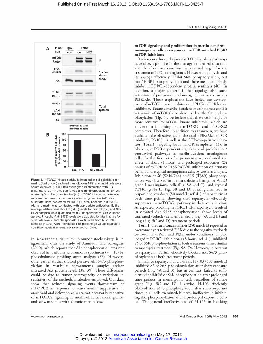
signaling observed in merlin knockdown arachnoid cellsand Schwann cells could be due to diminished mTORC2kinase activity, we directly assayedmTORC2 kinase activity.Endogenous mTORC2 was isolated from control and mer-lin knockdown arachnoid cell lysates by immunoprecipitat-ing Rictor, and its kinase activity was assayed using anexogenous Akt1 substrate. Similar amounts of mTOR wasimmunoprecipitated with Rictor in control and merlin-deficient cells stimulated with EGF, however, mTORC2
mTORC2 Signaling in NF2
www.aacrjournals.org Mol Cancer Res; 10(5) May 2012 653
American Association for Cancer Research Copyright © 2012 on May 17, 2012mcr.aacrjournals.orgDownloaded from
Published OnlineFirst March 16, 2012; DOI:10.1158/1541-7786.MCR-11-0425-T

kinase activity was greatly impaired in merlin knockdownarachnoid cells indicating that merlin loss inhibitsmTORC2 kinase activity (Fig. 3A). Akt S473 phosphory-lation levels for control and NF2 RNAi samples from 2independent mTORC2 kinase assays were quantified. Theaverage decrease in EGF-stimulated mTORC2 kinase activ-ity in merlin knockdown arachnoidal cells compared withcontrol cells was approximately 50% (Fig. 3B).
Heterogeneous expression of PKCa and Akt S473phosphorylation in merlin-deficient meningiomas andschwannomasOur data indicate that acute merlin downregulation by
RNAi in NF2 target cell types such as normal arachnoid andSchwann cells results in elevated mTORC1 signaling anddecreased mTORC2 signaling. We have shown earlier thatmTORC1 signaling is activated in NF2-deficient meningi-omas and schwannomas (14). To determine whether NF2tumors exhibit reduced mTORC2 signaling, we examined
PKCa and phospho-Akt S473 expression levels in merlin-deficient meningioma and schwannoma tumors comparedwith normal arachnoid tissue by immunohistochemicalstaining. We detected heterogeneity in both PKCa andphospho-Akt S473 expression levels across multiplebenign meningioma (n ¼ 5) and schwannoma (n ¼ 4)samples compared with TSC AML (Fig. 4 and Table 1).Previous studies showed that TSC AMLs display highphospho-S6 levels but do not express detectable levels ofPKCa or phospho-Akt (27). PKCa immunoreactivity wasmoderate to strongly positive in normal arachnoid tissue aswell as benign meningiomas and schwannomas. Weak tomoderate levels of phospho-Akt S473 positivity weredetected in both normal arachnoid and benign meningi-oma samples relative to TSC AML tissue. In schwanno-mas, however, phospho-Akt S473 staining was uniformlyreduced across tumor samples relative to normal arachnoidand meningioma tissues, and more comparable with TSCAMLs. The lack of detectable Akt S473 phosphorylation
Schwann cells
S6
Merlin
pAkt
(S473)
pS6
(S240/244)
Akt
pAkt
(T450)
PKCαα
pPKCα(S657)
GAPDH
SF/0.1% Full Full + Rapa.
NDRG1
pNDRG1
(T346)
38-
31-
52-
52-
76-
76-
52-
52-
31-
52-
52-
S6
Merlin
pAkt
(S473)
pS6
(S240/244)
SF/0.1% Full Full + Rapa.
Akt
pAkt
(T450)
PKCα
pPKCα(S657)
α-Tubulin
pNDRG1
(T346)
NDRG1
52-
31-
52-
52-
76-
76-
52-
52-
31-
Arachnoid cells
52-
52-
BARNAi scr NF2 scr NF2 scr NF2 RNAi scr NF2 scr NF2 scr NF2
α-Tubulin α-Tubulin
S6S6
pS6
(S240/244)
pS6
(S240/244)
Merlin
pAkt
(S473)
AktAkt
pAkt
(S473)
Merlin
RNAi scr NF2 scr NF2 scr NF2 scr NF2
SF/0.1% Full
Rapa. 0 0 24 h1 h
RNAi scr NF2 scr NF2 scr NF2 scr NF2
SF/0.1% Full
Rapa. 0 0 24 h1 h
Schwann cellsArachnoid cells
DC
52-
52-
52-
52-
31-
31-
52-
52-
52-
52-
31-
31-
Figure 2. Phosphorylation ofmTORC2 substrates in merlin-deficient arachnoid and Schwanncells. Control (scr) and merlinknockdown (NF2) arachnoid (A andC) or immortalized Schwann(pn02.3; B and D) cells were serumstarved (0.1% FBS) or cultured infull serum with and withoutrapamycin for 18 hours (A and B) or1 and 24 hours (C and D). A and B,phosphorylation of Akt S473, AktT450, and PKCa S657 is impairedin merlin knockdown arachnoidand pn02.3 Schwann cellscompared with control cells.Phosphorylation ofNDRG1T346, adirect substrate of SGK1, isincreased in merlin knockdowncells compared with controlcells. Expression and/orphosphorylation levels also weredetermined for merlin, Akt, PKCa,NDRG1, phospho-S6 (pS6 S240/244), and S6 by immunoblotting.Expression of a-tubulin or GAPDHwas used as loading controls. CandD, Akt S473 phosphorylation isdifferentially sensitive to short-term (1 hour) and long-term (24hours) rapamycin treatment inarachnoid and immortalizedpn02.3 Schwann cells.
James et al.
Mol Cancer Res; 10(5) May 2012 Molecular Cancer Research654
American Association for Cancer Research Copyright © 2012 on May 17, 2012mcr.aacrjournals.orgDownloaded from
Published OnlineFirst March 16, 2012; DOI:10.1158/1541-7786.MCR-11-0425-T
in schwannoma tissue by immunohistochemistry is inagreement with the study of Ammoun and colleagues(2010), which reports that Akt phosphorylation was notobserved in vestibular schwannoma specimens (n¼ 10) byphosphokinase profiling array analysis (37). However,other earlier studies showed positive Akt S473 phosphor-ylation in vestibular schwannoma samples and/orincreased Akt protein levels (38, 39). These differencescould be due to tumor heterogeneity or variations insensitivity of the methods/antibodies employed. Our datashow that reduced signaling events downstream ofmTORC2 in response to acute merlin suppression inarachnoid and Schwann cells are not necessarily reflectiveof mTORC2 signaling in merlin-deficient meningiomasand schwannomas with chronic merlin loss.
mTOR signaling and proliferation in merlin-deficientmeningioma cells in response to mTOR and dual PI3K/mTOR inhibitorsTreatments directed against mTOR signaling pathways
have shown promise in the management of solid tumorsand therefore may constitute a potential target for thetreatment of NF2 meningiomas. However, rapamycin andits analogs effectively inhibit S6K phosphorylation, butnot 4E-BP1 phosphorylation and therefore incompletelyinhibit mTORC1-dependent protein synthesis (40). Inaddition, a major concern is that rapalogs also causeactivation of prosurvival and oncogenic pathways such asPI3K/Akt. These trepidations have fueled the develop-ment of mTOR kinase inhibitors and PI3K/mTOR kinaseinhibitors. Because merlin-deficient meningiomas exhibitactivation of mTORC2 as detected by Akt S473 phos-phorylation (Fig. 4), we believe that these cells might bemore sensitive to mTOR kinase inhibitors, which areefficient in inhibiting both mTORC1 and mTORC2complexes. Therefore, in addition to rapamycin, we haveevaluated the effectiveness of the dual PI3K/Akt–mTORinhibitor, PI-103, as well as the ATP-competitive inhib-itor, Torin1, targeting both mTOR complexes (41), inblocking mTOR-dependent signaling and proliferation/prosurvival pathways in merlin-deficient meningiomacells. In the first set of experiments, we evaluated theeffect of short (1 hour) and prolonged exposures (24hours) of mTOR or P13K/mTOR inhibition on primarybenign and atypical meningioma cells by western analysis.Inhibition of S6 (S240/244) or S6K (T389) phosphory-lation was observed in merlin-deficient benign or WHOgrade I meningioma cells (Fig. 5A and C), and atypical(WHO grade II; Fig. 5B and D) meningioma cells inresponse to low doses (50 nmol/L; ref. 41) of rapamycin atboth time points, showing that rapamycin effectivelysuppresses the mTORC1 pathway in these cells in vitro.As expected, blocking mTORC1 with rapamycin resultedin elevated Akt S473 phosphorylation above levels ofuntreated (vehicle) cells under short (Fig. 5A and B) andlong (Fig. 5C and D) treatment periods.Torin1, used at a concentration (250 nmol/L) reported to
overcome hyperactivated PI3K due to the negative feedbackbetween mTORC1 and PI3K under conditions of pro-longed mTORC1 inhibition (>5 hours; ref. 41), inhibitedS6 or S6K phosphorylation at both treatment times, similarto rapamycin treatment (Fig. 5A–D). However, in contrastto rapamycin, Torin1, effectively blocked Akt S473 phos-phorylation at both treatment periods.Similar to rapamycin and Torin1, PI-103 (500 nmol/L)
inhibited S6 or S6K phosphorylation after short exposureperiods (Fig. 5A and B), but in contrast, failed to suffi-ciently inhibit S6 or S6K phosphorylation after prolongedtime periods in meningioma cells regardless of tumorgrade (Fig. 5C and D). Likewise, PI-103 efficientlyblocked Akt S473 phosphorylation after short exposuretimes in all cells examined, but was ineffective in inhibit-ing Akt phosphorylation after a prolonged exposure peri-od. The general ineffectiveness of PI-103 in blocking
Akt
Akt
Total
lysates
Merlin
Rictor
mTOR
IP Ab: IgG Rictor
RNAi: con con NF2
pAkt
(S473)
pAkt
(S473)
Rictor
mTOR
IP
kinase
In vitro
assay
EGF-stimulated
arachnoid cells
0
20
40
60
80
100
120
con RNAi NF2 RNAi
Avera
ge r
ela
tive A
kt
S473
ph
osp
ho
ryla
tio
n levels
B
A
Figure 3. mTORC2 kinase activity is impaired in cells deficient formerlin. Control (con) and merlin knockdown (NF2) arachnoid cells wereserum deprived (0.1% FBS) overnight and stimulated with EGF(5 ng/mL) for 30 minutes before lysis and immunoprecipitation (IP) withcontrol IgG or Rictor antibodies (Ab). mTORC2 kinase activity wasassessed in these immunoprecipitates using inactive Akt1 as asubstrate. Immunoblotting for mTOR, Rictor, phospho-Akt (S473),Akt, and merlin was conducted with appropriate antibodies. B, theaverage relative phospho-Akt (S473) levels for control (con) and NF2RNAi samples were quantified from 2 independent mTORC2 kinaseassays. Phospho-Akt (S473) levels were adjusted to total inactive Aktsubstrate levels, and phospho-Akt (S473) levels from NF2 RNAisamples (49.9%) were represented as percentage values relative tocon RNAi levels that were arbitrarily set to 100%.
mTORC2 Signaling in NF2
www.aacrjournals.org Mol Cancer Res; 10(5) May 2012 655
American Association for Cancer Research Copyright © 2012 on May 17, 2012mcr.aacrjournals.orgDownloaded from
Published OnlineFirst March 16, 2012; DOI:10.1158/1541-7786.MCR-11-0425-T

phosphorylation of Akt and S6K/S6 after 24 hours maybe, at least in part, due to the short half-life and rapidmetabolism of this drug that has been reported in vivo(42).We next evaluated the effect of rapamycin or Torin1 on
cell proliferation/viability of merlin-deficient meningiomacells (n ¼ 4) treated over a period of 6 days. In comparisonwith untreated cells, we observed that Torin1 (250 nmol/L)was most effective in inhibiting proliferation of all menin-gioma cells tested, and rapamycin (50 nmol/L) significantlyreduced the rate of proliferation. These data are providedin a representative growth curve of a benign meningioma(Fig. 6A). When we compared the average proliferative ratesof 2 independent cultures of benign meningioma or atypicalmeningioma cells at one time point (day 7), we observed thatTorin1 elicited the greatest inhibitory response in all celllines (Fig. 6B).
DiscussionOur previous work showed that merlin deficiency in NF2
target cells results in aberrant activation of mTORC1-mediated signaling as well as attenuation of Akt S473phosphorylation upon insulin treatment (14), consistentwith negative feedback regulation of IRS1 by activatedmTORC1 (22, 23, 43). In this study, we show that AktS473 phosphorylation is impaired in merlin-deficienthuman arachnoid cells in response to stimulation withgrowth factors such as EGF and PDGF, and in merlin-deficient human Schwann cells, in response to EGF, but notPDGF. These results clearly show that merlin-deficientarachnoid and Schwann cells may differ from each other inPDGF-mediated signaling. Attenuation of Akt S473 phos-phorylation by growth factors other than insulin/IGF sug-gest that in NF2, similar to TSC, more than one mechanismcould be operative to downregulate Akt activation. Further-more, recent studies have shown that mTORC1 activation,functioning through S6K phosphorylates Rictor to inhibitmTORC2 (44, 45).Here, we examined signaling downstream of mTORC2,
including phosphorylation of Akt (S473, T450) as well asother well-characterized substrates of mTORC2 such asPKCa (S657) and NDRG1, the latter being a specificreadout for mTORC2 and SGK1 activation (21). Theobserved decrease in phospho-AKT (S473, T450) andphospho-PKCa (S657) in merlin-suppressed arachnoid andSchwann cells resemble TSC1- or TSC2-deficient cells (27).
Table 1. Summary of results fromimmunostaining of PKCa and phospho-AktS473 in 5 benign (WHO grade I) merlin-deficient meningiomas and 4 vestibularschwannomas
Case no. PKCa Phospho-Akt (S473)
MN 1034 þþ þþMN 2805 þþ þMN 2896 þþ þ/þþMN 3633 þþ/þþþ þMN 1670 þþ þ
VS 3924 þþ –
VS 4206 þ/þþ –
VS 4231 þþ –
VS 4787 þþ –/þ
Arachnoid þþ þ/þþTSC-AML – –
NOTE: Staining was scored semiquantitatively: �, negative;þ, weak; þþ, medium; þþþ, strong.Abbreviations: MN, merlin-deficient meningiomas; VS, ves-tibular schwannomas.
pAkt S473PKCαα
Ara
ch
no
ida
l
tis
su
e
Be
nig
n
me
nin
gio
ma
TS
C A
ML
Sch
wan
no
ma
Figure 4. Normal arachnoid and benign (WHO grade I) merlin-deficientmeningioma and vestibular schwannoma sections were immunostainedfor PKCa andphospho-Akt (S473) expression. Normal arachnoid, benignmerlin-deficientmeningioma, and vestibular schwannoma tissues exhibitsimilar, moderate to strong PKCa expression. Phospho-Akt (S473) (pAktS473) staining is weak tomoderate in normal arachnoid andmeningiomatissue, and negative to weak in vestibular schwannomas. Data arerepresentative of results for 5 benignmerlin-deficientmeningiomas and 4vestibular schwannomas (Table 1). Sections of TSC AML (negativecontrols) show negative to weak PKCa and phospho-Akt (S473) stainingconsistent with previous reports (27). Arrow indicates normal arachnoid"cap" cells [magnification, �20, except PKCa staining of benignmeningioma and TSC AML (�40)].
James et al.
Mol Cancer Res; 10(5) May 2012 Molecular Cancer Research656
American Association for Cancer Research Copyright © 2012 on May 17, 2012mcr.aacrjournals.orgDownloaded from
Published OnlineFirst March 16, 2012; DOI:10.1158/1541-7786.MCR-11-0425-T

The decrease inmTORC2 kinase activity observed inmerlinknockdown cells is consistent with growth factor–indepen-dent targets of mTORC2 (Akt T450, PKCa S657) beingdefective in these cells. Future studies are necessary tounderstand the precise molecular mechanisms of mTORC2regulation by NF2 and whether this regulation is dependentor independent of TSC proteins. It is intriguing that con-trary to other mTORC2 targets, NDRG1 is aberrantlyactivated (independent of growth factors) in merlin knock-down arachnoid and Schwann cells. Interestingly, a veryrecent study has shown that knockout of Protor-1, aninteractor of Rictor results in a decrease in phosphorylationof SGK and its physiologic substrate NDRG1, without
influencing the phosphorylation of Akt and PKCa at theirhydrophobic or turn motifs (46). It is therefore tempting tospeculate whether Protor-1 is involved in mediatingNDRG1 activation in merlin deficiency, and further studiesare necessary to understand the mechanism and conse-quences of NDRG1 activation in merlin-deficient cells.Our results suggest that primary meningioma cells and
meningioma tumors with long-term (chronic) loss of merlinmay exhibit heterogeneity in mTORC2 signaling whencompared with acute loss of merlin achieved by RNAi incontrol normal arachnoid and Schwann cells in vitro. Phos-phorylation of Akt S473 is not dramatically reduced inmeningiomas when compared with normal arachnoid tissue.
Figure 5. Torin1 blocks mTORC1andmTORC2 signaling in benign andatypical merlin-deficientmeningioma cells. Immunoblotanalysis of phospho-S6 (pS6 S240/244) and phospho-Akt S473 (pAktS473) in primary cell cultures ofbenign (A and C) and atypical (B andD) meningioma cells treated with PI-103 (P; 500 nmol/L), rapamycin (R, 50nmol/L), Torin1 (T; 250 nmol/L), orvehicle (-; DMSO) for 1 hour (A and B)or 24 hours (C and D). Arrowindicates phospho-p70 S6K (T389)signal.
pAkt
S473
S6
pS6
S240/244
MN1 MN3
Inhibitor: - P R T - P R T
-31
-31
-52
Akt -52
-31
-31
-52
-52
MN1 MN3
Inhibitor: - P R T - P R T
S6
pS6
S240/244
Akt
-31
-52
-31
-52
MN5 MN6
MN5 MN6
-76
-52
-52
-52
Inhibitor: - P R T - P R T
pAkt
S473
S6
pS6
S240/244
Akt
Inhibitor: - P R T - P R T
pAKT S473
pp70 S6K
T389
Akt
pAKT S473
(long exposure)
1 h
24 h
BA
pAkt
S473
DC
p70 S6K
1 h
24 h
0
10
20
30
40
50
1 4 5 7
Vehicle
Rapamycin
Torin1
% c
ell
via
bili
ty (
x 1
,000)
(RLU
)
Days
A B
% c
ell
via
bili
ty (
x 1
,000)
(RLU
)
Vehicle Rapa Torin1
Cell proliferation (day 7)
Benign MN
Atypical MN
100
80
60
40
20
0
Figure 6. Torin1 effectively inhibits proliferation of merlin-deficient meningioma cells. A and B, mTOR inhibition by Torin1 prevents the proliferation of merlin-deficientmeningioma cells more effectively than rapamycin at concentrations tested. A, representative growth curve of a benignmerlin-deficientmeningiomacell line (MN1) grown in the presence of vehicle, 50 nmol/L rapamycin, or 250 nmol/L Torin1 for 6 days. Cell proliferation was measured in triplicate atindicated time points using the CellTiterGlo viability assay. B, cell proliferation of benign and atypical meningioma cells are inhibited most effectively inresponse to Torin1. Bar graphs represent the average RLU values of 2 benign and 2 atypical meningiomas treated for 6 days with indicatedcompounds as described in (A). Data are presented as average RLU values � SE from 2 individual cultures treated in triplicate.
mTORC2 Signaling in NF2
www.aacrjournals.org Mol Cancer Res; 10(5) May 2012 657
American Association for Cancer Research Copyright © 2012 on May 17, 2012mcr.aacrjournals.orgDownloaded from
Published OnlineFirst March 16, 2012; DOI:10.1158/1541-7786.MCR-11-0425-T

However, interestingly, phospho-Akt S473 staining inschwannomas is dramatically reduced, more closely resem-bling the staining pattern of TSC tumors thanmeningiomas.The lack of strong attenuation of mTORC2 signaling,particularly Akt S473 phosphorylation, which is a driver ofcell proliferation and survival in primary meningiomas,suggests that either compensation mechanisms exist oradditional genetic events cooperate with merlin loss inmeningiomas. This is in agreement with a recent study,which elegantly documents frequent chromosome altera-tions in NF2-associated grade 1 meningiomas (47). We alsospeculate that similar to TSC lesions, the lack of Akt S473phosphorylation in schwannomas may explain the benignnature of these tumors as well as the difficulty in establishingcells from them in culture. On the contrary, the atypicalfeatures commonly seen in benign meningiomas may beelicited by additional genetic events and/or Akt activation.Clinical trials with the allosteric inhibitor, rapamycin and
its analogs, known as rapalogs, are shown to be effective forvarious TSC-associated benign tumors including subepen-dymal giant cell astrocytomas (48, 49). However, the feed-back activation of oncogenic pathways by rapamycin andrapamycin-resistant phosphorylation of 4E-BP1 has led tothe development of kinase inhibitors of mTOR as well asdual PI3K/Akt and mTOR inhibitors (50). We tested theefficacy of mTORC1 inhibitor rapamycin; an ATP-com-petitive mTOR inhibitor Torin1, which is shown to be apotent inhibitor of bothmTORC1 andmTORC2 complex;and the dual PI3K/mTOR inhibitor PI-103 on primarybenign and atypical meningioma cells. We conclude thatTorin1 is more effective in blocking mTORC1 and Aktactivation in meningioma cells in vitro than rapamycin andPI-103 and more effective than rapamycin in inhibiting cell
proliferation. Although benign in nature, we believe thatheterogeneity exists in NF2-associated tumors adding com-plexity to signaling events. Therefore, therapeutic strategiesemploying Torin1 or equivalent mTOR kinase inhibitors incombination with other pathway inhibitors may be prom-ising for treating NF2.
Disclosure of Potential Conflicts of InterestNo potential conflicts of interest were disclosed.
Authors' ContributionsConception and design: M.F. James, J.F. Gusella, V. Ramesh.Development of methodology: M.F. James, E. Stivison.Acquisition of data: M.F. James, E. Stivison, S. Han, A.O. Stemmer-Rachamimov.Analysis and interpretation of data: M.F. James, R. Beauchamp, A.O. Stemmer-Rachamimov, V. Ramesh.Writing, review, and/or revision of the manuscript:M.F. James, R. Beauchamp, S.Han, M. Wallace, J.F. Gusella, A.O. Stemmer-Rachamimov, V. Ramesh.Administrative, technical, or material support: E. Stivison, R. Beauchamp, H. Li,M. Wallace.Study supervision: V. Ramesh.
AcknowledgmentsThe authors thank BrendanD.Manning andChristian C.Dibble (Harvard School
of Public Health/Brigham and Women's Hospital) for valuable discussions; Sun Kimfor technical assistance, and Stephen Ranney, and James C. Kim for valuable assistancein obtaining tissue samples.
Grant SupportThis work was supported by the NIH grants NS024279, Department of Defense
(DOD) Neurofibromatosis Research Program, S. Sydney De Young Foundation,Neurofibromatosis, Inc., New England, and Children's Tumor Foundation DrugDiscovery Initiative.
The costs of publication of this article were defrayed in part by the payment of pagecharges. This article must therefore be herebymarked advertisement in accordance with18 U.S.C. Section 1734 solely to indicate this fact.
Received September 7, 2011; revised March 5, 2012; accepted March 5, 2012;published OnlineFirst March 16, 2012.
References1. Evans DG. Neurofibromatosis 2 [Bilateral acoustic neurofibromatosis,
central neurofibromatosis, NF2, neurofibromatosis type II]. GenetMed2009;11:599–610.
2. Trofatter JA, MacCollin MM, Rutter JL, Murrell JR, Duyao MP, ParryDM, et al. A novel moesin-, ezrin-, radixin-like gene is a candidatefor the neurofibromatosis 2 tumor suppressor. Cell 1993;72:791–800.
3. RouleauGA,Merel P, LutchmanM, SansonM, Zucman J,Marineau C,et al. Alteration in a new gene encoding a putative membrane-orga-nizing protein causes neurofibromatosis type 2. Nature 1993;363:515–21.
4. Fehon RG, McClatchey AI, Bretscher A. Organizing the cell cortex: therole of ERM proteins. Nat Rev Mol Cell Biol 2010;11:276–87.
5. Lallemand D, Manent J, Couvelard A, Watillilaux A, Siena M,Chareyre F, et al. Merlin regulates transmembrane receptor accu-mulation and signaling at the plasma membrane in primary mouseSchwann cells and in human schwannomas. Oncogene 2009;28:854–65.
6. McClatchey AI, Fehon RG. Merlin and the ERM proteins–regulators ofreceptor distribution and signaling at the cell cortex. Trends Cell Biol2009;19:198–206.
7. Kaempchen K, Mielke K, Utermark T, Langmesser S, Hanemann CO.Upregulation of the Rac1/JNK signaling pathway in primary humanschwannoma cells. Hum Mol Genet 2003;12:1211–21.
8. Rong R, Tang X, Gutmann DH, Ye K. Neurofibromatosis 2 (NF2) tumorsuppressormerlin inhibits phosphatidylinositol 3-kinase through bind-ing to PIKE-L. Proc Natl Acad Sci U S A 2004;101:18200–5.
9. Hamaratoglu F, Willecke M, Kango-Singh M, Nolo R, Hyun E, Tao C,et al. The tumour-suppressor genes NF2/Merlin and expanded actthrough Hippo signalling to regulate cell proliferation and apoptosis.Nat Cell Biol 2006;8:27–36.
10. Morrison H, Sperka T, Manent J, Giovannini M, Ponta H, Herrlich P.Merlin/neurofibromatosis type 2 suppresses growth by inhibiting theactivation of Ras and Rac. Cancer Res 2007;67:520–7.
11. Zhao B,Wei X, Li W, Udan RS, YangQ, Kim J, et al. Inactivation of YAPoncoprotein by the Hippo pathway is involved in cell contact inhibitionand tissue growth control. Genes Dev 2007;21:2747–61.
12. Yi C, Troutman S, Fera D, Stemmer-Rachamimov A, Avila JL, ChristianN, et al. A tight junction-associated Merlin-angiomotin complex med-iates Merlin's regulation of mitogenic signaling and tumor suppressivefunctions. Cancer Cell 2011;19:527–40.
13. Li W, Giancotti FG. Merlin's tumor suppression linked to inhibition ofthe E3 ubiquitin ligase CRL4 (DCAF1). Cell Cycle 2010;9:4433–6.
14. James MF, Han S, Polizzano C, Plotkin SR, Manning BD, Stemmer-Rachamimov AO, et al. NF2/merlin is a novel negative regulator ofmTOR complex 1, and activation of mTORC1 is associated withmeningioma and schwannoma growth. Mol Cell Biol 2009;29:4250–61.
James et al.
Mol Cancer Res; 10(5) May 2012 Molecular Cancer Research658
American Association for Cancer Research Copyright © 2012 on May 17, 2012mcr.aacrjournals.orgDownloaded from
Published OnlineFirst March 16, 2012; DOI:10.1158/1541-7786.MCR-11-0425-T

15. Guertin DA, Sabatini DM. Defining the role of mTOR in cancer. CancerCell 2007;12:9–22.
16. Ma XM, Blenis J. Molecular mechanisms of mTOR-mediated transla-tional control. Nat Rev Mol Cell Biol 2009;10:307–18.
17. Huang J, Dibble CC, Matsuzaki M, Manning BD. The TSC1-TSC2complex is required for proper activation of mTOR complex 2. Mol CellBiol 2008;28:4104–15.
18. Sarbassov DD, Ali SM, Sengupta S, Sheen JH, Hsu PP, Bagley AF,et al. Prolonged rapamycin treatment inhibits mTORC2 assembly andAkt/PKB. Mol Cell 2006;22:159–68.
19. Pearce LR, Komander D, Alessi DR. The nuts and bolts of AGC proteinkinases. Nat Rev Mol Cell Biol 2010;11:9–22.
20. GuertinDA,StevensDM,ThoreenCC,BurdsAA,KalaanyNY,Moffat J,et al. Ablation in mice of the mTORC components raptor, rictor, ormLST8 reveals thatmTORC2 is required for signaling toAkt-FOXOandPKCalpha, but not S6K1. Dev Cell 2006;11:859–71.
21. Garcia-Martinez JM, Alessi DR. mTOR complex 2 (mTORC2) controlshydrophobic motif phosphorylation and activation of serum- andglucocorticoid-induced protein kinase 1 (SGK1). Biochem J 2008;416:375–85.
22. Shah OJ, Hunter T. Turnover of the active fraction of IRS1 involvesraptor-mTOR- and S6K1-dependent serine phosphorylation incell culture models of tuberous sclerosis. Mol Cell Biol 2006;26:6425–34.
23. Harrington LS, Findlay GM, Gray A, Tolkacheva T, Wigfield S, RebholzH, et al. The TSC1-2 tumor suppressor controls insulin-PI3K signalingvia regulation of IRS proteins. J Cell Biol 2004;166:213–23.
24. Zhang H, Bajraszewski N, Wu E, Wang H, Moseman AP, Dabora SL,et al. PDGFRs are critical for PI3K/Akt activation and negativelyregulated by mTOR. J Clin Invest 2007;117:730–8.
25. Yu Y, Yoon SO, Poulogiannis G, Yang Q, Ma XM, Villen J, et al.Phosphoproteomic analysis identifies Grb10 as an mTORC1 sub-strate that negatively regulates insulin signaling. Science 2011;332:1322–6.
26. Hsu PP, Kang SA, Rameseder J, Zhang Y, Attina KA, Lim D, et al. ThemTOR-regulated phosphoproteome reveals a mechanism ofmTORC1-mediated inhibition of growth factor signaling. Science2011;332:1317–22.
27. Huang J, Wu S, Wu CL, Manning BD. Signaling events downstream ofmammalian target of rapamycin complex 2 are attenuated in cells andtumors deficient for the tuberous sclerosis complex tumor suppres-sors. Cancer Res 2009;69:6107–14.
28. Efeyan A, Sabatini DM.mTORand cancer:many loops in onepathway.Curr Opin Cell Biol 2010;22:169–76.
29. JamesMF, Lelke JM,Maccollin M, Plotkin SR, Stemmer-RachamimovAO, Ramesh V, et al. Modeling NF2 with human arachnoidal andmeningioma cell culture systems: NF2 silencing reflects the benigncharacter of tumor growth. Neurobiol Dis 2008;29:278–92.
30. Fishbein L, Zhang X, Fisher LB, Li H, Campbell-Thompson M,Yachnis A, et al. In vitro studies of steroid hormones in neurofibro-matosis 1 tumors and Schwann cells. Mol Carcinog 2007;46:512–23.
31. Li H, Chang L-J, Muir D, Wallace MR. Immortalization of normal andNF1 neurofibroma-derived Schwann cell cultures. Manuscript inpreparation.
32. Sarbassov DD, Guertin DA, Ali SM, Sabatini DM. Phosphorylation andregulation of Akt/PKB by the rictor-mTOR complex. Science 2005;307:1098–101.
33. Huang J, Manning BD. The TSC1-TSC2 complex: a molecular switch-board controlling cell growth. Biochem J 2008;412:179–90.
34. Lopez-Lago MA, Okada T, Murillo MM, Socci N, Giancotti FG. Loss ofthe tumor suppressor gene NF2, encoding merlin, constitutively acti-vates integrin-dependent mTORC1 signaling. Mol Cell Biol 2009;29:4235–49.
35. Fraenzer JT, Pan H, Minimo L Jr., Smith GM, Knauer D, Hung G.Overexpression of theNF2gene inhibits schwannomacell proliferationthrough promoting PDGFR degradation. Int J Oncol 2003;23:1493–500.
36. Ammoun S, Flaiz C, Ristic N, Schuldt J, HanemannCO.Dissecting andtargeting the growth factor-dependent and growth factor-independentextracellular signal-regulated kinase pathway in human schwannoma.Cancer Res 2008;68:5236–45.
37. AmmounS,CunliffeCH,Allen JC,ChiribogaL,Giancotti FG, ZagzagD,et al. ErbB/HER receptor activation and preclinical efficacy of lapatinibin vestibular schwannoma. Neuro Oncol 2010;12:834–43.
38. Jacob A, Lee TX, Neff BA, Miller S, Welling B, Chang LS. Phospha-tidylinositol 3-kinase/AKT pathway activation in human vestibularschwannoma. Otol Neurotol 2008;29:58–68.
39. Hilton DA, Ristic N, Hanemann CO. Activation of ERK, AKT and JNKsignalling pathways in human schwannomas in situ. Histopathology2009;55:744–9.
40. Choo AY, Yoon SO, Kim SG, Roux PP, Blenis J. Rapamycin differen-tially inhibits S6Ks and 4E-BP1 to mediate cell-type-specific repres-sion ofmRNA translation. ProcNatl AcadSciUSA2008;105:17414–9.
41. ThoreenCC, KangSA,Chang JW, LiuQ, Zhang J,GaoY, et al. AnATP-competitive mammalian target of rapamycin inhibitor reveals rapamy-cin-resistant functions of mTORC1. J Biol Chem 2009;284:8023–32.
42. Donev IS, Wang W, Yamada T, Li Q, Takeuchi S, Matsumotot K, et al.Transient PI3K inhibition induces apoptosis and overcomes HGF-mediated resistance to EGFR-TKIs in EGFR mutant lung cancer. ClinCancer Res 2011;17:2260–9.
43. Huang J,ManningBD. A complex interplay betweenAkt, TSC2 and thetwo mTOR complexes. Biochem Soc Trans 2009;37:217–22.
44. Dibble CC, Asara JM, Manning BD. Characterization of Rictor phos-phorylation sites reveals direct regulation of mTOR complex 2 byS6K1. Mol Cell Biol 2009;29:5657–70.
45. Julien LA, Carriere A, Moreau J, Roux PP. mTORC1-activated S6K1phosphorylates Rictor on threonine 1135 and regulates mTORC2signaling. Mol Cell Biol 2010;30:908–21.
46. Pearce LR, Sommer EM, Sakamoto K, Wullschleger S, Alessi DR.Protor-1 is required for efficientmTORC2-mediatedactivation ofSGK1in the kidney. Biochem J 2011;436:169–79.
47. Goutagny S, Yang HW, Zucman-Rossi J, Chan J, Dreyfuss JM, ParkPJ, et al. Genomic profiling reveals alternative genetic pathways ofmeningioma malignant progression dependent on the underlying NF2status. Clin Cancer Res 2010;16:4155–64.
48. TyburczyME, Kotulska K, Pokarowski P, Mieczkowski J, Kucharska J,Grajkowska W, et al. Novel proteins regulated by mTOR in subepen-dymal giant cell astrocytomas of patients with tuberous sclerosiscomplex and new therapeutic implications. Am J Pathol 2010;176:1878–90.
49. Campen CJ, Porter BE. Subependymal giant cell astrocytoma (SEGA)treatment update. Curr Treat Options Neurol 2011;13:380–5.
50. Wander SA, Hennessy BT, Slingerland JM. Next-generation mTORinhibitors in clinical oncology: how pathway complexity informs ther-apeutic strategy. J Clin Invest 2011;121:1231–41.
mTORC2 Signaling in NF2
www.aacrjournals.org Mol Cancer Res; 10(5) May 2012 659
American Association for Cancer Research Copyright © 2012 on May 17, 2012mcr.aacrjournals.orgDownloaded from
Published OnlineFirst March 16, 2012; DOI:10.1158/1541-7786.MCR-11-0425-T