adamts1 and mmp1 proteolytically engage egf-like ligands
TRANSCRIPT

ADAMTS1 and MMP1 proteolyticallyengage EGF-like ligands in an osteolyticsignaling cascade for bone metastasis
Xin Lu,1 Qiongqing Wang,2 Guohong Hu,1 Catherine Van Poznak,3,7 Martin Fleisher,4 Michael Reiss,5
Joan Massague,2,6 and Yibin Kang1,5,8
1Department of Molecular Biology, Princeton University, Princeton, New Jersey 08544, USA; 2Cancer Biology and GeneticsProgram, Memorial Sloan-Kettering Cancer Center, New York, New York 10021, USA; 3Department of Medicine, MemorialSloan-Kettering Cancer Center, New York, New York 10021, USA; 4Department of Clinical Laboratories, Memorial Sloan-Kettering Cancer Center, New York, New York 10021, USA; 5Breast Cancer Program, Cancer Institute of New Jersey, NewBrunswick, New Jersey 08903, USA; 6Howard Hughes Medical Institute, Memorial Sloan-Kettering Cancer Center, New York,New York 10021, USA
Bone metastasis is mediated by complex interactions between tumor cells and resident stromal cells in the bonemicroenvironment. The functions of metalloproteinases in organ-specific metastasis remain poorly defineddespite their well-appreciated role in matrix degradation and tumor invasion. Here, we show a mechanismwhereby two distinct metalloproteinases, a disintegrin and metalloproteinase with thrombospondin motifs(ADAMTS1) and matrix metalloproteinase-1 (MMP1), orchestrate a paracrine signaling cascade to modulate thebone microenvironment in favor of osteoclastogenesis and bone metastasis. Proteolytic release of membrane-bound epidermal growth factor (EGF)-like growth factors, including Amphiregulin (AREG), heparin-binding EGF(HB-EGF), and transforming growth factor a (TGFa) from tumor cells suppress the expression of osteoprotegerin(OPG) in osteoblasts and subsequently potentiate osteoclast differentiation. EGF receptor (EGFR) inhibitors blockosteolytic bone metastasis by targeting EGFR signaling in bone stromal cells. Furthermore, elevated MMP1 andADAMTS1 expression is associated with increased risk of bone metastasis in breast cancer patients. This studyestablished MMP1 and ADAMTS1 in tumor cells, as well as EGFR signaling in osteoblasts, as promisingtherapeutic targets for inhibiting bone metastasis of breast cancer.
[Keywords: EGFR; bone metastasis; breast cancer; metalloprotease; osteoclastogenesis]
Supplemental material is available at http://www.genesdev.org.
Received May 25, 2009; revised version accepted June 22, 2009.
Bone metastasis is a frequent complication of breastcancer, and is often accompanied by debilitating bonefracture, severe pain, nerve compression, and hypercalce-mia (Mundy 2002; Roodman 2004; Guise et al. 2005;Gupta and Massague 2006). Effective therapies for bonemetastases rely on a better molecular understanding ofthe pathological process. An important concept hasemerged in recent years that osteolytic bone metastasisis driven by intricate cellular and molecular interactionsamong tumor cells and host stromal cells commonlyfound in the bone milieu (Mundy 2002; Roodman 2004;Guise et al. 2005). Tumor-derived factors alter the finebalance of osteoclast and osteoblast activities that are
necessary for maintaining normal bone homeostasis.Increased osteoclast activity leads to bone matrix degra-dation and the release of bone-derived growth factors thatcan further stimulate the metastatic function of tumorcells. Central to the control of this so-called ‘‘viciouscycle’’ of bone metastasis is the modulation of osteoclastactivity by the tumor necrosis factor (TNF) family mem-ber receptor activator of nuclear kB ligand (RANKL)(Mundy 2002). RANKL is expressed in both membrane-bound and soluble forms by osteoblasts, while its cognatereceptor RANK is expressed on the surface of osteoclastsand controls a key signaling pathway essential for oste-oclast differentiation. Osteoprotegerin (OPG), a solubledecoy receptor of RANKL, is also produced by osteoblaststo antagonize the activity of RANKL. The function oftumor-derived bone metastasis factors, such as parathy-roid hormone-related peptide (PTHrP), hinges upon theirability to increase RANKL production or decrease OPGsecretion by osteoblasts, ultimately promoting osteoclastdifferentiation and activation (Mundy 2002).
7Present address: Department of Internal Medicine, University ofMichigan, Ann Arbor, MI 48109, USA.8Corresponding author.E-MAIL [email protected]; FAX (609) 258-2340.Article published online ahead of print. Article and publication date areonline at http://www.genesdev.org/cgi/doi/10.1101/gad.1824809
1882 GENES & DEVELOPMENT 23:1882–1894 � 2009 by Cold Spring Harbor Laboratory Press ISSN 0890-9369/09; www.genesdev.org
Cold Spring Harbor Laboratory Press on December 20, 2021 - Published by genesdev.cshlp.orgDownloaded from

Although metalloproteinases are well known to havecritical functions in tissue remodeling, tumor progres-sion, and metastasis through their proteolytic activitiesfor extracellular matrix (ECM) degradation, invasion, andcytokine mobilization (Egeblad and Werb 2002; Overalland Lopez-Otin 2002; Page-McCaw et al. 2007), thespecific functional mechanism of metalloproteinases inpromoting bone metastasis has not been well character-ized. In an attempt to identify breast cancer bone metas-tasis genes, we previously used an in vivo selection ap-proach to derive highly bone metastatic variants from theMDA-MB-231 breast cancer cell line (Kang et al. 2003).Gene expression profiling of these variants identified a setof 11 putative breast cancer bone metastasis genes, in-cluding two genes encoding metalloproteinases, a disinte-grin and metalloproteinase with thrombospondin motifs(ADAMTS1) and matrix metalloproteinase-1 (MMP1).Thus, this model system provides an ideal opportunityto explore the functional role of these two metalloprotei-nases in bone metastasis.
ADAMTS1, a member of the ADAMTS family (Kunoet al. 1997), has been shown to be capable of binding toand degrading ECM components, including the proteo-glycans aggrecan and versican (Porter et al. 2005). Over-expression of ADAMTS1 promotes pulmonary metas-tasis of TA3 mammary carcinoma and Lewis lung carci-noma cells (Liu et al. 2005). However, the functional andclinical relevance of ADAMTS1 in bone metastasisremains unknown. MMP1 (also known as interstitialcollagenase) is the prototype of the large family of MMPs(Page-McCaw et al. 2007). Although the main substratesof MMP1 are fibrillar collagens, it can cleave many othercomponents in the ECM, including perlecan, laminin,aggrecan, and versican, some of which are also substratesfor ADAMTS1. Additionally, signaling molecule precur-sors, such as pro-TNFa, can be shed from cell surfaceby MMP1 (McCawley and Matrisian 2001; Egeblad andWerb 2002). MMP1 overexpression was found in a widevariety of malignant tumors (Brinckerhoff et al. 2000;Egeblad and Werb 2002) and was almost invariably cor-related with increased tumor stage, presence of distantmetastasis, and poor survival (Egeblad and Werb 2002). Inaddition to its ability to degrade ECM, MMP1 can alsopromote tumor invasion through proteolytic activation ofG protein coupled receptor PAR1 (Boire et al. 2005).Despite the well-known function of MMP1 in tumorprogression, its role in bone metastasis has remainedundefined.
In this study, we uncovered a functional role of MMP1and ADAMTS1 in promoting osteolytic bone metastasis.The combined actions of these two proteases activateosteoclast differentiation by shedding tumor-derived epi-dermal growth factor (EGF)-like factors to reduce osteo-blast production of OPG in the local bone microenvi-ronment. EGF receptor (EGFR) inhibitors block osteolyticbone metastasis by targeting EGFR signaling in bone stro-mal cells, and therefore may have therapeutic benefits inthe treatment of bone metastasis even for patients whoseprimary tumors are nonresponsive to EGFR inhibitors.Simultaneous overexpression of MMP1 and ADAMTS1
were observed in a large proportion of breast tumor sam-ples and is associated with increased risk of bone metas-tasis. These results suggest that molecular targeting ofmetalloproteinases and EGFR signaling in the bone stro-ma may become effective therapeutics for breast cancerbone metastasis.
Results
Combined silencing of ADAMTS1 and MMP1 reducesbone metastasis
Northern blot analysis of MDA-MB-231 sublines with dif-ferent metastasis abilities to bone revealed that ADAMTS1and/or MMP1 were highly expressed only in the highlymetastatic sublines (Fig. 1A; Kang et al. 2003; Minn et al.2005a). In order to test the functional importance ofADAMTS1 and MMP1 in metastasis, the expression ofthese two genes was silenced either individually orin combination by sshRNAs in SCP20, a highly bonemetastatic single-cell-derived progeny (SCP) from MDA-MB-231 (Fig. 1B; Kang et al. 2003). The control andknockdown derivatives of SCP20 were inoculated intonude mice by intracardiac injection, and metastasisburden was measured by weekly bioluminescence im-aging (BLI) using a firefly luciferase reporter stably ex-pressed in these cells (Minn et al. 2005b). Kaplan-Meiercurves and normalized BLI signals were plotted to analyzethe kinetics of the metastasis development (Fig. 1C;Supplemental Fig. S1A,B). Whereas the single knockdownof either ADAMTS1 or MMP1 did not significantly reducethe incidence of bone metastases or prolong survival(P > 0.1), the combined silencing of both genes dramati-cally inhibited bone metastasis formation (P = 0.0041)(Fig. 1C) and increased the survival rate (P = 0.0037)(Supplemental Fig. S1A). As indicated by BLI signalcurves, the single knockdowns caused an appreciablereduction of metastasis burden, yet a highly significantreduction was observed only when the expression ofboth genes was silenced (Supplemental Fig. S1B; repre-sentative mice from each group are shown in Fig. 1D).X-ray imaging revealed extensive osteolytic bone lesionsformed by the control and single-knockdown cells,whereas mice injected with double-knockdown cellsmaintained their bone integrity (Fig. 1D). The osteolyticnature of the metastases was confirmed by hematoxylinand eosin (H&E) staining, and the presence of osteo-clasts along the tumor–bone interface was visualizedand quantified by tartrate-resistant acid phosphatase(TRAP) staining (Fig. 1D; Supplemental Fig. S2 for en-larged images). Consistent with the pattern of the re-duction in metastasis burden by single and doubleknockdowns revealed by BLI, osteoclast number washighest in the control group and significantly lower inthe knockdown groups, with the most dramatic reduc-tion observed in the double-knockdown group (Fig. 1E;Supplemental Fig. S2). In the rare bone metastases de-veloped in mice inoculated with the double knockdowncells, the bone lesions had a smooth tumor–bone inter-face and very few TRAP-positive cells (Fig. 1D,E). Most of
ADAMTS1 and MMP1 promote bone metastasis
GENES & DEVELOPMENT 1883
Cold Spring Harbor Laboratory Press on December 20, 2021 - Published by genesdev.cshlp.orgDownloaded from

small bone lesions developed by double-knockdown cellregressed a few days after detection by BLI (see example inSupplemental Fig. S1C). ADAMTS1 and MMP1 do notplay a direct role in the proliferation of tumor cells, as nosignificant difference in primary tumor growth wasobserved for all three knockdown lines compared withthe control line after mammary fat pad injection (P > 0.4)(Supplemental Fig. S1D). This was consistent with a re-cent report revealing no correlation between MMP1mRNA level and tumor size in breast cancer (Chenget al. 2008). To rule out the possibility that the metastaticbehavior changes observed in the knockdown cells couldbe due to nonspecific gene silencing, we rescued theexpression of ADAMTS1 and MMP1, either individuallyor simultaneously, in the double-knockdown cells bystably expressing shRNA-resistant coding sequences(Supplemental Fig. S3A). When the modified cells wereevaluated for the bone metastasis ability, rescue of eitherADAMTS1 or MMP1 expression to the level of the orig-
inal SCP20 cells recovered the metastasis ability to thelevel of SCP20 (Supplemental Fig. S3B).
Overexpression of ADAMTS1 and MMP1 promotesbone metastasis
To further validate the effect of ADAMTS1 and MMP1 inpromoting osteolytic metastasis, a weakly metastaticclonal subline of MDA-MB-231, SCP6 (Fig. 1A), wasengineered to overexpress these two genes either individ-ually or in combination (Fig. 2A), and subjected to in vivometastasis assays. Only the combined overexpression ofboth genes strongly promoted osteolytic bone metastases(Fig. 2B; Supplemental Fig. S4A). No effect on primarytumor growth was observed when one or both of the twogenes were overexpressed (Supplemental Fig. S4B). Tofurther prove that modulating the activity of ADAMTS1and MMP1 could change the bone metastasis behavior oftumor cells, we used a mildly metastatic MDA-MB-231
Figure 1. Inhibition of breast cancer bone metastasis by combined knockdown of ADAMTS1 and MMP1. (A) Northern blot analysis ofADAMTS1 and MMP1 expression in MDA-MB-231 derivatives with different bone metastasis abilities (Kang et al. 2003). ATCC denotesthe parental MDA-MB-231 cell line. Sublines designated by four-digit numbers were obtained from in vivo selection of MDA-MB-231.SCP6 and SCP20 were SCPs of MDA-MB-231 derived in vitro by dilution cloning. Sublines are color-coded for their previously reportedmetastatic ability to bone: strong (red), intermediate (orange), and weak (green) (Kang et al. 2003). (B) Effective knockdown of ADAMTS1and MMP1 in the MDA-MB-231 subline SCP20 was confirmed by Northern blot analysis of total RNA or Western blot analysis of theCM. In A and B, white dividing lines separating lanes denote grouping of images from different parts of the same gel. (C) Kaplan-Meiercurve of the incidence of bone metastases in the hindlimbs of the nude mice after intracardiac injection of various cell lines, includingSCP20 stably infected with control viruses, or viruses expressing MMP1 shRNA, ADAMTS1 shRNA, or both. n = 10. (**) P < 0.01 basedon log-rank test. (D) Bioluminescent (BLI), radiographic and histological (H&E and TRAP staining) analyses of bone lesions in fourrepresentative mice of each experimental group at day 50 after cell injection. The BLI images are shown under the same color scale. Inthe radiograph images, white dotted lines show the contour of the bone lesions. Rare small bone metastasis observed in mice injectedwith double-knockdown cells has smooth tumor–bone interface and very few TRAP-positive cells. Bars, 200 mm. (E) Quantification ofTRAP-positive osteoclasts along the tumor–bone interface. Data represent average 6 SD. (**) P < 0.01; (***) P < 0.001 based on two-sided Student’s t-test.
Lu et al.
1884 GENES & DEVELOPMENT
Cold Spring Harbor Laboratory Press on December 20, 2021 - Published by genesdev.cshlp.orgDownloaded from
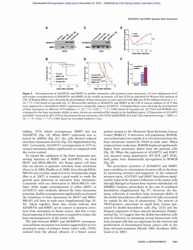
subline, 2279, which overexpresses MMP1 but notADAMTS1 (Fig. 1A). When MMP1 expression was si-lenced by shRNA (Fig. 2C), the cells showed reducedosteolytic metastasis activity (Fig. 2D; Supplemental Fig.S4C). Conversely, ADAMTS1 overexpression in 2279 in-creased metastasis ability significantly as compared withthe vector control.
To extend the validation of the bone metastasis pro-moting function of MMP1 and ADAMTS1, we usedMCF7 and MDA-MB-435, two breast cancer cell linesthat are known to generate osteolytic bone metastasis(Rucci et al. 2004; Phadke et al. 2006). Even though MDA-MB-435 was recently suspected to be of melanoma origin(Rae et al. 2007), it remains a good model to study thegeneral gene functions in osteolytic bone metastasis.Consistent with our observation in MDA-MB-231 sub-lines, while single overexpression of either MMP1 orADAMTS1 only modestly affected the bone metastasispotential, double overexpression significantly acceleratedthe formation of bone metastases by MCF7 and MDA-MB-435 cell lines in nude mice (Supplemental Figs. S5,S6). Taken together, these data clearly indicate thatADAMTS1 and MMP1 act in concert to promote osteo-lytic bone metastasis in breast cancer, and that a com-bined targeting of both proteases is required to reduce thebone metastagenicity of the tumor cells.
The link between MMP1 and ADAMTS1 overexpres-sion and bone metastasis was further supported by in vivometastasis assays of primary breast tumor cells, CN34,isolated from the pleural effusion of a breast cancer
patient treated at the Memorial Sloan-Kettering CancerCenter (MSKCC). A derivative cell population, BOM2B,was isolated after two rounds of in vivo selection from thebone metastasis formed by CN34 in nude mice. Whenreinjected into nude mice, BOM2B displayed significantlyhigher bone metastatic ability than the parental cells(Fig. 2E). When the expression of ADAMTS1 and MMP1was assessed using quantitative RT–PCR (qRT–PCR),both genes were dramatically up-regulated in BOM2B(Fig. 2F).
The proteolytic activities of ADAMTS1 and MMP1may contribute to colonization of tumor cells in the boneby promoting invasion and migration. In the transwellinvasion assay, ADATMS1 and MMP1 knockdown signif-icantly reduced the invasive behaviors of the tumor cellsthrough Matrigel or human bone marrow endothelial cell(HMBEC) barriers, particularly in the case of combinedknockdown (Supplemental Fig. S7). However, the dra-matic reduction of osteolytic bone metastasis ability ofthe double-knockdown cells could not be fully accountedfor simply by the loss of invasiveness. The paucity ofTRAP-positive osteoclasts in small bone lesions gen-erated by double-knockdown cells (Fig. 1D,E) and thefrequent regression of these nascent metastases (Supple-mental Fig. 1C) suggest that the double-knockdown cellsmay be defective in sustaining crucial interactions withbone stromal cells that are important for the survival andproliferation of disseminated breast cancer cells in thebone microenvironment (Mundy 2002; Roodman 2004;Guise et al. 2005).
Figure 2. Overexpression of ADAMTS1 and MMP1 in weakly metastatic cells promotes bone metastasis. (A) Low endogenous leveland ectopic overexpression of ADAMTS1 and MMP1 in the weakly metastatic cell line SCP6 as confirmed by Western blot analysis ofCM. (B) Kaplan-Meier curve showing the development of bone metastases in mice injected with different SCP6 sublines listed in A. n =
10. (*) P < 0.05 based on log-rank test. (C) Western blot analysis of ADAMTS1 and MMP1 in the CM of various sublines of 2279 thatwere engineered to knockdown MMP1 expression or ectopically express ADAMTS1. (D) Kaplan-Meier curve showing the developmentof bone metastases in different 2279 sublines. n = 10. (*) P < 0.05; (***) P < 0.001 based on log-rank test. (E) CN34 and BOM2B wereevaluated for the bone metastatic ability in mice, shown are normalized BLI signals in the hindlimb region. (F) Expression of ADAMTS
and MMP1 assessed by qRT–PCR in the primary breast carcinoma cells CN34 and BOM2B. In E and F data represent average 6 SEM. n =
10. (**) P < 0.01; (***) P < 0.001 based on two-sided Student’s t-test.
ADAMTS1 and MMP1 promote bone metastasis
GENES & DEVELOPMENT 1885
Cold Spring Harbor Laboratory Press on December 20, 2021 - Published by genesdev.cshlp.orgDownloaded from

Secreted factors from ADAMTS1- andMMP1-overexpressing cancer cells alter the balanceof RANKL and OPG production by osteoblasts
As ADAMTS1 and MMP1 expression was correlated withosteolytic activity and the number of osteoclasts in bonemetastases, we investigated the role of these genes intumor-stimulated osteoclast activation. Mouse primarybone marrow cells were treated with the conditionedmedia (CM) from the control and knockdown sublines ofSCP20 in an in vitro osteoclastogenesis assay, and TRAP-positive multinucleated mature osteoclasts were scored(Fig. 3A,B). Primary bone marrow cells that were culturedwithout CM from tumor cells had very few TRAP-positive cells. In contrast, the CM of the SCP20 cell lineinduced extensive differentiation of osteoclasts. How-ever, the osteoclast-activating ability of the CM wassignificantly reduced when the expression of MMP1 andADAMTS1 was suppressed either individually, or moresignificantly, in combination. To determine whetherADAMTS1 and MMP1 can influence osteoclast differen-tiation by altering the expression or release of RANKLand OPG in osteoblasts, we used the SCP20 CM toculture the murine osteoblast cell line MC3T3-E1 thathad been induced to fully differentiate with L-ascorbicacid and determined the level of RANKL and OPGreleased from osteoblasts by immunoblotting (Fig. 3C).Mature MC3T3-E1 cells secreted a low level of RANKLand relatively high level of OPG (RANKL/OPG ratio =0.16). This pattern was reversed by CM from tumor cells(RANKL/OPG ratio = 2.27). However, when MC3T3-E1was cultured with CM from ADAMTS1/MMP1 double-knockdown cells, OPG became the dominant species
again (RANKL/OPG ratio = 0.45). Soluble RANKL andOPG were clearly derived from MC3T3-E1 cells ratherthan tumor cells as they were absent in the tumor cellCM (data not shown), although we could not rule out thepossibility that tumor cells may also be induced to secretRANKL or OPG in the bone microenvironment.
ADAMTS1 and MMP1 release EGF-like ligandsfrom breast cancer cells to repress OPG expressionin osteoblasts
There could be two possible mechanisms by whichADAMTS1 and MMP1 regulate RANKL and OPG pro-duction from osteoblasts. They may alter the abundanceof RANKL and OPG by direct proteolytic processing ofthese two osteoblast-derived cytokines, or by targetingtheir tumor cell-derived paracrine regulatory factors. Weruled out the first possibility since direct treatmentof MC3T3-E1 osteoblasts by activated MMP1 andADAMTS1 proteases did not change the production ofOPG or RANKL (Supplemental Fig. S8). To identifytumor-secreted cytokines that are under proteolytic reg-ulation by MMP1 and ADAMTS1, we used a humancytokine antibody array (120 cytokine species included)to identify differentially represented cytokine(s) in theCM from control and the single- or double-knockdownSCP20 cells. Only one cytokine, Amphiregulin (AREG),was found to be consistently reduced in abundance inthe single-knockdown and, more significantly, double-knockdown sublines (Fig. 3D). AREG is a member of theEGF family cytokines. An antibody against EGF was alsopresent in the array but did not reveal any detectable levelof expression. Based on our previous microarray analysis
Figure 3. ADAMTS1 and MMP1 promote osteo-clastogensis by shedding EGF-like growth factorsfrom tumor cells. (A,B) Induction of osteoclasto-genesis in murine primary bone marrow cells byCM from various SCP20 sublines. Differentiatedmultinucleated osteoclasts were revealed by TRAPstaining. Representative images of TRAP-stainingare shown in A and quantification of TRAP-positivecells is shown in B. Data represent average 6 SD. (*)P < 0.05; (***) P < 0.001 with two-sided Student’st-test. (C) Alteration of RANKL/OPG abundance ratioin osteoblasts by ADAMTS1 and MMP1 to favorosteoclast differentiation. RANKL and OPG weredetected with immunoblotting in the culture mediaof osteoblast MC3T3-E1 in different conditions. (D)Identification of differentially expressed cytokines inthe CM using a human cytokine antibody array.Signal intensities corresponding to each cytokine inthe CM from knockdown cells were plotted againstits intensity from the control cells. AREG washighlighted with a dotted circle to show its consis-tent down-regulation in MMP1 and/or ADAMTS1knockdown cells. (E) Western blot showing the levelof three EGF-like factors, AREG, HB-EGF, and TGFa,in the CM from SCP20 sublines. (F) Cleavage of thein vitro translated full-length EGF-like factors byrecombinant MMP1. An Escherichia coli endoge-nous biotinylated protein served as loading control.
Lu et al.
1886 GENES & DEVELOPMENT
Cold Spring Harbor Laboratory Press on December 20, 2021 - Published by genesdev.cshlp.orgDownloaded from

of SCP20 and other MDA-MB-231 variants (Kang et al.2003), we used immunoblotting to analyze several addi-tional EGF family members that are expressed in appre-ciable levels in these cells. Heparin-binding EGF (HB-EGF) and transforming growth factor a (TGFa) were alsofound to be significantly lower in the CM of single- and/ordouble-knockdown lines compared with the control line(Fig. 3E). The changes in the expression of these EGF-likeligands were not regulated at the mRNA level, as de-termined by Northern blot analysis (Supplemental Fig.S9). Taken together, these results suggest that the differ-ent abundance of these EGF-like growth factors in CM iscontrolled at the post-translational level.
Metalloproteinases have been known to participate inthe shedding and maturation of membrane-bound growthfactors (Page-McCaw et al. 2007). ADAMTS1 and MMP1may increase the bioavailability of EGF family growthfactors in the CM by mediating their shedding from thecell membrane. Recently, EGF-like ligands were shown tobe released by ADAMTS1 (Liu et al. 2005). We tested ifMMP1 could also proteolytically cleave EGF-like ligandsby incubating in vitro translated and biotinynated AREG,HB-EGF, and TGFa with activated MMP1. The translatedproducts were indeed specifically cleaved after incuba-tion with MMP1 (Fig. 3F).
EGF-like ligands reduce OPG expression in osteoblastsand potentiate osteoclastogenesis
Having confirmed that AREG, HB-EGF, and TGFa bio-availability was regulated by ADAMTS1 and MMP1, wetested whether these tumor-derived EGF-like ligandscould elicit a signaling cascade in osteoblasts to alterthe expression of RANKL and OPG. This is biochemi-cally possible as osteoblasts express functional EGFR, thereceptor for AREG, HB-EGF, and TGFa (Normanno et al.
2005a; Zhu et al. 2007). In contrast, osteoclasts have beenshown to lack the expression of EGFR (Tanaka et al.1998). Soluble recombinant human AREG, HB-EGF, andTGFa were added individually or in combination intomature osteoblast cultures of two murine osteoblast celllines, MC3T3-E1 and 7F2, and one human osteoblast cellline hFOB1.19. Immuonblotting and ELISA were used tomeasure RANKL and OPG levels in the CM (Fig. 4A,B).While RANKL level was unaffected (data not shown),OPG expression was significantly reduced by the EGF-like ligands, and the regulation was on the mRNA level(Supplemental Fig. S10). Down-regulation of OPG secre-tion from osteoblasts by EGF-like factors may potentiateosteoclast differentiation. Indeed, we observed treatmentof three EGF-like factors, either individually or simulta-neously, significantly increased the number of matureosteoclasts in the mouse bone marrow culture (Fig. 4C),and this increase could be ablated by EGFR-neutralizingantibody cetuximab or EGFR kinase inhibitor gefitinib(Fig. 4D). More importantly, these agents also suppressthe ability of the tumor CM to potentiate osteoclasto-genesis (Fig. 4E), suggesting that inhibiting EGFR sig-naling in the osteoblasts may reduce the osteoclast-activating ability of tumor cells.
Targeting EGFR activity in the bone stroma blocksthe development of bone metastasis
Despite a high level of EGFR expression, MDA-MB-231lacks proliferative response to EGF (Price et al. 1999) andis resistant to the growth arrest by the EGFR kinase in-hibitor gefitinib in vitro (Davidson et al. 1987) and in vivo(Wakeling et al. 2002). Consistent with these previousobservations, we found that in vitro proliferation of MDA-MB-231 and its derivative SCP20 was unaffected bycetuximab or gefitinib at concentrations that significantly
Figure 4. EGF-like growth factors inhibitOPG expression in osteoblasts and pro-mote osteoclastogenesis. (A,B) Inhibitionof the OPG expression in murine osteo-blasts MC3T3-E1 and 7F2 as well as hu-man osteoblast hFOB treated with recom-binant AREG, HB-EGF, and TGFa indi-vidually or in combination, measured byimmunoblotting (A) and ELISA (B) of theCM. (C) EGF-like ligands increase osteo-clastogenesis in murine primary bone mar-row cells. (D) Inhibition of EGF-like ligandinduced osteoclastogenesis by anti-EGFRantibody cetuximab or EGFR kinase inhib-itor gefitinib. (E) Inhibition of SCP20 CMinduced osteoclastogenesis by cetuximabor gefinitib. In B–E, data represent average6 SD. (*) P < 0.05; (**) P < 0.01; (***) P <
0.001 with two-sided Student’s t-test.
ADAMTS1 and MMP1 promote bone metastasis
GENES & DEVELOPMENT 1887
Cold Spring Harbor Laboratory Press on December 20, 2021 - Published by genesdev.cshlp.orgDownloaded from

inhibit growth of various EGFR inhibitor-sensitive celllines, including BT474 (breast cancer) and A549 (lungcancer) (Fig. 5A,B). When tested as xenograft mammarytumors, SCP20 failed to show response to cetuximab,gefitinib, or the combined treatment (Fig. 5C). However,when SCP20 was inoculated into nude mice by intracar-diac injection to form bone metastasis, mice treated witheither cetuximab and/or gefitinib showed dramaticallylower bone metastasis burdens (P < 0.0001) (Fig. 5D–F).Overall, 100% of mice in the control group (treatmentwith drug vehicle only) developed osteolytic bone metas-tases, whereas only 30% of the mice from the groupstreated with a single drug developed bone metastases(with significantly lower metastasis burdens). Combinedtreatment with both drugs completely abolished the for-mation of metastasis, suggesting a superior performanceof the combinatorial regime over single agent targeting ofEGFR. To further evaluate whether EGFR inhibitorssuppress bone metastasis by blocking EGFR-dependenttumor–stroma interaction in the bone microenvironmentor by blocking earlier steps of metastasis following in-tracardiac injection (i.e., survival in the circulation andextravasation), we inoculated SCP20 cells directly intothe tibial metaphysis through intratibial injection andmonitored the bone metastasis formation with or with-out EGFR inhibitor treatments. Similar to the resultsfrom the intracardiac injection experiment, mice treatedwith cetuximab or/and gefitinib did not develop anysignificant metastasis in tibia, whereas the control micedeveloped massive osteolytic metastases (P < 0.01) (Fig.5G,H). Taken together, the results indicate that osteolytic
bone metastasis can be successfully controlled by usingclinically available EGFR inhibitors to target tumor–stroma interaction in the bone microenvironment, evenwhen tumor cells do not have anti-proliferative responseto EGFR inhibitors.
Elevated expression of MMP1 and ADAMTS1 in breastcancer increases risk of bone metastasis
To investigate the prevalence of MMP1 and ADAMTS1overexpression in breast cancer and their clinical rele-vance to bone metastasis, we first examined the frequencyof MMP1 and ADAMTS1 overexpression in a breastcancer tissue array consisting of a total of 146 samples:four cases of stromal tissue, two cases of hyperplasia, fivecases of ductal carcinoma in situ (DCIS), 100 cases ofprimary invasive ductal carcinoma, 18 cases of invasivelobular carcinoma, and 17 cases of poorly differentiatedbreast carcinoma. Immunohistochemical analysis ofMMP1 and ADAMTS1 revealed that MMP1 was foundto be expressed by both carcinoma cells and stromalcells in nearly all of the breast tumor samples (>98%).However, the expression of ADAMTS1 was largely re-stricted to cancer cells in a subset of the breast carcinomasamples (39.7%) and was not found to be expressed instromal breast tissues or hyperplasia. Thus, MMP1 andADAMTS1 were simultaneously expressed in a signifi-cant proportion of primary breast tumors (Fig. 6A).
MMP1 was previously reported to be a putative breastcancer predictive marker (Poola et al. 2005), yet its con-nection with tissue-specific metastasis of breast cancer
Figure 5. Targeting EGFR signaling activ-ity in the bone stroma blocks the forma-tion of bone metastasis. (A,B) In vitroproliferation of MDA-MB-231 and its de-rivative SCP20 was unaffected by treat-ment with cetuximab or gefitinib. Asa control, the proliferation of humanbreast cancer cell BT474 and lung carci-noma cell A549 was significantly inhibitedby the drugs. Data represent average 6 SD;(*) P < 0.05; (**) P < 0.01 with two-sidedStudent’s t-test. (C) Primary tumor growthof SCP20 was unaffected by treatmentwith cetuximab or/and gefitinib in vivo.(D) Single or combined treatment withcetuximab and gefitinib dramatically de-creases the development of bone metas-tasis, as quantified by BLI analysis, afterintracardiac injection of SCP20 cells. Datarepresent average 6 SD; (***) P < 0.001with Mann-Whitney test. n = 10. (E) Re-duction of the frequency of bone metasta-ses formation in the mice treated with thedrugs. (F) Representative mice in the con-trol and treatment cohorts with BLI andX-ray images taken at day 41. (G) Treatmentwith cetuximab or/and gefitinib inhibits theformation of bone metastasis after intratibial injection of SCP20 cells, as quantified by BLI analysis. Data represent average 6 SEM withn = 5 at day 45; (**) P < 0.01; (***) P < 0.001 with Mann-Whitney test. (H) Representative BLI images of hindlimbs of mice at day 45.
Lu et al.
1888 GENES & DEVELOPMENT
Cold Spring Harbor Laboratory Press on December 20, 2021 - Published by genesdev.cshlp.orgDownloaded from

has not been described. Since relevant bone metastasisinformation of the samples in the breast cancer tissuearray was not available, we instead performed MMP1ELISA on the serum samples from 250 breast cancerpatients for whom clinical data of their bone metastasisstatus had been collected. A high level of serum MMP1(>20 ng/mL) was significantly correlated with the pres-ence of bone metastasis in breast cancer patients (P =0.0046) (Fig. 6B). We were unable to perform a similaranalysis on serum ADAMTS1 as an effective ELISA assayfor ADAMTS1 could not be established. Nevertheless,by using the microarray data from a previously publisheddata set of 82 primary tumor samples obtained frombreast cancer patients treated at the MSKCC (Minnet al. 2005a), ADAMTS1 mRNA was found to be ex-pressed at a significantly higher level in the primarytumors from patients who developed bone metastasis(Fig. 6C). Together, these results validated overexpressionof MMP1 and ADAMTS1 in a significant proportion ofbreast tumors and their association with higher risk ofbone metastasis.
Discussion
Metalloproteinases, including MMP, ADAM, and ADAMTSfamily members, play important roles in development,tissue remodeling, and cancer progression (Egeblad andWerb 2002; Mochizuki and Okada 2007; Page-McCawet al. 2007). Their perceived functions in metastasishave been associated mainly with their ability to col-
lectively degrade all components of the ECM, therebypromoting tumor cell migration and invasion. In recentyears, it has become increasingly clear that the sub-strates for metalloproteinase also include many non-matrix extracellular and membrane-bound proteins,including protease precursors and inhibitors, cytokines,latent growth factors, growth factor-binding proteins, andadhesion molecules (McCawley and Matrisian 2001).Understanding how metalloproteinases promote organ-specific metastasis through altering the signaling milieuin the tumor–stroma microenvironment is crucial fordeveloping better molecular therapeutics for metastaticcancer.
The involvement of MMPs and related metalloprotein-ases in bone metastasis has long been speculated based onthe bone remodeling defect of MMP-deficient animals(Page-McCaw et al. 2007), the production of MMPs byosteoclasts during bone resorption (Reponen et al. 1994;Tezuka et al. 1994), the ability of MMPs to degrade majorcomponents of bone matrix (Bonfil et al. 2004), the highlevel of MMP expression in tumors, and their correlationswith poor clinical outcomes (Coussens et al. 2002). Broad-spectrum MMP inhibitors have been tested in preclinicalmodels of bone metastasis and proven to be effective inreducing bone destruction. For example, tissue inhibitorof metalloproteinase-2 (TIMP-2) and broad-spectrumMMP inhibitors, including Batimastat (BB-94), GM6001, and Neovastat (AE-941), were found to significantlyreduce bone metastasis of the MDA-MB-231 breastcancer cells in vivo (Yoneda et al. 1997; Lee et al. 2001;
Figure 6. Expression and prognosticvalue of ADAMTS1 and MMP1 in humanbreast cancer and a model for their mech-anism of action in promoting bone metas-tasis. (A) Immunohistochemical stainingof ADAMTS1 and MMP1 on a tissue arrayof human breast cancer samples. Shown isa typical case of infiltrating ductal carci-noma with evident expression of bothMMP1 and ADAMTS1 in the tumor cells(left and middle images), as well as a tumorsample with negative staining for ADAMTS1(right image). (B) Kaplan-Meier curve show-ing that high level of MMP1 in the serumof breast cancer patients is correlated witha higher risk of bone metastasis. (C) Ex-pression level of ADAMTS1 in primarybreast tumors with or without subsequentdevelopment of bone metastasis. Expres-sion levels of ADAMTS1 were extractedfrom the microarray data in a previousstudy (Minn et al. 2005a). Data representaverage 6 SD with P-value shown fromtwo-sided Student’s t-test. (D) A schematicmodel for the function of ADAMTS1 and
MMP1 in bone metastasis of breast cancer. ADAMTS1 and MMP1 synergistically promote the invasion of breast cancer cellsthrough the endothelium and the underlying connective tissue in the parenchyma of the metastasis site. The two metalloproteinasesalso shed EGF-like ligands, including HB-EGF, AREG, and TGFa, from the surface of the tumor cells. These growth factors down-regulate the expression of OPG in the osteoblasts. MMP1 and ADAMTS1 also increase the production of RANKL through anunknown mechanism. Increase of the RANKL-to-OPG ratio in the bone microenvironment promotes osteoclast differentiation andbone destruction.
ADAMTS1 and MMP1 promote bone metastasis
GENES & DEVELOPMENT 1889
Cold Spring Harbor Laboratory Press on December 20, 2021 - Published by genesdev.cshlp.orgDownloaded from

Nemeth et al. 2002; Weber et al. 2002; Winding et al.2002). However, the specific involvement and functionalmechanisms of individual metalloproteinase in bonemetastasis remain poorly characterized. In this study,we show that MMP1 and ADAMTS1 act in concert to notonly enhance invasion through the ECM and endothe-lium, but also to promote tumor colonization in the bonemicroenvironment through an intricate pro-osteolyticsignaling cascade that involves tumor cells, osteoblasts,and osteoclasts (Fig. 6D). MMP1 and ADAMTS1 pro-teolytically release EGF-like ligands, including AREG,HB-EGF, and TGFa, from tumor cells. These EGF familygrowth factors signal through the EGFR pathway inosteoblasts to inhibit the expression of OPG. MMP1and ADAMTS1 also increase the release of RANKL byosteoblasts through an unknown mechanism, as RANKLsecretion from cultured osteoblasts was not alteredby direct treatment of either the two proteases or theEGF-like growth factors. Overall, ADAMTS1/MMP1overexpression in tumor cells significantly increasedthe production ratio of RANKL to OPG by osteo-blasts, tipping the balance toward osteoclast activation(Fig. 6D).
In this osteolytic paracine scheme, the central role ofEGF-like growth factor signaling in osteoblast suggesteda potential therapeutic window for applying EGFR inhib-itors in the treatment of bone metastasis. The oncogenicfunction of the EGF family growth factors and theirreceptors has been extensively validated in experimentaland clinical settings (Hynes and Lane 2005). The in-volvement of the EGFR signaling pathway in bonemetastasis is less well characterized but has been impli-cated by a number of studies. For example, EGFR de-ficiency in mice results in delayed primary endochondralossification because of impaired recruitment and forma-tion of osteoclasts (Wang et al. 2004). Conversely, EGFtransgenic mice show postnatal growth retardation asa result of abnormal bone turnover (Chan and Wong2000). EGF and TGFa can strongly stimulate bone re-sorption in vitro (Ibbotson et al. 1986; Lorenzo et al. 1986;Takahashi et al. 1986), and EGFR signaling has beenshown to affect the expression of OPG and monocytechemoattractant protein-1 in osteoblast cells and pro-mote osteoclast differentiation in vitro (Zhu et al. 2007).Intriguingly, although EGFR inhibitors have been provento have little activity in breast cancer patients as singleagents (Normanno et al. 2005b), dramatic reduction ofbone pain has been documented in phase II clinical trialsof EGFR inhibitors in breast cancer patients with bonemetastasis (Albain et al. 2002; von Minckwitz et al. 2003).Our study linked the proteolytic shedding of EGF-likegrowth factors to an EGFR-dependent paracrine pro-osteolysis signaling cascade, and provided a molecularbasis to place EGF family ligands in the vicious cycle ofbone metastasis (Fig. 6D). Importantly, the osteolyticsignaling pathway revealed in our current study dependson the EGFR signaling pathway in osteoblasts, suggestingthat EGFR inhibitors may be effective for treatment ofbone metastasis, even for patients whose primary tumorsare nonresponsive to EGFR inhibitors. Indeed, we ob-
served that EGFR inhibitors can effectively suppress thedevelopment of bone metastasis by an EGFR inhibitor-nonsensitive breast cancer cell line, MDA-MB-231 (Fig.5). Further functional characterization of the EGF familyligands in preclinical models of bone metastasis willprovide additional rationale for clinical trials of EGFRinhibitors in bone metastasis.
In conclusion, our study revealed the functional im-portance of MMP1 and ADAMTS1 as well as stromalEGFR signaling in bone metastasis. Elevated levels ofboth MMP1 and ADAMTS1 were detected in nearly 40%of breast cancers. This observation, together with theprevalence of TGFa, AREG, and HB-EGF in breast tumors(37%–80%) (Normanno et al. 2005b), suggest that molec-ular targeting of MMP1 and ADAMTS1, combined withinhibition of EGFR, may potentially reduce the risk ofbone metastasis for a significant number of breast can-cer patients. Furthermore, a high level of serum MMP1strongly correlates with higher risk of bone metastasisand may serve as a predictive marker for the applicationof MMP or EGFR inhibitors in the treatment of bonemetastasis.
Materials and methods
Cell culture
The SCP20, SCP6, and 2279 sublines were derived from theparental cell line MDA-MB-231 (American Type Culture Collec-tion [ATCC]) (Kang et al. 2003). These sublines and theirgenetically modified variants were maintained in DMEM sup-plemented with 10% FBS, antibiotics, and appropriate selectiondrugs for transfected plasmids. H29 cells, a packaging cell line forretrovirus production, were maintained in DMEM supplementedwith 10% FBS, 2 mM glutamine, and antibiotics. MCF7 andMDA-MB-435 were purchased from ATCC and cultured follow-ing ATCC’s instructions. The human bone marrow endothelialcells line HBMEC-60, generously provided by Dr. C. Ellen van derSchoot (University of Amsterdam, Amsterdam, The Nether-lands), was maintained in Basal Medium 200 (Cascade Biolog-icals) supplemented with 20% FBS, 1 mg/mL hydrocortisone, 3ng/mL human fibroblast growth factor, 10 mg/mL heparin, and 10ng/mL human epidermal growth factor (Rood et al. 2000;Glinsky et al. 2001). Human umbilical vein endothelial cells(HUVEC) (Lonza) were maintained in the EBM-2 basal mediumsupplemented with the EGM-2 SingleQuot growth factors(Lonza). The murine cell line MC3T3-E1 subclone 4 (ATCC)was maintained as preosteoblasts in growth medium a-MEMsupplemented with 10% FBS and antibiotics. When switched tothe differentiation condition (growth medium supplementedwith 50 mg/mL L-ascorbic acid) with medium changed every2 d, the cells differentiated into mature osteoblasts in 7 d asevidenced by von Kossa staining. The murine osteoblast cell line7F2 (ATCC) was cultured in a-MEM with 10% FBS (Thompsonet al. 1998). The human cell line hFOB1.19 (ATCC) was main-tained as preosteoblasts in DMEM:F12 without phenol redsupplemented with 0.3 mg/mL G418 and 10% FBS at 34°C.When switched to 39.5°C for 2 d after confluence, the cellsdifferentiated into mature osteoblasts and the medium wascollected (Harris et al. 1995). BT474 cells were maintained inRPMI1640 with 10% FBS, 10 mg/mL bovine insulin, and anti-biotics. A549 cells were maintained in DMEM with 10% FBS andantibiotics.
Lu et al.
1890 GENES & DEVELOPMENT
Cold Spring Harbor Laboratory Press on December 20, 2021 - Published by genesdev.cshlp.orgDownloaded from

Generation of knockdown, overexpression,and expression-rescued cells
Stable shRNA-mediated knockdown was achieved with thepSuper-Retro system (with puromycin or hydromycin as selec-tion markers) (OligoEngine) targeting the following sequences:59-GGGTCCCAGACATGTGATA-39 for ADAMTS1 and 59-AGCGGAGAAATAGTGGCCC-39 for MMP1. shRNA retroviral vec-tors were transfected into the packaging cell line H29. After 48 hviruses were collected, filtered, and used to infect target cells inthe presence of 5 mg/mL polybrene. The infected cells wereselected with 0.5 mg/mL puromycin. ADAMTS1 and MMP1overexpression were achieved using the retroviral expressionvector pMSCVneo and pMSCVhyg, respectively. Viruses weregenerated and used to infect target cells as above and the infectedcells were selected with 1.0 mg/mL G418 or 0.4 mg/mL hygro-mycin. ADAMTS1 rescue was obtained by creating four silentpoint mutations in the shRNA targeted sequence (59-GGGAGCCAAACGTGTGATA-39) in the ADAMTS1-pMSCVneo overex-pression plasmid. MMP1 was rescued in the similar way (59-AGCGGAGGAACTCTGGCCC-39). In order to avoid clonal var-iations, a pooled population of at least 500 independent clones ofeach transfection/transduction was used to generate each stablecell line used in this study
Tumor xenografts and analysis
All procedures involving mice, such as housing and care, and allexperimental protocols were approved by Institutional AnimalCare and Use Committee (IACUC) of Princeton University. Forintracardiac injections, cells were harvested from subconfluentcell culture plates, washed with PBS twice, and resuspended at1 3 106 per milliliter in PBS. Suspended cells (0.1 mL) wereinjected into the left cardiac ventricle of 4-wk-old, female nudemice (NCI) using 26-gauge needles as described previously(Arguello et al. 1992). Mice were anesthesized with ketamine(100 mg/kg body weight) and xylazine (10 mg/kg body weight)before injection. For intratibial injection, 30-gauge needles wereused to inject 1 3 105 cells in 10 mL into the tibia in anesthetizedmice following the method described previously (Bakewell et al.2003). Development of metastases in bone and other organs wasmonitored by BLI. Anesthetized mice were retroorbitally in-jected with 75 mg/kg D-Luciferin (6900-XR-1001, Caliper Life-Science). Bioluminescence images were acquired with the IVISImaging System (Xenogen). Analysis was performed with LivingImage software (Xenogen) by measuring photon flux of the wholemouse body or a region of interest. Data were normalized to thesignal obtained immediately after injection (day 0). X-ray radi-ography analysis of bone lesions was performed using proceduresas described previously (Kang et al. 2003). For the orthotopicxenograft model, mammary fat pad injections and tumor sizemeasurement were performed following the procedure describedpreviously (Minn et al. 2005a). Caliper measurements of tumormammary tumors were used to quantify tumor growth, as strongBLI signals quickly become saturated by the rapid growth ofprimary tumors.
Histological analysis
Hindlimb bones were excised, fixed in 10% neutral-bufferedformalin, decalcified, and embedded in paraffin for H&E andTRAP staining (Kos et al. 2003). Osteoclast number was assessedas multinucleated TRAP-positive cells along the tumor–boneinterface on TRAP-stained sections and expressed as osteoclastnumber per millimeter of interface (Yin et al. 2003).
Human cytokine array
CM from 24-h incubation of confluent cells was applied to theHuman Cytokine Antibody Array C Series 1000 (Raybiotech)following the manufacturer’s instruction.
Osteoclastogenesis assay in vitro
Bone marrow cells were flushed out from femora and tibiae from6-wk-old FVB/N mice and plated in basal culture medium(a-MEM supplemented with 10% FBS, 2 mM L-Glutamine, andantibiotics) overnight. The next day, the nonadherent cells wereplated at 1 3 106 per well in 24-well plates in a 1:1 mixture ofbasal culture medium/filtered CM (harvested from 24-h incuba-tion of confluent tumor cells) supplemented with recombinantmurine sRANKL (50 ng/mL) and recombinant murine MCSF (50ng/mL). Alternatively, the cells were cultured in the supple-mented basal medium added with recombinant human EGF-likefactors at 10 nM. Medium was changed every 3 d. TRAP stainingwas performed on day 6 using a leukocyte acid phosphotase kitfrom Sigma (Mitchell et al. 1996). TRAP-positive multinucleatedcells were counted as mature osteoclasts and reported as thenumber of osteoclasts/well.
Tissue array immunohistochemistry
A breast cancer tissue microarray composed of 146 primarytumors was used to stain for ADAMTS1 and MMP1 expression.Tumor specimens were obtained from the Cancer Institute ofNew Jersey with informed consent from all subjects in accor-dance with the Institutional Review Boards of Princeton Univer-sity and the University of Medicine and Dentistry of New Jersey.Immunohistochemistry was performed by the Tissue AnalyticCore Facility in the Cancer Institute of New Jersey. Anti-humanADAMTS1 (a gift from Richard Leduc) and anti-human MMP1(Chemicon) were first optimized on regular human breast tissueslides using Discovery XT automated immunostainer (VentanaMedical Systems). Breast cancer tissue array slides were depar-affinized and antigen retrieval was performed using either CC1(Cell Conditioning Solution, Ventana Medical Systems) withextended time of 72 min for ADAMTS1 detection or protease I(Ventana Medical Systems) for MMP1 detection. ADAMTS1 andMMP1 antibodies were applied at the dilution of 1:10 and 1:50,respectively, and incubated at room temperature for 2 h. Uni-versal secondary antibody (Ventana Medical Systems) was ap-plied for 12 min followed by chromogenic detection kit DABMap(Ventana Medical Systems). Slides were counterstained withhematoxylin and dehydrated with xylene.
Each tumor was represented on the arrays by four cores. Eachcore was scored as negative (0), low (1), medium (2), or high (3)according to staining intensities. The core scores from the sametumor were averaged to produce a single score. A tumor with anaverage score >1 was considered as positive for the expression ofthe gene.
Serum samples and ELISA analysis of serum MMP1
The serum samples used in the study came from breast cancerpatients who have had serum stored at �70°C within theMSKCC Clincal Chemistry repository. The Clinical Chemistryserum repository includes samples from patients who had serialassessment of CA-15.3 (or BR2729) and CEA over the past 7 yr forclinical indications. In the adjuvant setting, these blood samplesare drawn every 3–12 mo as part of MSKCC adjuvant care. Inpatients with metastatic disease, samples are routinely drawnmonthly during follow up visits. Two-hundred-fifty serum
ADAMTS1 and MMP1 promote bone metastasis
GENES & DEVELOPMENT 1891
Cold Spring Harbor Laboratory Press on December 20, 2021 - Published by genesdev.cshlp.orgDownloaded from

specimens were selected from patients with or without evidenceof metastases. A database with the clinicopathologic data ofinterest, including age, tumor characteristics (TNM, ER/PR/HER2 status), involvement of metastasis, and conditions thatmay affect bone, as well as treatments administered, wereconstructed. Following the retrieval of samples, all patientidentifiers were removed from the specimens to assure patientconfidentiality. The identification, data collection, and materialtransfer were performed under a Waiver of Authorization thatwas reviewed and approved by the MSKCC Institutional ReviewBoard and Princeton University Institutional Review Panel.
Serum MMP1 levels were assayed using a quantitative ELISAkit (Oncogene, catalog no. QIA55). The personnel performing theassay were blinded with regard to the patients’ clinical status.
Pharmacological inhibitors
Cetuximab (Imclone) was obtained from the MSKCC pharmacy.Gefitinib (Iressa) was obtained from LC Laboratories. For in vitroproliferation assay, cells were seeded at 2 3 104 per well in 24-well plates. Twenty-four hours later, the cells were treated withcontrol human IgG or cetuximab (10 and 100 mg/mL), and DMSOor gefitinib (1 and 10 mM), respectively, for 5 d before the cellnumber was measured by counting with trypan blue as theviability dye. For osteoclastogenesis assay, cetuximab (50 mg/mL)or gefitinib (5 mM) were added and renewed simultaneously withEGF-like factors or tumor cell CM. For xenograft experiments,mice were pretreated with the agents 1 d before tumor cellinoculation and subsequently treated in the following regime: (1)control: 0.05% Tween 80 vehicle by oral gavage daily and 1 mgper mouse nonspecific purified human IgG in sterile PBS by i.p.injection twice a week; (2) gefitinib in 0.05% Tween 80 admin-istered at 100 mg/kg per day and human IgG twice a week; (3) 1mg per mouse cetuximab twice a week and 0.05% Tween 80; and(4) gefitinib and cetuximab using the doses as described in 2 and 3(Moulder et al. 2001; Gupta et al. 2007).
Statistical analysis
Results were reported as mean 6 SD (standard deviation) ormean 6 SEM (standard error of the mean), indicated in the figurelegend. Comparisons between Kaplan-Meier curves were per-formed using the log-rank test. Other comparisons were per-formed using unpaired two-sided Student’s t-test without equalvariance assumption or nonparametric Mann-Whitney test.
Additional experimental procedures, including invasion andtransendothelial assays, Northern blot, and Western blot analy-sis, osteoblast assay, in vitro translation and proteolytic assay,qRT–PCR, in vivo selection, and characterization of bone-tropicCN34 variants are listed in the Supplemental Material.
Acknowledgments
We thank L. Qin, Y. Wei, N. Sethi, and members of our labo-ratories for helpful discussions; C. DeCoste for assistance withflow cytometry; M. Bisher for assistance with histology; M.A.Blanco for comments of the manuscript; J. Yan, M. Yuan, andC. Yan for technical assistance; A.B. Olshen for statistical advice;T.A. Guise and K.S. Mohammad for training and technical advicein bone histology; R. Leduc for ADAMTS1 antibodies; C.E. vander Schoot for HMBEC-60 cell line; O. Matveeva for shRNA oligodesigns; and L. Cong, J. Friedman, and L. Goodell at TissueAnalytic Service Core of Cancer Institute of New Jersey forassistance with immunohistochemistry analysis of tissue arrays.Y.K. is a Champalimaud Investigator and a Department ofDefense Era of Hope Scholar Award recipient. This research
was additionally supported by grants from the American CancerSociety, the Susan G. Komen for the Cure, and the New JerseyCommission on Cancer Research. J.M. is an Investigator of theHoward Hughes Medical Institute and is funded by grants fromthe National Institutes of Health, the Kleberg Foundation, theHearst Foundation, and the BBVA Foundation. X.L. is a recipientof a Harold W. Dodds Fellowship from Princeton University.
References
Albain K, Elledge R, Gradishar WJ, Hayes DF, Rowinsky E,Hudis C, Pusztai L, Tripathy D, Modi S, Rubi S. 2002. Open-label, phase II, multicenter trial of ZD1839 (‘Iressa’) inpatients with advanced breast cancer. Breast Cancer ResTreat 76: S33 (Abstract 20). doi: 10.1023/A:1021560101414.
Arguello F, Furlanetto RW, Baggs RB, Graves BT, Harwell SE,Cohen HJ, Frantz CN. 1992. Incidence and distribution ofexperimental metastases in mutant mice with defectiveorgan microenvironments (genotypes Sl/Sld and W/Wv).Cancer Res 52: 2304–2309.
Bakewell SJ, Nestor P, Prasad S, Tomasson MH, Dowland N,Mehrotra M, Scarborough R, Kanter J, Abe K, Phillips D,et al. 2003. Platelet and osteoclast b 3 integrins are criticalfor bone metastasis. Proc Natl Acad Sci 100: 14205–14210.
Boire A, Covic L, Agarwal A, Jacques S, Sherifi S, Kuliopulos A.2005. PAR1 is a matrix metalloprotease-1 receptor thatpromotes invasion and tumorigenesis of breast cancer cells.Cell 120: 303–313.
Bonfil RD, Osenkowski P, Fridman R, Cher ML. 2004. Matrixmetalloproteinaes and bone metastasis. Cancer Treat Res
118: 173–195.Brinckerhoff CE, Rutter JL, Benbow U. 2000. Interstitial colla-
genases as markers of tumor progression. Clin Cancer Res 6:4823–4830.
Chan S-Y, Wong RW-C. 2000. Expression of epidermal growthfactor in transgenic mice causes growth retardation. J BiolChem 275: 38693–38698.
Cheng S, Tada M, Hida Y, Asano T, Kuramae T, Takemoto N,Hamada J, Miyamoto M, Hirano S, Kondo S, et al. 2008. HighMMP-1 mRNA expression is a risk factor for disease-free andoverall survivals in patients with invasive breast carcinoma.J Surg Res 146: 104–109.
Coussens LM, Fingleton B, Matrisian LM. 2002. Matrix metal-loproteinase inhibitors and cancer: Trials and tribulations.Science 295: 2387–2392.
Davidson NE, Gelmann EP, Lippman ME, Dickson RB. 1987.Epidermal growth factor receptor gene expression in estrogenreceptor-positive and negative human breast cancer celllines. Methods Enzymol 1: 216–223.
Egeblad, M. and Werb, Z. 2002. New functions for the matrixmetalloproteinases in cancer progression. Nature Reviews 2:161–174.
Glinsky VV, Glinsky GV, Rittenhouse-Olson K, Huflejt ME,Glinskii OV, Deutscher SL, Quinn TP. 2001. The role ofThomsen-Friedenreich antigen in adhesion of human breastand prostate cancer cells to the endothelium. Cancer Res 61:4851–4857.
Guise TA, Kozlow WM, Heras-Herzig A, Padalecki SS, Yin JJ,Chirgwin JM. 2005. Molecular mechanisms of breast cancermetastases to bone. Clin Breast Cancer 5: S46–S53.
Gupta GP, Massague J. 2006. Cancer metastasis: Building aframework. Cell 127: 679–695.
Gupta GP, Nguyen DX, Chiang AC, Bos PD, Kim JY, Nadal C,Gomis RR, Manova-Todorova K, Massague J. 2007. Media-tors of vascular remodelling co-opted for sequential steps inlung metastasis. Nature 446: 765–770.
Lu et al.
1892 GENES & DEVELOPMENT
Cold Spring Harbor Laboratory Press on December 20, 2021 - Published by genesdev.cshlp.orgDownloaded from

Harris SA, Enger RJ, Riggs BL, Spelsberg TC. 1995. Developmentand characterization of a conditionally immortalized humanfetal osteoblastic cell line. J Bone Miner Res 10: 178–186.
Hynes NE and Lane HA. 2005. ERBB receptors and cancer: Thecomplexity of targeted inhibitors. Nature Reviews 5: 341–354.
Ibbotson KJ, Harrod J, Gowen M, D’Souza S, Smith DD, WinklerME, Derynck R, Mundy GR. 1986. Human recombinanttransforming growth factor a stimulates bone resorptionand inhibits formation in vitro. Proc Natl Acad Sci 83: 2228–2232.
Kang Y, Siegel PM, Shu W, Drobnjak M, Kakonen SM, Cordon-Cardo C, Guise TA, Massague J. 2003. A multigenic programmediating breast cancer metastasis to bone. Cancer Cell 3:537–549.
Kos CH, Karaplis AC, Peng JB, Hediger MA, Goltzman D,Mohammad KS, Guise TA, Pollak MR. 2003. The calcium-sensing receptor is required for normal calcium homeostasisindependent of parathyroid hormone. J Clin Invest 111:1021–1028.
Kuno K, Kanada N, Nakashima E, Fujiki F, Ichimura F, Matsush-ima K. 1997. Molecular cloning of a gene encoding a newtype of metalloproteinase–disintegrin family protein withthrombospondin motifs as an inflammation associated gene.J Biol Chem 272: 556–562.
Lee J, Weber M, Mejia S, Bone E, Watson P, Orr W. 2001. Amatrix metalloproteinase inhibitor, batimastat, retards thedevelopment of osteolytic bone metastases by MDA-MB-231human breast cancer cells in Balb C nu/nu mice. Eur J
Cancer 37: 106–113.Liu Y-j, Xu Y, Yu Q. 2005. Full-length ADAMTS-1 and the
ADAMTS-1 fragments display pro- and antimetastatic activ-ity, respectively. Oncogene 25: 2452–2467.
Lorenzo JA, Quinton J, Sousa S, Raisz LG. 1986. Effects of DNAand prostaglandin synthesis inhibitors on the stimulation ofbone resorption by epidermal growth factor in fetal rat long-bone cultures. J Clin Invest 77: 1897–1902.
McCawley LJ, Matrisian LM. 2001. Matrix metalloproteinases:They’re not just for matrix anymore! Curr Opin Cell Biol 13:534–540.
Minn AJ, Gupta GP, Siegel PM, Bos PD, Shu W, Giri DD, VialeA, Olshen AB, Gerald WL, Massague J. 2005a. Genes thatmediate breast cancer metastasis to lung. Nature 436: 518–524.
Minn AJ, Kang Y, Serganova I, Gupta GP, Giri DD, DoubrovinM, Ponomarev V, Gerald WL, Blasberg R, Massague J. 2005b.Distinct organ-specific metastatic potential of individualbreast cancer cells and primary tumors. J Clin Invest 115: 44–55.
Mitchell PG, Magna HA, Reeves LM, Lopresti-Morrow LL,Yocum SA, Rosner PJ, Geoghegan KF, Hambor JE. 1996.Cloning, expression, and type II collagenolytic activity of ma-trix metalloproteinase-13 from human osteoarthritic carti-lage. J Clin Invest 97: 761–768.
Mochizuki S, Okada Y. 2007. ADAMs in cancer cell prolifera-tion and progression. Cancer Sci 98: 621–628.
Moulder SL, Yakes FM, Muthuswamy SK, Bianco R, Simpson JF,Arteaga CL. 2001. Epidermal growth factor receptor (HER1)tyrosine kinase inhibitor ZD1839 (Iressa) inhibits HER2/neu(erbB2)-overexpressing breast cancer cells in vitro and invivo. Cancer Res 61: 8887–8895.
Mundy, G.R. 2002. Metastasis to bone: Causes, consequencesand therapeutic opportunities. Nature Reviews 2: 584–593.
Nemeth JA, Yousif R, Herzog M, Che M, Upadhyay J, ShekarrizB, Bhagat S, Mullins C, Fridman R, Cher ML. 2002. Matrixmetalloproteinase activity, bone matrix turnover, and tumor
cell proliferation in prostate cancer bone metastasis. J Natl
Cancer Inst 94: 17–25.Normanno N, De Luca A, Aldinucci D, Maiello MR, Mancino
M, D’Antonio A, De Filippi R, Pinto A. 2005a. Gefitinibinhibits the ability of human bone marrow stromal cells toinduce osteoclast differentiation: Implications for the path-ogenesis and treatment of bone metastasis. Endocr Relat
Cancer 12: 471–482.Normanno N, De Luca A, Maiello MR, Mancino M, D’Antonio
A, Macaluso M, Caponigro F, Giordano A. 2005b. Epidermalgrowth factor receptor (EGFR) tyrosine kinase inhibitors inbreast cancer: Current status and future development. Front
Biosci 10: 2611–2617.Overall CM, Lopez-Otin C. 2002. Strategies for MMP inhibition
in cancer: Innovations for the post-trial era. Nat Rev Cancer
2: 657–672.Page-McCaw A, Ewald AJ, Werb Z. 2007. Matrix metalloprotei-
nases and the regulation of tissue remodelling. Nat Rev Mol
Cell Biol 8: 221–233.Phadke PA, Mercer RR, Harms JF, Jia Y, Frost AR, Jewell JL,
Bussard KM, Nelson S, Moore C, Kappes JC, et al. 2006.Kinetics of metastatic breast cancer cell trafficking in bone.Clin Cancer Res 12: 1431–1440.
Poola I, DeWitty RL, Marshalleck JJ, Bhatnagar R, Abraham J,Leffall LD. 2005. Identification of MMP-1 as a putative breastcancer predictive marker by global gene expression analysis.Nat Med 11: 481–483.
Porter S, Clark IM, Kevorkian L, Edwards DR. 2005. TheADAMTS metalloproteinases. Biochem J 386: 15–27.
Price JT, Tiganis T, Agarwal A, Djakiew D, Thompson EW. 1999.Epidermal growth factor promotes MDA-MB-231 breastcancer cell migration through a phosphatidylinositol 39-kinase and phospholipase c-dependent mechanism. Cancer
Res 59: 5475–5478.Rae J, Creighton C, Meck J, Haddad B, Johnson M. 2007. MDA-
MB-435 cells are derived from M14 Melanoma cells—A lossfor breast cancer, but a boon for melanoma research. Breast
Cancer Res Treat 104: 13–19.Reponen P, Sahlberg C, Munaut C, Thesleff I, Tryggvason K.
1994. High expression of 92-kDa type IV collagenase (gelat-inase) in the osteoclast lineage during mouse development.Ann N Y Acad Sci 732: 472–475.
Rood PML, Calafat J, Von dem Borne AEGK, Gerritsen WR, vander Schoot CE. 2000. Immortalisation of human bone mar-row endothelial cells: Characterisation of new cell lines. Eur
J Clin Invest 30: 618–629.Roodman GD. 2004. Mechanisms of bone metastasis. N Enl J
Med 350: 1655–1664.Rucci N, Ricevuto E, Ficorella C, Longo M, Perez M, Di
Giacinto C, Funari A, Teti A, Migliaccio S. 2004. In vivobone metastases, osteoclastogenic ability, and phenotypiccharacterization of human breast cancer cells. Bone 34: 697–709.
Takahashi N, MacDonald BR, Hon J, Winkler ME, Derynck R,Mundy GR, Roodman GD. 1986. Recombinant human trans-forming growth factor-a stimulates the formation of osteo-clast-like cells in long-term human marrow cultures. J ClinInvest 78: 894–898.
Tanaka S, Takahashi T, Takayanagi H, Miyazaki T, Oda H,Nakamura K, Hirai H, Kurokawa T. 1998. Modulation ofosteoclast function by adenovirus vector-induced epidermalgrowth factor receptor. J Bone Miner Res 13: 1714–1720.
Tezuka K, Nemoto K, Tezuka Y, Sato T, Ikeda Y, Kobori M,Kawashima H, Eguchi H, Hakeda Y, Kumegawa M. 1994.Identification of matrix metalloproteinase 9 in rabbit osteo-clasts. J Biol Chem 269: 15006–15009.
ADAMTS1 and MMP1 promote bone metastasis
GENES & DEVELOPMENT 1893
Cold Spring Harbor Laboratory Press on December 20, 2021 - Published by genesdev.cshlp.orgDownloaded from

Thompson DL, Lum KD, Nygaard SC, Kuestner RE, Kelly KA,Gimble JM, Moore EE. 1998. The derivation and character-ization of stromal cell lines from the bone marrow of p53�/�
mice: New insights into osteoblast and adipocyte differenti-ation. J Bone Miner Res 13: 195–204.
von Minckwitz G, Jonat W, Beckmann M, De Bois A, KleebergU, Kuhnie H, Kettner E, Hilfrich J, Torode J, Schneeweiss A.2003. A multicenter phase II trial to evaluate gefitinib (Iressa,ZD1839) (500mg/day) in patients with metastatic breastcancer after previous chemotherapy treatment. Eur J Cancer
1: S133.Wakeling AE, Guy SP, Woodburn JR, Ashton SE, Curry BJ,
Barker AJ, Gibson KH. 2002. ZD1839 (Iressa): An orallyactive inhibitor of epidermal growth factor signaling withpotential for cancer therapy. Cancer Res 62: 5749–5754.
Wang K, Yamamoto H, Chin JR, Werb Z, Vu TH. 2004.Epidermal growth factor receptor-deficient mice havedelayed primary endochondral ossification because of de-fective osteoclast recruitment. J Biol Chem 279: 53848–53856.
Weber MH, Lee J, Orr FW. 2002. The effect of Neovastat (AE-941) on an experimental metastatic bone tumor model. Int J
Oncol 20: 299–303.Winding B, NicAmhlaoibh R, Misander H, Hoegh-Andersen P,
Andersen TL, Holst-Hansen C, Heegaard AM, Foged NT,Brunner N, Delaisse JM. 2002. Synthetic matrix metallopro-teinase inhibitors inhibit growth of established breast cancerosteolytic lesions and prolong survival in mice. Clin Cancer
Res 8: 1932–1939.Yin JJ, Mohammad KS, Kakonen SM, Harris S, Wu-Wong JR,
Wessale JL, Padley RJ, Garrett IR, Chirgwin JM, Guise TA.2003. A causal role for endothelin-1 in the pathogenesis ofosteoblastic bone metastases. Proc Natl Acad Sci 100:10954–10959.
Yoneda T, Sasaki A, Dunstan C, Williams PJ, Bauss F, De ClerckYA, Mundy GR. 1997. Inhibition of osteolytic bone metas-tasis of breast cancer by combined treatment with thebisphosphonate ibandronate and tissue inhibitor of thematrix metalloproteinase-2. J Clin Invest 99: 2509–2517.
Zhu J, Jia X, Xiao G, Kang Y, Partridge NC, Qin L. 2007. EGF-like ligands stimulate osteoclastogenesis by regulating ex-pression of osteoclast regulatory factors by osteoblasts. J BiolChem 282: 26656–26664.
Lu et al.
1894 GENES & DEVELOPMENT
Cold Spring Harbor Laboratory Press on December 20, 2021 - Published by genesdev.cshlp.orgDownloaded from

10.1101/gad.1824809Access the most recent version at doi: originally published online July 16, 200923:2009, Genes Dev.
Xin Lu, Qiongqing Wang, Guohong Hu, et al. osteolytic signaling cascade for bone metastasisADAMTS1 and MMP1 proteolytically engage EGF-like ligands in an
Material
Supplemental
http://genesdev.cshlp.org/content/suppl/2009/07/17/gad.1824809.DC1
Related Content
Genes Dev. September , 2009 23: 2117-2123
Theresa A. Guiseosteolysis mediated by metalloproteinasesBreaking down bone: new insight into site-specific mechanisms of breast cancer
References
http://genesdev.cshlp.org/content/23/16/1882.full.html#related-urls
Articles cited in:
http://genesdev.cshlp.org/content/23/16/1882.full.html#ref-list-1This article cites 57 articles, 18 of which can be accessed free at:
License
ServiceEmail Alerting
click here.right corner of the article or
Receive free email alerts when new articles cite this article - sign up in the box at the top
Copyright © 2009 by Cold Spring Harbor Laboratory Press
Cold Spring Harbor Laboratory Press on December 20, 2021 - Published by genesdev.cshlp.orgDownloaded from